2. anatomy and physiology part 1 - ocw.snu.ac.krocw.snu.ac.kr/sites/default/files/note/3221.pdf ·...
TRANSCRIPT

Intro. To BMEIntro. To BME
2. Anatomy and Physiology2. Anatomy and Physiologypart 1part 1

Intro. To BMEIntro. To BME
- Objectives in chapter 2.- To get familiar with medical terminologies
and accompanying concepts.
- Think of this as a reference.
- More reference terminologies in the additional reference book to be distributed.
- Will highlight most relevant words for students to remember.
.

Intro. To BMEIntro. To BME
- Multimedia Resources for this chapter.- We use Four Videos for this chapter.
• 1. Skeletal and tissues at Berkeley• http://medical-education.blogspot.com/2006/11/video-
anatomy-skeletal-system.html• 2. Allen Weiner, U Washington, “Biology is Nanotech”• http://www.uwtv.org/programs/displayevent.aspx?rID=2505&f
ID=497• 3. A Biomimetics Video and an article in IEEE Sectrum, Mar.
2008 “Fly, Robot Fly”• http://micro.seas.harvard.edu/
• 4. A Neuroscience for kid” Video• http://www.uwtv.org/programs/displayevent.aspx?rID=4909

Intro. To BMEIntro. To BME
Contents2.1. INTRO.2.2. CELLUAR ORGANIZATION2.2.1. PLASMA MEMBRANE2.2.2. CYTOPLASM AND ORGANELLES2.2.3. DNA AND GENE EXPRESSION
2.3. TISSUES2.4. MAJOR ORGAN SYSTEM2.4.1. CIRCULATORY SYSTEM2.4.2. RESPIRATORY SYSTEM2.4.3. NERVOUS SYSTEM2.4.4. SKELETAL SYSTEM2.4.5. MUSCULAR SYSTEM

Intro. To BMEIntro. To BME
- anatomy : the internal and external structure of the body and their physical relationship
- physiology : the study of the function of those structures

Intro. To BMEIntro. To BME
Medical Terminology
• Bacteria -- Back door of cafeteriaCat scan -- Searching for kittyComa -- A punctuation markDiarrhea -- Journal of daily eventsDilate --Impotent -- Distinguished, well knownPelvis -- Cousin of ElvisSecretion -- Hiding anythingSeizure -- Roman emperorTerminal illness --

Intro. To BMEIntro. To BME
FIG 2.1. (a) anterior view of male body in anatomical position (b) lateral view of female body (c) posterior view of male body in anatomical position. Relative directions (proximal and distal, superior and inferior, and medial and lateral) are also shown.
anatomy
Ipsilateral
contralateral
Ventral
Dorsal

Intro. To BMEIntro. To BME
FIG 2.2.The body can be divided into sections by the frontal, sagittal, and transverse planes. The midsagittal plane goes through the midline of the body.
planes
Intro. To BMEIntro. To BME
▶ anatomical division of human body
•axial part : head, neck, thorax, abdomen, pelvis
•abdominal region divided into nine regions or four quadrants
•appendicular part
• upper extremities
•(limbs: shoulders,upper arms,forearms.wrists,hands),
•lower extremities
•(hips,thighs,lower legs,ankles,feet)

Intro. To BMEIntro. To BME
▶ cell theory -developed by Theodor Schwann and Matthias Jakob
Schleiden
- formalized by Rudolf Virchow in the mid-1800s.(1) All organisms are composed of one or more
cells⑵ the cell is the smallest unit of life⑶ all cells come from previously existing cellThus cells are the basic building blocks of life.
2. 2. CELLUAR ORGANIZATION

Intro. To BMEIntro. To BME
FIG 2.5. The plasma membrane surrounds all cells. It consists of a double layer of phospholipids interspersed with proteins and cholesterol.

Intro. To BMEIntro. To BME
FIG.2.8. substances that are too large to pass through the integral proteins in the plasma membrane can be moved into the cell by means of endocytosis (a) and out of the cell by means of exocytosis (b)
- Endocytosis : The outer materials move to inner cell with form of a vesicle. Material outside the cell is engulfed by a portion of the plasma membrane which encircles it to form a vesicle.The vesiclethen pinches off from the plasma membrane and moves its contents to the inside of the cell.
- Exocytosis : The material within the cell is surrounded by a membrane to from a vesicle. The vesicle moves to the edge of thecell, then its membrane fuses with the plasma membrane and its contents are released.
Transport of large molecules

Intro. To BMEIntro. To BME
DNA’s are found in Chromosomes . Genes are functional parts of DNA molecules. DNA’s consist of nucleotides.
- DNA: tightly coiled strands that are found in chromosomes.– Chromosomes: Human somatic cell has 46 chromosomes.
Stretched out to be about 2nm wide and 2m long.– Genes: segments of DNA.Each has a particular location in
a specific chromosome and contains the code for producing one of the three forms of RNA
2.2.3. DNA and Gene Expression
Intro. To BMEIntro. To BME
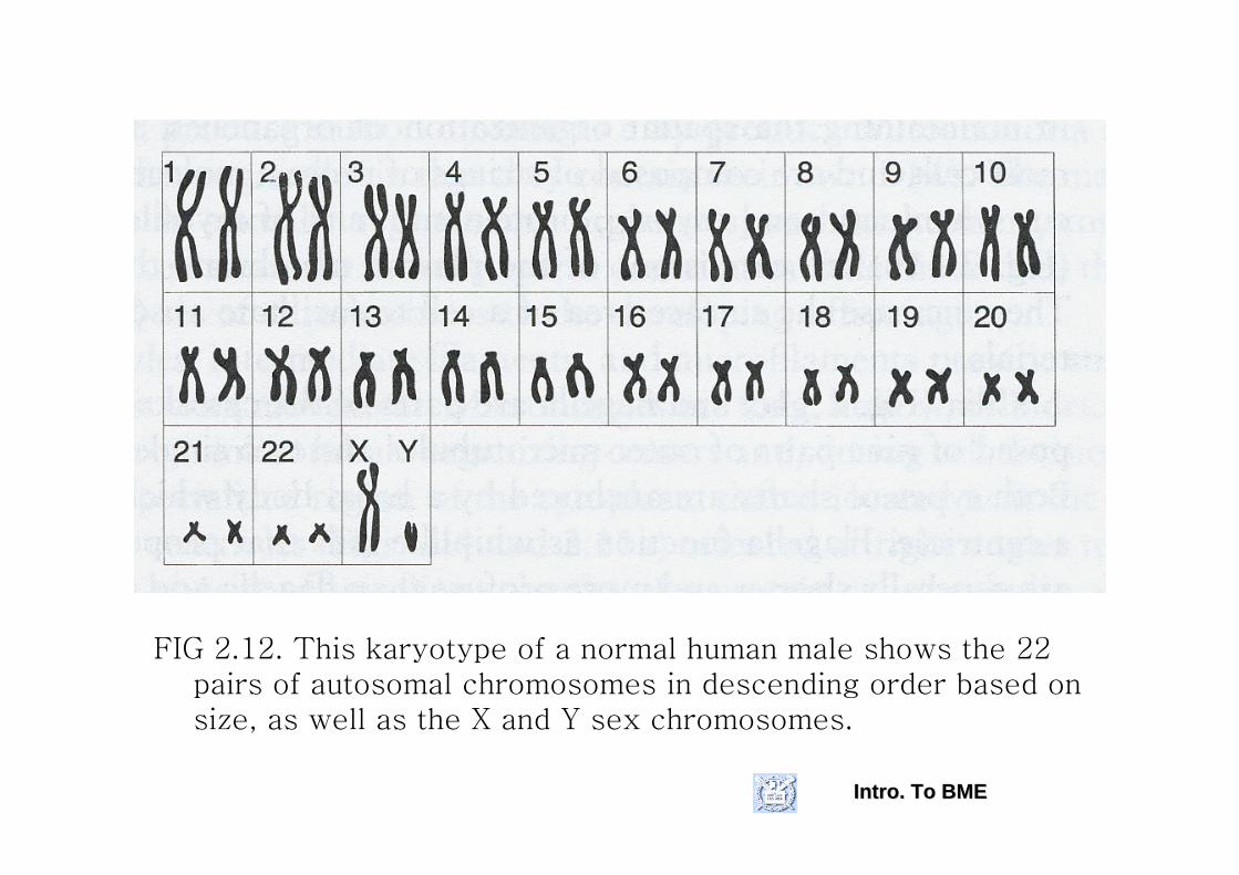
FIG 2.12. This karyotype of a normal human male shows the 22 pairs of autosomal chromosomes in descending order based on size, as well as the X and Y sex chromosomes.

Intro. To BMEIntro. To BME
▶ DNA- contains information from both parents(the one in the
mitochondria comes from mother)- wrapped around protein spools (nucleosomes) in
nucleus.- organized into pairs of chromosomes- each gene has a particular location in a specific
chromosome and contains the code for producing one of the three forms of RNA▪ ribosomal RNA ▪ messenger RNA ▪ transfer RNA

Intro. To BMEIntro. To BME
▶ human Genome Project- began in 1990, functional Genomics began in
2001 (originally planned for 15 years)- First to identify the location of at least 3000
specific human genes - Then to determine the sequence of
nucleotides (about 3 billion) in a complete set of haploid human chromosomes

Intro. To BMEIntro. To BME
▶ DNA replication
- occur during cell division- each strand of DNA is duplicated so that two double
helices now exist.- Each consists of one strand of the original DNA and
one new strand.- Some enzymes check for accuracy so that the error
rate is reduced to approximately one per billion.

Intro. To BMEIntro. To BME
FIG 2.13. During replication, DNA helicase (shown as a black wedge in b) unzips the double helix (a).
Another enzyme, DNA polymerase, then copies each side of the unzipped chain in the 5’ to 3’ direction.
One side of the chain (5’ to 3’) can be copied continuously, whereas the opposite side (3’ to 5’) is copied in small chunks in the 5’ to 3’ direction that are bound together by another enzyme, DNA ligase.
Two identical double strands of DNA are produced as a result of replication.
Intro. To BMEIntro. To BME
▶ transcription - DNA is in the nucleus and proteins are made on
ribosomes outside of nucleus and in the cytoplasm—need to send message out.
- Sequence of nucleotides in a gene that codes for a protein is transferred to mRNA through complementary base pairing of the nucleotide sequence in the gene.
- Difference from replication▪ only a certain stretch of DNA acts as the template and not the whole strand▪ different enzymes are used▪ only a single strand is produced

Intro. To BMEIntro. To BME
FIG 2.14. During transcription, RNA is formed from genes in the cell’s DNA by complementary base pairing to one of the strands. RNA contains uracil(U) rather than thymine(T) so the T in the first two pairs of the DNA become Us in the single-stranded RNA.

Intro. To BMEIntro. To BME
Translation(after transription)-Codon:a triplet in mRNA:coden(and
anticoden) can organize 64(43) associationsfor nucleotides(A,U,C,G) in each of the three places.
-tRNA contains an anticoden (3-base set), and binds at an area away from the triplet to an amino acid that is sprecific for that particular anticodon.
- DNA>mRNA>Codon>tRNA>Anticodon>specific_amino_acid>poly_peptide
FIG 2.15. Following transcription from DNA and processing in the nucleus, mRNA moves from the nucleus to the cytoplasm. In the cytoplasm, the mRNA joins with a ribosome to begin the process of translation.
During translation, tRNA delivers amino acids to the growing polypeptide chain. Which amino acid is delivered depends on the anticodon of a specific tRNA. Each tRNA binds to a particular amino acid at a site that is opposite thelocation of the anticodon.
For example, the codon CUG in mRNA is complementary to the anticodon GAC in the tRNA that carries leucine and will result in adding the amino acid leucine to the polypeptide chin.

Intro. To BMEIntro. To BME
• Central Dogma (Watson&Crick) 1955

Intro. To BMEIntro. To BME
- Each codon codes for a specific amino acid, but some amino acids are specified by more than one coden.
- Examples- ▪ coden for metihionine : AUG
▪ codons for leucine : UUA, UUG, CUU, CUC, CUA, CUG
- ▪ anticodon on the tRNA that delivers the methionineto the ribosome : UAC▪ tRNA with anticodons of AAU, AAC, GAA, GAG, GAU, GAC deliver leucine

Intro. To BMEIntro. To BME
▶ More on the translation
- mRNA binds to a ribosome and tRNA delivers amino acids to the growing polypeptide chain.
- Peptide bonds are formed between each new amino acid and the previous one.
- tRNA moves off into the cytoplasm where an amino acid joins with its anticodon.
- Process continues until a stop codon (UAA,UAG,or UGA) is reached on the mRNA
- The protein is then released into the cytoplasm (for transportation out of cell) or into the rough ER (for further modifications).