19960103 172 - dtic. · pdf filethe overall goals of this proposal are to characterize the...
TRANSCRIPT

T T?
JAN 0 5 i996F
GRANT NO: DAMD17-94-J-4120
TITLE: Ret Receptor: Functional Consequences of Oncogenic
Rearrangements
PRINCIPAL INVESTIGATOR(S): Susan S. Taylor, Ph.D.
CONTRACTING ORGANIZATION: University of California, San DiegoLa Jolla, California 92093-0934
REPORT DATE: October 1995
TYPE OF REPORT: Annual
PREPARED FOR: U.S. Army Medical Research and Materiel CommandFort Detrick, Maryland 21702-5012
DISTRIBUTION STATEMENT: Approved for public release;distribution unlimited
The views, opinions and/or findings contained in this report arethose of the author(s) and should not be construed as an officialDepartment of the Army position, policy or decision unless sodesignated by other documentation.
19960103 172DTIC QUALITY INSPECTED I

DISCLAIMER NOTICE
THIS DOCUMENT IS BEST
QUALITY AVAILABLE. THE COPY
FURNISHED TO DTIC CONTAINED
A SIGNIFICANT NUMBER OF
COLOR PAGES WHICH DO NOT
REPRODUCE LEGIBLY ON BLACK
AND WHITE MICROFICHE.

3 ~ ~ ~ ~ ~ ~ ~ ~ ~ ~ ~ a ,, I3 hlcv, , ... ~o ' ' rc o-cee four p.,: respones, enclu-I.g dhe See"1 fo:: Lvevm3,g3, .3. 3 7o ~ .r , eiconpo and ' .oevi ng the collection of information. Send comments regae.d. i :ci .d ci hu E. -C cr oh
ol1 ci. th , i opc g 'ool:r-u i s e burden. to 3'Ieshncion Headquarters Services, Direcioote 'o 30 cr Inf orm- 12cr i3 hehi., '..3 ~ ~ ~ ~ ~ ~ ~ ~~~ý th ' co,3'32S-~2 n e Office 03 Man'tgement and Budget, Paperwork Redlucti~oe Projec, (01--o33ý o
'October 1995 _ Annual 15 Sep 94 -14 Sep 95
1,Ret Receptor: Functional Consequences of Oncogenicý;Rearrangements DAMD17-94-J--4120
Susan S. Taylor, Ph.D.
I 'c~~jW~~c.~ [(F .2 ') 5 1))~1.
University of California, San Diego'La Jolla, California 92093-0934
C / ' 3 > "L,.. 'ol~~i3~ .0.
iU.S. Army Medical Research and Materiel Command ' .
!Fort Detrick, Maryland 21702-5012
!Approved for public release; distribution unlimited
The overall goals of this proposal are to characterize the gene product of a novel oncogene ret/ptc2. Thisiprotein is a fusion of the RIux subunit of cAMP-dependent protein kinase and the kinase domain of areceptor tyrosine kinase. The ligand for the RET receptor is still unknown, however, this gene isassociated with many inherited dominant cancers such as MEN2A, MEN2B, and Hirschprung'sdisease. Ret/ptc2 is a constitutively active form of the RET receptor, and we believe it will be aprototype for other oncogene receptor tyrosine kinases. Our goals are to express large amounts ofretlptc2 to establish the structural basis for its activation, to establish how the RET receptor functions invivo by identifying the proteins it interacts with, and to model the ret kinase domain and map the sites ofmutation that are known to be associated with various diseases. We are also constructing chimeras thatresemble ret/ptc2 using the kinase domains of the EGF receptor and the insulin receptor.
5' ..; i E
Breast cancer, ret/ptc2, oncogenes, receptor tyrosine kinases, structure/function 27
VLAF~~lF]CA~l0N ,1c 19 3C2n, C-
jUnclassified Unclassified jUnclassified "Unlimited
lr 5c-cý 172,30-5500 St~andard, Fom 2913 (ceov 2-9))Presc ' bed oy ANSI Std Z39-18253-102

FOREWORD
Opinions, interpretations, conclusions and recommendations arethose of the author and are not necessarily endorsed by the USArmy.
-Where copyrighted material is quoted, permission has beenobtained to use such material.
- Where material from documents designated for limiteddistribution is quoted, permission has been obtained to use thematerial.
Citations of commercial organizations and trade names inthis report do not constitute an official Department of Armyendorsement or approval of the products or services of theseorganizations.
In conducting research using animals, the investigator(s)d-lered to the "Guide for the Care and Use of LaboratoryVAnimals , prepared by the Committee on Care and Use of Laboratory
Animals of the Institute of Laboratory Resources, NationalResearch Council (NIH Publication No. 86-23, Revised 1985).
----- For the protection of human subjects, the investigatoris)adhered to policies of applicable Federal Law 45 CFR 46.
In conducting research utilizing recombinant DNA technology,investigator(s) adhered to current guidelines promulgated by
the National Institutes of Health.
JJ• In the conduct of research utilizing recombinant DNA, the" fiiestigator(s) adhered to the NIH Guidelines for ResearchV Involving Recombinant DNA Molecules.
4~U~In the conduct of research involving hazardous organisms,e investigator(s) adhered to the CDC-NIH Guide for Biosafety in
Kicrobiological and Biomedical Laboratories.
Acces'on For
DT, C ',•..:,
-Signature Datef

Principal Investigator: Taylor. S.S.
TABLE OF CONTENTS
Page
TABLE OF CONTENTS 1
INTRODUCTION and BACKGROUND 2
SPECIFIC AIMS 4
RESEARCH DESIGN AND EXPERIMENTAL RESULTS 4
CONCLUSION 8
REFERNECES 8-11
APPENDIX 12

Principal Investigator: Taylor. S.S.
INTRODUCTION and BACKGROUND: Protein phosphorylation is probably themost important mechanism for regulation in eukaryotic cells. The tightly regulatedenzymes that catalyze the phosphorylation of proteins, the protein kinases, areimportant components of signaling pathways that regulate normal cellularfunctions such as the cell cycle, metabolism, differentiation, memory and responseto hormones, to name only a few. Over 400 are now known[I], and mutations thatgenerate unregulated or constitutively activated protein kinases are typicallyoncogenic.
One of the simplest members of the protein kinase family is cAMP-dependentprotein kinase, cAPK [2]. Being one of the best understood members of the proteinkinase family, cAPK also serves as a template for the others since all of theseenzymes have evolved from a common ancestor and contain a conserved catalyticcore. cAPK, in the absence of cAMP, contains two types of subunits, a regulatory (R)subunit and a catalytic subunit (C). The R 2C 2 holoenzyme is catalytically inactive.In the presence of cAMP the complex dissociates into an R2-(cAMP) 4 dimer and 2free and active C-subunits. The crystal structure of the C-subunit, solved in ourlaboratory, serves as a structural template for the entire family of protein kinases [3].It defines the folding of the polypeptide chain as well as the positions of theinvariant residues that mostly cluster around the active site [4].
The objective of this grant is to characterize a novel oncogenic tyrosinekinase, Ret/ptc2, found in human papillary thyroid carcinomas. Specifically, wewant to understand the molecular basis for its constitutive activation and the basisfor its oncogenic properties. Ret/ptc2 is a rearranged gene product composed of thecAMP-dependent protein kinase (cAPK) regulatory subunit Ia (RIh) at its N-terminus fused to the tyrosine kinase core of the Ret proto-oncogene.
Ret Proto-Oncogene. The ret proto-oncogene (proto-ret) was cloned from aTHP-1 human monocytic leukemia cDNA library, and is expressed in a number ofhuman neuroblastoma and leukemia cell lines as 140-190 kDa glycoproteins [5].Although its ligand is still unknown, sequence analysis identified it to be a memberof the receptor tyrosine kinases. The putative extracellular domain contains Ca2 +binding domains similar to those in the cadherin proteins suggesting a role forproto-Ret in cell-cell recognition during development. In situ hybridizationindicates that proto-ret expression is important for neurogenesis, and normalkidney organogenesis in the mouse [6; 7; 8] . Point mutations, prematuretruncations, and chromosomal rearrangements of human proto-ret with othergenes have been linked to a number of human cancers.
The Ret Oncogene Family. The family of ret oncogenes can be divided intothree separate classes. The first class of ret oncogenes was produced in vitro bytransfecting NIH 3T3 cells with high molecular weight DNA from human celllymphomas [9], human colon carcinoma [10], and human stomach cancer tissue [11].The high propensity of proto-ret to rearrange with other genes is reflected in itsname for rearranged upon transfection [9].
The second class consists of missense mutations and truncated forms ofproto-ret that are proposed to result in either hyper- or hypoactivity. These areassociated with three dominantly inheirited human cancer syndromes: MEN 2A,
2

Principal Investigator: Taylor. S.S.
MEN 2B, familial medullary thyroid carcinoma (FMTC) [12; 13], andHirschsprung's disease [14; 15]. Germ-line mutations of proto-ret are dominant andare associated specifically with MEN 2A [16].
The third class of ret oncogenes, isolated from human papillary thyroidcarcinomas [17]; Bongarzone; 1993 #2580; Grieco, 1994 #2892; Bongarzone,1994 #2893, consists of 3 types: retiptcl, ret/ptc2 and ret/ptc3. The 5' end of theeach oncogene is a portion of an unrelated gene fused in frame to the identical splicesite of the proto-ret gene resulting in an intact functional Ret kinase.
The ret/ptcl and ret/ptc2 oncogenes each produce 2 isoforms as a result ofalternative splicing, and unlike proto-Ret are completely cytosolic, phosphorylatedon tyrosine residues, and constitutively active [18]. The 5' end of retiptcl is afragment of a new gene designated, H4(DS1OS170) [19; 20], and the 5' end ofret/ptc3 encodes a gene designated elel or ret fused gene, whose gene product doesnot show sequence identity to known proteins [21; 22; 23]. Unlike ret/ptcl andret/ptc3, the N-terminal sequence of ret/ptc2 gene [18] encodes approximately 60% ofa biochemically well characterized protein, the RIa subunit of cAPK. Comparison ofproto-Ret, the cAPK RIa-subunit, and Ret/ptc2 genes is illustrated in Figure 1.
TM1 proto- ret
E-Cadherin-like Cysteine Tyrosine Kinase Domainregion Repeats ret/ptc2
Inhibitor Tyrosine Kinase DomainSite
I WOW cAPK-RI a
Dimerization A BDomain cAMP Binding
Sites
Figure 1. Comparison of proto-ret, ret/ptc2, and cAPK RIa subunit
Regulatory Subunit of cAPK. The regulatory subunit of cAPK maintains theC subunit in an inactive state by forming a stable R2C2 tetramer. A pseudo-substrateinhibition sequence in the R-subunit mimics peptide substrates and fills the peptidebinding site of the C-subunit. The binding of cAMP to R causes the complex todissociate and to release two active C-subunits.
Although there are several unique gene products in the R-subunit family, allshare a well-defined domain structure. The RIa subunit begins with a dimerizationdomain close to its amino terminus followed by a pseudo-substrate inhibitoryregion and ends with two tandem cAMP binding domains. The Rla dimerizationdomain is stabilized by two antiparallel interchain disulfide bonds [24]. Circulardichoism studies of the proteolytically isolated RIa dimerization domain indicate it
is predominantly a-helical, and extremely stable to thermal denaturation [25]. The
crystal structure of the bovine RIa was recently solved in our laboratory [26]. Thesplice site of the RIa fragment in Ret/ptc2 is at the beginning of the aC-A helix, thus
3

Principal Investigator: Taylor. S.S.
deleting the last 21% of the A site cAMP-binding domain and the entire B-site cAMPbinding domain.
SPECIFIC AIMS
Our overall long terms goals are to understand the molecular basis for theconstitutive activation of ret/ptc2 and to characterize the physiological functioningof both ret/ptc2 and proto-ret. Our specific aims are the following:1. To understand the structural features of ret/ptc2 that are required for itsoncogenic properties. To achieve this we have developed an in vivo assay tomeasure a mitogenic response of ret/ptc2 by microinjecting it into the nucleus of ratfibroblast 1OT 1 / 2 cells.2. To characterize the biochemical properties of ret/ptc2 by overexpressing theprotein in E. coli and human kidney 293 cells. Phosphorylation sites will be mappedand kinetic properties characterized.3. To identify Ret/ptc2 binding proteins identify using a yeast two-hybridsystem.4. To construct homologs of ret/ptc2 using the tyrosine kinase domains of theEGF receptor and the insulin receptor.5. To model the kinase core of Ret/ptc2 based on the crystal structure of the C-subunit of cAPK and on the kinase domain of the insulin receptor.
We have made excellent progress in each of these areas, as indicated below,during this first year of the grant. Because much of this work is still unpublished,we shall describe it in some detail. This is an entirely new project for our laboratorybut it is quickly becoming a major focus.
RESEARCH DESIGN AND EXPERIMENTAL RESULTS
Expression and Purification of Ret/ptc2: We have succeeded in expressing the76 kDa isoform of Ret/ptc2 both in E. coli and in human kidney 293 cultured cells.In the bacterial expression system, ret/ptc2 was inserted behind a hexahistidinesequence in the bacterial expression vector, pET15b (Novagen), and transformedinto the host strain, NovaBlue(DE3). Yields are 1-2 milligrams per liter.
Ret/ptc2 is detected with a polyclonal rabbit antipeptide antibody to residues535-551 in the kinase domain. Recombinant rRet/ptc2 is soluble, stable and isrecognized by both the C-terminal Ret peptide antibody and a monoclonal mousephosphotyrosine antibody. Substitution of the bovine RIa sequence for the humanRia sequence in Ret/ptc2 results in increased levels of expression. Expression ofRet/ptc2 both in E.coli and in human kidney 293 cells yields a gene product which ismultiply phosphorylated on tyrosine residues.
His6-rRet/ptc2 was purified by affinity chromatography using a nickel resin(Fig. 2, panel A, lane 3). Ret/ptc2 autophosphorylates in vitro , and labeling with [T-32p]-ATP is 2-3x higher in the presence of Mn+2 than Mg+2 . Autophosphorylation ofRet/ptc2 results in a molecular weight shift of the major band (Fig. 2, panel A,compare lanes 2 and 3). Autoradiography of autophosphorylated Ret/ptc2 in Fig. 2,reveals 5 labeled protein bands (Fig. 2, panel B, lane 2) suggesting multiple
4

Principal Investigator: Taylor. S.S.
phosphorylation sites. The phosphorylated Ret/ptc2 isoforms observed in Figure 2,panel A, lanes 2 & 3 are all recognized by the C-terminal Ret peptide antibody andthe antiphosphotyrosine antibody. Preliminary results from inhibition assaysindicate that both the cAPK C-subunit and cAMP bind to Ret/ptc2.
A B
1 2 3 1 2
Figure 2. Purified SDS-PAGE separated His6-Ret/ptc2, electroblotted onto PVDFmembrane, and stained with Ponceau S. Panel A: lane 1, molecularweight standars, lane 2: autophosphorylated [32p]-His6-Ret/ptc2, lane 3:purified His6-Ret/ptc2. The arrow on the left identifies the 74.25 kDa pre-stained bovine serum albumin molecular weight standard, and thearrowhead identifies His6-Ret/ptc2. Panel B: lane 1: molecular weightstandards, lane 2: autoradiography of autophosphorylated [32p]-His6-Ret/ptc2 from Panel A, lane 2. The arrowhead points to thephosphorylated His6-Ret/ptc2 isoforms.
Biological Assay of Ret/ptc2 Mutants: Microinjection of ret/ptc2 and mutantsof ret/ptc2 has provided us with an in vivo assay by which the induction of DNAsynthesis by Ret/ptc2 and mutants can be qualitatively evaluated. Developing thisin vivo assay for the mitogenic effects of ret/ptc2 was a critical step. In this assay,ret/ptc2 was inserted into the mammalian expression vector, pRc/CMV(Invitrogen), behind the human cytomegalovirus promoter and microinjected intorat fibroblast cells (1OT 1/ 2). Injected cells are visualized by fluorescence, and DNAsynthesis is measured by the incorporation of 5-bromo-deoxyuridine [27]. The lossof mitogenic activity of inactive Ret/ptc2 mutants is not attributable to the inabilityof the cells to express these proteins since similar levels of protein expression were
5
Principal Investigator: Taylor. S.S.
visualized in all injected cells by immunofluorescence. With this system we haveestablished: a) Ret/ptc2 requires only the RIa dimerization domain to induce DNAsynthesis, b) site-specific mutations in Ret/ptc2 of kinase domain residuescorresponding to the MEN 2B and Hirschsprung's disease mutations yield resultssimilar that observed in the human disease phenotypes, c) selective mutation ofTyr350, Tyr424, Tyr505,Tyr586 to Phe show reduced levels or loss of DNA synthesis,d) substitution of the bovine RIa gene for the human RIa gene in ret/ptc2 does notaffect its ability to induce DNA synthesis [27].
The Ret/ptc2 Hirschsprung's disease mutants did not induce DNA synthesiswhereas the MEN 2B mutant elicited a mitogenic response. These results areconsistent with the human Ret oncogene disease phenotypes wherebyHirschsprung's disease is presumed to be due to loss of function, and the MEN 2Bmutation has been proposed to elicit an altered substrate specificity such thatinappropriate mitogenic signaling pathways are activated [28; 29]. Recentidentification of two other Ret kinase domain mutants associated with FMTC,Glu768Asp [30], and Val804Leu [31], which correspond to residues Glu292 and Va1328in Ret/ptc2, respectively, are now being introduced into the Ret/ptc2 kinase domainand tested in our in vivo microinjection assay. Glu292 resides above the glycineloop in our Ret/ptc2 model. The Va1328 residue in our Ret/ptc2 model resides atthe end of 3-strand 5 in the small lobe, approximately 5 residues from the hingeregion which separates the small and large lobes. Mutation of this residue from Valto Leu shortens the distance of this residue to within 3 A of Leu303, which is locatedon the aC-helix in the small lobe. Va1328 corresponds to Met120 in the cAPK C-subunit. Met120 is one of several residues which interacts with the adenine ring ofATP via hydrophobic interactions and hydrogen bonding [32]. Kinetic analysis ofall the Ret/ptc2 missense mutants will define their catalytic efficiencies andelucidate the molecular basis of their disease phenotypes.
Mutations of Tyr350 and Tyr424 to Phe decreased the mitogenic response inour assay [27]. Tyr350 resides within an insert sequence in the PDGF kinase domainwhich contains two SH2 binding sites, and thus this phosphorylated residue may beinvolved in downstream signaling. Phosphorylation of Tyr424, corresponding toTyr1158 in the InsR, is necessary for full InsR activity [33]. Mutation of Tyr429,which corresponds to Thr197 in the cAPK C-subunit, Tyr416 in src, and Tyr1163 inthe InsR [34], did not result in the induction of DNA synthesis. This result suggeststhe activation of the Ret/ptc2 kinase domain is unlike that of the src, a cytosolictyrosine kinases, and the Ser/Thr kinase, cAPK. In addition, this result indicatesthat phosphorylation of specific tyrosyl residues which serve to activate receportyrosine kinases (RTKs) differ amongst the RTKs. Molecular modeling studies ofthe Ret/ptc2 kinase core indicate Tyr586 resides at an exposed site of the proposedstructure, whereas Tyr505 is less exposed albeit highly conserved in other RTKswhich suggests it may be important for structural stability.
Phosphorylation of Tyr350 and Tyr586 in vivo, may provide docking sites forSH2 containing proteins. Immunoprecipitation of both Ret/ptcl and Ret/ptc2expressed in NIH 3T3 cells have shown that phosphorylated Shc is associated withall four isoforms of the 2 proteins [35]. In vitro binding assays of cell lysates to
6

Principal Investigator: Taylor. S.S.
immobilized glutathione-fused Grb2 or Grb2 SH2 domains indicate the SH2domains of Grb2 may also be associated with the anti-Shc immunoprecipitatedRet/ptcl and Ret/ptc2 isoforms as well as other phosphorylated unidentifiedproteins [35]. The reduction or loss of mitogenic activity in the Ret/ptc2 tyrosinemutants may stem from effects of structural instability, alterations in catalyticactivity, or decreased interactions with downstream signaling proteins. Furtherinvestigation of these Ret/ptc2 tyrosine mutants is underway.
Yeast Two Hybrid System: We have begun to use the yeast two hybrid systemto isolate RET-binding proteins. Using ret/ptc2 as the bait, we have been able toidentify several proteins that specifically bind to the RET portion of ret/ptc2. Theseproteins are now being expressed as fusion proteins and binding is being confirmedin vitro. Several of these proteins contain SH2 domains and, using a series ofTyr/Phe mutants, the binding of each of these proteins is being correlated with aspecific tyrosine residue. The effect of each of these proteins in the in vivo assay issimultaneously being tested. From these studies is emerging a comprehensive set ofRBP's (Ret Binding Proteins) that dock at specific sites along the polypeptide chain.Binding is being confirmed by surface plasmon resonance using the expressedprotein and the expressed and purified binding domains.
Identification of Phosphorylated Tyrosine Residues: To identify thephosphorylation sites in Ret/ptc2, we are labelling the protein in vivo with [32pi].Western analysis of SDS-PAGE separated His6-Ret/ptc2 indicates it is multiplyphosphorylated on tyrosine residues. Two dimensional peptide maps will be usedto identify 32p-labelled peptides. Peptides will also be separated by reverse-phaseHPLC, and the [32p]-peptide(s) will be sequenced. The phosphorylation sites in therecombinant ret/ptc2 will then be compared with the sites that are labeled whenret/ptc2 is explressed in eukaryotic cells to make certain that the sites are the same.Phosphaorylation sites will also be indentified by electrospray mass spectrometry.
Identification of phosphorylated tyrosine residues in Ret/ptc2 will enable usto design and synthesize an optimum consensus peptide substrate for use as aRet/ptc2-specific peptide substrate. By identifying a consensus peptide based on theautophosphorylation sequences, we expect to gain a higher specific activity and thusa greater sensitivity in our in vitro assay.
An enzymatic assay for Ret/ptc2, based on a modification of theautophoshorylation assay whereby the [32 p]-phosphorylated peptide substrate isbound to a phosphocellulose membrane disk for quantitation by liquid scintillationcounting, is now being established for routine use. Steady state parameters such asKm, Kcat and Vmax will be determined from plots of initial velocity versussubstrate concentration. This kinetic analysis will be essential for quantifying thespecific effects of the various mutations as well as for under-standing, in general, thebiological properties of this enzyme.
Modelling of the Ret Tyrosine Kinase Domain: The ret kinase domain hasbeen modeled based on the structure of the catalytic subunit of cAPK and therecently solved structures of the insulin receptor kinase [36]. We are done mappingthe sites that are associated with the various malignancies. By mapping these sitesand correlating the sites with the phenotype displayed by the malignancy, we hopeto better understand ret/ptc2 as well as proto-ret functions.
7

Principal Investigator: Taylor. S.S.
CONCLUSION
This represents a totally new project for my laboratory, and the progressduring this first year has been excellent. With the biological assay we have been ableto establish what is essential for the mitogenic properties of ret/ptc2. In conjunctionwith this, we are now using the yeast two-hybrid system to identify Ret bindingproteins and are developing a comprehensive picture of this receptor tyrosinekinase and its in vivo partners. At the same time we have developed an E. coliexpression system that yields mg quantities of proteins both for Ret/ptc2 and for theEGF and insulin receptor homologs. Finally, we have been modeling the kinaseportion of Ret based on our structure of the cAPK C-subunit and of the crystalstructure of the insulin receptor kinase domain. Of particular importance now ismapping of the various mutation sites in Ret that are known to be linked with awide variety of malignancies.
REFERENCES
1. Hanks, S.K., & Hunter, T. Protein kinases 6. The eukaryotic protein kinasesuperfamily: kinase (catalytic) domain structure and classification. Faseb J. 9 (8),576-596 (1995).
2. Taylor, S.S.,Buechler, J.A., & Yonemoto, W. cAMP-dependent Protein Kinase:Framework for a Diverse Family of Regulatory Enzymes. Annu. Rev. Biochem. 59971-1005 (1990).
3. Knighton, D.R.,Zheng, J.,Ten Eyck, L.F.,Ashford, V.A.,Xuong, N.-h.,Taylor, S.S., &Sowadski, J.M. Crystal Structure of the Catalytic Subunit of cAMP-dependentProtein Kinase. Science 253 407-414 (1991).
4. Taylor, S.S.,Knighton, D.R.,Zheng, J.,Ten Eyck, L.F., & Sowadski, J.M. StructuralFramework for the Protein Kinase Family. Annu. Rev. Cell Biol. 8 429-462 (1992).
5. Takahashi, M.,Buma, Y., & Taniguchi, M. Identification of the ret proto-oncogene in neuroblastoma and leukemia cells. Oncogene 6 297-301 (1991).
6. Pachis, V.,Mankoo, B., & Constantini, F. Expression of the c-ret proto-oncogeneduring mouse embryogenesis. Development 119 1005-17 (1993).
7. Sugaya, R.,Ishimaru, S.,Hosoya, T.,Saigo, K., & Emori, Y. A Dropsophila homologof human proto-oncogene ret transiently expressed in embryonic meuronalprecursor cells including neuroblasts and CNS cells. Mech. Dev. 45 139-45 (1994).
8. Schuchardt, A.,D'Agati, V.,Larsson, B.L.,Constantini, F., & Pachnis, V. Defects inthe kidney and enteric nervous system of mice lacking the tyrosine kinase receptorRet [see comments]. Nature 367 380-3 (1994).
8

Principal Investigator: Taylor. S.S.
9. Takahashi, M.,Ritz, J., & Cooper, G.M. Activation of a Novel HumanTransforming Gene, ret, by DNA Rearrangement. Cell 42 581-588 (1985).
10. Ishizaka, Y.,Tahira, T.,Ochiai, M.,Ikeda, I.,Sugimura, T., & Nagao, M. MolecularCloning and Characterization of Human ret-II Oncogene. Oncogene Res. 3 193-7(1988).
11. Kunieda, T.,Marsui, M.,Nomura, N., & Ishizaki, R. Cloning of an ActivatedHuman ret Gene With a Novel 5' Sequence Fused by DNA Rearrangement. Gene107 323-8 (1991).
12. Hofstra, R.M.,Landsvater, R.M.,Ceccherini, I.,Stulp, R.P.,Stelwagen, T.,Luo,Y.,Pasini, B.,Hoppener, J.W.M.,Ploos van Amstel, H.K.,Romeo, G.,Lips, C.J.M., &Buys, C.C.C.M. A mutation in the RET proto-oncogene associated with multipleendocrine neoplasia type 2B and sporadic medullary thyroid carcinoma. Nature 367375-376 (1994).
13. Mulligan, L.M.,Eng, C.,Healey, C.S.,Clayton, D.,Kwok, J.B.,Gardner, E.,Ponder,M.A.,Frilling, A.,Jackson, C.E., & Lehnert, H. Specific mutations of the RET proto-oncogene are related to disease phenotype in MEN 2A and FMTC. Nat. Gent. 6 70-4(1994).
14. Edery, P.,Lyonney, S.,Mulligan, L.M.,Pelet, A.,Dow, E.,Abel, L.,Holder, S., &Nihoul. Mutations of the RET proto-oncogene in Hirschsprung's disease. Nature367 378-80 (1994).
15. Romeo, G.,Ronchetto, P.,Luo, Y.,Barone, V.,Seri, M.,Ceccherini, I.,Pasini,B.,Bocciardi, R.,Lerone, M., & Kaarianinen, H. Point mutations affecting thetyrosine kinase domain of the RET proto-oncogene in Hirschsprung's disease.Nature 367 377-8 (1994).
16. Mulligan, L.M.,Kwok, J.B.J.,Healey, C.S.,Elsdon, M.J.,Eng, C.,Gardner, E.,Love,D.R.,Mole, S.E.,Moore, J.K.,Papi, L.,Ponder, M.A.,Telenius, H.,Tunnacliffe, A., &Ponder, B.A.J. Germ-Line Mutations of the RET Proto-Oncogene in MultipleEndocrine Neoplasia Type 2A. Nature 363 458-460 (1993).
17. Grieco, M.,Santoro, M.,Berlingieri, M.T.,Melillo, R.M.,Donghi, R.,Bongarzone,I.,Pierotti, M.A.,Della Porta, G.,Eusco, A., & Vecchio, G. PTC is a novel rearrangedform of the ret proto-oncogene and is frequently detected in vivo in human thyroidpapillary carcinomas. Cell 60 557-563 (1990).
18. Bongarzone, I.,Monzini, N.,Borrello, M.G.,Carcano, C.,Ferraresi, G.,Arighi,E.,Mondellini, P.,Della Porta, G., & Pierotti, M.A. Molecular characterization of athyroid tumor-specific transforming sequence formed by the fusion of ret tyrosine
9

Principal Investigator: Taylor. S.S.
kinase and the regulatory subunit RI alpha of cyclic AMP-dependent protein kinaseA. Mol. Cell. Biol. 13 358-366 (1993).
19. Grieco, M.,Cerrato, A.,Santoro, M.,Fusco, A.,Melillo, R.M., & Vecchio, G.Cloning and characterization of H4 (D1OS170), a gene involved in RETrearrangements in vivo. Oncogene 9 2531-5 (1994).
20. Tong, Q.,Li, Y.,Smanik, P.A.,Fithian, L.J.,Xing, S.,Mazzaferri, E.L., & Jhiang, S.M.Characterization of the promoter region and oligomerization domain of H4(D1OS170), a gene frquently rearranged with the ret proto-oncogene. Oncogene 101781-7 (1995).
21. Bongarzone, I.,Butti, M.G.,Coronelli, S.,Borello, M.G.,Santoro, M.,Mondellini,P.,Pilotti, S.,Fusco, A.,Della, P.G., & Pierotti, M.A. Frequent activation of ret proto-oncogene by fusion with a new activating gene in papillary thyroid carcinomas.Cancer Res. 54 2879-85 (1994).
22. Santoro, M.,Dathan, N.A.,Berlingieri, M.T.,Bongarzone, I.,Paulin, C.,Grieco,M.,Pierotti, M.A.,Vecchio, G., & Fusco, A. Molecular characterization of RET/PTC3;a novel rearranged version of the RET proto-oncogene in a human thyroid papillarycarcinoma. Oncogene 9 509-16 (1994).
23. Minoletti, F.,Butti, M.G.,Coronelli, S.,Miozzo, M.,Sozzi, G.,Pilotti, S.,Tunnacliffe,A.,Poerotti, M.A., & Bongarzone, I. The two genes generating RET/PTC3 arelocalized in chromosomal band 10q11. 2 . Genes, Chromosomes and Cancer 11 51-7(1994).
24. Bubis, J.,Vedvick, T.S., & Taylor, S.S. Antiparallel Alignment of the TwoProtomers of the Regulatory Subunits of the Regulatory Subunit Dimer of cAMP-Dependent Protein Kinase I. J. Biol. Chem. 262 14961-14966 (1987).
25. Le6n, D.,Herberg, F.W.,Banky, P., & Taylor, S.S. Characterization of an (X- HelicalDimer from the Regulatory Subunit of cAMP-Dependent Protein Kinase.(Submitted 1995).
26. Su, Y.,Dostmann, W.R.G.,Herberg, F.W.,Durick, K.,Xuong, N.-h.,Ten Eyck,L.F.,Taylor, S.S., & Varughese, K.I. Regulatory (RIu) Subunit of cAMP-dependentProtein Kinase: Crystal Structure of a 1-91 Deletion Mutant Defines CooperativecAMP Binding Sites. In press Science (1995).
27. Durick, K.,Yao, V.J.,Borrello, M.G.,Bongarzone, I.,Pierotti, M.A., & Taylor, S.S.Tyrosines outside the kinase core and dimerization are required for the mitogenicactivity of RET/ptc2. in press J. Biol. Chem. (1995).
28. Songyang, Z.,Carraway, K.L.,Eck, M.J.,Harrison, S.C.,Feldman,R.A.,Mohammedi, M.,Schlessinger, J.,Hubbard, S.R.,Smith, D.P.,Eng, C.,Lorenzo,
10

Principal Investigator: Taylor. S.S.
M.J.,Ponder, B.A.J.,Mayer, B.J., & Cantley, L.C. Catalytic specificity of protein-tyrosine kinases is critical for selctive signalling. Nature 373 536-9 (1995).
29. Santoro, M.,Carlomagno, F.,Romano, A.,Bottaro, D.P.,Dathan, N.A.,Grieco,M.,Fusco, A.,Vecchio, G.,Matoskova, B.,Kraus, M.H., & DiFiore, P.P. Activation ofRET as a dominant transforming gene by germline mutations of MEN2A andMEN2B. Science 267 381-3 (1995).
30. Eng, C.,Smith, D.P.,Mulligan, L.M.,Healey, C.S.,Zvelebil, M.J.,Stonehouse,T.J.,Ponder, M.A.,Jackson, C.E.,Waterfield, M.D., & Ponder, B.A.J. A novel pointmutation in the tyrosine kinase domain of the RET proto-oncogene in sporadicmedullary thyroid carcinoma and in a family with FMTC. Oncogene 10 509-13(1995).
31. Bolino, A.,Schuffenecker, I.,Luo, Y.,Seri, M.,Silengo, M.,Tocco, T.,Chabrier,G.,Houdent, C.,Murat, A.,Schlumberger, M.,Tourniaire, J.,Lenoir, G.M., & Romeo,G. RET mutations in exons 13 and 14 of FMTC patients. Oncogene 10 2415-19(1995).
32. Zheng, J.,Knighton, D.R.,Ten Eyck, L.F.,Karlsson, R.,Xuong, N.-h.,Taylor, S.S., &Sowadski, J.M. Crystal Structure of the Catalytic Subunit of cAMP-dependentProtein Kinase Complexed with MgATP and Peptide Inhibitor. Biochemistry 32 (9),2154-2161 (1993).
33. White, M.,Shoelson, S.,Keutmann, H., & Kahn, C. A cascade of tyrosineautophosphorylations in the beta-subunit activates the phosphotransferase of theinsulin receptor. J. Biol. Chem. 263 2969-80 (1988).
34. Hanks, S.K.,Quinn, A.M., & Hunter, T. The Protein Kinase Family: ConservedFeatures and Deduced Phylogeny of the Catalytic Domains. Science 241 42-52 (1988).
35. Borrello, M.G.,Pelicci, G.,Arighi, E.,De, F.L.,Greco, A.,Bongarzone, I.,Rizzetti,M.,Pelicci, P.G., & Pierotti, M.A. The oncogenic versions of the Ret and Trk tyrosinekinases bind Shc and Grb2 adaptor proteins. Oncogene 9 1661-8 (1994).
36. Hubbard, S.R.,Wei, L.,Ellis, L., & Hendrickson, W.A. Crystal structure of thetyrosine kinase domain of the human insulin receptor [see comments]. Nature 372(6508), 746-754 (1994).
11

Principal Investigator: Taylor. S.S.
APPENDIX
1. Su, Y.,Dostmann, W.R.G.,Herberg, F.W.,Durick, K.,Xuong, N.-h.,Ten Eyck,L.F.,Taylor, S.S., & Varughese, K.I. Regulatory (RIa) Subunit of cAMP-dependentProtein Kinase: Crystal Structure of a 1-91 Deletion Mutant Defines CooperativecAMP Binding Sites. In press Science (1995).
2. Durick, K.,Yao, V.J.,Borrello, M.G.,Bongarzone, I.,Pierotti, M.A., & Taylor, S.S.Tyrosines outside the kinase core and dimerization are required for the mitogenicactivity of RET/ptc2. J. Biol. Chem. In Press (1995).
12

Reprint Series11 August 1995, Volume 269, pp. 807-813
Regulatory Subunit of ProteinKinase A: Structure of Deletion
Mutant with cAMP Binding Domains
Y. Su, W. R. G. Dostmann,* F. W. Herberg,t K. Durick,N-h. Xuong, L. Ten Eyck, S. S. Taylor, and K. I. Varughese
Copyright © 1995 by the American Association for the Advancement of Science

th two cAMP binding sites. It forms a tightegulaory Subunit of Protein complex rapidly with C, and cAMP medi-ates activation in a manner similar to theinase A: Structure of Deletion tetrameric holoenzyme. Full understandingof the molecular basis for cAMP bindingiand for holoenzyme activation requires highMutant with cAMP Binding Domains nstructures. Crystal structures ofthe C Subunit have been determined (I Wa.
Y. Su, W. R. G. Dostmann,* F. W. Herberg,t K. Durick, We describe here the structure of the Al-N-h. Xuong, L. Ten Eyck, S. S. Taylor, K. I. Varughese 91 deletion mutant of the recombinant bo-
vine RIa subunit (rRlc). Ever though theIn the molecular scheme of living organisms, adenosine 3',5'-monophosphate (cyclic dimerization domain is absent, this struc-AMP or cAMP) has been a universal second messenger. In eukaryotic cells, the primary tore nevertheless reveals the detailed fea-receptors for cAMP are the regulatory subunits of cAMP-dependent protein kinase. The tures of each cAMP binding site and pro-crystal structure of a 1-91 deletion mutant of the type Ia regulatory subunit was refined vides a molecular basis for the cooperativeto 2.8 A resolution. Each of the two tandem cAMP binding domains provides an extensive binding of cAMP and activation of thenetwork of hydrogen bonds that buries the cyclic phosphate and the ribose between two holoenzyme.P3 strands that are linked by a short cY helix. Each adenine base stacks against an aromatic Structure solution. The Al-91 rRIocring that lies outside the 03 barrel. This structure provides a molecular basis for under- subunit, (Al-91)rRlc, was expressed in E.standing how cAMP binds cooperatively to its receptor protein, thus mediating activation coli as described (11). Unlike the full-lengthof the kinase. R subunit, this deletion mutant is resistant to
proteolysis. A typical yield is 200 mrg from 4liters of Culture. To generate a heavy atomsubstitution site, we replaced the Cys residue
Protein phosphorylation and dephospho- complex to dissociate, thereby releasing an with Set at position 145 by the Kunkelrylation is one of the principal mechanisms R2 (cAMP)4 dimer and two free and active method (12). The expression, biochemicalby which cellular functions are regulated in C subunits. Both cytoplasmic and nuclear properties, and crystallization of (Al-91)-eukaryotic cells in response to external proteins are substrates for cAPK, and when rRIa(S145C) are similar to those ofstimuli (1). The enzymes catalyzing these C is not anchored to R in the RC, comn- (Al-91)rRlcx.phosphorylations, the protein kinases, are plex, it can enter the nucleus (7). Crystals of (Al -91 )rRIcx and (Al -91)-tightly regulated and maintained in an in- There are two general classes of R sub- rR(S145C) were grown by the vapor diffu-active state in the absence of the specific units, types I and II, and within each class sion method with the hanging-drop proce-activating signal. The mechanism for main- are at least two distinct gene products (2, 6). dure in Linbro plastic tissue culture platestaining the inhibited state of any protein All R subunits nevertheless retain a well- at 22°C (13). The drops contained 5 V1. ofkinase is at least as critical for its function as defined domain structure. At the amino ter- protein stock (8 mg/ml) and 5 pVl of reser-is catalytic efficiency. minus is a dimerization domain, followed by voir solution. Hexagonal crystals were pro-
Although the family of protein kinases an autoinhibitor site that resembles either a duced from a reservoir solution of 1.1 to 1.2now includes several hundred members (2), substrate or an inhibitor. This autoinhibitor M (NH 4)2SO 4 (grade III, Sigma), 10 tocyclic AMP-dependent protein kinase segment binds to the active site of the cata- 12.5 percent glycerol (Sigma) and 10 mM(cAPK) was among the first to be charac- lytic subunit. The R subunits are thus com- dithiothreitol buffered with 80 to 100 mMterized (3). The demonstration of the acti- petitive inhibitors of substrate proteins. The sodium acetate, pH 5.5; they grew to theirvation of cAPK by cAMP introduced the carboxyl terminus is comprised of two tan- maximum size, 0.15 by 0.15 by 1.5 mm,hormone second messenger concept, where- dem homologous cAMP binding domains, A over 3 weeks. They belonged to space groupby a hormone binding to the extracellular and B. Site A is masked in the holoenzyme P6122 (P6 522), and the unit cell dimen-surface led to the generation of a cytoplas- so that the cooperative activation is mediat- sions were a b = 88.9 A, c = 179.9 A.mic second messenger (4). Of the protein ed by cAMP binding first to site B (8). This There was one molecule per asymmetrickinases, cAPK is also one of the simplest triggers a conformational change that makes unit with a Matthews coefficient, V,,, of 2.9and best understood biochemically (5, 6), site A more accessible. Cyclic AMP binding A1 per dalton. The solvent content (14)largely because the regulatory (R) and cat- to site A then mediates dissociation of the was approximately 57 percent.alytic (C) components are coded for by complex. The two cAMP binding sites can Diffraction data for the native and heavyseparate genes, and the proteins can be be readily distinguished by several criteria, atom derivative crystals were collected ini-readily separated upon activation. The in- Site A has a faster off-rate and has a prefer- tially with the Xiong-Hamlin multiwireactive holoenzyme is an RzCz tetramer. Cy- ence for N6-sttbstituted analogs. Site B, with area detector system (15) at the NIH Na-clic AMP binding cooperatively causes the a slower off-rate, is preferred by C2- and tional Research Resource at UCSD. The
C8-substituted analogs (9). The cAMP native crystal diffracted to 2.9 A resolution.Y. Su, W. R. G. Dostmann, F. W. Herberg, K. Durick, and binding domains are also homologous to the Three heavy atom derivatives, namely, twoS. S. Taylor are in the Department of Chemistry andBiochemistry and N-h. Xuong and K. I. Varughese are in cAMP binding domain of the catabolite mercury derivatives and a gold derivative,the Department of Biology, University of California, San gene activator protein (CAP) in Escherichia were prepared, and diffraction data to 3.5 ADiego, La Jolla, CA 92093-0654, USA. L. Ten Eyck is at coli (10). were collected. The diffraction data for thethe San Diego Supercomputer Center, La Jolla, CA92186, USA. The domain structure of the R subunits, native protein and the derivatives were
aresntddresa: Institut fur Phrmakoge nn Toxi- first characterized by limited proteolysis, again measured to higher resolutions withcologie, Technsche Universitat Munchen, Biederstener was subsequently probed with deletion mu- an MAR image plate scanner at the Stan-Stral3e 29, 80802 Monchen, Germany. tants (5). One of the most stable of these ford Synchrotron Radiation LaboratorytPresent address: Ruhr-Universitft Bochum, Mediz- mutants has a deletion of 91 residues at the (SSRL). The statistics of data collectionnische Fakiltat, Institut fir Physiologische Chemie I,44780 Bochum, Germany. NH2-terminus (11). Although monomeric, and the multiple isomorphic replacementt:To whom correspondence should be addressed, it retains the autoinhibitor site as well as (MIR) phases computed with the program
SCIENCE • VOL. 269 * 11 AUGUST 1995 807

package PHASES (16) are listed in Table 1. 190 residues. The coordinates of this mod- could not be traced. Five other regions ofSolvent-flattening calculations (17) with el were improved by X-PLOR (20) refine- the chain consisting of residues 113 to2.8 A MIR phases gave an interpretable ment, which gave an R factor of 34.1 118, 275 to 279, 285 to 288, 302 to 311,map. The space group was assigned as P6522 percent for data between 10.0 to 3.0 A. At and 375 to 376 showed up in the map withfrom the right-handedness of the ot helices. this stage, using the program SIGMAA densities less well defined compared to the
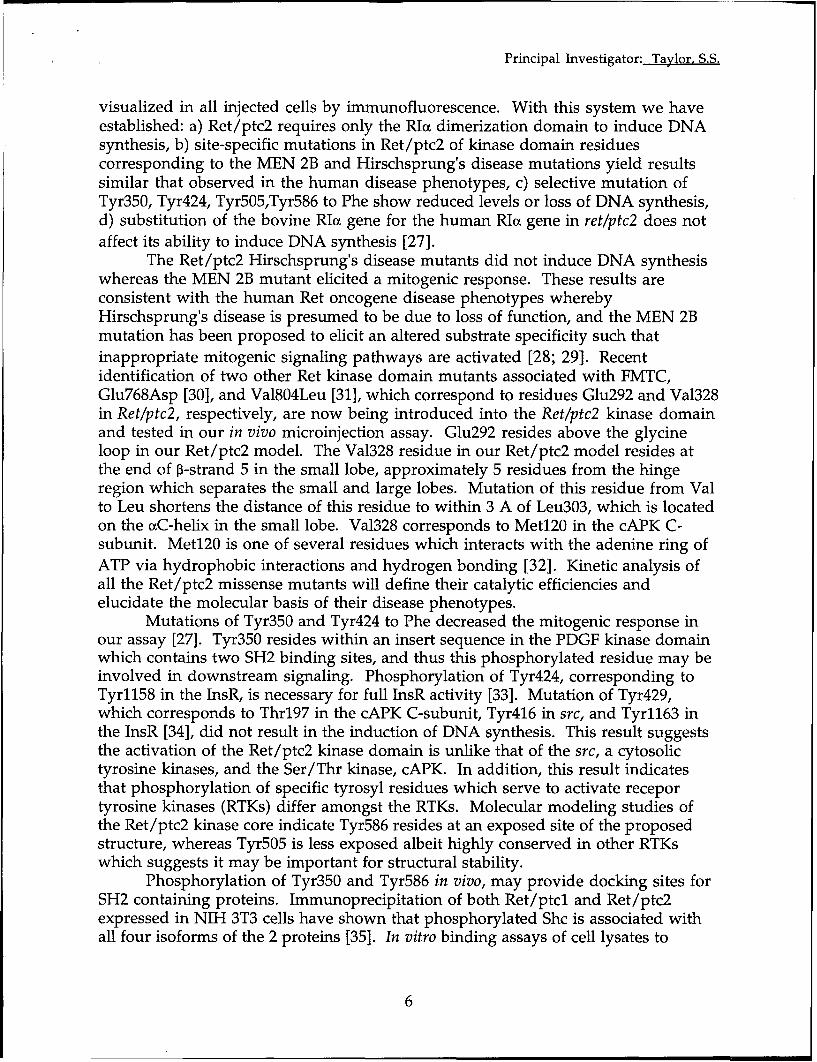
After the map was computed with the (21), we combined phases from the refined rest of the structure; however, these resi-solvent-flattened phases, model building partial model with the MIR phases, and dues could be traced and were included inwas done with the graphics programs the resultant map revealed better electron the refinements. These regions were allTOM (18) and 0 (19). The program 0 density for the unbuilt regions. Seventy- solvent-exposed. Residues 302 to 311, lo-was used to construct the backbone and four more residues were incorporated into cated in the surface loop connecting 34side chains based on the Cot trace that was the model after several rounds of model and P35 of domain B, have high B factors.manually generated with the use of TOM. building and phase combinations. The re- Side chains were assigned as follows.The map showed well-defined electron finement was done iteratively with Cys145 and Cys360 were identified from thedensity for cAMP bound to both domains X-PLOR and omit maps. At present, mod- positions of the mercury atoms in the de-A and B (Fig. 1). The initial model in- el consists of 2020 nonhydrogen atoms. rivatives. In each domain, conserved resi-cluded two molecules of cAMP and three The NH 2-terminal residues 92 to 112 and dues interacting with cAMP, Glu 2°°, andsegments of peptide chains consisting of the COOH-terminal residues 377 to 379 Arg2z 9 in domain A and Glu3 24 and Arg333
Table 1. Diffraction data and structure solution statistics. N, number of observa- These two data sets were merged with a native data set measured at SSRL (X =tions, Rderv = =F, F,, 1 FR -hNi , 1/=(h) - /(h)J/Yh.XNi l/(h)i, where 1.08 A) on a MAR image plate; the program MOSFLM (45), was used to reduce the/(h), is the ith measurement of reflection h and I(h) is the mean value of the N data from SSRL. The Hg atom position in the C2H5HgCI derivative was locatedequivalent reflections. The R derivative is calculated with respect to a merged data from the isomorphous difference Patterson map. The Au position and the posi-set obtained by merging the two data sets. FH, average root-mean-square (rms) tions of the two Hg atoms in the S145C mutant derivative were located withheavy atom structure factor amplitude. Faom, average anomalous dispersion cross-difference electron density maps. The solvent-flattening calculations werestructure factor amplitude of heavy atoms. E, rms closure error. Centric RPFph(Obs) done according to Wang et al. (17), on the basis of an assumed solvent content of- Fp(calc) X 1 00/111Fph(obs) - Fpj; Ea, rms anomalous closure error. The Au 50 percent. After convergence, the mean figure of merit was 0.84 with the mapderivative was formed by soaking the crystal in solutions of 2 mM KAuCi4.The Hg inversion R factor of 29 percent. An analysis of the diffraction pattern revealed thatderivative was obtained by cocrystallizing with C2H5HgCI. The protein has two the overall temperature factor of the crystal is anisotropic. Hence an anisotropiccysteines, one at residue 345 and the other at residue 360; however, only Cys360 correction was applied. After the correction, the refinement provided an R factor ofreacted with Hg. For obtaining another Hg derivative, a new mutant was created 22.1 percent from the previous R of 25 percent. Further refinement with theby replacing Ser145 with Cys. Soaking of crystals of this mutant with 6 mM program TNT (46) with all the data yielded the final R of 22.9 percent. As the highp-chloromercuribenzene sulfonate (PCMBS) yielded a derivative with two Hg sites, resolution data were affected by decay, we confirmed our present refinement toDiffraction data from two native crystals were measured at 0CSD (X= 1.518 A). 2.8 A data.
Data sets used in the structure determination
Reflections Observations n sym Overallderivcomplete N DeviceData set (N (N) (A,) (%) (%)(%
Native 1 8904 61857 3.0 4.8 99.3 MultiwireNative 2 15809 37561 2.4 5.5 88.5 MAR(SSRL)COH5 HgCI 14656 53743 2.5 7.5 95.0 7.8 MAR(SSRL)KAuCI4 9494 19257 2.7 6.9 83.8 11.8 MAR(SSRL)Hg derivative of S1 45C mutant 9801 52070 2.8 8.9 88.7 10.7 MAR(SSRL)
MIR statistics of the heavy atom derivatives
Average resolution of the shell (A)Derivative Measurement
Overall 9.16 5.57 4.63 4.12 3.77 3.51 3.31 3.15 3.01 2.87
C2HHgCI FHIE 1.96 1.81 2.29 1.90 1.72 1.92 1.96 2.07 1.97 2.08 2.11Fanom/Eanom 2.02 1.89 2.15 1.76 1.83 2.05 2.03 2.24 2.21 2.40 1.95Centric R 0.68
KAuCI4 FH/E 1.40 1.58 1.84 1.54 1.36 1.24 1.26 1.44 1.27 1.21 1.29Centric R 0.58
Hg derivative of S145C mutant FH/E 2.08 2.32 2.36 1.99 1.88 2.01 2.07 2.15 1.96 2.02 1.98Centric R 0.64
Figure of merit 0.66 0.737 0.714 0.684 0.686 0.674 0.648 0.656 0.622 0.576 0.563
Refinement
Model Final R B factor Data selection Programsfactor
190 residues + 2 cAMP 34.1 Overall 10.0-3.o A //a>2 X-PLOR247 residues + 2 cAMP 25.0 Individual 10.0-2.8 A I//> 2 X-PLOR264 residues + 2 cAMP 22.1 Individual 8.0-2.8 A I/cr>2 X-PLOR264 residues + 2 cAMP 22.9 Fixed 15.0-2.8 A all F TNT
808 SCIENCE ° VOL. 269 ° 11 AUGUST 1995
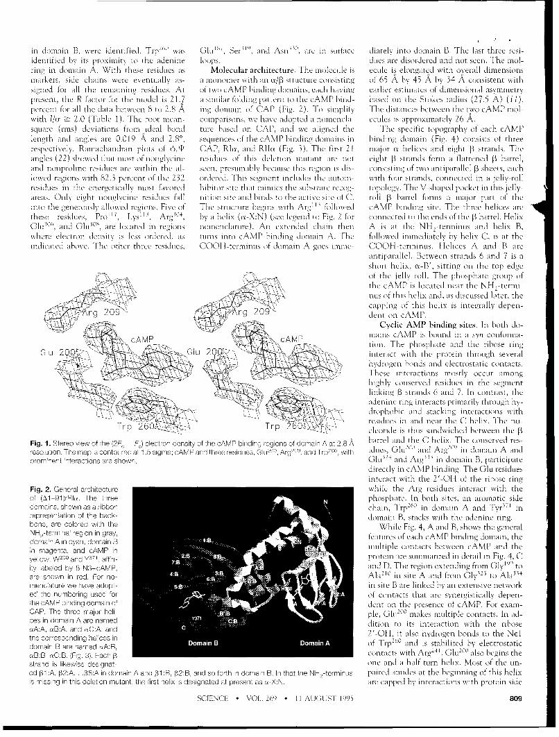
in domain B, were identified. Trp260 was GLuL , Ser• ", and Asn"', are in surface diately into domain B. The last three resi-identified by its proximity to the adenine loops, dues are disordered and not seen. The mol-ring in domain A. With these residues as Molecular architecture. The molecule is ecule is elongated with overall dimensionsmarkers, side chains were eventually as- a monomer with an x/p3 structure consisting of 65 A by 45 A by 34 A consistent withsigned for all the remaining residues. At of two cAMP binding domains, each having earlier estimates of dimensional asyrmmetrypresent, the R factor for the model is 21.7 a similar folding pattern to the cAMP bind- based on the Stokes radius (27.5 A) ( 1).percent for all the data between 8 to 2.8 A ing domain of CAP (Fig. 2). To simplify The distances between the two cAMP mol-wvith I/or Ž 2.0 (Table 1). The root-mean- comparisons, we have adopted a nomencla- ecules is approximately 26 A.square (tns) deviations from ideal bond ture based on CAP, and we aligned the The specific topography of each cAMPlength and angles are 0.019 A and 2.80, sequences of the cAMP binding domains in binding domain (Fig. 4) consists of threerespectively. Ramachandran plots of 4J,1lf CAP, RIa, and RI1lu (Fig. 3). The first 21 major cx helices and eight P3 strands. Theangles (22) showed that most of nonglycine residues of this deletion mutant are not eight 13 strands form a flattened 13 barrel,and nonproline residues are within the al- seen, presumably because this region is dis- consisting of two antiparallel 13 sheets, eachlowed regions with 82.3 percent of the 232 ordered. This segment includes the autoin- with four strands, connected in a jelly-rollresidues in the energetically most favored hibitor site that mimics the substrate recog- topology. The V-shaped pocket in this jelly-areas. Only eight nonglycine residues fall nition site and binds to the active site of C. roll 13 barrel forms a major part of theinto the generously allowed regions. Five of The structure begins with Arg' " followed cAMP binding site. The three helices arethese residues, Pro' 1, Lys'", Arg' 04, by a helix (a-X:N) (see legend to Fig. 2 for connected to the ends of the P3 barrel. HelixGlu1306, and GluILL3S, are located in regions nomenclature). An extended chain then A is at the NH,-terminus and helix B,where electron density is less ordered, as turns into cAMP binding domain A. The followed immediately by helix C, is at theindicated above. The other three residues, COOH-terminUs of domain A goes imme- COOH-terminus. Helices A and B are
antiparallel. Between strands 6 and 7 is ashort helix, a-B', sitting on the top edgeof the jelly roll. The phosphate group ofthe cAMP is located near the NH,-termi-
\\ nus of this helix and, as discussed later, thecapping of this helix is integrally depen-
0929 dent on cAMP.Cyclic AMP binding sites. In both do-
cMAP mains cAMP is bound in a syn conforma-tion. The phosphate and the ribose ring
GIL- 209_ý Gl 200 interact with the protein through several-•* .> -hydrogen bonds and electrostatic contacts.
.5 ' ~ ~ X \These interactions Mostly Occuir amonghighly conserved residues in the segmentlinking 3 strands 6 and 7. In contrast, theadenine ring interacts primarily through hy-drophobic and stacking interactions with
-- 1residues in and near the C helix. The nu-T, _. cleotide is thus sandwiched between the 13
Fig. 1. Stereo view of the (2F. F) electron density of the cAMP binding regions of domain A at 2.8 A barrel and the C helix. The conserved res-
resolution. The map is contoured at 1.5 sigma; cAMP and three residues, Glu2°, Arg2 '°, and Trp 6°, with iducs, GltU200 and Arg'ý9 in domain A andprominent interactions are shown. Chu 324 and Argt
3 in domain B, participatedirectly in cAMP binding. The Glui residuesinteract with the 2'-OH of the ribose ring
Fig. 2. General architecture while the Arg residues interact with theof (A1-91)rRlu. The three phosphate. In both sites, an aromatic sidedomains, shown as a ribbon chain, Trp-"' in domain A and Tyr5 I inrepresentation of the back domain B, stacks with the adenine ring.bone, are colored with the While Fig. 4, A and B, shows the generalNH2-terminal region in gray, featires of each cAMP binding domain, thedomain A in cyan, domain B . oa c bet din cAmand the
in magenta, and cAMP in 2 multiple contacts between cAMP and theyellow. W and y371, affin- protein are summarized in detail in Fig. 4, City laeled by 8 N3-cAMP, and D. The region extending from Gly19' toare shown in red. For no- 4Ali in site A and from Gly"' to Ala' 4
menclature we have adopt- in site B are linked by an extensive networked the numbering used for of contacts that are synergistically depen-the cAMP binding domain of dent on the presence of cAMP. For exam-CAP. The three maior hell pIe, Gliui° makes multiple contacts. In ad-ces in domain A are named dition to its interaction with the riboseaA:A, aB:A, and eC:A, and 2'-OH, it also hydrogen bonds to the NEIthe corresponding helices in 10toean co respo g nBei i, A of TrplD and is stabilized by electrostaticdomain B are named aA:B, ,4 0eB:B, rsC:B, (Fig. 3). Each 1 contacts with Arg' 41 . GluL°° also begins thestrand is likewise designat one-and-a-half-turn helix. Most of the tin-ed 31 :A, 132:A .. 138:A in domain A and 131:B, 132:B, and so forth in domain B. In that the NH2-terminus paired amides at the beginning of this helixis missing in this deletion mutant, the first helix is designated at present as n-X:N. are capped by interactions with protein side
SCIENCE * VOL. 269 ° 11 AUGUST 1995 809

. " I
chains or by cAMP. For example, the aNH the adenine base stacks with the indole ring Gly1 99 in A and Gly323 in B. A larger sideof Glu199, conserved so far in all of these of Trp260 while the six-member ring has van chain here could collide with the adeninecAMP binding sites, hydrogen bonds to the der Waals contacts with Leu 2°1. In cAMP ring. Although the fold is conserved, the2'-OH of cAMP. The aNH of Glu200 hy- binding site B, the adenine ring is sand- length of the C helix and its position rela-drogen bonds to the OE2 of its own side wiched by hydrophobic interactions be- tive to the P3 barrel differ in these threechain. The cNH of Ala2 0 2 hydrogen bonds tween the side chains of Leu316 , Tyr37", structures (Fig. 5). Most significant is theto the equatorial oxygen of the phosphate Ser373, and Ile325 on one side and Va13 0
1, displacement of the C helix in domain Ain cAMP. The oNH of Leu 20 3 hydrogen ValP 13, and Ala335 on the other. The phe- away from the P3 barrel. As discussed below,bonds to the a• carbonyl of Leu2°1. Thus, nol ring of Tyr37' also stacks with the ade- two residues, Trp 60 and Arg241 , specificallythe architecture of this site, and specifically nine ring. The stacking of the aromatic keep this helix extended away from thethe secondary structure that includes this rings of Trp260 and Tyr371 with the adenine barrel.short helix, is integrally dependent on the ring of cAMP also orients the dipole mo- The different conformation of cAMP,presence of cAMP itself. The stacking of ments of the adenine, Tyr, and Trp rings in syn in R and anti in CAP, is due mostthe adenine ring with Trp2 60 also depends an antiparallel alignment as predicted (23). likely to differences in the environmenton this network of interactions involving Whereas the dipole moments for the ade- surrounding the adenine rings. The stack-Glu 200 . The Arg20 9 plays a major structural nine and Trp rings are fixed, the dipole ing of the adenine ring with aromatic siderole in addition to binding cAMP. It con- moment of Tyr depends on the rotation of chains comparable to Trp260 and Tyr371,tributes to cAMP binding by interacting the phenolic hydroxyl group. The hydroxyl for example, is missing in CAP. Anotherwith the equatorial exocyclic oxygen of the group of Tyr371 is fixed by a hydrogen bond difference is that the short helix foundcAMP phosphate. This same N'q nitrogen to Glu324 so that the dipole of Tyr371 can- between P3 strands 6 and 7 in the R sub-also is only 3.5 A from the backbone car- not rotate. units is missing in CAP. CAP has onebonyl of Gly1 99. Thus Arg20 9 bridges the In prokaryotes the primary receptor for extra residue in the region that links P36conserved segment that links P3 strands 6 cAMP is CAP where cAMP binding direct- and P37, and this may prevent the helixand 7. However, by contacting the back- ly mediates gene expression (24). As pre- from forming. Cyclic AMP binding isbone carbonyl of Asn 171 in p3 strand 3 and dicted by sequence alignments (Fig. 3) (10), three orders of magnitude tighter in Rthe side chain carboxylate of Asp 170 , it the general features of the cAMP domains than CAP. The presence of the phos-bridges the cAMP binding domain and in CAP and R are conserved although we phate:helix interactions and the stackingtransmits a signal that extends beyond the were unable to solve the structure by mo- interactions between the adenine ringsimmediate cAMP binding site. The inter- lecular replacement with the coordinates of and the aromatic side chains probably ac-action with Asp 170 also contributes to the CAP. Each cAMP binding domain has count for this difference, at least in part.neutralization of the charge on Arg20 9. three main helices and an eight-stranded 13 In eukaryotes, homologous cAMP bind-
A similar network of contacts is found in barrel. There are three invariant Gly resi- ing motifs are conserved in the R subunitssite B (Fig. 4D). Glu32 4 hydrogen bonds dues in each domain. One of them (Gly166 of cAPK, in the guanosine 3',5'-monophos-with the 2'-OH of the ribose ring, to the in domain A and Gly2
15 in domain B) lies phate (cGMP)-dependent protein kinase
phenolic OH of Tyr371, and interacts with between 132 and 133 and is situated at the (cGPK), and in the cyclic nucleotide-gatedits own backbone amide. The aromatic side third corner of a type II 13 turn. Type II 13 channels (25). The difference in relativechain at the end of the C helix is thus turns prefer a Gly residue at this position. specificity for cAMP compared to cGMP ispositioned both by its stacking with the The second conserved Gly is located in a approximately a factor of 100 for cAPK andadenine ring of cAMP and by hydrogen loop connecting 133 and 134. The third in- cGPK (26). One residue that partially ac-bonding to a conserved residue in the 13 variant Gly begins the active site pocket, counts for this specificity is the equivalentbarrel. Likewise, the backbone amide ofGly323 also interacts with the 2'-OH of theribose, and Glu32 4 is followed by a short A 101 J32 P3r -- - -I I 1_ 4 ! _ --• -- Ihelix. Most of the unpaired uNH groups at CAP 8/DPTLEWFLSHCHIHKYPSKSTLIHQ•EKAETLYYIVKESVAVLIKDEE ------- GKEMILSYLNQG/67
the beginning of this helix also hydrogen a:A :•A 18 0 188
bond in a manner similar to that in domain RIa 141/DNERSDIFDAMFPVSFIAGETVI2QQDEGDNFYVIDIEMDVYVN -------- NEWATSVGEG/195
A. Unlike Glu200 , Glu 32 4 does not interact RiIa 141/PEQLSQVLDAMFERTVKVDEHVIDQ•DDGDNFYVIER TYDILVTK -------- DNQTRSVGQYDNH/199
with an Arg comparable to Arg2 41 . As in F-1:. PI:B 1 288 B 190 5 Bdomain A, conserved Arg333 forms a single RFC 259/KWERLTVADALEPVQFEDGQKIVVQEPGDEFFTII. 8SAAvLQRRSEN - EEFVEVGRLGPS/319
ion pair with the equatorial phosphate ox- RZCC 263/VSERMKIVDVIGEKVYKDGERIITQEKADSFYIIES•EVSILIKSKTKVNKDGENQEVEIARCHKG/329
ygen of cAMP but also plays a structural
role by interacting with the backbone car- P 7 B __
bonyls of Gly3 2 3 and Clu2 8 9, which is in 13 CAP 68/DFI•LGLFEEGQEMSqVRAKTACEVAEISYKKFRQLIQVNPDILMRLSAQMARRLQVTSEKVGN/133
strand 3, similar to domain A. In domain B 6A aB :BA 28 7 A PB:A . B.A333 2o 7 ooo V I I I I F.¢.there is no carboxylate near Arg333 to help RIa 196/GSTP VKAKTNVKLWGIDRDSYRRILMGSTLRKRKMYEEFLSKVSILESLD--/25S
neutralize its charge; but there are still po- R240 200/GSFELALMYN-T IVATSEGSLWGLDRVTFRRIIVKNNAKKRKMFESFIESVPLLKSLE--/262
tential interactions with residues that ex- 6:B bBB 7 -- I8 r8 :B B-B C B
tend from Gly28 7 to Glu
289. Rla 323/DYF AMN-RP vvARGPLKCVKLDRPRFERVLGPCSDILKRNIQQYNSFVSLSv/379
R 3b 330/ QYFLALVTN-KP- AYAVGDVKCLVMDVQAFERLLGPCMDIMKRNISHYEEQLVKMFGSSMD/394In addition to the electrostatic interac- 350 370
tions, the tight binding of cAMP with Kd'sof 10 to 100 nM involves a number f Fig. 3. Sequence alignment of the cAMP binding domains of RI, R1, and CAP. RI corresponds to bovine
0 RIv and RII corresponds to bovine Rila. Residues that are conserved in all three proteins are indicated bystrong hydrophobic interactions. In site A, black boxes. Sites of affinity labeling with 8-N3-cAMP in the native protein are indicated by filled stars.one side of the adenine base faces a hydro- Sites affinity labeled in the proteolyzed protein are indicated by open stars. Residues identified asphobic pocket formed by the side chains of important for cAMP-mediated activation by genetic screening are indicated by black dots. Single-letterAla'8 9 , Val184 , Val 18 2, Ala21 0 , and Ala2 1 1. abbreviations for the amino acid residues are A, Ala; C, Cys; D, Asp; E, Glu; F, Phe; G, Gly; H, His; I, lie;On the other side, the five-member ring of K, Lys; L, Leu; M, Met; N, Asn; P, Pro; Q, Gin; R, Arg; S, Ser; T, Thr; V, Val; W, Trp; and Y, Tyr.
810 SCIENCE * VOL. 269 * 11 AUGUST 1995

of Ala210 (domain A) and Ala. 34 (domain the OG1 of Thr and the N2 of cGMP. These two aromatic side chains in RIcx areB) in RIot. This is always a Thr in cGPK Correlation of the structure with chem- optimally aligned, which presumably ac-and an Ala in R. Replacement of these Ala ical data. Affinity labeling with 8-N3- counts for the exceptionally high efficiencyresidues with Thr in RIa did improve spec- cAMP was used to identify residues near the in labeling (Fig. 4, E and F).ificity for cGMP but did not weaken the cAMP binding sites. Two sites were labeled Affinity labeling of proteolytic frag-affinity for cAMP (27). On the basis of the in RIa: Trp26
1 in Site A and Tyr"71in Site B ments and deletion mutants provide an in-current structure, replacing Ala210 with Thr (28). In the RII subunit, Trp2"' is replaced dication of conformational flexibility. Forand adding an NH 2 at the position 6 of with a Set and only one site was labeled, example, when domain B (residues 260 tocAMP gives a distance of 2.4 A between Tyr"'1 , the equivalent of Tyr 71 in RI (29). 379) in Rlcu is deleted, all the residues
normally labeled are absent. In this protein,Tyr 244 was labeled by 8-N1-cAMP (30),indicating that, in the absence of domain B,the C helix in domain A moved closer tothe 13 barrel, thus resembling more closelythe orientation in both domain B and CAP
05 /(Fig. 5). When the NH2 -terminus of the,pI w7RII subunit was removed by proteolysis
(residues 1 to 94), the pattern of photoaf-finity labeling also was altered. In thiscase, Tyr1" il domain A was labeled, inaddition to Tyrl , indicating that theNH,-terminus also imposed some structur-
/ B al constraints on the COOH-terminal partof the molecule (31).
The chemical features of the two cAMPbinding sites were also mapped with cAMPanalogs (32). As predicted, no strong hy-drogen bonding interactions exist betweenthe adenine ring and the protein; most ofthe interactions are hydrophobic and stack-ing. The closest potential hydrogen bond isbetween the N6 in cAMP bound to domain
20 B and the backbone carbonyl of Asn 372.Whereas three H bonds between the 2'-,3'-, and 5'-ribose oxygens were predicted,only the 2'-OH hydrogen bond was ob-served in the structure. The importance ofthe exocyclic phosphate oxygens was alsocorrectly predicted. Although the axial ox-ygen interacts with the amide of Ala212 andAla334, respectively, in site A and B, theequatorial oxygen binds to the side chain ofArg20 9 and to the amide of Ala20 2 in site A
E and to Arg 53 and Ala 23 in site B. There is
no evidence, however, for a pentacovalentintermediate as was suggested. Analogs alsopredicted correctly that cAMP binds to Rin a syn conformation, in contrast to theanti conformation seen in CAP.
Analogs also can discriminate between`2 60 sites A and B (33). Site A can accept
analogs having substituents at the C6 posi-tion whereas substituents at the C8 positionare not well tolerated. The N6 of cAMPbound to site A is exposed, but the acces-sibility of the C8 position is blocked by thesix-member ring of Trp2 60 , as well as by
Fig. 4. (A and B): Overall features of the cAMP binding domains. Ribbon diagrams of domain of cAMP Vall"2 and Val'9 2. In contrast, site B prefersbinding domains A and B are shown in (A) and (B), respectively. The general orientation of cAMP (yellow), analogs substituted at the C8 position, andrelative to the 3 barrel and the C helix is shown. Conserved residues (R219 and E200 in domain A and R33 in this site, the accessibility of the N6 po-and E32
1 in domain B) are indicated. Stereo views of the hydrogen bonding interactions between cAMP in is site, by acces of co -
and the protein are shown in (C) (site A) and (D) (site b). Additional residues, R241, E26 7 , and D17 0 , that sition is blocked by van der Waals contactsinteract directly with cAMP domain A are also shown. Possible H bonds are indicated by dashed lines with four residues, Tyr" 7 , Asn t2 , Ser373,(distances < 3.3 A). Space-filling models of each cAMP binding site are shown in (E) (site A) and (F) (site and Valt"', the N7-C8 edge of cAMP isB). Atoms of cAMP and residues involved in cAMP binding are colored differently: carbon in yellow, accessible (Fig. 4F). Another feature thatnitrogen in blue, phosphorus in dark blue, oxygen in red, the others in pink. C8 is the site of attachment distinguishes the two sites is the relativeof the photoreactive azido moiety, off-rates for cAMP. In site B with its slower
SCIENCE - VOL. 269 • 11 AUGUST 1995 811

off-rate, the cyclic nucleotide is buried more the crystal structure the charge of Arg20 9 is (38), pairs with Glu200 , as predicted (39). Indeeply and is packed tightly against Tyr 371. neutralized in part by Asp170 . With (Rp)- domain B, Trp2 60 stacks with cAMP, andAt the base of the cAMP binding pocket, cAMPS bound, the charge of R209 would Ile293 is at the hydrophobic interface be-for example, Arg333 is completely buried by be neutralized predominantly by the nega- tween the two domains. Other mutations,the nucleotide (Fig. 4F). In contrast, cAMP tive charge on the sulfur leaving no anchor genetically engineered into the RIca sub-binds to domain A with a relatively fast off for Asp17 0 . The hydrogen bonding to unit, have provided insights into the chem-rate. In the crystal structure, this cAMP Ala20 2 would also be disturbed. The larger ical structure of the cAMP binding sites andbinding site is more open. size of the sulfur in both of the chiral the cooperativity between the two sites.
Of the hundreds of cAMP analogs tested, analogs probably causes steric hindrance Extensive in vitro analysis of mutants suchonly one group served as antagonists, the and accounts for their low affinity. At this as Arg209Lys, Gly1 9 9Glu, and Ala335Asp,chiral phosphorothioate analog (Rp)cAMPS, point, it is not clear why (Sp)cAMPS, and has confirmed the importance of these res-and its analogs (34). When the sulfur is in not (Rp)cAMPS, is an antagonist for CAP. idues for cAMP binding and signaling (40).the equatorial position, as in (Rp)cAMPS, Genetic approaches also identified func- Molecular basis for cooperativity. Adissociation of the holoenzyme was tionally important residues. The most ex- primary region of contact between domainsblocked, whereas (Sp)cAMPS was an ago- tensive genetic mapping has been done in A and B is an extended hydrophobic sur-nist. Sulfur has a larger van der Waals RIa of S49 mouse lymphoma cells (37). For face. Specifically, the hydrophobic surfaceradius (1.70 A) than oxygen (1.35 A) (35), these cells cAMP is toxic. By isolating mu- in domain B formed by Va1265, Leu 269 ,and the P-S bond length of the phosphoro- tants that were resistant to cAMP, a family Va1
3 4 6 , Leu2 9
4, Ile 292, Tyr321, Leu3 64, Leu 357,thioate group is accordingly longer (1.95 A of RIcE mutants defective in cAMP binding Va13 56, Cys 360 , and Ile 363 is covered bycompared to 1.5 A). Because most of the were identified. Most of these dominant Tyr2 44, Phe 247 , Leu 248 , Va1251, Ile2 53,charge resides on the sulfur, the resonance negative mutations (Fig. 3) are located in Leu2 54, and Leu 251 from domain A (Fig. 6).of the electrons is also reduced in these the highly conserved P36-37 loops. Addi- In addition, the ends of euA:B are anchoredchiral analogs (36). Sulfur also does not tional residues that lie outside this loop are by further electrostatic and hydrophobic in-form hydrogen bonds as well as oxygen. In Gly1'6 9 and Arg241 in domain A and Trp 26 0 teractions with domain A. Trp260 , which is(Rp)cAMPS, the sulfur replaces the equa- and Ile 293 in domain B. Gly169 precedes a critical residue that links the two do-torial oxygen that interacts with Arg20 9 and Asp' 70
, which ion pairs with Arg20 9, and mains, was first indicated to be important
the amide backbone of Ala202 in Site A. In Arg241, which is critical for cooperativity by affinity labeling (28). It lies at the be-ginning of cuA:B and is thus part of thesecondary structure of domain B, yet its sidechain interacts directly with cAMP bindingsite A by stacking with the adenine ringand by hydrogen bonding to the conservedGlu 2
11 (Fig. 6). Thus it is an integral part ofthe network of contacts that define cAMP
Fig. 5. Comparisons of each cAMP binding do- • Dmimain from RI and CAP. cu-C atoms of the l3 barrelof each cAMP binding domain were superim-posed using Program C. (A) Domain A (darker line)Caand CAP. R24 1 in domain A and R123 in CAP
site through salt bridges. (B) Domain B (darker /,•xline) and CAP. (C) Domain A and domain B (darker , 1
line). Tyr 244 is affinity-labeled when the domain B ••=is deleted. ([
Fig. 5.Fig. 6. Interactions between domains A and B.
The a• carbons of residues that participate in hy-drophobic interactions between the two domainsfare indicated as balls. Additional residues that
posdcould play a role in mediating allosteric interac-
tions are indicated (E2°°, i
2n
9, K
24°, A
24 1, W2
E2 67
, E32
, R33
3, y3 71
), as are the bound cAMPlin)molecules. The arrow indicates the site where
cAMP binds first in the holeoezyme.
812 SCIENCE • VOL. 269 ° ot AUGUST 1995

. 0
binding site A. Another interdomain hydro- R1I is more accessible in the holoenzymne 16. W. Furey and S. Swaminathan, Am. Crystallogr. As-soc. Mtg. Abstr. Ser. 2. 18, 73 (1990).gen bond interaction involves the side chain than in the dissociated R,(cAMP)4 (43) 17. B.-C. Wang, Methods EnzymoL 115, 90 (1985).
of Lys 241 in uC:A, which hydrogen bonds to (Fig. 6). As indicated earlier, this hydro- 18. C. Cambillau and E. Horjales, J. MoL Graphics 5,
the backbone carbonyls of Asp2t6 7 and phobic region is linked in multiple ways to 174(1987).Leu 26•' at the COOH-terminus of cA:B. Fi- cAMP binding site A with Asp26r7 and 19. T.A. Jones, J. Y. Zou, S. W. Cowan, M. Kjeldgaard,Acta Crystallogr. 47, 110 (1901 ).
nally, there is an electrostatic interaction Trp261 being of particular importance. Re- 20. A.aT. Brnger, XPLO Version 3.1 Manualt(Yale Uni-between Arg241 and Asp267 . Thus the A placement of Arg24
1, which binds directly versity, New Haven, CT, 1993).helix of domain B is anchored firmly to key to both Asp 2"7 in domain B and Glu 2°° in 21. R. Read, Acta Crystallogr. A42, 140 (1986).regions of site A by hydrophobic, electro- domain A, with Set or Lys demonstrated 22. R. A. Laskowski, M. W. Macarthur, D. S. Moss, J. M.
Thornton, J. Appi. Crystallogr. 26, 283 (1993).static, and hydrogen bonding interactions, that this residue is a key feature for the 23. K. Baldridge and W. R. G. Dostmann, unpublished
One question relating to the R subunits allosteric coupling between sites A and B data.of cAPK is how the cooperative binding of (38). Thus communication between the 24. A. Kolb, S. Busby. H. BuL, S. Garges, S. Adhya,
Annu. Rev. Biochem. 62. 749 (1993).cAMP leads to the dissociation, and thus two domains is likely carried out through 2 Shabb and J. D. Corbin, J. Biol. Chem. 267,activation, of the holoenzyme. Although the C helix of domain B to the interdomain 5723 (1992).we cannot fully understand this process Ln- hydrophobic interaction region and trans- 26. T. Lincoln and J. D. Corbin, Adv. Cyclic Nucleic Res.
til a structure of the holoenzyme is solved, mitted to site A through residues Asp• 27 .15. 139 (1983).we can begin to understand how communi- Arg241, Clu 200 , and Trp 2 60 or vice versa. B2ol. Chem. 266, 24320 (199 1).
cation between sites A and B and the C When holoenzyme forms, the initial 28. J. Bubis and S. S. Taylor, Biochemistry. 24, 2163
subunit might be mediated from the struc- docking of C to R involves interactions of (1985); bid. 26, 3478 (1987).utue of the R subunit. A deletion mutant the autoinhibitor site in R, with the active 29. A. R. Kerlavage and S. S. Taylor, J. Biol. Chem. 255,
8483 (1980).lacking domain B (A260-379) and even a site cleft in C. This autoinhibitor site in the 30. G. E. Ringheim, L. D. Saraswat, LD. Bubis, S. S.double mutant, where both the NH 2-termi- free R subunit is extremely labile to prote- Taylor, ibid. 263, 18247 (1988).nus (A1-91) and domain B are deleted, still olysis (5, 6) and, in our structure, the first 31. J. Bubis and S. S. Taylor, Biochemistry. 26, 5997(1987).bind C tightly ( 1, 41), demonstrating that 21 residues are disordered. This initial in- 32. R. J. W. DeWit etal., Eur, J. Biochem. 152, 255
domain B is not required for high affinity teraction is essential; however, it is not (1984); T. S. Yagura and J. P. Miller, Biochemistrybinding to C. On the basis of kinetic argu- sufficient to convey high affinity binding. 20, 879 (1981); B. Jastorff, J. Hoppe, M. Morr, Eur.
ments, site A is masked in the holoenzyme To achieve high affinity binding requires J3. Biochem. 101, 555 (1979).
(8). Thus, in the sequential cooperative interactions involving the surface of C that D. Cgrein etal., ibd. 15.0, 29 1985); S. B. Rannels
pathway for activation of cAPK, cAMP lies COOH-terminal to the consensus site and J. D. Corbin, J. Biol. Chem. 255, 7085 (1980).
binds first to site B, which "opens tip" site peptide. This is the surface that surrounds 34. P. J. M. Van Hastart etal., ibid. 259,1002011984);VW. Dostmann et al., ibid. 265, 10484 (1990).
A allowing cAMP to bind and C to be and includes the essential phosphorylation 35. T. E. Creighton, Proteins (Freeman, New York,released. site, Thr'1 7 (44). Point mutation of the 1984), p. 139.
There are only two stable conformations P-Thr itself as well as the basic residues that 36. P. A. Frey and R. D. Sammons, Science 228, 541
of the R subunit; the cAMP-saturated, dis- bind the phosphate all interfere with R (1985).37. G. S. McKnight et al., Rec. Prog. Hormone Res. 44,
sociated R andi the holoencyme. The inter- subunit binding. The second step in fore- 307 (1988). K. A. Gorman and R. A. Steinberg, So-actions between domains A and B as well as ing holoenzyme would thus be the docking mat. Cell Mol. Genet. 20, 301 (1994).the immediate environment of each cAMP of the C subunit, with its active site cleft 38. M. M. Symcox, R. D. Cauthron, D. Ogreid, R. A.
Steinberg, J. Biol. Chem. 269. 23025 (1994).binding site must be different in these two occupied, to a region on cAMP binding 39. R. A. Steinberg, K. B. Gorman, D. Ogreid, S. B.structures. Since no structure of any cAMP- domain A. With this structure we cart now Doskeland, I. T. Weber, ibid. 266, 3547 (1991); T. A.free domain is yet available, we can only begin to model these interaction sites. Woodford, L. A. Correll, G. S. McKnight, ibid. 262,
13321 (1987); E. Duprez et al., b•id. 268, 8832speculate on the conformational changes (1993).that take place. The C helices, however, REFERENCES AND NOTES 40. J. J. Neitzel, W. R. G. Dostmann, S. S. Taylor, Bio-probably play an important role in both chemistry 30, 733 (1991).
1. E. G. Krebs, Bioscience Rep. 13, 127 (1993). 41. G. E. Ringheim and S. S. Tayior, J. Biol. Chem. 265,domains. The aromatic side chains of 2. S. K. Hanks, Curr. Biol. 1,369 (1991). 4800 (1990).
Trp2
6° and Tyr37t , for example, are packed 3. D. A. Walsh. J. P. Perkins, E. G. Krebs, J. Biol. Chem. 42. J. Bubis, L. D. Saraswat, S. S. Taylor, Biochemistry
tightly against the cAMP ligands and have 243, 3763 (1968). 27, 1570 (1988).no contacts on the sides away from cAMP. 4. E. W. Sutherland and T. W. Rail, ibid. 232, 1077 43. N. Nelson and S. S. Taylor, J. Biol. Chem. 258,
(1958). 10981 (1983).They must therefore either collapse into the 5S. S. Taylor, J. A. Buechler, W. Yonemoto, Annu. 44. C. S. Gibbs, D. R. Knighton, J. M. Sowadski, S. S.cAMP binding pocket in the absence of Rev. Biochem. 59, 971 (1990). Taylor. M. J. Zoller, ibid. 267, 4806 (19921; 5. A.
cAMP or otherwise rearrange. The C helix 6. J. D. Scott, Pharmacol. Ther. 55, 123 (1991). Orellana, P. S. Amieux, X. Zhao, G. S. McKnight,in domain A also is slightly bent suggesting 7. E. A. Nigg, Adv. Cancer Res. 55, 271 (1990). D. A. ibid. 268, 6843 (1993).
Fantozzi etal., J. Biol. Chem. 269, 2676 (1994). 45. A. G. W. Leslie in joint CCCP4 and ESF-EACMBa strained conformation. When cAMP 8. D. Ogreid and S. .Doskeland, FEBS. Lett. 129,287 Newsletter on Protein Crystallography No. 26
binds to site B in the holoenzyme and stacks (1981). (Daresbury Laboratory, Warrington, UK, 1992).
with Tyr-"', the orientation of the C helix 9. D. Rgreid, R. Ekanger, R. H. Suva, J. P. Miller, S. B. 46. D. E. Tronrud, L. F. Ten Eyck, B. W. Matthews, Actarelative to the barrel has to change. The .Doskeland, Fur. J. Biochem. 181, 19 (1989). Crystallogr. A43, 489 (1987).
r10. . T. Weber, T. A. Steitz, J. Bubis, S. S. Taylor, BRo- 47. Supported in part by NIH grants GM34921 (S.S.T.)fact that cooperativity is lost when Tyr371 is chemistry. 26, 343 (1987). and RR01644 (N-h.X.), the Lucille P. Markey Chari-
replaced with Phe (42) confirms the impor- 11. L. Saraswat, G. E. Ringheim. J. Bubis, S. S. Taylor, J. table Trust (S.S.T., L.T.E., N-h.X.), NSF grant BIR-Biol. Chem. 263,18241 (1988). F. W. Herberg, W. R. 9223760 (L.T.E.), and Public Health Service Training
tance of this initial binding of cAMP to Dostmann, M. Zorn, S. J. Davis, S. S. Taylor, Bio- Grant GM07313 (Y.S.). We thank the San Diegodomain B and suggests that the strong di- chemistry 33, 7485 (1994). SupercomputerCentertorseoftheAdvancedsci-
pole-dipole interaction between cAMP and 11 aD. R. Knighton et at, Science 253, 407 (1991); ibid.. entific Visualization Laboratory and the Cray C90, thep. 414. Stanford Synchrotron Radiation Laboratory for pro-
Tyr371 is important. The cAMP binding to 12. T. A. Kunkel, K. Benebek, J. McClary, Methods En- viding us beam time for data coalection, and Xiaoping
site B will also influence the position of the zymol. 204, 125 (1991). Dai for his help in data collection and data process-
NHI-terminus of oC:B. Two residues here, 13. Y. Su, S. S. Taylor, W. R. Dostman, N.-h. Xuong, K. ing. Coordinates have been deposited on the Brook-CyS360 and lie363, are directly involved in I. Varughese, J. MoL Biol. 230. 1091 (1993). haven Protein Data base, with tracking number
14. B. W. Matthews, ibid. 33, 491 (1968). RGS.the hydrophobic interactions between the 15. N.-h. Xuong, C. Nielson, R. Hamlin, D. Anderson, J.two domains. The equivalent of Cys 311 in Appl. Crystallogr. 18, 342 (1985). 15 February 1995; accepted 28 June 1995
SCIENCE • VOL. 269 - 11 AUGUST 1995 813

JOBNAME: BC PAGE: 1 SESS: 4 OUTPUT: Fri Sep 8 13:24:00 1995/kJsb27aisf,6Ibc/bc/bc4295/bcO733-95a
TO AVOID DELAY BC, C5-0808, 352754 POrfRNAd & SeurRETURN PROOFSWITHIN I DAYBY EXPRESS MAlL j AUTHOR: PLEASE SEE QUERIES
THROUGHOUT YOUR MANUSCRIPT
TIlE JOURNAL OF BIOIWOICAL CIIEMISMirYCom m unicationVol. 270. NO. 42, issue o ? pp. 1-4,m 1995 by The American Society for Biochemistry and Molecular Biology. Inc.Printed in U.S.A.
Tyrosines outside the Kinase proto-oncogene encodes a novel receptor tyrosine kinase with acadherin-like motif in its extracellular domain (2).
SThe c-ret proto-oncogene is responsible for four human dis-Required for th&: i t? ease syndromes: Ilirschsprung's disease, a developmental dis-g n R Tporder, and the dominantly inherited cancer syndromes FMTC,'Activity of RET/ptc21 MEN2A, and MEN2B. Ilirschsprung's disease occurs at a rate
of approximately 1 in 6000 live births and is characterized by a(Received for publication, July 26, 1995) lack of parasympathetic innervation of the lower intestine.
Kyle Durlcktk ,]irginla J. Yaot, Occurrence of this disease corresponds to c-ret gene deletionsMaria Grazia'#lorrcllo¶l, Italia Bongarzonell and nonsense point mutations leading to truncation of theMarco A. Pierottil, and Susan S. Taylor expressed protein (3, 4). Also, three mutations resulting inFrom the tDepartment of Chemistry and single amino acid substitutions in the kinase region of the c-retBiochemistry, University of California, gene have been reported in Hirschsprung's patients (3, 4). InSan Diego, La Jolla, California 92093-0654 addition to medullary thyroid carcenoma, the unique patho-and ¶Divisione di Oncologia Sperimentale A logic feature of FMTC, MEN2A, and MEN2B patients display
del stituto Nazional~entr MilaItay JIeu aia Tumnor Milan,Ital tyadditional hyperplasias. These syndromeshave been linked to
Defects in the c-ret proto-oncogene, a member of point mutations in the c-ret gene (5, 6, 7). Recent evidencethe protein tyrosine kinase receptor family, have re- suggests that, in the case of the MEN2A, these mutationscently been linked to two types of genetic syndromes, result in the expression of constitutively activated forms ofHirschsprung's disease and the multiple endocrine neo- c-ret (8).plasia family of inherited cancers. RET/ptc2 is the prod- It has also been demonstrated that constitutively activeuct of a papillary thyroid carcinoma translocation event forms of the c-ret proto-oncogene are present in nearly half ofbetween the genes coding for c-ret and the type la reg- papillary type thyroid carcinomas (9). These oncogenic forms ofulatory cubunit of protein kinase A (RIa) (Lanzi, C., ret are the result of somatic chromosome translocations or1lorrello, M., Ilongarzone, I., Migilazza, A., Fusco, A., inversions, which fused the c-ret tyrosine kinase domainCrieco, M., Santoro, M., Gambetta, IL, Zunino, F., Della (RETtk) with different genes. One of the resultant transformingPorta, G., and Pierotti, M. (1992) Oncogene 7,2189-2194). sequences, REr/ptc2 (papillary thyroid carcinoma), was theThe resulting 596-residue protein contains the first two- product of a crossover between the genes coding for c-ret andthirds of Ria and the entire tyrosine kinase domain of the type la regulatory subunit ofcyclic-AMP-dependent proteinc-ret (RETIk). An in vivo assay of growth stimulatory kinase (RIa) (10). The chimeric gene encodes a protein of 596effects was developed, which consisted of microinject- residues, which contains the first two-thirds of RIct and theing a RET/ptc2 expression plasmid Into the nuclei of entire tyrosine kinase domain of c-ret (11). RET/ptc2 is trans-1OT1/2 mouse fibroblasts and observing the Incorpora- forming, presumably due to constitutive tyrosine kinase activ-tion of 5-bromodeoxyuridine. This assay was used todetermine that only the dimerization domain of Ria ity, but the structural basis for RETtk activation via fusion tofused to RETck Is required for RET/ptc2's mitogenic ac- RIa remains unclear.tivity. In addition, all of the reported Hiirschsprung's Here we report the development and use of a microinjectiondisease point mutations in the RETlh (S289P, R421Q, and assay to study the mitogenic activity RET/ptc2. Deletion mu-R496G) Inactivate RET/ptc2 in our assay, confirming tants were tested in the assay to determine which portions ofthat these are loss of function mutations. Two tyrosines RIa were required to elicit the mitogenic activity of RET/ptc2.outside the conserved kinase core were also Identified Only the amino-terminal region of tIoa, which encodes a dimer-that are essential for full mitogenie activity of RET/ptc2. ization domain (12), fused to the REt~k was necessary. ThisThese two tyrosines, Tyr-350 and Tyr-586, are potential portion of RIa was also capable of activating the tyrosine ki-sites for Sre homology 2 and phosphotyrosine binding nase domain of the epidermal gVfwth factor receptor in thisdomain interactions. assay system. In addition, all of tile reported Hirschsprung's
disease point mutations in the RETtk inactivated RET/ptc2 inour assay. To begin the search for interaction sites with other
The ret oncogene was discovered in transfection experiments signaling proteins, the mitogenic activity of RET/ptc2 mutantswhere DNA from human lymphomas was introduced into lacking single tyrosines was tested. Two tyrosines were foundN1113T3 cells (1). This oncogene was not expressed in the to be essential for the mitogenic activity of RET/ptc2.original lymphoma, but instead had arisen from a rearrange.ment during the rransfection, hence the name ret. The c-rct EXPERIMENTAL PROCEDURES
Construction of Mammalian Expression Jloamids--The cDNA cod-ing for wild-type RET/ptc2 was excised from a pMAM-neo expression
* This research was supported in part by United States Army Grant vector previously described (11) using the restriction enzyme Xbal. ThisAIDS1762 (to S. S. T.) and by AIRC and CNR Special Project ACRO (toM. A. P.). The costs of publication of this article were defrayed in partby the payment of page charges. This article must therefore be hereby iThe abbreviations used are: FMTC, familial medullary thyroid car-marked "advertisement" in accordance with 18 U.S.C. Section 1734 cinoma; MEN, multiple endocrine neoplasia; RIa, type la regulatorysolely to indicate this fact. subunit of cAMP-dependent protein kinase (protein kinase A); RETrky
§ Supported by the Markey Charitable Trust as a Fellow; currently tyrosine kinase domain of the c-Ret receptor; S112, Src homology 2supported by National Institutes of Health Training Grant NCI T32 domain; DMEM, Dulbecco's modified Eagle's medium; FBS, fetal bovineCA09523. serum; FITC, fluorescein isothiocyanate; BrdUrd, bromodeoxyuridine;
I To whom correspondence should be addressed. EGF, epidermal growth factor.
Orig. Op. OPERATOR jPROOF: PE's: A's: COMME NTS, . . "- " "
1st K35, 2nd judy I 352754

JOBNAME: BC PAGE: 2 SESS: 4 OUTPUT: Fri Sep 8 13:24:00 1995/csbZ]disk6/ 'cbc/bc4295/bc0733 - 95a
4 A|
2 RETlptc2 Mitogenic Activity
PrriC mI-I10 TR &atl,-lhk 40
35
VCATS20
PM."1 -|o "rR sell Wll-•d 5
pRETI/P2 5
0
2
Fla. 2. Mitogenic activity of RET/ptc2 point mutants. The frac-tion of injected cells that stained positive for BrdUrd incorporation is
FiG. 1. Immunostaining for RET/ptc2 expression and 5-bro- shown for various plasmids expressing either wild-type RET/ptc2 ormodcoxyurldine (BrdUrd) Incorporation in 10TI/2 cells Injected RET/ptc2 point mutants. In each case the plasmids were injected at awith pRET/ptc2 (a and b, e and fl or pCAT (c and d, g and h) concentration of 100 gg/ml. The column labelea background representsexpression vectors, a, lgG injection marker stained with FITC anti- uninjected cells under the assay conditions. The error bars show theGuinea pig IgG. b, same field of cells as a, but showing Texas Red 95% confidence interval calculated using the standard error of propor-anti-RET,k staining. c, lgG injection marker stained with FITO anti- tion. The number in parentheses is the total number of injected cells.guinea pig IgG. d, Same field of cells as c, but showing Texas Redanti-RET,, staining. e, IgG injection marker stained with FITC anti-rabbit IgG. f, Same field of cells as e, but showing Texas Red anti- min, and then washed with phosphate-buffered saline. Incorporation ofBrdUrd staining. g, IgG injection marker stained with FITC anti-rabbit 5-bromodeoxyuridine was visualized by successively incubating theIgG. h, Same field of cells as g, but showing Texas Red anti-BrdUrd fixed cells with mouse anti-bromodeoxyuridine (undiluted, Ainersham),staining. biotinylated donkey anti-mouse IgO (dilution 1:500, Jackson), Texas
Red streptavidin (dilution 1:100, Amersham), and FITC anti-rabbit IgG(dilution 1:100, Jackson).
fragment was then subcloned into the Xbal restriction site of the pRo/CMV mammalian expression vector (Invitrogen). Restriction digests RESULTS AND DISCUSSIONwere performed to screen for orientation, and then the entire cDNA Development of Mitogenic Assay-To evaluate which struc-sequence was verified using dideoxy sequencing (13) with Sequenase tural features of RET/ptc2 are essential for its mitogenic activ-version 2.0 (U. S. Biochemical Corp.).
Site-directed mutagenesis was performed with the Kunkle method ity, an expression plasmid microinjection assay was developed.(14) using the Mutagene kit (Bio-Rad). Constructs expressing deletion This assay allows for both rapid screening of protein expressionmutants of RET/ptc2 were made by introducing NheI restriction sites and evaluation of mitogenic activity. The expression vector,flanking the segment of DNA to be deleted, digesting with NheI, and pRET/ptc2, was co-injected into nuclei of serum-starved 10T1/2then ligating the new ends back together. All mutant constructs were fibroblasts with rabbit or guinea pig IgG, which served as ansequenced to verify mutagenesis. Supercoiled plasmid DNA expressing injection marker. After incubation for 24 h in 5-bromodeoxyuri-various constructs were prepared by double banding in cesium chloride ine marker.)Afteingsaation medium t cl weregradients (15). dine (BrdUrd)-containing starvation medium, the cells were
Cell Culture and Microinjection-Mouse 10TI/2 fibroblasts were fixed and stained for the, 4iftj tion marker and either forplated in Dulbecco's modified Eagle's medium containing 10% fetal RETtk protein expressionK(Fig. 1ý,and d)or DNA synthesis asbovine serum (DMEM + 10% FBS). The cells were maintained at 37 "C assessed by BrdUrd incbrspowet on (Fig. 1, fand h). An identicalin a 10% CO2 atmosphere and split just before reaching confluence. construct containing the chloramphenicol acetyltransferase
For microinjection, cells were plated on glass coverslips and grown to70% confluence in DMEM + 10% FBS. The coverslips were then trans- gene (pCAT) was used as a negative control.ferred to DMEM containing 0.05% calf serum. After 24 h of incubation Cells injected with pRET/ptc2 expressed protein detectablein the FBS-free medium, the cells were injected into their nuclei with by anti-RETi antibodies as earl?~ 5 h post-injection (Fig. 1b).solutions of injection buffer (20 mm Tris, p11 7.2, 2 mM MgCl2 , 0.1 mm The expressed protein was cytoplasmic, and the amount andEDTA, 20 mM NaCI) containing 100 pg/mi expression plasmid DNA and distribution of expressed protein was indistinguishable from6 mg/ml either guinea pig or rabbit IgG (Sigma). All microinjection that observed in pRET/ptc2-injected cells for all of the mutantsexperiments were performed using an automatic micromanipulator discussed in this paper (data not shown). A plasmid concentra-(Eppendorf, Fremont, CA), with glass needles pulled on a verticalpipette puller (Kopf, Tujunga, CA). lion of 100 fg/mi was chosen for injections. Under these con-
Immunostaining-For detection of RET/ptc2 protein, cells were f'xed dition ver 30% of the pRET/ptc2-injected cells entered Sin 3.7% formaldehyde 5 h after injection for 5 min, and then washed e, corpared to less than 6% of the pCAT-injected cellswith phosphate-buffered saline. The cells were then incubated succes-sively with rabbit anti-RET1 k (dilution 1:500), biotinylated donkey anti- Co•.equences of Mutations in ret-2Using this assay, themi-
rabbit IgG (dilution 1:400, Jackson ImmunoResearel, West Grove, PA), togenic activity of various RET/ptc2 point mutants was testedTexas Red streptavidin (dilution 1:100, Amersham Corp.), and FITCanti-guinea pig IgG (dilution 1:100, Jackson). The anti-RET,, antibody (Fig. 2). To determine whether cAMP binding was importantis a rabbit polyclonal antibody raised against a synthetic peptide cor- for RET/ptc2 activity, Arg-211 was mutated to Lys. This changeresponding to residues 535-551 of RET/ptc2 conjugated to keyhole eliminates high affinity cAMP binding in the A binding site oflimpet hemocyanin (Calbiochem). RIa (17); however, this point mutation had no significant effect
DNA synthesis was assessed through incorporation of the thymidine on RET/ptc2 mitogenic activity. In contrast, elimination of aanalog 5-bromodeoxyuridine (BrdUrd) and its subsequent detection by conservimmunostaining (16). Following nuclear microinjection, 0.1% BrdUrd ed Lys in the kinase domain (18), K282R, eliminatedlabeling reagent (Amersham) was added to the starvation medium in vivo mitogenic activity. Likewise, the three reported(DMEM + 0.05% calf serum), and the cells were incubated for an Hirschsprung's disease point mutations located in the c-Retadditional 24 h. Cells were fixed in 95% ethanol, 5% acetic acid for 30 tyrosine kinase domain, S289P, R421Q, and R496G, all inacti-
Orig. Op. OPERATORnjd PROOt:, PE's: A's: ICOMMENTS: ARTNO:1st K35, 2nd I judy k .JW 71 1352754
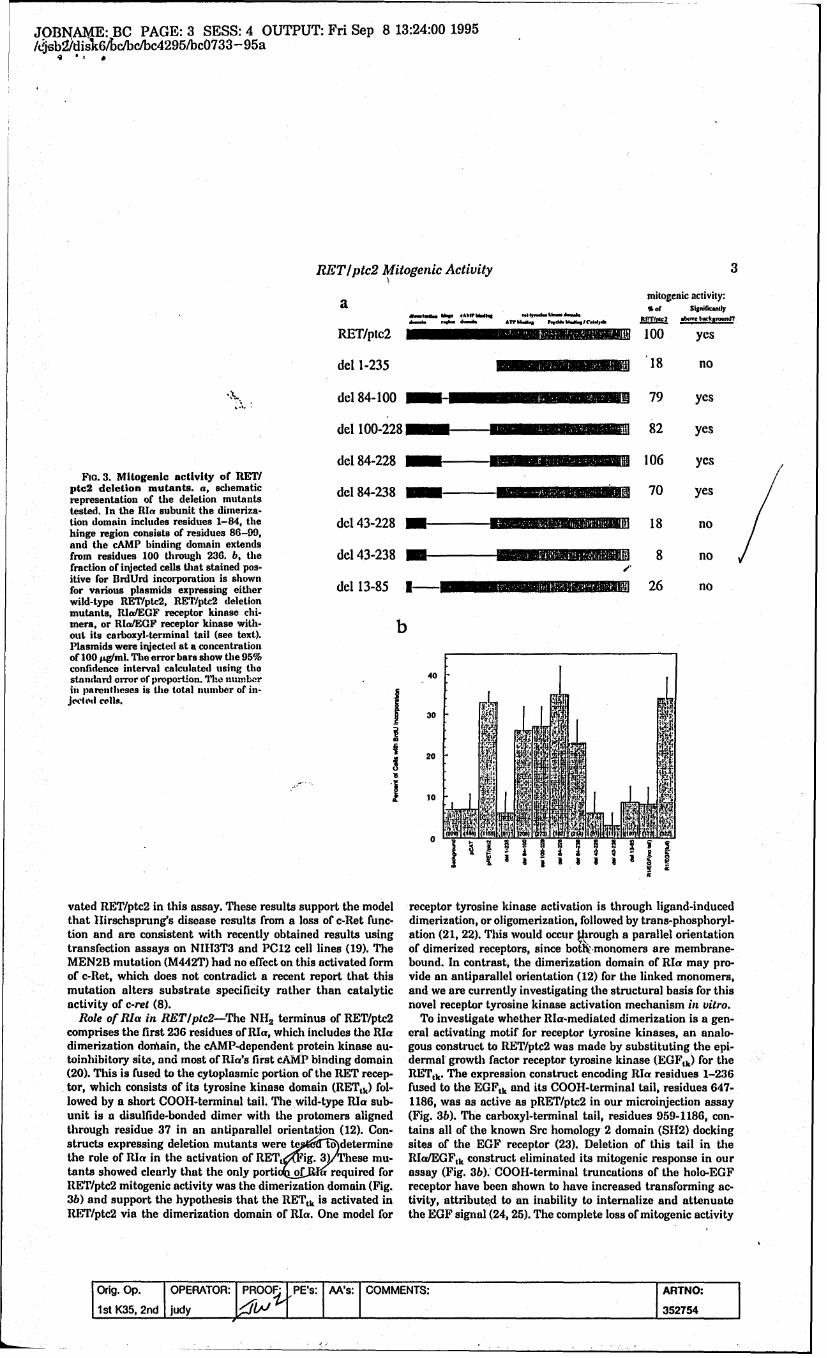
JOBNAME: BC PAGE: 3 SESS: 4 OUTPUT: Fri Sep 8 13:24:00 1995/j6b2Idis]k6/ cbc/bc4295/bc0733- 95a
RET/ptc2 Mitogenic Activity 3
a mi(ogenic activity:
RET/ptc2 1 100 yes
del 1-235 18 no
"dcl 84-100 79 yes
dcl 100-228 82 yes
del 84-228 106 yesFia. 3. Mitogenic activity of RET/
ptc2 deletion mutants, a, schematic dl84_3_---___-_-"___ 70 yesrepresentation of the deletion mutants del 84-238tested. In the Rla subunit the dimeriza-tion domain includes residues 1-84, the dcl 43-228 18 nohinge region consists of residues 86-99,and the cAMP binding domain extendsfrom residues 100 through 236. b, the dcl 43-238 8 nofraction of injected cells that stained pos- OPitive for BrdUrd incorporation is shownfor various plasmids expressing either del 13-85 I 26 nowild-type RET/ptc2, RET/ptc2 deletionmutants, RIlaEGF receptor kinase chi-mera, or RIaEGF receptor kinase with- bout its carboxyl-terminal tail (see text).Plasmids were injected at a concentrationof 100 pg/ml. The error bars show the 95%confidence interval calculated using thestandard orror of proportion. Th1e nxunber 40in parentheses is the total number of in-jeded colls.
30 -
520 ,
10 B
0
vated RET/ptc2 in this assay. These results support the model receptor tyrosine kinase activation is through ligand-inducedthat Hlirschsprung's disease results from a loss of c-Ret func- dimerization, or oligomerization, followed by trans-phosphoryl-tion and are consistent with recently obtained results using ation (21, 22). This would occur through a parallel orientationtransfection assays on NIH3T3 and PC12 cell lines (19). The of dimerized receptors, since botlkmonomers are membrane-MEN2B mutation (M442T) had no effect on this activated form bound. In contrast, the dimerization domain of RIa may pro-of c-Ret, which does not contradict a recent report that this vide an antiparallel orientation (12) for the linked monomers,mutation alters substrate specificity rather than catalytic and we are currently investigating the structural basis for thisactivity of c-ret (8). novel receptor tyrosine kinase activation mechanism in vitro.
Role of Rla in RETIptc2-The N'L2 terminus of RET/ptc2 To investigate whether RIa-mediated dimerization is a gen-comprises the first 236 residues of RIa, which includes the RIa eral activating motif for receptor tyrosine kinases, an analo-dimerization doiain, the cAMP-dependent protein kinase au- gous construct to RET/ptc2 was made by substituting the epi-toinhibitory site, and most of RIa's first cAMP binding domain dermal growth factor receptor tyrosine kinase (EGFtk) for the(20). This is fused to the cytoplasmic portion of the RET recep- RETtk. The expression construct encoding RIa residues 1-236tor, which consists of its tyrosine kinase domain (RETtk) fol- fused to the EGFtk and its COOH-terminal tail, residues 647-lowed by a short COOH-terminal tail. The wild-type RIa sub- 1186, was as active as pRET/ptc2 in our microinjection assayunit is a disulfide-bonded dimer with the protomers aligned (Fig. 3b). The carboxyl-terminal tail, residues 959-1186, con-through residue 37 in an antiparallel orientation (12). Con- tains all of the known Src homology 2 domain (SIt2) dockingstructs expressing deletion mutants were t o &)determine sites of the EGF receptor (23). Deletion of this tail in thethe role of RIcy in the activation of RETt7lFig. 3)These mu- RII/EGFtk construct eliminated its mitogenic response in ourtants showed clearly that the only porti of a required for assay (Fig. 3b). COO1-terminal truncations of the holo-EGFRET/ptc2 mitogenic activity was the dimerization domain (Fig. receptor have been shown to have increased transforming ac-3b) and support the hypothesis that the RETtk is activated in tivity, attributed to an inability to internalize and attenuateRET/ptc2 via the dimerization domain of RIa. One model for the EGF signal (24, 25). The complete loss of mitogenic activity
Orig. Op. OPERATOR: I PROOF,/PE's: AA's: COMMENTS: ARTNO:
I1st K(35, 2nd IJudy L~1'l I352754

.ýOINAME BCPAGE: 4 SESS: 4 OUTPUT: Fri Sep 8 13:24:00 1995
4 ~RET/ptc2 Mitogenic Activity
a the 1-I-helix of the insulin receptor tyrosine kinase (18, 28). Thisso tyrosine is highly conserved in the receptor tyrosine kinases
(26) and could serve a structural role rather than as an S112docking site. Tyr-586, which completely eliminated mitogenic
40 71activity when mutated to Phie, is in the carboxyl-terminal tail of~~ the RETtk.
A that provide S112 or phosphotyrosine binding domain docking
.. j~j ~.sites essential for the mitogenic response of RET/ptc2. Muta-20 tion of the other Tyr in the 00011-terminal tail, Tyr-553, had
no effect on mitogentic activity. Results from the Y586F mutantindicate that kinase activity alone is not sufficient for the
S 10 mitogenic response of RET/ptc2. Work is under way to investi-gate RET/ptc2 phosphorylation sites and to search for interact-ing proteins.
REFERENCESI l 1. Takahashi, M., Ritz. J., and Cooper, G. M. (1985) Cell 42, 581-5882. Takahnashi, M., Butna, Y., and lust, 11. (1989) Oncogene 4. 805-8063. Romeo, G., Ronchetto, P. Luo, Y., Barone, V., Seri, M., Ceccherini, I., Pasini,
B., Bocciardi, R., Lerone, M., Kaansianeu, H.. and Martucciello, G. (1994)Nature 3117, 377-378 i
M ~~ON M~ $$I.~ Ion1 4. Edery, P., Lyonnet, S., Mulligan, L., Pelet, A., Dow, E., Abel, L., Holder, S.,c -RET Nihoul-Fekete, C., Ponder, B., and Munnich, A. (1994) Nature 367, 378-380
T- 5. Mulligan, L., Eng, C., Hlealey, C., Clayton, D., Kwok, J., Gardner, E., Ponder,M., Frilling, A., Jacksaon, C., Lehnert, Ht., Neumann, H., Thibodeau, S., andPonder, 1.. (1994) Nat. Genet. 6, 70-74
6. Eng, C.. Smith, D., Mulligan, L., Nagai. M., Hlealey, C., Ponder, M., Gardner,0 N.k. 6 I1dh E ., Scheuniann, G., Jackson, C., Tunnaclilre, A., and Ponder, B. (1994)
310 414 43 410M 30 3 IMAnw n no" Hum. Mat. Genet. 3,237-241
RET/ptc2 7. Hofatra, I., Landavater, It, Ceccherinu, I., Stulp, R., Stelwagen, T., Luo, Y.,C.-Aw Psaini, B., Hoppener, J., van Ainstel, I., Romeo, G., Lips, C., and Buys, C.
o (1994) Nature 367, 375-376Effdd.0~o~lh.o,,~o,,,gd~iy,* 8. Santoro. M., Carlornagno, F., Romano. A., Bottaro, D.. Dathan, N.,.Grieco, M.,
Fusco, A., Veecchio, G., Matoakova, B., Kraus, M., and Di Fiore, P. (1995)* ' Science 267,.381-383
9. ongro I., Butti M.. Coronelli, S., Ilorrello, M., Santoro, M., Mondellini,Fin.4. itoeni aciviy o RETptc tyosie t phnylla- P.. Pilotti, S., Fusco, A., Della Porta, G., and Pierotti, M. (1994) Cancer les.
nine mnutants.a, the fraction of injected cells which atained poaitive for 54i,2979-2985UrdUrd incorporation ia shown for various plasmids expreasing either 1o. Lanzi, C., Ilorrello, M., Bongarzone, I., Migliazza, A., Fusco, A., Grieco. M.,wild-type RET/ptc2 or RET/ptc2 Tyr to Phe point mutants, Plasmids Santoro, M., Gambetta, R., Zunino, F., Della Porte, G., and Pierotti, M.were injected at a concentration of 100 pg/ml. The column labeled (1992) Oncogene 7, 2189-2194background represents uninjected celia under the assay conditions. The 11. Bongarzone, I., MonzintiN., Borrello, M., Carcano, C., Ferraresi, G., Arighi, E.,error bars show the 95% confidence interval calculated using the stand- Mondellini, P., Della Ports, 0., and Pierotti, M. (1993) Mot. Cell Biol. 13,
arderrr o prporion Th nuberin arethees s te ttalnumer 2. 358-366ard rro ofproortin. he umbr i parnthsesis he ttalnumer 2. ubis, J., Vedvick, T., and Taylor, S. (1987) J. Biol. Chem. 262, 14961-14966of injected cells. b, schematic representation of RET/ptc2 showing the 13 Sanger, F., Nicklen, S., and Coulson, A. R. (1977) Proc. Nall. Acad. Sci. U. S.A.locations of the seven tyrosines mutated to phienylalanine. The kinase 74,543-46domain of RET extends from residues 237 to 545 of RETr/ptc2, the 14, Kunkle, T. A., Bebenelc, K, and McClary, J. (1991) Methods Enzymol. 204,COOII-terminal. tail ends at residue 596, and the platelet-derived 125-139growtha factor-like insert of RET encompasses residues 344-367. The 15. Ausubel, F., Brent, It., Kingston, It. Moore, D., Seidman, J., Smith, J., andcorresponding tyrosines are shown on the c-Ret schematic (2). Struhl, K. (1994) Current Protocols in Molecular Biology, John Wiley &
Sons, Inc., Brooklyn, NY16. Meinkoth, J., Goldsmith, P., Spiegel, A., Feramnisco, J., and Burrow, G. (1992)
for our COOH-terminal EGFtk deletion suggests that SH2 do- J. Biol. Chem. 267, 13239-13245main interactions are more imiportant for the mitogenic re- 17. Bubis, J., Neitasl, J., Saraswat, L,, and Taylor, S. (1988) J. Bial. Chem. 263,
9668-9673sponse of Rla/EGFlk than for 00011-terminal truncations of 18. Hanks, S. K., Quinn, A. M., and Hlunter, T. (1988) Science 241, 42-52the holo-EGF receptor. 19. Pasini, B., Borrello, M., Greco. A., Bongarzone, I., Luo, Y., Mondellini, P.,
Rol ofTyrsins n R T~pc2-o ienifyposibl SI12 Alberti, L., Miranda, C., Arighi, Wl ,Bocciardi, R., Seri, M., Barone, V.,Roleof yroine in E~ltc2 To dentfy ossbleSH2 Romeo, G., and Pierotti, M. (1995);Na: Genet. 10, 35-40interaction sites in RET/ptc2, several tyrosines were mutated 20. Taylor, S., Buechler, J., and Yonemoto, W. (1990) Annu. Rev. fliochem. 59,
ha~e-d the resultant mutants tested for mitogenic activity 2 971-1005it.)I 21. Wich, A., and Schlessinger, J. (1990) Cell 61, 203-212
ýWig. 4./f the seven Tyr mutants, two, Y35OF and Y424F, 22: Schlessinger, J. (1988) Trends Biochem. Sci. 13, 443-447"kexkibied significantly reduced mitogenic activity. Mutations 23. Koch, C., Anderson, A., Moran, M., Ellis, C., and Pawson, T. (1991) Science
two the tyrsins, 505FandY58F, aolihedthe ito 24 252,668-674at toohrtrsnsY55 an 86,aoihdtemt 2.Wells, A., Welsh, J., Lazar, C., Wiley, H., Gill, G., and Rosenfeld, M. (1990)genie effect. Tyr-350 is located in a variable insert region of Science 247, 962-964receptor tyrosind kinases, and in the platelet-derived growth 25. Reddy, C., Wells, A., and Lauffenburger, D. (1994) Biotech. Prog. 10.,377-384
26. van der Geer, P., Hunter, T., and Lindberg, IL (1994) Annu. Rev. Cell Biot. 10,fact-or recepttor this insert -region contains two S112' binding' 261-337sites (26). Tyr-424 in RET/ptc2 corresponds to Y1158 in the 27. White, M., Shoelson, S., Keutmann, H., and Kahn, C. (1988) J. Biol. Chem.
required 263, 2969-2980insulin receptor (26), and phosphorylation at Y1 158 is reurd 28. Hubbard. S., Wei, L., Ellie, L., and Hendrickson, W. (1994) Nature 372,for full activity (27). T~yr-505 is in a sequence that aligns with 746-754
Orig. Op. OPERATOR: jPROOF: IPE's: M~s: COMMENTS: ARTNO:I1st K(35, 2nd Judy 1./~1 I352754