18 filling defects in the ureter
TRANSCRIPT

18 Filling Defects in the Ureter
CLINICAL IMAGAGINGAN ATLAS OF DIFFERENTIAL DAIGNOSIS
EISENBERG
DR. Muhammad Bin Zulfiqar PGR-FCPS III SIMS/SHL

• Fig GU 18-1 Nonopaque ureteral calculus (arrows).

• Fig GU 18-2 Blood clot in a proximal ureter (arrows).

• Fig GU 18-3 Renal vein thrombosis. (A) Excretory urogram shows characteristic notching (arrow) of the upper ureter. There is enlargement of the left kidney with poor calyceal function due to compression from parenchymal engorgement. (B) In another patient with renal vein thrombosis and ureteral notching, a venogram demonstrates exuberant periureteral collaterals (arrows).16
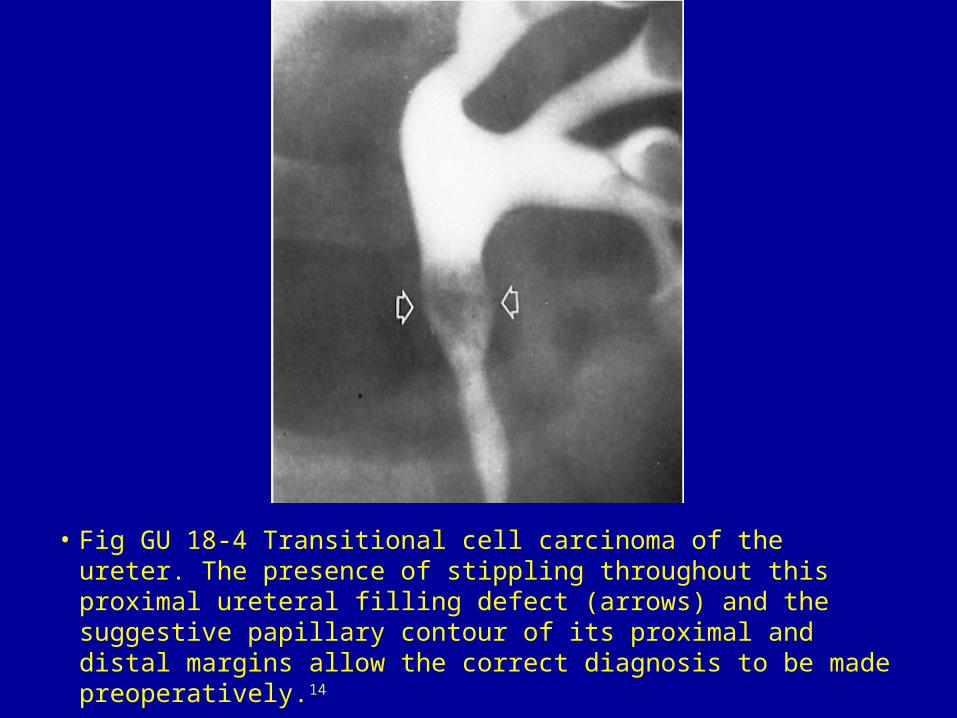
• Fig GU 18-4 Transitional cell carcinoma of the ureter. The presence of stippling throughout this proximal ureteral filling defect (arrows) and the suggestive papillary contour of its proximal and distal margins allow the correct diagnosis to be made preoperatively.14
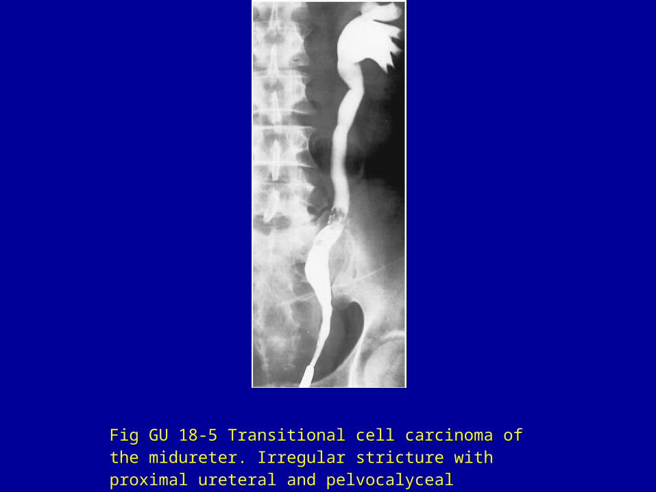
Fig GU 18-5 Transitional cell carcinoma of the midureter. Irregular stricture with proximal ureteral and pelvocalyceal dilatation.
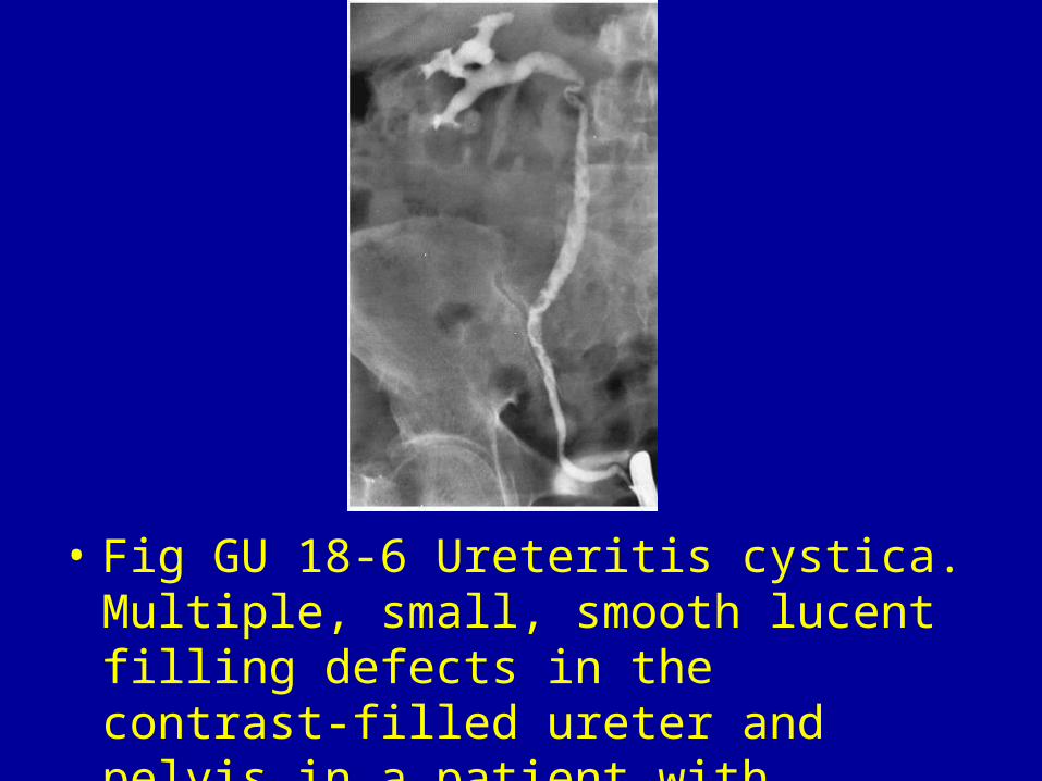
• Fig GU 18-6 Ureteritis cystica. Multiple, small, smooth lucent filling defects in the contrast-filled ureter and pelvis in a patient with chronic urinary tract infection.
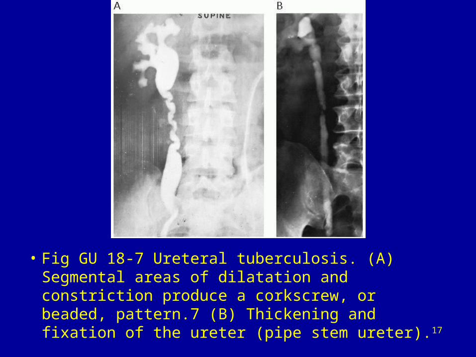
• Fig GU 18-7 Ureteral tuberculosis. (A) Segmental areas of dilatation and constriction produce a corkscrew, or beaded, pattern.7 (B) Thickening and fixation of the ureter (pipe stem ureter).17

Fig GU 18-8 Malacoplakia of the ureter. Magnified view of the distal right ureter shows multiple filling defects simulating ureteritis cystica.18

