1770 research article - journal of cell...
TRANSCRIPT

The IplA Ca2+ channel of Dictyostelium discoideum isnecessary for chemotaxis mediated through Ca2+, butnot through cAMP, and has a fundamental role innatural aggregation
Daniel F. Lusche, Deborah Wessels, Amanda Scherer, Karla Daniels, Spencer Kuhl and David R. Soll*W. M. Keck Dynamic Image Analysis Facility, Department of Biology, University of Iowa, Iowa City, IA 52242, USA
*Author for correspondence ([email protected])
Accepted 5 December 2011Journal of Cell Science 125, 1770–1783� 2012. Published by The Company of Biologists Ltddoi: 10.1242/jcs.098301
SummaryDuring aggregation of Dictyostelium discoideum, nondissipating, symmetrical, outwardly moving waves of cAMP direct cells towardsaggregation centers. It has been assumed that the spatial and temporal characteristics of the front and back of each cAMP wave regulateboth chemokinesis and chemotaxis. However, during the period preceding aggregation, cells acquire not only the capacity to chemotax
in a spatial gradient of cAMP, but also in a spatial gradient of Ca2+. The null mutant of the putative IplA Ca2+ channel gene, iplA–,undergoes normal chemotaxis in spatial gradients of cAMP and normal chemokinetic responses to increasing temporal gradients ofcAMP, both generated in vitro. However, iplA– cells lose the capacity to undergo chemotaxis in response to a spatial gradient of Ca2+,suggesting that IplA is either the Ca2+ chemotaxis receptor or an essential component of the Ca2+ chemotaxis regulatory pathway. In
response to natural chemotactic waves generated by wild-type cells, the chemokinetic response of iplA– cells to the temporal dynamicsof the cAMP wave is intact, but the capacity to reorient in the direction of the aggregation center at the onset of each wave is lost. Theseresults suggest that transient Ca2+ gradients formed between cells at the onset of each natural cAMP wave augment reorientation towards
the aggregation center. If this hypothesis proves correct, it will provide a more complex contextual framework for interpreting D.
discoideum chemotaxis.
Key words: cAMP chemotaxis, Microfluidic chamber, Ca2+ binding, Inositol trisphosphate receptor, Mechanoreceptor
IntroductionExtracellular cations play essential roles in cell polarity, motility
and chemotaxis (Soll et al., 2011). For that reason, they must be
present at optimum concentrations in the natural environment
(Soll et al., 2011). For the soil amoeba Dictyostelium discoideum
to achieve maximum velocity either the extracellular
concentration of Ca2+ must reach 10 mM or the concentration
of K+ reach 40 mM (Lusche et al., 2009; Lusche et al., 2011;
Scherer et al., 2010). Moreover, for D. discoideum amoebae to
orient in a spatial gradient of the chemoattractant cAMP, either
Ca2+ must reach 5 mM, or K+ or Na+ reach 15 mM (Lusche et al.,
2009; Lusche et al., 2011). In addition to its role in facilitating
motility and chemotactic orientation, Ca2+ also acts as a
chemoattractant. When developing cells attain chemotactic
responsiveness to spatial gradients of extracellular cAMP, they
also attain chemotactic responsiveness to spatial gradients of
extracellular Ca2+ (Scherer et al., 2010). Deletion of the gene for
the putative sodium/hydrogen exchanger Nhe1 (NheA) (Patel and
Barber, 2005) resulted in the loss of K+ as a facilitator of polarity
and motility, and the loss of Na+ or K+ as a facilitator of
chemotactic orientation in a spatial gradient of cAMP (Lusche
et al., 2011). Deletion of Nhe1, however, did not affect Ca2+
facilitation of these behaviors (Lusche et al., 2011). These results
indicated that one or more plasma membrane proteins other than
Nhe1 was involved in the facilitation of motility and chemotaxis
by Ca2+ (Lusche et al., 2011).
To identify these membrane proteins, we first searched for, but
did not find in D. discoideum, an ortholog of CaR, a bona fide G-
protein-coupled Ca2+ receptor (Brown et al., 1993; Garrett et al.,
1995; Brown and MacLeod, 2001; Khan and Conigrave, 2010)
involved in motility and chemotaxis in higher animal cells
(Boudot et al., 2010). We therefore turned our attention to
inositol 1,4,5-triphosphate receptor-like protein A (IplA), a
membrane protein in D. discoideum with homology to the
inositol trisphosphate receptors (InsP3Rs) of higher eukaryotes
(Traynor et al., 2000). These six transmembrane proteins function
as homotetrameric Ca2+ channels (Taylor and Laude, 2002;
Foskett et al., 2007; Foskett, 2010), possessing a long
cytoplasmic N-terminal region with a binding site for InsP3
and other regulatory proteins, and a shorter C-terminal region
with putative binding sites for additional regulatory proteins
(Patterson et al., 2004; Foskett et al., 2007; Foskett, 2010).
InsP3Rs are usually located in the membrane of endoplasmic
reticulum and vesicles, but are also found at low levels in the
plasma membrane (Furuichi et al., 1989; Yule et al., 2010; Dellis
et al., 2006; Taylor et al., 2009a; Taylor et al., 2009b; Barrera
et al., 2007; Bezprozvanny, 2005; Delmas et al., 2002; Joseph,
1996; Tanimura et al., 2000; Tojyo et al., 2008; Rossier et al.,
1770 Research Article
Journ
alof
Cell
Scie
nce

1991; Fadool and Ache, 1994). In the endoplasmic reticulum,
they play roles in Ca2+ homeostasis (Maeda et al., 1991; Ferriset al., 1989; Foskett et al., 2007). Deletion of the iplA gene in D.
discoideum has been shown to block Ca2+ influx across the
plasma membrane in response to a global cAMP signal (Traynoret al., 2000; Schaloske et al., 2005; Shanley et al., 2006). MutantiplA– cells, however, were found to undergo chemotaxis in aspatial gradient of cAMP released from a micropipette, leading to
the conclusion that Ca2+ influx stimulated by cAMP wasnot necessary for cAMP chemotaxis. It was subsequentlydiscovered that iplA– cells lost shear-induced motility (i.e.
mechanoreception) (Fache et al., 2005; Lombardi et al., 2008).Together, these results suggested that IplA was a strong candidatefor a cell surface molecule or a molecule in the signal
transduction pathway that mediated the facilitating effects ofextracellular Ca2+ on motility and cAMP chemotaxis, and/orCa2+ chemotaxis (Lusche et al., 2009; Scherer et al., 2010).
Here, we provide evidence that IplA plays selective roles in the
extracellular effects of Ca2+ on cell behavior. It is necessary forCa2+ facilitation of increased velocity, but not for the increasescaused by the alternative cation K+. More interestingly, IplA is
essential for chemotaxis in a spatial gradient of Ca2+, but not forchemotaxis in a spatial gradient of cAMP. It is also not essentialfor the chemokinetic responses to temporal waves of cAMP that
mimic the temporal dynamics of natural cAMP waves. Given thatiplA– cells chemotax normally in spatial gradients of cAMPgenerated in vitro and exhibit normal chemokinetic responses totemporal waves of cAMP generated in vitro, we fully expected
iplA– cells to behave normally to natural waves of cAMPgenerated in an aggregation territory. This was based on theassumption that the complete behavior of amoebae in natural
aggregation territory could be explained by their behavior inspatial and temporal gradients of cAMP. Instead, we found thatalthough iplA– cells undergo a normal chemokinetic surge in
velocity in response to the increasing phase of each naturallyrelayed cAMP wave in wild-type aggregation territories, theylose the ability to reorient towards the aggregation center at the
onset of the front of each wave. These results support ahypothesis, previously entertained (Scherer et al., 2010), thattransient Ca2+ gradients might be generated between cells at theonset of the front of each natural relayed cAMP wave that
augment orientation towards the center of an aggregationterritory, the source of each wave. If this hypothesis proves tobe true, it would provide a more complex context for interpreting
mutations that affect natural chemotaxis.
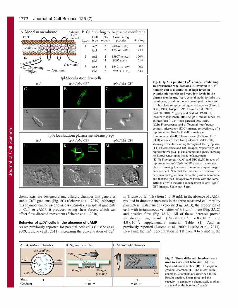
ResultsIplA localization
In D. discoideum, IplA, a presumed Ca2+ channel, contains six
transmembrane domains (Fig. 1A) (Traynor et al., 2000). The loopthat includes the membrane spanning regions 5 and 6 has beenshown in human IplAs to include the Ca2+ transport (pore) domain
(Fig. 1A) (Ramos-Franko et al., 1999; da Fonseca et al., 2003).Models generated for the human IplA ortholog suggest that the N-and C-terminal regions are probably cytoplasmic, whether in the
membrane of the endoplasmic reticulum or the plasma membrane(Fig. 1A) (Foskett et al., 2007; Foskett, 2010). To assess thelocalization of IplA, we generated the mutant strain iplA–
complemented with a plasmid in which wild-type iplA+ waslinked to GFP, iplA–/iplA+-GFP. The iplA+-GFP construct wasunder the regulation of the actin promoter. In living iplA–/iplA+-GFP
cells, IplA–GFP localized in vesicles throughout the cytoplasm(Fig. 1E–H). These vesicles also abutted the plasma membrane.
When brightness was enhanced to assess plasma membranelocalization, the increased brightness of the associated vesiclesprecluded visualization of low plasma membrane fluorescence.
To visualize membranes, we isolated plasmid membrane ghostsby Triton extraction according to the methods of Condeelis(Condeelis, 1979). Image enhancement revealed fluorescence inthe plasma membranes (Fig. 1K–N). Membranes from iplA–
cells analyzed similarly exhibited no autofluorescence(Fig. 1I,J). A control strain was generated in the iplA–
background in which only the GFP gene was placed under the
regulation of the actin promoter. Fluorescence of GFP in strainiplA–-GFP was diffuse throughout the cytoplasm of living cells(supplementary material Fig. S1) and not associated with the
plasma membrane. Together, these results indicate that IplA islocalized primarily in vesicular membranes in the cytoplasm andsuggest low levels in the plasma membrane.
Ca2+ binding
To test whether IplA plays a role in Ca2+ binding at the cell
surface, cells were treated with NaN3 for 30 minutes, whichinhibits Ca2+ transport, and then incubated with 45Ca2+ for 5minutes (Wick et al., 1978; Milne and Coukell, 1991; Milne and
Devreotes, 1993; Groner and Malchow, 1996). In three separateexperiments, iplA– cells exhibited respective decreases in Ca2+
binding to 73, 61 and 64% that of control cells, suggesting that at
least a portion of Ca2+ binding to the plasma membrane ismediated by IplA (Fig. 1B).
Behavioral assays
To test for selective Ca2+-dependent defects in the iplA– mutant,we employed three chambers (Fig. 2). The Sykes–Moore
chamber (Fig. 2A) is round with upper and lower glass walls,and a metal clamped rim with inlet and outlet ports. The ports areattached to pumps (Varnum et al., 1985; Varnum-Finney et al.,
1987; Varnum-Finney et al., 1988), allowing perfusion withbuffer in order to assess basic cell motility in the absence ofcAMP. This chamber can also be used to assess behavior in
response to temporal waves of cAMP, generated by a pumpsystem. These waves mimic the temporal dynamics of a series ofnatural cAMP waves, without establishing spatial gradients ofcAMP (Varnum et al., 1985; Varnum-Finney et al., 1987;
Wessels and Soll, 1990; Wessels et al., 1992; Wessels et al.,2009).
However, because the Sykes–Moore chamber is round, itgenerates randomly directed shear forces that can affect thebehavior of cells by activating IplA, a putative mechanoreceptor(Fache et al., 2005; Lombardi et al., 2008). The Zigmond
chamber (Fig. 2B) consists of a bridge, which supports cells,bordered by two wells (Zigmond, 1977; Varnum and Soll, 1984).If one well is filled with buffer alone (‘sink’) and the other with
buffer plus cAMP (‘source’), a cAMP gradient is generatedacross the bridge and chemotaxis can then be assessed at thesingle cell level. If both wells are filled with the same buffer
solution lacking cAMP, basic motile behavior can be assessed atlow cell density in the absence of shear forces. Although theZigmond chamber is effective in assessing chemotaxis in a
spatial gradient of cAMP, it is ineffective in assessing Ca2+
chemotaxis, given that Ca2+ gradients rapidly dissipate becauseof the high diffusion rate of the cation. To test for Ca2+
Ca2+ chemotaxis receptor in Dictyostelium 1771
Journ
alof
Cell
Scie
nce
chemotaxis, we designed a microfluidic chamber that generates
stable Ca2+ gradients (Fig. 2C) (Scherer et al., 2010). Although
this chamber can be used to assess chemotaxis in spatial gradients
of Ca2+ or cAMP, it produces strong shear forces, which can
effect flow-directed movement (Scherer et al., 2010).
Behavior of iplA– cells in the absence of cAMP
As we previously reported for parental Ax2 cells (Lusche et al.,
2009; Lusche et al., 2011), increasing the concentration of Ca2+
in Tricine buffer (TB) from 5 to 10 mM, in the absence of cAMP,
resulted in dramatic increases in the three measured cell motility
parameters: instantaneous velocity (Fig. 3A,B), the proportion of
cells with instantaneous velocities of $9 mm/minute (Fig. 3A,C)
and positive flow (Fig. 3A,D). All of these increases proved
statistically significant (P57.061027, 6.061025 and
4.061027; supplementary material Table S1). And as
previously reported (Lusche et al., 2009; Lusche et al., 2011),
increasing the Ca2+ concentration in TB from 0 to 5 mM in the
Fig. 2. Three different chambers were
used to assess cell behavior. (A) The
Sykes–Moore chamber. (B) The Zigmond
gradient chamber. (C) The microfluidic
chamber. Chambers are described in the
Results section. Shear force and the
capacity to generate a chemotactic gradient
are noted at the bottom of panels.
Fig. 1. IplA, a putative Ca2+ channel, containing
six transmembrane domains, is involved in Ca2+
binding and is distributed at high levels in
cytoplasmic vesicles and very low levels in the
plasma membrane. (A) A general model for IplA in a
membrane, based on models developed for inositol
trisphosphate receptors in higher eukaryotes (Furuichi
et al., 1989; Joseph, 1996; Foskett et al., 2007;
Foskett, 2010; Mignery and Sudhof, 1990). IP3,
inositol trisphosphate. (B) The iplA– mutant binds less
extracellular 45Ca2+ than parental Ax2 cells.
(C,D) Fluorescence and differential interference
contrast microscopy (DIC) images, respectively, of a
representative live iplA– cell, showing no
fluorescence. (E–H) Fluorescence (E,G) and DIC
(D,H) images of two live iplA–/iplA+-GFP cells,
showing vesicular staining throughout the cytoplasm.
(I,J) Fluorescence and DIC images, respectively, of a
representative iplA– plasma membrane ghost, showing
no fluorescence upon image enhancement.
(K–N) Fluorescent (K,M) and DIC (L,N) images of
representative iplA–/iplA+-GFP plasma membrane
ghosts, showing low-level fluorescence upon image
enhancement. Note that the fluorescence of whole live
cells was far higher than that of the plasma membrane,
and that the iplA– images were taken with the same
settings or with the same enhancement as iplA–/iplA+-
GFP images. Scale bar: 5 mm.
Journal of Cell Science 125 (7)1772
Journ
alof
Cell
Scie
nce

absence of cAMP resulted in an increase in the directional
persistence of translocating cells (Fig. 3A,E) that was statistically
significant (P55.061023; supplementary material Table S1).
Mutant iplA– cells did not show increases in velocity parameters
comparable with the increases in wild-type cells when the Ca2+
concentration in TB was increased to 10 mM (Fig. 3A–D).
Mutant iplA– also did not undergo an increase in directional
persistence when the Ca2+ concentration was increased from 0 to
5 mM, although they did undergo the increase when the Ca2+
concentration was increased to 10 mM (Fig. 3A,E). The defects
in responsiveness of iplA– cells to 10 mM Ca2+ were discernible
in computer-generated perimeter tracks. The tracks of iplA– cells
(Fig. 3G) were on average more contracted than those of Ax2
cells (Fig. 3F). No major differences in cell shape were evident
(see inserts in Fig. 3F,G).
If IplA plays a selective role in the cationic facilitation of cell
motility by Ca2+ in the absence of cAMP, then cells of the null
mutant iplA– and its parent strain Ax2 should exhibit no
behavioral differences from wild-type cells when translocating
in TB containing 40 mM K+, the facilitating concentration of this
monovalent cation in the absence of added Ca2+ (Lusche et al.,
2009). This prediction is predicated on the discovery that K+, but
not Ca2+, facilitation is mediated by Nhe1 (Lusche et al., 2011).
When perfused with a 40 mM K+ solution in a Sykes–Moore
Fig. 3. Mutant iplA– cells exhibit selective defects
in the facilitation of velocity by Ca2+. The
behavior of mutant iplA– cells was compared with
that of parental Ax2 and complemented iplA–/iplA+
cells in a Sykes–Moore chamber perfused with TB
containing different concentrations of Ca2+, in the
absence of cAMP. (A) Motility parameters (means
6 standard deviation) at different concentrations of
Ca2+. (B) Instantaneous velocity plotted as a
function of Ca2+ concentration. (C) Proportion of
cells with instantaneous velocities $9 mm/minute
plotted as a function of Ca2+ concentration.
(D) Positive flow plotted as a function of Ca2+
concentration. (E) Directional persistence plotted as
a function of Ca2+ concentration. (F,G) Perimeter
tracks of representative parental Ax2 and iplA–
cells, respectively, translocating in TB containing
10 mM Ca2+. Insets in F and G reveal no
differences in shape. (H) Motility parameters of
Ax2 and iplA– cells in a Sykes–Moore chamber
perfused with TB containing 40 mM K+ in the
absence of cAMP.
Ca2+ chemotaxis receptor in Dictyostelium 1773
Journ
alof
Cell
Scie
nce

chamber, all motility parameters were similar in iplA– and
wild-type cells (Fig. 3H), supporting the conclusion that in the
absence of cAMP, K+ facilitation is mediated by Nhe1, not IplA
and that Ca2+ facilitation is mediated by IplA, not Nhe1.
Motility and chemotactic orientation in a spatial gradient
of cAMP
As we previously reported for parental Ax2 cells (Lusche et al.,
2009), increasing the concentration of Ca2+ in TB to 10 mM in a
spatial gradient of cAMP caused an increase in the three motility
parameters, instantaneous velocity (Fig. 4A,B), percentage of
cells with instantaneous velocities of $9 mm/minute (Fig. 4A,C),
and positive flow (Fig. 4A). Increasing the concentration from
0 to 5 mM caused an increase in directional persistence
(Fig. 4A,D). In the case of iplA– cells in a spatial gradient of
cAMP, there were less pronounced increases in the velocity
parameters (Fig. 1A–C). Directional persistence, however,
increased at 5 mM, as it did in Ax2 cells (Fig. 4A,D). The
differences between iplA– and Ax2 cells were either less
pronounced or negligible in a spatial gradient of cAMP than in
the absence of cAMP (Fig. 3; supplementary material Table S1).
Only the differences in instantaneous velocity and percentage of
cells with instantaneous velocities of $9 mm/minute were
significant (P55.061023, 4.061023; supplementary material
Table S1). These results are consistent with previous
observations, especially on lateral pseudopodium formation and
turning, indicating that a spatial gradient of cAMP enhances the
effect of Ca2+ (Lusche et al., 2009).
As previously reported (Lusche et al., 2009; Lusche et al.,
2011; Scherer et al., 2010), when Ca2+ was not added (0 mM
Ca2+) to a spatial gradient of cAMP generated in TB, Ax2 cells
still underwent chemotaxis, but with a reduced chemotactic index
Fig. 4. Mutant iplA– cells exhibit
selective defects in Ca2+ facilitation of
velocity, but undergo normal
chemotaxis in a spatial gradient of
cAMP. Studies were performed in
spatial gradients of cAMP generated in
TB containing different Ca2+
concentrations in a Zigmond chamber.
(A) Motility parameters (means 6
standard deviation) at different
concentrations of Ca2+.
(B) Instantaneous velocity as a function
of Ca2+ concentration. (C) Proportion of
cells with instantaneous velocities
$9 mm/minute as a function of Ca2+
concentration. (D) Directional
persistence as a function of time.
(E) Chemotactic index as a function of
time. (F,G) Perimeter tracks of Ax2 and
iplA– cells, respectively, in cAMP
gradients generated in 10 mM Ca2+.
Insets are for shape comparisons.
(H) Motility parameters of Ax2 and
iplA– in spatial gradients generated in
TB containing 40 mM K+ in TB.
Journal of Cell Science 125 (7)1774
Journ
alof
Cell
Scie
nce

(CI) of 0.3460.35 and a percentage positive chemotaxis value of
80% (Fig. 4A,E). Also, as previously reported (Lusche et al.,2009; Lusche et al., 2011), Ax2 cells attained maximum
chemotactic orientation (CI5+0.6960.21; percentage positive
chemotaxis598%) when the Ca2+ concentration was raised to5 mM (Fig. 4A,E) (Lusche et al., 2009; Lusche et al., 2011;
Scherer et al., 2010). Although relatively mobile, iplA– cells
exhibited negligible chemotactic orientation (CI5+0.0160.30;percentage positive chemotaxis570%) in a spatial gradient of
cAMP at 0 mM Ca2+, but exhibited maximum chemotactic
orientation (CI5+0.7560.14), similar to that of Ax2 cells, when
the concentration of Ca2+ was raised to 5 mM (Fig. 4A,E). Therelatively high efficiency of chemotaxis by iplA– cells in a spatial
gradient of cAMP generated in TB containing 10 mM Ca2+ was
evident in representative perimeter tracks (Fig. 4F,G). Theseresults support the earlier observations by Traynor et al. (Traynor
et al., 2000). They also indicate that as is the case for Ax2 cells,
chemotactic orientation of iplA– cells in a nonfacilitatingconcentration of K+ or Na+ requires 5 mM Ca2+, and this latter
requirement is independent of IplA.
If IplA functions specifically in the facilitation of maximum
velocity, then the iplA– and Ax2 cells should exhibit similar
maximum motility and chemotactic orientation in a spatialgradient of cAMP generated in TB containing 40 mM K+. When
a spatial gradient of cAMP was generated in TB containing 40 mM
K+ with no added Ca2+, the motility and chemotaxis parameters of
iplA– cells were similar for Ax2 and iplA– cells (Fig. 4H).
Responses to temporal gradients of cAMP
In a naturally aggregating population of D. discoideum cells the
pulses of cAMP released from cells at an aggregation center
(Gerisch et al., 1966; Konijn et al., 1967; Devreotes and Steck,
1979) are relayed through the population as outwardly moving,
nondissipating, symmetrical waves (Tomchik and Devreotes,
1981; Devreotes et al., 1983). In the front of each wave, an
amoeba experiences an increasing temporal as well as an
increasing spatial gradient of cAMP and in the back of each
wave, it experiences a decreasing temporal as well as a
decreasing spatial gradient of cAMP (Soll et al., 2002). At the
peak and in the back of each wave, amoebae undergo a decrease
polarity and directionality, and a dramatic decrease in velocity
(Varnum et al., 1985; Wessels et al., 1992; Soll et al., 2002). At
the onset of the front of next relayed wave, an amoeba reorients
towards the aggregation center, and then moves in a rapid and
persistent fashion towards the aggregation center, in what appears
to be a relatively semi-blind fashion in response to the increasing
temporal gradient of cAMP, which serves as chemokinetic signal
(Wessels et al., 1992; Soll et al., 2002). To test whether iplA–
cells were capable of assessing the temporal dynamics of a wave,
we challenged them with a series of four temporal waves
generated in buffered salt solution with K+ as the facilitating
cation. The waves were generated in the absence of an
established spatial gradient in a Sykes–Moore chamber
(Fig. 2A) attached to a pump system (Varnum et al., 1985;
Wessels et al., 1992). Mutant iplA– cells underwent a transient
increase in velocity in the increasing phase, and a decrease at the
peak and in the decreasing phase, of a majority of the temporal
cAMP waves (Fig. 5C,D), in a manner similar to that of
parental Ax2 cells (Fig. 5A,B). Ten additional Ax2 and 10
additional iplA– cells responded in a similar fashion. Therefore,
chemokinetic responsiveness to the increasing phases of cAMP
waves generated in vitro, as well as chemotactic orientation in a
spatial gradient of cAMP generated in vitro, are intact in iplA–
cells (Fig. 4).
Fig. 5. Mutant iplA– cells undergo increases in instantaneous
velocity in the increasing phase (f, blue zone) of a series of
temporal waves of cAMP generated in a Sykes–Moor chamber.
The temporal waves of cAMP approximate the temporal dynamics of
natural cAMP waves in the absence of a spatial gradient. These
experiments were performed in a buffered salt solution containing
40 mM K+ as the facilitating cation.
Ca2+ chemotaxis receptor in Dictyostelium 1775
Journ
alof
Cell
Scie
nce

Ca2+ chemotaxis is lost in iplA– cells
To test for Ca2+ chemotaxis, a microfluidic chamber was
employed (Fig. 2C) in which stable Ca2+ gradients were
established (Scherer et al., 2010). We first compared the
behavior of Ax2 and iplA– cells in the chamber in uniform
10 mM Ca2+. The parameter ‘rightward directionality’ (RD),
movement in the direction of flow (shear force), was computed as
the net distance traveled rightward (x) divided by the total
distance traveled (y; Fig. 6A). An RD of +1.00 represents
absolute rightward directionality, an RD of 0.00 represents
random directionality, and an increasing positive RD, from +0.01
to +0.99, reflects increasing rightward directionality. At a global
concentration of 10 mM Ca2+, in the absence of a Ca2+ gradient,
the mean instantaneous velocity of iplA– cells was 6.062.6 mm/
minute, compared with 10.965.3 mm/minute for Ax2 cells, a
reduction of 45% (Fig. 6D). The proportion of iplA– cells with
instantaneous velocities $9 mm/minute was also well below that
of Ax2 cells (Fig. 6D). The RD of Ax2 cells was +0.5960.34,
whereas the RD of iplA– cells was close to 0.00 (+0.0460.27;
Fig. 6D). The percentage of Ax2 cells exhibiting rightward
movement was 87%, whereas that of iplA– cells was close to
50%, demonstrating that the direction of iplA– cells under strong
Fig. 6. Mutant iplA– cells lose the capacity
to undergo chemotaxis in a spatial
gradient of Ca2+ generated in a
microfluidic chamber. (A) Computing
rightward directionality (RD). x, net distance
a cell moves to the right, the direction of
flow; y, total distance a cell moves.
(B) Rightward movement of representative
parental Ax2 cells in response to the
rightward shear force in the absence of a
chemotactic gradient, in uniform 10 mM
Ca2+ in TB. (C) The absence of rightward
movement of representative mutant iplA–
cells in response to rightward shear force in
the absence of a chemotactic gradient in
uniform 10 mM Ca2+. (D) Motility
parameters in the absence of a chemotactic
gradient in the microfluidic chamber in
uniform 10 mM Ca2+ in TB. (E) Chemotaxis
up a cAMP gradient by parental Ax2 cells.
The cAMP gradient was generated in 10 mM
Ca2+ in TB. (F) Chemotaxis up a cAMP
gradient by iplA– cells. The cAMP gradient
was generated in 10 mM Ca2+ in TB.
(G) Chemotaxis up a Ca2+ gradient by
parental Ax2 cells. The Ca2+ gradient was
generated in TB. (H) Random movement in a
Ca2+ gradient by iplA– cells. The Ca2+
gradient was generated in TB. (I) Motility
and chemotaxis parameters in a cAMP or
Ca2+ gradient generated in a
microfluidic chamber.
Journal of Cell Science 125 (7)1776
Journ
alof
Cell
Scie
nce

flow was random (Fig. 6D). The loss of flow-induced directional
movement and submaximal velocity by iplA– cells (Fig. 6C) wasapparent in a comparison of their perimeter tracks with those ofAx2 (Fig. 6B,C). Similar results for iplA– cells were obtained at
20 and 30 mM Ca2+ (data not shown). These defects wereconsistent with those previously reported using very differentassays (Fache et al., 2005; Lombardi et al., 2008).
We then compared the behavior of iplA– and Ax2 cells in
spatial gradients of cAMP generated in the microfluidic chamber(Fig. 2C). As previously demonstrated (Scherer et al., 2010),although Ax2 cells translocated in the direction of flow in the
absence of a cAMP gradient, they translocated in the direction ofincreasing cAMP in a spatial gradient of cAMP, with almost nobias to the right due to the direction of flow (Fig. 6E,I). MutantiplA– cells moved up spatial gradients of cAMP with the same
directionality as Ax2 cells (Fig. 6E,F,I). The chemotactic indicesof Ax2 and iplA– cells in cAMP gradients in the microfluidicchamber were +0.3560.35 and +0.3060.30, respectively, and the
percentage positive chemotaxis 90 and 84%, respectively(Fig. 6I). These results reinforce those obtained with theZigmond chamber (Fig. 4) and micropipette assays (Traynor
et al., 2000), demonstrating again that chemotaxis in a spatialgradient of cAMP is intact in iplA– cells.
We next tested whether iplA– cells underwent chemotaxis inCa2+ gradients generated in the microfluidic chamber (Fig. 2C).
Parental Ax2 cells had a chemotactic index of +0.2060.25,percent cells with a positive chemotactic index of 86% and aninstantaneous velocity of 10.562.9 mm/minute (Fig. 6I), values
very close to those we reported previously (Scherer et al., 2010).The RD of Ax2 cells in a Ca2+ gradient was +0.2560.38,showing rightward bias (Fig. 6I). The bias can be recognized in
representative perimeter tracks of Ax2 cells in a Ca2+ gradient(Fig. 6G). In marked contrast, the chemotactic index of iplA–
cells in similar Ca2+ gradients was +0.0160.29, a valueindicating the loss of Ca2+ chemotaxis (Fig. 6I). The
percentage iplA– cells with a positive chemotactic index was49% (Fig. 6I), close to 50%, the value for directionalrandomness. The differences between Ax2 and iplA– cells were
statistically significant (P5261026, 661028; supplementarymaterial Table S1). We further tested a variety of Ca2+ gradientsvarying in shape and concentration range, but observed no Ca2+
chemotaxis (data not shown). The RD of iplA– cells was–0.0460.33, very close to 0.00, indicating again the loss offlow-induced directionality (Fig. 6I). These results demonstrate
that although iplA– cells have fully retained the capacity toundergo positive chemotaxis in a spatial gradient of cAMP, theyhave lost the capacity to undergo chemotaxis in a spatial gradientof Ca2+. The directional movement of Ax2 cells with a rightward
bias in a spatial gradient of Ca2+ and the random movement ofiplA– cells with no rightward bias are reflected in a comparison ofrepresentative perimeter tracks (Fig. 6G and 6H, respectively).
Complementation of iplA– rescues defective phenotype
To test for complementation, we generated strain iplA–/iplA+-GFP, in which iplA+ was under the regulation of the actin
promoter in a nonintegrating plasmid. The three velocityparameters, instantaneous velocity, the proportion of cells withinstantaneous velocities of $9 mm/cell, and positive flow,
returned to wild-type levels in 10 mM Ca2+ in the absence ofcAMP (Fig. 3A) and the chemotactic and mechanoreceptiondefects were also rescued (Fig. 6I). The rescued levels (Fig. 3A,
Fig. 4A, Fig. 6I) were indistinguishable statistically from that ofthe wild-type parent strain, Ax2 (i.e. P,0.05). These results
demonstrate that the defects of iplA– cells were the direct result ofthe deletion of iplA+ and not a second site mutation.
Chemotaxis of iplA– cells in natural Ax2 aggregationterritories
If chemotaxis in a natural aggregation territory is regulated solelyby the spatial and temporal dynamics of relayed waves of cAMP,
as we have previously argued (Soll et al., 2002), then iplA– cellsshould undergo normal chemotaxis in natural chemotactic wavesgenerated by wild-type cells. This prediction is predicated on
their normal responses to spatial and temporal gradients of cAMPassayed in vitro. To test this prediction, we analyzed the behaviorof vitally stained minority iplA– cells seeded in majority wild-type cell aggregation territories (Wessels et al., 2000a; Wessels
et al., 2000b; Wessels et al., 2004; Wessels et al., 2007; Heidet al., 2004; Stepanovic et al., 2005). Under these conditions,minority mutant cells are challenged with natural waves relayed
by majority wild-type cells. Mutant iplA– cells were stained withDiI during growth, mixed with unlabeled Ax2 cells at a ratio of1:9 and the mixture of cells allowed to undergo aggregation on a
plastic surface in buffered salt solution (Sussman, 1987), inwhich K+ was the facilitating cation (Lusche et al., 2011). Inthe carpet of mixed cells established on the plastic surface,precocious cells, which act as aggregation centers, release pulses
of cAMP and these signals are then relayed through thepopulation as nondissipating outwardly moving waves(Tomchik and Devreotes, 1981) (Fig. 7A,B). The relayed
waves can be deduced from the behavior of majority, unlabeledAx2 cells, which undergo transient increases in velocity towardsthe aggregation center in the front of each wave, and decreases at
the peak and in the back of each wave (Wessels et al., 1992;Wessels et al., 2000a; Wessels et al., 2000b; Wessels et al., 2007;Heid et al., 2004; Stepanovic et al., 2005; Soll et al., 2002). The
behavior of minority iplA– cells in relayed waves could then beassessed through comparison with the behavior of Ax2 cellslocated within a 30 mm radius, which were responding to thesame relayed waves. As revealed in time plots of instantaneous
velocity, Ax2 cells (black dot plots) surged towards theaggregation center in the deduced front of each successivewave for approximately 2.5 minutes (Fig. 7C,E). The time
interval between waves was approximately 7 minute(Fig. 7C,E). Neighboring mutant iplA– cells (red dot plots)surged in a similar fashion in the front of the same successive
waves (Fig. 7C,E). There was, however, a defect in iplA– cellsdiscerned in centroid tracks. Whereas Ax2 cells (black dot tracks)made relatively constant net progress towards the aggregation
center in the front of each successive wave (Fig. 7D,F), iplA–
cells (red dot tracks) surged relatively in random directions in thefront of waves, thus making less net progress towards theaggregation center (Fig. 7D,F). The iplA– cells making the most
progress towards the aggregation center was used for comparisonin experiment 1 (Fig. 7D) and experiment 2 (Fig. 7F). Similarresults were obtained in three independent experiments.
To quantify the apparent defect in orientation, we measuredthe angle, in degrees, between the direction of the wave (a) andthe net direction of translocation (b) of individual majority wild-
type cells and minority mutant iplA– cells in the front of each offour to five successive cAMP waves generated by the majorityAx2 cells in natural aggregation territories. Net direction directly
Ca2+ chemotaxis receptor in Dictyostelium 1777
Journ
alof
Cell
Scie
nce

towards the aggregation center would be 0 , net direction directly
away from the aggregation center would be 180 , and net random
direction would be 90 . Models of a net angle (,) of
translocation of 30˚ and 90˚ are presented in Fig. 8A and 8B,
respectively. Although the mean angle of direction of the
majority wild-type cells was 33612 , that of minority iplA–
cells was 101622˚ (Fig. 8C), the latter close to the measure of
randomness (i.e. 90 ). These results demonstrate that even though
iplA– cells can chemotax up a spatial gradient of cAMP generated
in vitro, and even though they correctly surge in the increasing
phase of temporal waves generated in vitro, they are impaired in
their capacity to reorient correctly and move in the front of each
natural cAMP wave in a directed fashion towards the source of a
natural, relayed waves (i.e. the aggregation center).
DiscussionFollowing cloning of the first InsP3R over 20 years ago from the
mouse cerebellum (Furuichi et al., 1989), numerous inositol
trisphosphate receptor genes have been identified and grouped
into a family of transient receptor potential (TRP) channels
(Vannier et al., 1998; Blondel et al., 1993; Maranto, 1994; Ross
et al., 1992; Sudhof et al., 1991). Although most commonly
found in the endoplasmic reticulum (Furuichi et al., 1989; Yule,
et al., 2010), InsP3Rs have also been found associated with the
Fig. 7. Mutant iplA– cells do not reorient in the front of natural waves generated by parental Ax2 cells. To assess how iplA– cells respond to outwardly
moving, nondissipating relayed waves of chemoattractant, DiI-labeled iplA– cells and unlabeled parental Ax2 cells were mixed in a 1:9 ratio and allowed to
aggregate. Before streaming, the behavior of individual iplA– and Ax2 cells was analyzed. (A) 2D model of naturally relayed, outwardly moving, nondissipating
waves of cAMP in an aggregation territory. (B) 1D model of naturally relayed waves. (C,E) Velocity plots from two independent experiments of individual
majority parental Ax2 (black dots) and neighboring minority mutant iplA– (red dots) cells responding to natural waves. The waves diagrammed at top, were
deduced from Ax2 cell behavior. (D,F) Centroid tracks from two independent experiments, of representative Ax2 cells (black dots) and iplA– cells (red dots). Agg.
center, aggregation center. W1, W2, W3. Waves 1, 2 and 3. P, velocity peaks.
Journal of Cell Science 125 (7)1778
Journ
alof
Cell
Scie
nce

plasma membrane, but at far lower density (Fujimoto et al., 1995;
Joseph, 1996; Dellis et al., 2006; Rossier et al., 1991; Fadool and
Ache, 1994; Taylor et al., 2009a; Taylor et al., 2009b; Barrera
et al., 2007; Bezprozvanny, 2005; Delmas et al., 2002; Tanimura
et al., 2000; Tojyo et al., 2008). The possible role of InsP3Rs in
Ca2+ chemotaxis had never before been considered or tested.
The role of IplA in Ca2+-facilitated motility and cAMPchemotaxis
By mutational analysis we previously demonstrated that in buffer
lacking added Ca2+, the facilitation of motility by K+ and the
requirement of K+ or Na+ for chemotactic orientation in a spatial
gradient of cAMP (Lusche et al., 2011) are regulated by Nhe1
(Lusche et al., 2011), a putative monovalent cation/hydrogen
exchanger in the plasma membrane of D. discoideum (Patel and
Barber, 2005). The alternative cationic facilitation of these
behaviors by Ca2+ in the null mutant nhe1–, however, was
completely intact (Lusche et al., 2011), leading us to concludethat Nhe1 mediates monovalent cation effects on behavior, andthat one or more other surface molecules mediate alternative
Ca2+ effects. In the present study we found that IplA plays a rolein the Ca2+ facilitation of velocity in buffer and in a spatialgradient of cAMP, but it plays no role in the Ca2+ or K+
requirement for chemotactic orientation in a spatial gradient of
cAMP. We also found that IplA plays no role in the chemokineticresponsiveness of cells to increasing temporal gradients of cAMPgenerated in vitro (Varnum et al., 1985; Soll et al., 2002). Our
previous (Lusche et al., 2010) and present results, therefore,demonstrate that although all of the K+ or Na+ effects on basicbehavior and cAMP chemotaxis are mediated by Nhe1, only
selective Ca2+ effects on basic behavior are mediated by IplA.These results indicate that one or more molecules other than IplAplay a role in the Ca2+ requirement for chemotactic orientation, a
response that involves both the capacity to sense the direction ofa cAMP gradient and suppression of lateral pseudopodiumformation and turning (Soll et al., 2002).
The localization of IplA
Our analysis of living cells expressing IplA–GFP revealed thatfluorescence was primarily cytoplasmic and vesicular. Thegeneration of plasma membrane ghosts revealed very low level
staining. Because the preparation of ghosts involved treatment ofcells with Triton X-100, a detergent that can disrupt lipids andpossibly result in the contamination of plasma membranes with
vesicular membranes, we can only tentatively conclude thatIplA might be localized in the plasma membrane. But otherobservations add support to a cell surface role for IplA. Results
by Traynor et al. suggested that IplA was involved inextracellular Ca2+ uptake and therefore might function as aCa2+ channel at the surface of the cell (Traynor et al., 2000).Uptake, however, could also be mediated through the
intracellular regulation of Ca2+ stores. Schaloske et al. havepresented evidence that the main role of IplA could be inintracellular homeostasis (Schaloske et al., 2005). Shanley
et al. subsequently demonstrated that iplA– cells underwentelectrotaxis, which depends on regulated Ca2+ influx, indicatingthat IplA is not the cell surface Ca2+ influx channel
for electrotaxis (Shanley et al., 2006). However, themechanoreception defect in iplA– cells lends support to apossible role of IplA at the plasma membrane. Mutant iplA–
cells lose the orientation response in the direction of fluid flowand cannot attain shear-induced maximum velocity (Fache et al.,2005; Lombardi et al., 2008), suggesting that it functions as amechanoreceptor at the surface of the cell. However, these results
can be just as cogently explained by a role for IplA in asignal transduction pathway downstream of an unidentifiedmechanoreceptor. Our observation that there is a reduction in
Ca2+ binding at the surface of iplA– cells are consistent with cellsurface localization, but could just as well reflect an indirectdecrease in another plasma membrane Ca2+-binding protein that
depends upon IplA for membrane localization. Therefore,although there is no definitive proof that IplA is a surfacereceptor, the possibility remains viable.
IplA is necessary for Ca2+ chemotaxis
Perhaps the most interesting defect of the iplA– mutant is the lossof Ca2+ chemotaxis. We generated a variety of Ca2+ gradients in
Fig. 8. Measurements of defective reorientation in the front of natural
chemotactic waves. Mutant iplA– cells and parental Ax2 cells were mixed at
a 1:9 ratio and allowed to aggregate. Orientation angles (,) were computed in
the front of each relayed wave. (A,B) Method for measuring orientation angle.
In A, the angle demonstrates orientation in the general direction (,30 ) of the
aggregation center, the source of the wave. In B, the angle (,90 ) suggests
random direction. (C) Measurements of angles for parental Ax2 (WT) and
iplA– cells. Average angle 6 standard deviation is calculated for each cell in
four to five successive waves. (D) Model in which a short-lived Ca2+ gradient
(red line) is generated between cells at the onset of each cAMP wave, the
latter relayed through the population.
Ca2+ chemotaxis receptor in Dictyostelium 1779
Journ
alof
Cell
Scie
nce

microfluidic chambers with different shapes and differentconcentration ranges, but iplA– chemotaxis was never observed
(data not shown). These results suggest that either IplA functionsat the cell surface as a receptor for Ca2+ chemotaxis, or another,unidentified molecule in the plasma membrane acts as the Ca2+
chemotaxis receptor and transduction of the signal produced byan intracellular Ca2+ gradient through this unidentified receptordepends on IplA, either at the cell surface or intracellularly.
If IplA proves to be the bona fide surface receptor for Ca2+
chemotaxis, it must be coupled to relevant signal transductionpathways in order to elicit directed movement up a spatial
gradient of Ca2+ (Brown and MacLeod, 2001; Brown et al., 1993;Khan and Conigrave, 2010; Magno et al., 2011; Huang et al.,2002; Huang et al., 2004; Handlogten et al., 2001; Ward, 2004).Given the similarities between cell behavior during chemotaxis in
a spatial gradient of Ca2+ and behavior in a spatial gradient ofcAMP (Scherer et al., 2010), one would expect downstreamconvergence of the Ca2+ and cAMP signal transduction
pathways. Coupling of the receptors to the G-protein complex(Wang et al., 2011; Garcia and Parent, 2008; Swaney et al., 2010)might represent an upstream point of convergence. Because IplA
is an inositol trisphosphosphate receptor-like protein (Traynoret al., 2000) and phospholipase C is activated to form InsP3 whencAMP receptors are stimulated by a global cAMP signal (Europe-Finner and Newell, 1987; Drayer et al., 1994; Kortholt et al.,
2007; King et al., 2009), IplA might function downstream of asimilar Ca2+-induced system. Hence, convergence of the cAMPand Ca2+ pathways might occur downstream of that interaction.
The possible convergence of the signal transduction pathways ofthe chemotactic response to spatial gradients of cAMP and Ca2+,and, hence, the sharing of downstream components of a common
signal transduction pathway, could explain, in part, why the twochemotactic systems are acquired at roughly the same time in thedevelopmental program of D. discoideum (Scherer et al., 2010).
IplA is required for chemotaxis in a natural wave
In vitro analyses performed here and previously (Traynor et al.,
2000) revealed that chemotaxis up a spatial gradient of cAMP isintact in the iplA– mutant. Additional analyses performed hererevealed that the chemokinetic response to the increasing phase
of a temporal camp wave generated in vitro was also intact. Wepreviously argued that these in vitro assays tested the fullcomplement of responses to the spatial and temporal dynamics of
a naturally relayed cAMP wave (Soll et al., 2002). We thereforefully expected iplA– cells to function normally during naturalaggregation, based on the assumption that the cAMP wave
represented the exclusive natural chemotactic signal. However,the response of minority iplA– cells seeded in a naturalaggregation territory of majority parental wild-type Ax2 cellswas highly aberrant. Although iplA– cells exhibited increases in
velocity (i.e. velocity surges) that correlated with the velocitysurges of neighboring majority Ax2 cells in the front ofconsecutive natural cAMP waves relayed by the majority wild-
type cell populations, they did not reorient towards theaggregation center at the onset of each new wave, as was thecase for neighboring parental wild-type cells (Wessels et al.,
1992; Soll et al., 2002).
A new model for chemotaxis
Because Bonner and colleagues (Konijn et al., 1969) firstdemonstrated that the chemotactic agent in natural D. discoideum
aggregation territories was cAMP, and Tomchik and Devreotes
then provided fixed images of cAMP waves (Tomchik and
Devreotes, 1981), it has been assumed that cAMP is the sole
chemoattractant during natural aggregation. Upon discovering
that D. discoideum amoebae also underwent chemotaxis in Ca2+
gradients, and that cAMP and Ca2+ chemotaxis were acquired
at similar times in the developmental program, we suggested
that Ca2+ chemotaxis also plays a role in natural aggregation,
although at the time of that suggestion, we had no evidence that it
indeed played such a role (Scherer et al., 2010). Because global
stimulation by cAMP causes a rapid uptake and then release of
Ca2+ by aggregation-competent cells (Bumann et al., 1984;
Bumann et al., 1986; Wick et al., 1978), we considered the
possibility that this represents a process that takes place at the
onset of each natural wave, leading to the establishment of a
transient Ca2+ gradient between cells (Fig. 8D). In turn, a rapid
chemotactic response of cells at the onset of a natural wave to
such a transient Ca2+ gradient might mediate or augment
reorientation towards the aggregation center (Scherer et al.,
2010). Because Ca2+ has so high a diffusion rate, the possibility
of a relayed Ca2+ wave cannot be entertained (Scherer et al.,
2010). This hypothesis (Fig. 8D) would explain our observation
that iplA– cells surge in response to the increasing temporal
gradient in the front of each natural cAMP wave, but have trouble
reorienting in the direction of the aggregation center. This
hypothesis warrants further investigation. If it proves correct, it
would alter our view of natural D. discoideum chemotaxis, and
provide an expanded contextual framework for interpreting
motility and chemotaxis mutants.
Materials and MethodsStrain maintenance, growth and development
The parental Dictyostelium discoideum Ax2 strain and the iplA– mutant, HM1038(Traynor et al., 2000), were obtained from the Dictyostelium stock center (http://dictybase.org/StockCenter/StockCenter.html) and subcloned. Cells were reconsti-tuted from frozen stocks every 2 weeks (Varnum et al., 1986), and grown in HL-5medium (http://dictybase.org/techniques/index.html) supplemented with 10 mg/mlof blasticidin S (Sigma-Aldrich). The iplA–/iplA+-GFP derivative (see below) wasgrown in both 10 mg/ml blasticidin S and 20 mg/ml of G418 (Sigma-Aldrich).Aggregation-competent cells were obtained according to methods previouslydescribed (Soll, 1979).
Generating iplA–/iplA+-GFP cells
Total RNA was extracted using Trizol (Invitrogen, Carlsbad, CA) and RT-PCRperformed using the Long Range RT-PCR kit (Qiagen, Hilden, Germany)according to manufacturer’s instructions. The 9534 bp iplA cDNA was amplifiedusing the Roche Long Template PCR Kit (Roche Applied Sciences, Indianapolis,IN) according to manufacturer’s instructions. The primers used were Kpn forward(59-AAATCGGGGTACCATGGAAGAGAAAAATGTTAATTTGAAA-39) andiplA KPN reverse (59-CGGGGTACCTTTTTGTTGTTGTTTTAAATCACTAAC-39). Underlines indicate restriction sites. The ligated plasmid was transformed intoDH5a max efficiency Escherichia coli. The amplified plasmid was then isolatedusing the Rapid Plasmid Purification Kit (Marligen, Ijamsville, MD). The Gatewayrecombination cloning system (Invitrogen) was used for tagging the N-terminus ofthe iplA cDNA with GFP. PCR was performed with the pTOPO-XL-iplA plasmidas template, using the primers 59-CACCATGGAAGAGAAAAATGTTAATT-TGAAAACC-39 (cloning site underlined) and 59-CGTTCGTTGCCGGCTT-ATTTTTGTTGTTGTTTTAAATCACTAACTTGTTGTC-39 (restriction siteunderlined). The resulting cDNA fragment was purified using the Qiagen PCRPurification Kit (Qiagen) and digested with NaeI (NEB, Ipswich, MA) to generatea blunt end. The digested fragments were dephosphorylated using calf intestinephosphatase (NEB) and the fragments purified in a TAE (40 mM Tris-HCl,20 mM acetic acid, 1 mM EDTA, pH 8.0) gel. The resulting iplA cDNA wasligated into the plasmid pENTR-D (Invitrogen) at a 1:2 ratio and the ligationconstruct transformed into TOP10-competent cells. To derive the N-terminal GFP–iplA fusion, the D. discoideum extrachromosomal plasmid pDM351 (Veltman et al.,2009), obtained from the Dictybase stock center http://dictybase.org/StockCenter/StockCenter.html), was used for recombination with the plasmid pENTR-D-iplA.pDM351 carries pENTR-D-specific recombination sites, an actin15-driven
Journal of Cell Science 125 (7)1780
Journ
alof
Cell
Scie
nce

N-terminal GFP expression cassette and a neomycin resistance cassette for positiveselection. To perform recombination, equal amounts of pENTR-D-iplA andpDM351 were incubated overnight in the presence of Gateway-LR Clonase(Invitrogen). The reaction was terminated by addition of protein kinase Kaccording to the manufacturer’s (Invitrogen) instructions. Cells were transformedinto TOP10-competent cells and positive clones were identified by colony PCR.The N-terminal fusion of GFP to iplA+ in pDM351-iplA was confirmed bysequencing. The plasmid pDM351-iplA+ was transformed into iplA– cells asdescribed previously (Lusche et al., 2011; Wessels et al., 2007). Positive cells(iplA–/iplA+-GFP) from growth plates were sorted by fluorescent-activated cellsorting (FACS).
45Ca2+ binding
Aggregation-competent cells were washed in Tricine buffer (TB) and incubated for1 hour in TB containing 10 mM Ca2+ (Lusche et al., 2009; Scherer et al., 2010;Lusche et al., 2011). Cells were then washed in TB + 10 mM Ca2+ and 66106
cells incubated in 5 mM NaN3 in TB. After 30 minutes, 1 mCi 45CaCl2 (AmericanRadiolabeled Chemicals, St. Louis, MO) was added, the cells were incubated for5 minutes on ice and pelleted, and the pellet resuspended in 100 ml TB. Thesuspension was then transferred immediately to 10 ml Ultima Gold liquidscintillation cocktail (Perkin Elmer, Waltham, MA) and dissolved overnight.Parental control and iplA– cells were treated in parallel and measured in duplicate.Samples were counted using a liquid scintillation counter (Packard, Perkin Elmer).Background counts were assessed by counting an aliquot of cells that had beenincubated in the absence of 45Ca2+. The protein content of the samples wasmeasured using the NanoDrop (Thermo Scientific, Wilmington, DE). Thepercentage reduction in binding by mutant cells was assessed by comparingbinding to wild-type cells (100%) in each experiment.
GFP localization by direct detection
For direct detection of GFP fluorescence, iplA–/iplA+-GFP cells were grown in lowfluorescence axenic medium (http://dictybase.org/techniques/index.html). Cellswere then developed to aggregation stage, washed as previously described (Luscheet al., 2009) and dispersed on a coverslip in 10 mM Ca2+. Images of live cells weregathered with a Bio-Rad Radiance 2100MP laser scanning system (Bio-RadMicroscience Ltd., Hemel Hempstead, UK) attached to a TE2000E microscope(Nikon USA Ltd., Melville, NY) using a Nikon 606 PLAN APO 1.2 immersionobjective. GFP was excited at 488 nm with an argon laser at 10% power. Images oflive cells were collected in a single optical plane 1.4 mm above the substratum byaccumulated scans over an 8-second period. Simultaneously, a differentinterference contrast image was also collected for each accumulated fluorescenceimage. All images were processed using Adobe PhotoshopTM software (San Jose,
CA). To test for localization at the plasma membrane, Triton-X-100-extractedplasma membrane ghosts were generated by the method of Condeelis (Condeelis,1979). Briefly, iplA–/iplA+-GFP or iplA– cells were developed on nitrocellulosefilters. Aggregation-competent cells were washed in 20 mM Na+/K+ phosphatebuffer. Cells were incubated for 2 minutes in 50 mg/ml of concavalin A (Sigma-Aldrich) and resuspended in ice-cold Tris-EDTA buffer pH 7.6. Cells wereimmediately lysed in an ice-cold solution containing 1 mM EDTA, 5 mM Tris and0.2% Triton X-100, pH 7.6. Unlysed cells were removed by centrifugation at480 g. The supernatant was then centrifuged at 2000 g and the denser pelletremoved. The pellet of plasma membrane ghosts was washed in Tris-EDTA buffer.Ghosts were resuspended and visualized using a Bio-Rad Radiance 2100MP laserscanning system (Bio-Rad Microscience Ltd.) attached to a TE2000E microscope(Nikon USA Ltd., Melville, NY) using Nikon Plan Fluor 206 objective. GFP wasexcited with an argon laser line at 488 nm. All images were processed usingAdobe PhotoshopTM software.
DIAS analysis of behavior
Cell images were digitally acquired using iStopMotion (Boinx Software,www.boinx.com) software and converted to QuickTimeTM (www.apple.com/quicktime) format for analysis with two-dimensional (2D) DIAS [Dynamic ImageAnalysis System (Soll and Voss, 1998)] software as previously described (Soll,1995; Wessels et al., 2009). Instantaneous velocity, percentage cells $9 mm/minute, directional persistence, chemotactic index and percentage positivechemotaxis were computed from centroid positions (Soll, 1995; Soll and Voss,1998; Wessels et al., 2009; Scherer et al., 2010). Instantaneous velocity wascomputed at 4-second intervals between each consecutive pair over a 2-minuteperiod by centroids methods, previously described in detail (Maron, 1982; Soll,1995; Soll and Voss, 1998). The mean instantaneous velocity was computed fromthe averages of over 20 cells, each individually analyzed. The parameterpercentage cells $9 mm/minute was computed as the proportion of cells in apopulation moving with average instantaneous velocity greater than or equal to9 mm/minute. The percentage was computed for over 20 cells, each individuallyanalyzed. The positive flow parameter was computed by overlapping perimeteroutlines of two consecutive cell images, calculating the area in the second of thetwo images that did not overlap the first, and expressing that non-overlapping areaas a percentage of the area of the first image. This was performed at 4-second
intervals over a 10-minute period for each cell, and the mean (6 standarddeviation) computed from the data of over 20 cells. Directional persistence wascomputed as the net distance between the first and last centroid of a centroid trackdivided by the summed distances between consecutive centroid positions of thetrack. The centroids were computed for each of over 20 cells at 4-second intervalsover a 10-minute period. The chemotactic index (CI) was computed as the netdistance traveled in the direction of the cAMP source divided by the total distancetraveled. The mean CI (6 standard deviation) was then computed from the data ofover 20 cells. A CI of –1.00 indicated direct movement down the gradient, +1.00indicated direct movement up the gradient, 0.00 indicated random movement, and+0.01 to +0.99 indicated increasing levels of positive chemotaxis. The rightwarddirectionality (RD) parameter is defined in the results section. The percentagepositive chemotaxis parameter was measured as the proportion of cells in apopulation with a chemotactic index greater than 0.00.
Mixing experiments for natural aggregation
To analyze the behavior of iplA– cells in wild-type aggregation territories, mutantcells were labeled with the vital dye DiI (Invitrogen), mixed with a majority ofunlabeled Ax2 cells, and motion was analyzed during aggregation according tomethods described in detail elsewhere (Wessels et al., 2004; Heid et al., 2004).Briefly, iplA– cells were labeled by incubation in HL-5 containing 0.05 mM DiI(Invitrogen) for 24 hours in the dark. Control Ax2 cells were treated similarly, butin the absence of DiI. HL-5 was then removed from labeled iplA– cells andunlabeled vegetative Ax2 cells by washing in buffered salt solution (20 mM KCl,2.5 mM MgCl2, 20 mM KH2PO4 and 5 mM Na2HPO4, pH 6.4) (Sussman, 1987) inwhich K+ was the facilitating cation (Lusche et al., 2011). The Ax2 and labeled iplA–
cell populations were then mixed at a 9:1 ratio to yield 56106 cells per 2 ml. Thissuspension was inoculated into a 35 mm Petri dish and the dish placed on the stage of aNikon Eclipse TE-2000 microscope connected to a Bio-Rad Radiance 2100MP laserscanning confocal microscope (Bio-Rad Microscience Ltd.). After 6 hours, imageacquisition was begun using a green HeNe laser at 543 nm, a 106 objective and a 2.2digital zoom. A signal-enhancing lens and an emission filter HQ 590/70 were includedin the light path. Transmitted light images were continuously collected through atransmitted light detector at 543 nm. Cells were exposed to laser light every 30seconds with a laser intensity of 19.7% for DiI excitation and 2.8% for the transmittedlight. Transmitted and fluorescence images were collected through the photomultipliertube with LaserSharp 2000 software (release 5.2) and converted to a QuickTimeTM
format. Labeled iplA– cells and unlabeled Ax2 cells were outlined from theQuickTimeTM movie and motion analyzed using 2D-DIAS as described above.
AcknowledgementsWe are grateful to Kristin Weigel and Vanja Stojkovic for help withthe Ca2+-binding studies.
FundingThis work was supported by the Developmental Studies HybridomaBank, a National Resource originally established by the NationalInstitutes of Health. Deposited in PMC for release after 12 months.
Supplementary material available online at
http://jcs.biologists.org/lookup/suppl/doi:10.1242/jcs.098301/-/DC1
ReferencesBarrera, N. P., Morales, B. and Villalon, M. (2007). ATP and adenosine trigger the
interaction of plasma membrane IP3 receptors with protein kinase A in oviductalciliated cells. Biochem. Biophys. Res. Commun. 364, 815-821.
Bezprozvanny, I. (2005). The inositol 1,4,5-trisphosphate receptors. Cell Calcium 38,261-272.
Blondel, O., Takeda, J., Janssen, H., Seino, S. and Bell, G. I. (1993). Sequence andfunctional characterization of a third inositol trisphosphate receptor subtype, IP3R-3,expressed in pancreatic islets, kidney, gastrointestinal tract, and other tissues. J. Biol.
Chem. 268, 11356-11363.
Boudot, C., Saidak, Z., Boulanouar, A. K., Petit, L., Gouilleux, F., Massy, Z.,
Brazier, M., Mentaverri, R. and Kamel, S. (2010). Implication of the calciumsensing receptor and the Phosphoinositide 3-kinase/Akt pathway in the extracellularcalcium-mediated migration of RAW 264.7 osteoclast precursor cells. Bone 46, 1416-1423.
Brown, E. M. and MacLeod, R. J. (2001). Extracellular calcium sensing andextracellular calcium signaling. Physiol. Rev. 81, 239-297.
Brown, E. M., Gamba, G., Riccardi, D., Lombardi, M., Butters, R., Kifor, O.,
Sun, A., Hediger, M. A., Lytton, J. and Hebert, S. C. (1993). Cloning andcharacterization of an extracellular Ca(2+)-sensing receptor from bovine parathyroid.Nature 366, 575-580.
Bumann, J., Wurster, B. and Malchow, D. (1984). Attractant-induced changes andoscillations of the extracellular Ca++ concentration in suspensions of differentiatingDictyostelium cells. J. Cell Biol. 98, 173-178.
Ca2+ chemotaxis receptor in Dictyostelium 1781
Journ
alof
Cell
Scie
nce

Bumann, J., Malchow, D. and Wurster, B. (1986). Oscillations of Ca++ concentration
during the cell differentiation of Dictyostelium discoideum. Differentiation 31, 85-91.
Condeelis, J. (1979). Isolation of concanavalin A caps during various stages of
formation and their association with actin and myosin. J. Cell Biol. 80, 751-758.
da Fonseca, P. C., Morris, S. A., Nerou, E. P., Taylor, C. W. and Morris, E. P.
(2003). Domain organization of the type 1 inositol 1,4,5-trisphosphate receptor as
revealed by single-particle analysis. Proc. Natl. Acad. Sci. USA 100, 3936-3941.
Dellis, O., Dedos, S. G., Tovey, S. C., Taufiq-Ur-Rahman, Dubel, S. J. and Taylor,
C. W. (2006). Ca2+ entry through plasma membrane IP3 receptors. Science 313, 229-
233.
Delmas, P., Wanaverbecq, N., Abogadie, F. C., Mistry, M. and Brown, D. A. (2002).
Signaling microdomains define the specificity of receptor-mediated InsP(3) pathways
in neurons. Neuron 34, 209-220.
Devreotes, P. N. and Steck, T. L. (1979). Cyclic 39,59 AMP relay in Dictyostelium
discoideum. II. Requirements for the initiation and termination of the response. J. Cell
Biol. 80, 300-309.
Devreotes, P. N., Potel, M. J. and MacKay, S. A. (1983). Quantitative analysis of
cyclic AMP waves mediating aggregation in Dictyostelium discoideum. Dev. Biol. 96,
405-415.
Drayer, A. L., Van der Kaay, J., Mayr, G. W. and Van Haastert, P. J. (1994). Role
of phospholipase C in Dictyostelium: formation of inositol 1,4,5-trisphosphate and
normal development in cells lacking phospholipase C activity. EMBO J. 13, 1601-
1609.
Europe-Finner, G. N. and Newell, P. C. (1987). Cyclic AMP stimulates accumulation
of inositol trisphosphate in Dictyostelium. J. Cell Sci. 87, 221-229.
Fache, S., Dalous, J., Engelund, M., Hansen, C., Chamaraux, F., Fourcade, B.,
Satre, M., Devreotes, P. and Bruckert, F. (2005). Calcium mobilization stimulates
Dictyostelium discoideum shear-flow-induced cell motility. J. Cell Sci. 118, 3445-
3457.
Fadool, D. A. and Ache, B. W. (1994). Inositol 1,3,4,5-tetrakisphosphate-gated
channels interact with inositol 1,4,5-trisphosphate-gated channels in olfactory
receptor neurons. Proc. Natl. Acad. Sci. USA 91, 9471-9475.
Ferris, C. D., Huganir, R. L., Supattapone, S. and Snyder, S. H. (1989). Purified
inositol 1,4,5-trisphosphate receptor mediates calcium flux in reconstituted lipid
vesicles. Nature 342, 87-89.
Foskett, J. K. (2010). Inositol trisphosphate receptor Ca2+ release channels in
neurological diseases. Pflugers Arch. 460, 481-494.
Foskett, J. K., White, C., Cheung, K. H. and Mak, D. O. (2007). Inositol trisphosphate
receptor Ca2+ release channels. Physiol. Rev. 87, 593-658.
Fujimoto, T., Miyawaki, A. and Mikoshiba, K. (1995). Inositol 1,4,5-trisphosphate
receptor-like protein in plasmalemmal caveolae is linked to actin filaments. J. Cell
Sci. 108, 7-15.
Furuichi, T., Yoshikawa, S., Miyawaki, A., Wada, K., Maeda, N. and Mikoshiba, K.
(1989). Primary structure and functional expression of the inositol 1,4,5-trisphos-
phate-binding protein P400. Nature 342, 32-38.
Garcia, G. L. and Parent, C. A. (2008). Signal relay during chemotaxis. J. Microsc.
231, 529-534.
Garrett, J. E., Capuano, I. V., Hammerland, L. G., Hung, B. C., Brown, E. M.,
Hebert, S. C., Nemeth, E. F. and Fuller, F. (1995). Molecular cloning and
functional expression of human parathyroid calcium receptor cDNAs. J. Biol. Chem.
270, 12919-12925.
Gerisch, G., Normann, I. and Beug, H. (1966). [Rhythm of cell orientation and
velocity of movement in the chemotactic reaction system of Dictyostelium
discoideum]. Naturwissenschaften 53, 618.
Groner, M. and Malchow, D. (1996). Calmodulin-antagonists inhibit vesicular Ca2+
uptake in Dictyostelium. Cell Calcium 19, 105-111.
Handlogten, M. E., Huang, C., Shiraishi, N., Awata, H. and Miller, R. T. (2001). The
Ca2+-sensing receptor activates cytosolic phospholipase A2 via a Gqalpha-dependent
ERK-independent pathway. J. Biol. Chem. 276, 13941-13948.
Heid, P. J., Wessels, D., Daniels, K. J., Gibson, D. P., Zhang, H., Voss, E. and Soll,
D. R. (2004). The role of myosin heavy chain phosphorylation in Dictyostelium
motility, chemotaxis and F-actin localization. J. Cell Sci. 117, 4819-4835.
Huang, C., Handlogten, M. E. and Miller, R. T. (2002). Parallel activation of
phosphatidylinositol 4-kinase and phospholipase C by the extracellular calcium-
sensing receptor. J. Biol. Chem. 277, 20293-20300.
Huang, C., Hujer, K. M., Wu, Z. and Miller, R. T. (2004). The Ca2+-sensing receptor
couples to Galpha12/13 to activate phospholipase D in Madin-Darby canine kidney
cells. Am. J. Physiol. Cell Physiol. 286, C22-C30.
Joseph, S. K. (1996). The inositol triphosphate receptor family. Cell. Signal. 8, 1-7.
Khan, M. A. and Conigrave, A. D. (2010). Mechanisms of multimodal sensing by
extracellular Ca(2+)-sensing receptors: a domain-based survey of requirements for
binding and signalling. Br. J. Pharmacol. 159, 1039-1050.
King, J. S., Teo, R., Ryves, J., Reddy, J. V., Peters, O., Orabi, B., Hoeller, O.,
Williams, R. S. and Harwood, A. J. (2009). The mood stabiliser lithium suppresses
PIP3 signalling in Dictyostelium and human cells. Dis. Model. Mech. 2, 306-312.
Konijn, T. M., Van De Meene, J. G., Bonner, J. T. and Barkley, D. S. (1967). The
acrasin activity of adenosine-39,59-cyclic phosphate. Proc. Natl. Acad. Sci. USA 58,
1152-1154.
Konijn, T. M., van de Meene, J. G., Chang, Y. Y., Barkley, D. S. and Bonner, J. T.
(1969). Identification of adenosine-39,59-monophosphate as the bacterial attractant for
myxamoebae of Dictyostelium discoideum. J. Bacteriol. 99, 510-512.
Kortholt, A., King, J. S., Keizer-Gunnink, I., Harwood, A. J. and Van Haastert,
P. J. (2007). Phospholipase C regulation of phosphatidylinositol 3,4,5-trisphosphate-
mediated chemotaxis. Mol. Biol. Cell 18, 4772-4779.
Lombardi, M. L., Knecht, D. A. and Lee, J. (2008). Mechano-chemical signaling
maintains the rapid movement of Dictyostelium cells. Exp. Cell Res. 314, 1850-1859.
Lusche, D. F., Wessels, D. and Soll, D. R. (2009). The effects of extracellular calcium
on motility, pseudopod and uropod formation, chemotaxis, and the cortical
localization of myosin II in Dictyostelium discoideum. Cell Motil. Cytoskeleton 66,
567-587.
Lusche, D. F., Wessels, D., Ryerson, D. E. and Soll, D. R. (2011). Nhe1 is essential for
potassium but not calcium facilitation of cell motility and the monovalent cation
requirement for chemotactic orientation in Dictyostelium discoideum. Eukaryot. Cell
10, 320-331.
Maeda, N., Kawasaki, T., Nakade, S., Yokota, N., Taguchi, T., Kasai, M. and
Mikoshiba, K. (1991). Structural and functional characterization of inositol 1,4,5-
trisphosphate receptor channel from mouse cerebellum. J. Biol. Chem. 266, 1109-
1116.
Magno, A. L., Ward, B. K. and Ratajczak, T. (2011). The calcium-sensing receptor: a
molecular perspective. Endocr. Rev. 32, 3-30.
Maranto, A. R. (1994). Primary structure, ligand binding, and localization of the human
type 3 inositol 1,4,5-trisphosphate receptor expressed in intestinal epithelium. J. Biol.
Chem. 269, 1222-1230.
Maron, M. (1982). Numerical Analysis: A Practical Approach. New York: Macmillan.
Mignery, G. A. and Sudhof, T. C. (1990). The ligand binding site and transduction
mechanism in the inositol-1,4,5-triphosphate receptor. EMBO J. 9, 3893-3898.
Milne, J. L. and Coukell, M. B. (1991). A Ca2+ transport system associated with the
plasma membrane of Dictyostelium discoideum is activated by different chemoattractant
receptors. J. Cell Biol. 112, 103-110.
Milne, J. L. and Devreotes, P. N. (1993). The surface cyclic AMP receptors, cAR1,
cAR2, and cAR3, promote Ca2+ influx in Dictyostelium discoideum by a G alpha 2-
independent mechanism. Mol. Biol. Cell 4, 283-292.
Patel, H. and Barber, D. L. (2005). A developmentally regulated Na-H exchanger in
Dictyostelium discoideum is necessary for cell polarity during chemotaxis. J. Cell
Biol. 169, 321-329.
Patterson, R. L., Boehning, D. and Snyder, S. H. (2004). Inositol 1,4,5-trisphosphate
receptors as signal integrators. Annu. Rev. Biochem. 73, 437-465.
Ramos-Franco, J., Galvan, D., Mignery, G. A. and Fill, M. (1999). Location of the
permeation pathway in the recombinant type 1 inositol 1,4,5-trisphosphate receptor. J.
Gen. Physiol. 114, 243-250.
Ross, C. A., Danoff, S. K., Schell, M. J., Snyder, S. H. and Ullrich, A. (1992). Three
additional inositol 1,4,5-trisphosphate receptors: molecular cloning and differential
localization in brain and peripheral tissues. Proc. Natl. Acad. Sci. USA 89, 4265-4269.
Rossier, M. F., Bird, G. S. and Putney, J. W., Jr (1991). Subcellular distribution of the
calcium-storing inositol 1,4,5-trisphosphate-sensitive organelle in rat liver. Possible
linkage to the plasma membrane through the actin microfilaments. Biochem. J. 274,
643-650.
Schaloske, R. H., Lusche, D. F., Bezares-Roder, K., Happle, K., Malchow, D. and
Schlatterer, C. (2005). Ca2+ regulation in the absence of the iplA gene product in
Dictyostelium discoideum. BMC Cell Biol. 6, 13.
Scherer, A., Kuhl, S., Wessels, D., Lusche, D. F., Raisley, B. and Soll, D. R. (2010).
Ca2+ chemotaxis in Dictyostelium discoideum. J. Cell Sci. 123, 3756-3767.
Shanley, L. J., Walczysko, P., Bain, M., MacEwan, D. J. and Zhao, M. (2006). Influx
of extracellular Ca2+ is necessary for electrotaxis in Dictyostelium. J. Cell Sci. 119,
4741-4748.
Soll, D. and Voss, E. (1998). Two and three dimensional computer systems for
analyzing how cells crawl. In Motion Analysis Of Living Cells (ed. D. Soll and D.
Wessels), pp. 5-52. New York: Wiley-Liss.
Soll, D., Wessels, D., Lusche, D. F., Kuhl, S., Scherer, A. and Grimm, S. (2011). Role
of extracellular cations in cell motility, polarity, and chemotaxis. Res. Rep. Biol. 2,
69-88.
Soll, D. R. (1979). Timers in developing systems. Science 203, 841-849.
Soll, D. R. (1995). The use of computers in understanding how animal cells crawl. Int.
Rev. Cytol. 163, 43-104.
Soll, D. R., Wessels, D., Heid, P. J. and Zhang, H. (2002). A contextual framework for
characterizing motility and chemotaxis mutants in Dictyostelium discoideum. J.
Muscle Res. Cell Motil. 23, 659-672.
Stepanovic, V., Wessels, D., Daniels, K., Loomis, W. F. and Soll, D. R. (2005).
Intracellular role of adenylyl cyclase in regulation of lateral pseudopod formation
during Dictyostelium chemotaxis. Eukaryot. Cell 4, 775-786.
Sudhof, T. C., Newton, C. L., Archer, B. T., 3rd, Ushkaryov, Y. A. and Mignery,
G. A. (1991). Structure of a novel InsP3 receptor. EMBO J. 10, 3199-3206.
Sussman, M. (1987). Cultivation and synchronous morphogenesis of Dictyostelium
under controlled experimental conditions. Methods Cell Biol. 28, 9-29.
Swaney, K. F., Huang, C. H. and Devreotes, P. N. (2010). Eukaryotic chemotaxis: a
network of signaling pathways controls motility, directional sensing, and polarity.
Annu. Rev. Biophys. 39, 265-289.
Tanimura, A., Tojyo, Y. and Turner, R. J. (2000). Evidence that type I, II, and III
inositol 1,4,5-trisphosphate receptors can occur as integral plasma membrane
proteins. J. Biol. Chem. 275, 27488-27493.
Taylor, C. W. and Laude, A. J. (2002). IP3 receptors and their regulation by
calmodulin and cytosolic Ca2+. Cell Calcium 32, 321-334.
Journal of Cell Science 125 (7)1782
Journ
alof
Cell
Scie
nce

Taylor, C. W., Taufiq-Ur-Rahman and Pantazaka, E. (2009a). Targeting andclustering of IP3 receptors: key determinants of spatially organized Ca2+ signals.Chaos 19, 037102.
Taylor, C. W., Rahman, T., Tovey, S. C., Dedos, S. G., Taylor, E. J. and
Velamakanni, S. (2009b). IP3 receptors: some lessons from DT40 cells. Immunol.
Rev. 231, 23-44.Tojyo, Y., Morita, T., Nezu, A. and Tanimura, A. (2008). The clustering of inositol
1,4,5-trisphosphate (IP(3)) receptors is triggered by IP(3) binding and facilitated bydepletion of the Ca(2+) store. J. Pharmacol. Sci. 107, 138-150.
Tomchik, K. J. and Devreotes, P. N. (1981). Adenosine 39,59-monophosphate waves inDictyostelium discoideum: a demonstration by isotope dilution-fluorography. Science
212, 443-446.Traynor, D., Milne, J. L., Insall, R. H. and Kay, R. R. (2000). Ca(2+) signalling is not
required for chemotaxis in Dictyostelium. EMBO J. 19, 4846-4854.Vannier, B., Zhu, X., Brown, D. and Birnbaumer, L. (1998). The membrane topology
of human transient receptor potential 3 as inferred from glycosylation-scanningmutagenesis and epitope immunocytochemistry. J. Biol. Chem. 273, 8675-8679.
Varnum, B. and Soll, D. R. (1984). Effects of cAMP on single cell motility inDictyostelium. J. Cell Biol. 99, 1151-1155.
Varnum, B., Edwards, K. B. and Soll, D. R. (1985). Dictyostelium amebae altermotility differently in response to increasing versus decreasing temporal gradients ofcAMP. J. Cell Biol. 101, 1-5.
Varnum, B., Edwards, K. B. and Soll, D. R. (1986). The developmental regulation ofsingle-cell motility in Dictyostelium discoideum. Dev. Biol. 113, 218-227.
Varnum-Finney, B., Edwards, K. B., Voss, E. and Soll, D. R. (1987). Amebae ofDictyostelium discoideum respond to an increasing temporal gradient of thechemoattractant cAMP with a reduced frequency of turning: evidence for a temporalmechanism in ameboid chemotaxis. Cell Motil. Cytoskeleton 8, 7-17.
Varnum-Finney, B., Schroeder, N. A. and Soll, D. R. (1988). Adaptation in themotility response to cAMP in Dictyostelium discoideum. Cell Motil. Cytoskeleton 9,9-16.
Veltman, D. M., Akar, G., Bosgraaf, L. and Van Haastert, P. J. (2009). A new set ofsmall, extrachromosomal expression vectors for Dictyostelium discoideum. Plasmid
61, 110-118.
Wang, Y., Chen, C. L. and Iijima, M. (2011). Signaling mechanisms for chemotaxis.
Dev. Growth Differ. 53, 495-502.
Ward, D. T. (2004). Calcium receptor-mediated intracellular signalling. Cell Calcium
35, 217-228.
Wessels, D. and Soll, D. R. (1990). Myosin II heavy chain null mutant of Dictyostelium
exhibits defective intracellular particle movement. J. Cell Biol. 111, 1137-1148.
Wessels, D., Murray, J. and Soll, D. R. (1992). Behavior of Dictyostelium amoebae is
regulated primarily by the temporal dynamic of the natural cAMP wave. Cell Motil.
Cytoskeleton 23, 145-156.
Wessels, D., Reynolds, J., Johnson, O., Voss, E., Burns, R., Daniels, K., Garrard, E.,
O’Halloran, T. J. and Soll, D. R. (2000a). Clathrin plays a novel role in the
regulation of cell polarity, pseudopod formation, uropod stability and motility in
Dictyostelium. J. Cell Sci. 113, 21-36.
Wessels, D. J., Zhang, H., Reynolds, J., Daniels, K., Heid, P., Lu, S., Kuspa, A.,
Shaulsky, G., Loomis, W. F. and Soll, D. R. (2000b). The internal phosphodiester-
ase RegA is essential for the suppression of lateral pseudopods during Dictyostelium
chemotaxis. Mol. Biol. Cell 11, 2803-2820.
Wessels, D., Brincks, R., Kuhl, S., Stepanovic, V., Daniels, K. J., Weeks, G., Lim,
C. J., Spiegelman, G., Fuller, D., Iranfar, N. et al. (2004). RasC plays a role in
transduction of temporal gradient information in the cyclic-AMP wave of
Dictyostelium discoideum. Eukaryot. Cell 3, 646-662.
Wessels, D., Lusche, D. F., Kuhl, S., Heid, P. and Soll, D. R. (2007). PTEN plays a
role in the suppression of lateral pseudopod formation during Dictyostelium motility
and chemotaxis. J. Cell Sci. 120, 2517-2531.
Wessels, D. J., Kuhl, S. and Soll, D. R. (2009). Light microscopy to image and quantify
cell movement. Methods Mol. Biol. 571, 455-471.
Wick, U., Malchow, D. and Gerisch, G. (1978). Cyclic-AMP stimulated calcium influx
into aggregating cells of Dictyostelium discoideum. Cell Biol. Int. Rep. 2, 71-79.
Yule, D. I., Betzenhauser, M. J. and Joseph, S. K. (2010). Linking structure to
function: Recent lessons from inositol 1,4,5-trisphosphate receptor mutagenesis. Cell
Calcium 47, 469-479.
Zigmond, S. H. (1977). Ability of polymorphonuclear leukocytes to orient in gradients
of chemotactic factors. J. Cell Biol. 75, 606-616.
Ca2+ chemotaxis receptor in Dictyostelium 1783
Journ
alof
Cell
Scie
nce