· 162 ragnar fange and david grove i. introduction fishes are the dominating vertebrate group as...
TRANSCRIPT

DIGESTION
RAGNAR FANGE and DAVID GROVE
I . Introduction . . . . . . . . . . . . . . . . . . . . . . . . . . . . . . . . . . . . . . . . . . . . . . . . . . I1 . Feeding Mechanisms ........................ I11 . Anatomy and Histology of the Alimentary Cana
. 4. Esophagus . . . . . . . . . . . . . . . . . . . . . . . . . . . . . . . . . . . . . . . . . . . . . . . . B . Stomach ....................... . . . C . Intestine ...................................... 11 . Liver and
E . Pancreas . . . . . . . . . . . . . F . Endocrine Cells and Hormones
(: . Blood and Lymph Vessels . 13 . Lymphoid Tissue . . . . . . . . .
IV . Digestive Fluids and Enzymes . A . Gastric Secretion . . . . . . . . . . . . . . . . . . . . . . . . . . . . . . . . . . . . . . . . . . B . Pancreatic Secretion . . . . . . . . . . . . . . . . . . . . . . . . . . . . . . . . . . . . . . .
. Intestinal Enzymes . . . . . . . ............................. E . Regulation of Secretory Activities . . . . . . . . . . . . . . . . . . . . . . . . . . . F . Intestinal Microorganisms . . . . . . . . . . . . . . . . . . . . . . . . . . . . . . . . . .
V . Digestion and Absorption . . . . . . . . . . . . . . . . . . . . . . . . . . . . . . . . . . . . . . A . Digestion . . . . . . . . . . . . . . . . . . . . . . . . . . . . . . . . . . . . . . . . . . . . . . . . . B . Absorption ....................
VI . Movement of Food through the Alim
A . Methods Used to Measure the Ti
Emptying . . . . . . . . . . . . . . . . . . . . . . . . . . . . . . . . . . . . . . . . . . . . . . . . .
C . Feeding Niches . . . . . . . . . . . . . . . . . . . . . . . . . . . . . . . . . . . . . . . . . . . D . Other Factors That Influence Gastric Motility . . . . . . . . . . . . . . . . E . Conclusions .................... . . . . . . .
Motility . . . . . . . . . . . . . . . . . . . . . . . . . . . . . . . . . . . . . . . . . . . . . . . . . . . . . . A . Introduction . . . . . . . . . . . . . . . . . . . . . . . . . . . . . . . . . . . . . . . . . . . . . . . 13 . Cyclostomes . . . . . . . . . . . . . . . . . . . . . . . . . . . . . . . . . . . . . . . . . . . . . . . C . Elasmobranchs . . . . . . . . . . . . . . . . . . . . . . . . . . . . . . . . . . . . . . . . . . . . D . Teleosts . . . . . . . . . . . . . . . . . . . . . . . . . . . . . . . . . . . . . . . . . . . . . . . . . . . E . Summary
V1 Perspectives
References . . . . . . .
13 . Effect of Temperature .................... . . . . . . . .
VII . Physiological Studies on Fish Gastrointestinal
162
162
165
165
166
170
173
174
175
178
178 179
179
181
184
185 186
188
189
189
190
192
192 200
205
206
212
215
215
219
220
222
238 240
241
161
FISH PHYSIOLOGY. VOL . VIII Copyright 0 1979 by Academic Prers. Inc .
All rights of reproduction in any fonn reserved . ISBN 0-12-350408-2

162 RAGNAR FANGE AND DAVID GROVE
I. INTRODUCTION
Fishes are the dominating vertebrate group as far as number of
species is concerned, and in their immense variety have adopted many
nutritional habits. One can distinguish piscivores, insectivores,
molluscivores, large plant feeders (herbivores), phyto- and zooplank-
tivores, mud feeders (detritivores), cleaner fish, and, especially in the
primitive Cyclostomata, parasites and feeders on carcasses. Some
species are extremely specialized in their feeding habits while others
are omnivorous. As described elsewhere (Chapter 3) fishes have
specific amino acid, lipid, carbohydrate, vitamin, inorganic ion, and
water requirements. A wide variety of structural and physiological
adaptations permit fishes to capture, digest and absorb these require-
ments from their food. Several previous surveys deal with digestion
and digestive organs in fishes: Biedermann (1911), anatomy, digestive
physiology; Pernkopf and Lehner (1937), anatomy of the intestine;
Jacobshagen (1937), anatomy of the stomach; Suyehiro (1942),
anatomy, feeding habits; Al-Hussaini ( 1949a,b), functional anatomy;
Bernard (1952), digestion; Barrington (1957, 1962), digestion; Bertin
( 1958), anatomy; Creac’h (1963), proteolytic enzymes; Smit (1968), gastric digestion in lower vertebrates; Barnard ( 1973), comparative
biochemistry; Prosser ( 1973), comparative physiology; Kapoor et al. (1975a,b), digestion, gustatory system.
Our aim in this article is to review the current information on the
structure and physiology of the fish alimentary canal but particularly
to stress the mechanisms controlling the movement and digestion of food. The rate at which fish digest their food is of primary importance
in determining the rates of feeding and growth (see Chapters 3 and
11).
11. FEEDING MECHANISMS
Feeding mechanisms in the jawless Cyclostomata are different
from those of other vertebrates. The round suctorial mouth of the adult
Petromyzon and Lampetra is armed with horny teeth. Anticoagulant
secretions allow tissue fluids and blood to be ingested and passed
directly to the intestine, since cyclostomes possess no stomach.
Juvenile (ammocoete) lampreys survive in freshwater for several years
by microphagous feeding. At metamorphosis pouchlike gills are formed inside the gill arches which generate a tidal water flow quite

4. DIGESTION 163
independent of the mouth to facilitate respiration while the fish is
attached to the host. Hagfish, which possess barbels, have horny teeth on the palate and tongue.
In the gnathostomata, the anterior visceral arches have formed jaws which are relatively simple in the Chondrichthyes. Ectodermal folds inside the jaws produce a series of teeth which move upward to re- place those that are lost. The teeth may be homodont (Ruju) or heterodont (Heterodontus) in relation to the diet (Reif, 1976).
In the osteichthyes, the membrane and cartilage bones which form the jaws have a more complex structure provided with an equally complex arrangement of muscles, nerves, and ligaments. Jaw movements in both groups are associated with respiration, biting,
scraping, chewing, and rejection of particles. Ballentijn et al. (1972) gave a detailed description of the architecture of the jaws of the carp, showing how the movements of the premaxilla and maxilla allow flex-
ible protrusor movements of the mouth to change its shape and posi- tion under the influence of the adductor mandibulae 1 alpha and beta, 2 and 3. The mouth can be turned ventrally for feeding and ejection of particles but can be closed without compressing the buccal cavity when full of food. Similar analyses of jaw movements are given in Alexander (1970) and Osse (1969). Keast and Webb (1966) made a
detailed comparison of the mouth and body structure of fifteen species of teleosts of one Canadian lake; Hatanaka et ul. (1954) made a similar study of flatfish, while Hobson (1974) undertook a survey of the feed- ing relationships among more than 100 species of marine teleosts of the Coral Reefs at Hawaii. The latter author concluded that, in this marine community, the carnivorous habit is central to teleostean evo- lution. The relatively unspecialized carnivores have limited prey species which are vulnerable to attack. These are mainly nocturnal in activity as are the fish predators. After the final development of modern lithothamnion-scleractinian coral reefs some 50 million years ago, an explosive radiation of acanthopterygian teleosts occurred al- lowing them to become diurnal carnivores and planktivores, coral eaters, benthos foragers on large echinoids and mollusks or cleaner fish. In contrast to freshwater evolution, herbivorous species probably appeared relatively late. Recognizable adaptations to the new feeding niches are the following.
1. New positions of the paired fins for increased maneuverability 2. Reorganization of the premaxilldmaxilla for greater flexibility
and protrusibility
164 RAGNAR FANGE AND DAVID GROVE
3. Changes in jaw and snout shape such as elongation for snipping off coral polyps (Chaetodon) or other sessile invertebrates (For- cipeger) leading to the evolution of cleaner fishes (Labroides phthiriphagus)
4 . Changes in tooth shape such as the delicate incisors of Chaeto- don, the crushing teeth of blennies, or even the fused plates of the tetraodontiformes
5. Development of accessory structures such as the barbels ofMul- Zus to detect prey buried in the substratum
6. Behavioral changes allowing Cork to roll over stones to detect small prey or Suflamen to expose buried prey with water jets from fins or gills.
These are only a few examples of adaptations of feeding mecha- nisms. Lips may be present (Catostomus, Mugil) or completely absent (Sparus). Accessory external organs bearing taste buds and used for detection and location of food are found as barbels on the snout or lower jaw (Cyprinidae, Siluridae, Gadidae, Mullidae, and others) or as
sensory fin areas (Gadidae, Triglidae) (Kapooret al., 1975a). Teeth may be found not only on the jaws (premaxilla, maxilla, mandible) but also on the prevomer, vomer, palatine, sphenoid, tongue, and the dorsal and ventral regions of the pharynx. Among the teleosts, a bewildering array of tooth and gill raker adaptations are encountered which allow successful ingestion of the preferred food. The teeth may be absent from the jaws (Cyprinidae), minute (planktivorous clupeids), flat and molariform (Raja, Brama), incisiform (Blennius), pointed or serrate (Sphyraena, sharks) or fused into crushing plates (Tetraodon). Gill rakers may form blunt “teeth” (most carnivores) or a filter basket (Dorosoma, Alosa, Polyodon, Labeo) (see, e.g., Suyehiro, 1942; Weisel, 1973). A tongue is not always present (Labeo). It is rarely freely mov-
able, yet in Dorosoma it is protrusible and in Plecoglossus it forms flaps producing mucus to entrap algal particles scraped off by the comblike teeth. In the pharynx, dorsal and ventral pharyngeal pads may be developed to crush the food, or to compress the algal or detrital ingesta before swallowing (Cyprinidae, Catostomidae, Cobitidae). Pharyngeal and epibranchial organs, with a lumen entering the esophagus, are believed to consolidate food particles and’are found in several genera among the .Osteoglossiformes, Cypriniformes, Gonorhychiformes, and Clupeiformes (Nelson, 1967). There are no multicellular salivary glands in fish, but solitary mucus-producing gland cells (goblet cells) lubricate the food to facilitate swallowing.
4. DIGESTION 165
111. ANATOMY AND HISTOLOGY OF THE ALIMENTARY
CANAL
The major divisions of the vertebrate alimentary canal are mouth, buccal cavity, pharynx, esophagus, stomach, intestine, rectum, and related organs. In some fishes the digestive canal constitutes a straight tube from the mouth to the anus. More often, however, the canal makes loops and is structurally divided into functionally different parts. Thus one can usually distinguish esophagus, stomach, and intes-
tine and often subdivisions of these. Valves or sphincters often sepa- rate different parts of the digestive canal. The principal layers of the gut wall are the mucosa (inner epithelium and adjacent tissues), sub- mucosa, muscularis (usually double layered), and serosa. Associated with the canal are two glands, the liver and the pancreas, which de- liver their secretions into the intestinal lumen through special ducts.
It is generally agreed that the structure of the regions of the alimen- tary canal in a given species is related to its diet but that modifications are superimposed on the basic gut plan of the group to which the species belongs. An example of this is given by Weisel (1962) who examined the cyprinid Ptychocheilus oregonense, which preys on young salmon, but which has inherited the toothless and stomachless condition from ancestors assumed to be catostomid suctorial feeders on fine particles.
A number of comprehensive articles have appeared in which the
morphology, histology, and cytology of the fish alimentary canal have been described. There are 'many studies in addition to the reviews mentioned in the Introduction (Ishida, 1935; Kirtisinghe, 1940; Girgis, 1952; Burnstock, 1959a,b; Weisel, 1962; Mohsin, 1962; Hale, 1965; Bishop and Odense, 1966; Keast and Webb, 1966; Bullock, 1963,1967; Chaichara and Bullock, 1967; Schmitz and Baker, 1969; Frantsusova, 1971; De Groot, 1971; Bucke, 1971; Vegas-Velez, 1972; Chakrabarte et al., 1973; Kayanja et al., 1975). Tanaka (1973) has investigated the structure and function of the digestive system of teleost larvae.
A. Esophagus
Posteriorly, the pharynx passes into a short, wide, muscular esophagus. In elasmobranchs, the esophageal mucosa is often provided with cone-shaped or branched papillae directed backward. Without marked boundaries the esophagus merges caudally with the

166 RAGNAR FANGE AND DAVID GROVE
stomach. In many species the submucosa contains voluminous masses
of lymphomyeloid tissue (“organ of Leydig”). In teleosts the mucosa is
dominated by characteristic large mucous cells (goblet cells) which
may give the epithelium a “frothy” appearance in histological
sections. The mucosal epithelium is said to be typically stratified
(Kapoor et al., 1975b), although Vegas-Velez (1972) found it to be sim-
ple in the species he examined. The mucosa, including the basement
membrane, and the stratum compactum are usually thrown into folds
which allow distension during swallowing. The muscular coat is typi-
cally of striated muscle. If a circular layer is present (Gadus, Labeo) it lies outside the longitudinal coat; Gasterosteus has only the circular
coat. The muscles are innervated by the Xth (vagus) nerve. Glands
similar to gastric glands have been observed in the caudal esophagus
of some species such as Mugil capito (Ghazzawi, 1935), Cottus gobio and (Par)enophrys bubalis (Western, 1969), and Dorosoma cepedianum (Schmitz and Baker, 1969). In a variety of fishes exam-
ined by Isokawa et al. (1965) and Khanna and Mehrotra (1970), esophageal sacs are reported with (Ariomma, Pampus) or without (Tet- ragonus, Zticus) teeth. The esophagus may terminate in a cardiac
sphincter or valve (Labeo) although such demarcation is not invariable
(Odense and Bishop, 1966; Schmitz and Baker, 1969). In stomachless
fishes the esophagus enters the intestine directly.
B. Stomach
Within the gnathostomata, the stomach is claimed to be a con-
comitant development with the jaws to receive and store newly in-
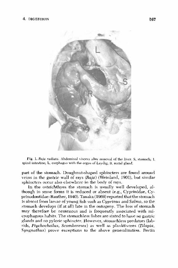
gested food, which may be of large size, and to initiate digestion with pepsin in an acid medium. In elasmobranchs, the stomach is present as a J-shaped organ consisting of a descending pars cardiaca and an ascending pars pylorica (Fig. 1). The inner epithelium of the mucosa
is simple and consists of cylindrical cells which stain with the periodic
acid Schiff (PAS) reagent and which probably secrete mucin. Tubular
multicellular glands running perpendicular to the luminal border
open into mucosal foveolae. The gland cells are of one type only
containing acidophilic granules. They may be designated oxyntic cells
(Hogben, 1967a,b). A muscularis mucosa is present in the cardiac re-
gion only. The muscularis (externa) consists of an inner circular and an
outer longitudinal layer of smooth muscles. The circular muscles are
strongly developed in the pyloric region to form the pyloric sphincter
(Petersen, 1908-1909; Oppel, 1896-1900). In some elasmobranchs, a chamberlike enlargement, the bursa entiana, is formed in the pyloric
4. DIGESTION 167
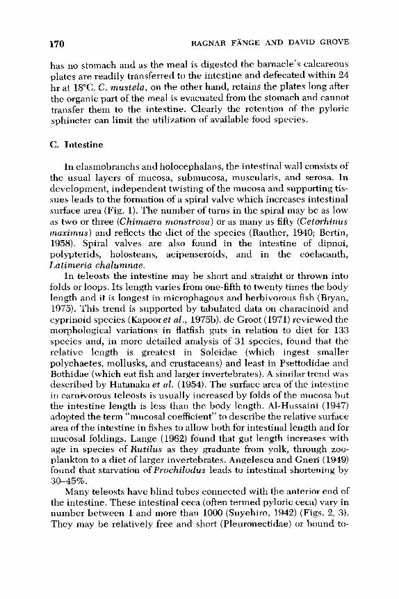
Fig. 1. Raja radiata. Abdominal viscera after removal of the liver. S, stomach; I, spiral intestine; L, esophagus with the organ of Leydig; R, rectal gland.
part of the stomach. Doughnut-shaped sphincters are found around
veins in the gastric wall of rays (Raja) (Weinland, 1901), but similar
sphincters occur also elsewhere in the body of rays.
In the osteichthyes the stomach is usually well developed, al-
though in some forms it is reduced or absent (e.g., Cyprinidae, Cy-
prinodontidae: Rauther, 1940). Tanaka (1969) reported that the stomach
is absent from larvae of young fish such as Cyprinus and Salmo, so the
stomach develops (if at all) late in the ontogeny. The loss of stomach
may therefore be neotenous and is frequently associated with mi-
crophagous habits. The stomachless fishes are stated to have no gastric
glands and no pyloric sphincter. However, stomachless predators (lab-
rids, Ptychocheilus, Scomberesox) as well as planktivores (Tilapia, S yngnczthus) prove exceptions to the above generalization. Bertin

168 RAGNAR FANGE AND DAVID GROVE
(1958) and Vegas-Velez (1972) point out that earlier reviews, tabulat- ing families all of whose members are “stomachless,” may be in error since histological examinations have demonstrated the presence of gastric glands in some species (e.g., Syngnathus, Mugil). Gupta (1971) observed cells resembling gastric gland cells in the digestive canal of the stomachless carnivorous fish, Xenentodon cancila.
Many of the stomachless fishes are provided with pharyngeal chewing devices which permit the ingested food to reach the intestine in a fragmented condition (Rauther, 1940; Bertin, 1958).
Where present, the stomach of teleosts varies greatly in shape. A straight tubelike stomach is found in Gobiidae, Gasterosteiformes, and Symbranchi (Pomatoschistus, Gasterosteus, Spinachia, S ymbran- chus). In flatfish and esocids (Limanda, Pleuronectes, Esox) it is curved, whereas in most fishes the stomach may be shaped like one of the letters U, J, or Y (Suyehiro, 1942). In cottids (Myoxocephalus, Enophrys) and Tilapia the stomach is saclike while in elopids, clupeids, G ymnarchus, Ophiocephalus, Anguilla and many others a
gastric cecum extends caudally. This cecum sometimes is very long (Regalecus, S tomias, Rauther, 1940).
The teleost stomach typically has a lining of columnar epithelial cells, without a striated border. Goblet cells are scattered through the epithelium. Tubular glands, sometimes in groups, are found in the cardiac and fundic regions. The tubules may be simple or occasionally branched, their presence thickening the depth of the mucosa. They open into foveolae. Mucus-producing neck cells may be distin- guished. As in elasmobranchs (and in fact in nonmammalian verte- brates generally) the main part of the gastric glands are made up of one type of cell only. This contains abundant secretory granules, probably pepsinogen (Tan and Teh, 1974), but the cells are believed at the same time to be producers of HC1 (Barrington, 1957; Iro, 1967). According to Weinreb and Bilstad (1955), gastric gland cells in the rainbow trout (Salmo gairdneri) structurally resemble chief cells in other animals. Changes in the microscopic structure of the gastric glands during se-
cretion were reported by Arcangeli (1908). The ultrastructure of gas- tric gland cells in teleosts has been studied by Ling and Tan (1975) in the coral fish, Chelmon rostratus, and by Noaillac-Depeyre and Gas (1978) in the perch, Perca fluuiatilis. The gastric gland cells in their apical region contain a compact system of tubules. The tubules show resemblances to tubular structures of oxyntic cells of amphibians and higher vertebrates and to structures in the chloride cells of cyclo-
stomes (lamprey). At their bases the cells contain zymogenlike secre- tory granules and a rich rough endoplasmic reticulum. The secretory
4. DIGESTION 169
granules are released apically by a process of exocytosis. The ultra-
structural features of the gastric gland cells are consistent with the
hypothesis that they are active both in acid production and in the synthesis of pepsinogen.
Supporting the gastric mucosa is a submucosa often containing a
stratum compactum and smooth muscle fibers (Rauther, 1940; Burnstock, 1959a). The muscularis consists of an inner circular and an outer longitudinal layer of smooth muscles, but striated esophageal muscles may extend into the cardiac portion of the stomach (Perca, Centropristes, Zeus, Solea, Blake, 1930; Rauther, 1940). In the stomiatid Cyclothone the stomach wall contains two layers of
diagonally-crossing striated muscle fibers (Nusbaum, 1923) and in the stomachless cyprinid Tinca a layer of inner circular and outer lon-
gitudinal striated muscle fibers surround the “normal” (smooth mus- cle) muscularis (Kilarski and Bigai, 1971). The inner circular layer of smooth muscles of the fish stomach is usually two to three times thicker than the longitudinal coat, and this is accentuated in fishes provided with a gizzard. The pyloric sphincter consists of thickening of the circular smooth muscle layer. In stomachless fishes the sphinc- ter may be absent or replaced by an esophageo-intestinal valve formed by a fold of the mucosa and submucosa.
The capacity of the stomach in relation to the body weight varies between species and is reflected in the size of the meal that can be taken voluntarily. The flatfish Limanda for example has a gastric vol- ume of 8 m1/100 g and can ingest up to 10% of its body weight in a meal. The stomachless Leuciscus rutilus can consume 15% of its body weight of chironomid larvae, and Carassius carassius 21% of its body weight. Sculpins may ingest 30-50% of their body weight at a single feeding.
Several teleosts possess a gizzardlike enlargement of the pars
p ylorica (Mugil spp., Coregonus, Osphromenus, Chanos, Sardinella, Chatoessus, Citharinus, Mormyrus, Notopterus). In Dorosoma the muscularis of the gizzardlike part of the stomach consists of three strongly developed layers of smooth muscles and has a thick mucous cuticula lining the lumen. Fish with gizzards usually are micropha- gous, detritivores, or herbivores. Mugil cephalus ingests microalgae and plant detritus together with mineral particles which act as a grind- ing paste (Rauther, 1940; Schmitz and Baker, 1969; Odum, 1970).
On of us (D.G.) has observed that when two species of small shore fish (Blennius pholis and Ciliata mustela) are offered barnacles (Balanus balanoides) as food, the fish ingest them readily although only the blenny takes this species in the wild. The blenny, however,
170 RAGNAR FANGE AND DAVID GROVE
has no stomach and as the meal is digested the barnacle’s calcareous plates are readily transferred to the intestine and defecated within 24 hr at 18°C. C . mustela, on the other hand, retains the plates long after the organic part of the meal is evacuated from the stomach and cannot transfer them to the intestine. Clearly the retention of the pyloric sphincter can limit the utilization of available food species.
C. Intestine
In elasmobranchs and holocephalans, the intestinal wall consists of the usual layers of mucosa, submucosa, muscularis, and serosa. In development, independent twisting of the mucosa and supporting tis- sues leads to the formation of a spiral valve which increases intestinal surface area (Fig. 1). The number of turns in the spiral may be as low as two or three (Chimaera monstrosa) or as many as fifty (Cetorhinus maximus) and reflects the diet of the species (Rauther, 1940; Bertin, 1958). Spiral valves are also found in the intestine of dipnoi, polypterids, holosteans, acipenseroids, and in the coelacanth, Latimeria chalumnae.
In teleosts the intestine may be short and straight or thrown into folds or loops. Its length varies from one-fifth to twenty times the body length and it is longest in microphagous and herbivorous fish (Bryan, 1975). This trend is supported by tabulated data on characinoid and cyprinoid species (Kapoor et al., 1975b). de Groot (1971) reviewed the morphological variations in flatfish guts in relation to diet for 133 species and, in more detailed analysis of 31 species, found that the relative length is greatest in Soleidae (which ingest smaller
polychaetes, mollusks, and crustaceans) and least in Psettodidae and Bothidae (which eat fish and larger invertebrates). A similar trend was described by Hatanaka et al. (1954). The surface area of the intestine in carnivorous teleosts is usually increased by folds of the mucosa but the intestine length is less than the body length. Al-Hussaini (1947) adopted the term “mucosal coefficient” to describe the relative surface area of the intestine in fishes to allow both for intestinal length and for mucosal foldings. Lange (1962) found that gut length increases with age in species of Rutilus as they graduate from yolk, through zoo- plankton to a diet of larger invertebrates. Angelescu and Gneri (1949) found that starvation of Prochilodus leads to intestinal shortening by 30-45%.
Many teleosts have blind tubes connected with the anterior end of the intestine. These intestinal ceca (often termed pyloric ceca) vary in number between 1 and more than 1000 (Suyehiro, 1942) (Figs. 2, 3). They may be relatively free and short (Pleuronectidae) or bound to-

4. DIGESTION 171
Fig. 2. Cyclopterus lumpus. Pyloric part of the digestive system in a freshly dis-
sected specimen. In the middle of the figure are numerous intestinal (pyloric) ceca. A
powerful ring-shaped muscle contraction is observed in the stomach (S).
gether to form a compact, glandlike mass (Thunnidae, Xiphidae). Very well developed ceca are found in certain malacopterygians (clupeids,
salmonids), gadids, and coryphaenoids. Their presence is not related
to the relative gut length nor to the general feeding niche of the spe-
cies, yet within a chosen group of fishes (e.g., Heterosomata), it seems possible to relate the number of ceca to the diet (de Groot, 1971).
The intestinal ceca closely resemble the intestine in structure,
with a well-developed muscularis consisting mainly of circular muscle
fibers. The inner epithelium contains goblet cells, but light and elec-
tron microscopic studies indicate that they lack cells that secrete di-
gestive enzymes (Jansson and Olsson, 1960; Luppa, 1966; Vegas- Velez, 1972). Between the ceca highly basophilic pancreatic exocrine
gland cells may occur in the connective tissue. The function of the
ceca is not clear. Since they originate from that region of the intestine

172 RAGNAR FANGE AND DAVID GROVE
Fig. 3. Micrornesistius poutassou. Intestinal (pyloric) ceca in a freshly dissected
preparation. The ceca to the right in the figure are dilated with semitransparent walls.
The ceca to the left show constrictions (arrows) due to contraction of circular smooth
muscles. (N, parasitic nematodes in the connective tissue surrounding the ceca).
where bile and pancreatic juices are released, the ceca may form di-
gestive compartments active in resorption of certain nutrients. It has
been supposed that they are of especial importance for the resorption
of fat and waxes (Greene, 1914; Janson and Olsson, 1960; Benson and
Lee, 1975). The ceca often harbor remarkably large numbers of para-
sites. Thus in the ceca of Coregonus lavaretus Reichenbach-Klinke
and Reichenbach-Klinke (1970) observed up to 300 individual
cestodes.
Stomachless fishes have no intestinal ceca. Other fishes without
ceca include Amia, Chirocentrus, S ymbranchus, Anguilla, Esox, Gas- terosteus, Agriopus, Megalops, Batrachus, silurids, and loricarids
(Jacobshagen, 1915; Pernkopf and Lehner, 1937).

4. DIGESTION 173
The mucosa of the intestine is lined by a simple columnar epithelium which possesses the brush border of microvilli (apical plate) typical of absorptive tissues (Odense and Bishop, 1966; Yamamoto, 1966; Krementz and Chapman, 1975). Cartier and Buclon
(1973) estimated the number of microvilli per mm2 as 34 x 106 in cyprinids. Mucus-secreting goblet cells occur scattered among the epithelial cells and show a positive PAS reaction (Gammon et al., 1972). Multicellular glands are not usually found. A stratum compac- tum is frequently present, but the teleostean intestine seems to lack a
muscularis mucosa. The submucosa is very thin and contains scattered collagen and elastic fibers, blood vessels, and nerves. The muscularis consists of inner circular and outer longitudinal smooth muscles. In cyprinids, cobitids, and S yngnathus, striped muscles may be found in the muscularis (Rauther, 1940). In the tench (Tincn tinca) the striated muscle layer extends over the whole intestine (Ohnesorge and Rauch, 1968; Kilarski and Bigai, 1971).
Ciliated epithelium has been reported in the intestine of a few
teleosts (S yngnathus) and some primitive fishes (Pol ypterus senegalis, Polyodon spathula ) (Ishida, 1935; Bucke, 1971; Weisel, 1973; Magid,
1975) although it may be difficult to differentiate cilia from long mi- crovilli on the basis of structure alone. Observations should always be made on living epithelial cells (Odense and Bishop, 1966). Where present, cilia are probably a primitive character since they are found in Amphioxus and in ammocoete larvae. In the cyclostome Myxine glutinosa the intestinal epithelium produces a peritrophic membrane (Adam, 1960).
Rectum. The terminal .part of the intestine is frequently differ- entiated as a wider “rectum,” often demarcated from the intestine by
an ileo-rectal valve largely formed by smooth muscles (Gadus, Gam- busia), but in many species no valve is present (Mohsin, 1962). The mucosa is typically richly endowed with goblet cells. The mucosal epithelium may be provided with microvilli indicating strong absorp- tive properties (Bullock, 1967). In elasmobranchs the epithelium may be stratified. The rectal gland of elasmobranchs (Fig. 1) is now be- lieved to secrete Na and C1 ions into the lumen as part of the osmo- regulatory mechanisms. The muscularis of the rectum is similar to that of the intestine, but striated muscles occur near the anus.
D. Liver and Gallbladder
The liver lies anteriorly in the body cavity and receives blood from the hepatic artery and from one or more portal veins which drain the gastric and intestinal mucosa, the swimbladder (gas gland), the spleen,

174 RAGNAR FANGE AND DAVID GROVE
and the pancreas. Embryologically, the liver develops as an epithelial outgrowth of the digestive tube, and in its simplest form is a branched tubular gland as in the cyclostome Myxine glutinosa. In some teleosts the liver is claimed to have a tubular structure (Anguilla anguilla, Muraena, Pleuronectes). However, in most vertebrates, including
fishes, the liver is a complicated structure consisting of anastomosing epithelial lamellae separating blood sinusoids (Elias and Sherrick, 1969). This “muralium” has been found in fishes as diverse as lam- preys (Bengelsdorf and Elias, 1950), Salmo gairdnerii (Scarpelli et al., 1963), Poecilia (Hale, 1965), Zctalurus (Hinton and Pool, 1976), and Micropterus (Hinton et al., 1972). As in other vertebrates, hepatocytes contain numerous mitochondria, rough endoplasmic reticulum (mi- crosomes), Golgi apparatus, lysosomes, peroxisomes, lipid and glyco- gen deposits. Intracellular bile canaliculi are present, with the single exception to date ofIctalurus (Hinton and Pool, 1976), which link up with intercellular canaliculi. The lining epithelial cells of the canali- culi possess numerous microvilli (David, 1961; Yamamoto, 1965a,b). According to Welsch and Storch (1973) the teleostean liver contains two categories of hepatocytes, lipid-rich and glycogen-rich. In some species (Tetraodon, Liinanda) lipid-rich cells predominate while in others (Chromis, Corydoras, Amphiprion) glycogen-rich cells are more common. Hinton and Pool (1976) concluded that, in Zctalurus,
large protruding cells, which differ from hepatocytes lining the sinusoids, are Kupffer cells of the reticulo-endothelial system. Lipids may be present in large quantities in some fish livers. In elasmo- branchs, squalene and other hydrocarbons accumulate in the liver, helping the fish to gain buoyancy (Corner et al., 1969).
Due to the presence of a large fat vacuole the hepatocytes of elasmobranchs are larger than those of teleosts. In most fishes the exocrine secretion of the liver, the bile, is stored in a gallbladder. The bile duct opens into the anterior intestine or into the intestinal ceca (Western, 1969). The wall of the gallbladder consists of columnar or
cuboidal epithelium, a thin submucosa and a muscularis of smooth muscle cells. The similarly constructed ductus choledochus is associ- ated with the pancreatic duct in many fish, and its entrance to the intestine is guarded by the smooth muscle sphincter Oddi.
E. Pancreas
In the cyclostome Myxine glutinosa (hagfish) the pancreas consists of exocrine gland cells, containing zymogen granules, in the intestinal mucosa. The homologue of the endocrine pancreatic tissue of higher

4. DIGESTION 175
vertebrates is concentrated around the bile duct. In elasmobranchs
and holocephalans, the exocrine pancreas forms a discrete organ with a duct emptying into the anterior intestine (duodenum). The micro- scopic structure may be much like that of the mammalian pancreas [e.g., in Chimaera monstrosa (Fujita, 1962)l and numerous scattered islets of endocrine cells are present. In elasmobranchs the spleen and the pancreas are often closely associated. This is also the case in Pro- toptems, the African lungfish, in which the pancreas is a large black- pigmented organ at the posterior end of the spleen and, like the latter, embedded in the gut wall. In teleosts the exocrine pancreas is usually diffuse. It consists of ramified tubules or acini scattered in the connec- tive tissue of the intestinal surface, the mesenteries, between the in- testinal ceca or within the liver or spleen (Nagase, 1964; Bishop and Odense, 1966; Western, 1969; Gammon et al., 1972; Hinton and Pool, 1976). In the mesenteries the pancreas forms sheaths around blood vessels. Several small pancreatic ducts open into the intestine or intes-
tinal ceca, although in some cases a single duct may join the bile duct.
In cyprinids (Cyprinus carpio), labrids (Crenilabrus melops), and cer- tain other forms, strands of pancreatic tissue are found within the portal canals of the liver, the pancreatic ducts probably opening into
the bile ducts. The combined liver and pancreatic tissue has been termed “hepatopancreas” but must not be confused with so-called hepatopancreas of invertebrates (It0 et al., 1962; Elias and Sherrick, 1969). The exocrine cells of the teleostean pancreas are strongly basophilic and contain zymogen granules. In a minority of teleosts the pancreas forms a distinct organ which is embedded in fat and connec- tive tissue. The compact pancreas accompanies portal vessels on the surface of the intestine (Esox, Silurus, Parasilurus, Anguilla, Conger, Acerina, Pleuronectes, Oppel, 1896-1900; Hill, 1926; Chesley, 1934;
Suyehiro, 1942; Kukla, 1954, 1958). The secretory cells form acini with narrow lumina, and a common pancreatic duct opens into the anterior intestine. Blood reaches the pancreas from three arteries and drains into the v. portae (Kukla, 1958). For a discussion of the endocrine elements associated with the pancreas, see Section II1,F.
F. Endocrine Cells and Hormones
The vertebrate alimentary canal and certain other organs contain large numbers of granulated endocrine cells. Two lines of these cells have been termed enterochromaffin (argentaffin) and entero- chromaffinlike (argyrophil) cells. When supplied with amine precur- sors these cells produce and store amines. For that reason they have

176 RAGNAR FANGE AND DAVID GROVE
been termed APUD cells [abbreviation for Amine Precursor Uptake and Decarboxylation (Pearse, 1977)l. Another term is gastro-entero- pancreatic (GEP) cells (Solcia et al., 1978). A classification on ultra-
structural grounds of mammalian GEP cells has been suggested (Solcia
et al., 1978). Several investigators have demonstrated GEP cells in nonmammalian vertebrates such as cyclostomes and true fishes (Ug- geri, 1938; Read and Burnstock, 1968; Bucke, 1971; Gabe and Matoja, 1971, 1972; Ling and Tan, 1975; Tue, 1975; Ostberg, 1976; Falkmer et al., 1978). These endocrine elements are also found in protochordates (amphioxus: Van Noorden and Pearse, 1976).
The GEP cells are assumed to synthesize low molecular weight polypeptides which act as hormones. The various polypeptides show structural resemblances to each other and the hypothesis has been forwarded that different polypeptide hormones (glucagon, secretin, gastrin) evolved together with insulin from an ancestral proinsulinlike molecule (Track, 1973). Several gastrointestinal polypeptide hor- mones occur in fish (Table I). Nilsson (1970, 1973) obtained evidence for the presence of secretin- and cholecystokininlike substances in the
Table I
Hormones and Hormonelike Substances in Fish Gut and Pancreas
Substance Tissue Species Reference
Insulin
Gastrin
Secretin Secretin
Secretin Cholecystokinin
(CCK) CCK
Cholecystokininlike
substance Cholecystokininlike
substance
Ceruleinlike substance
Substance P
Histamine
Histamine Histamine
Islets
Islets
Intestine Intestine
Intestine Intestine
Intestine
Intestine
Intestine
Stomach
Intestine
Stomach
Stomach Intestine
All
Rhinobatus productus
Myxine glutinosa Salmon, dogfish, ray
Esox, Gadus Myxine glutinosa
Chimaera monstrosa
Lampetra, Petromyzon
Esox, Anguilla
Gadus morhua
Gadus morhua, Squalus acnnthias
Salmo gairdnerii, Gadus morhua
Esox lucius
Myxine glutinosa
Hansen (1975)
Nilsson (1970, 1973) Bayliss and Starling
Dockray (1975) Nilsson (1973)
Nilsson (1970)
Barrington (1972)
Barrington and Dockray
Larsson and Rehfeld
Dahlstedt et al. (1959)
Reite (1969)
Lorentz et al. (1973) Reite (1965)
(1903)
(1972)
(1978)

4. DIGESTION 177
intestine of the hagfish (Myrine glutinosa) and the holocephalan
Chimaera monstrosa. Gabe and Matoja (1972) demonstrated cells
structurally resembling mammalian gastrin cells in the stomach mu-
cosa of the teleost, Mugil auratus. Ostberg et al. (1976) showed, in
immunofluorescence studies, that the intestinal mucosa of cyclo-
stomes contains cells that react with antisera against mammalian glu-
cagon and gastrin. Larsson and Rehfeld (1978) found that the gastric
mucosa of the cod (Gadus morhua) contains a rich population of cells
demonstrable with antisera against mammalian gastrin and chole-
cystokinin (CCK), but the immunogenic reactivity was due to a
ceruleinlike polypeptide. The authors concluded that, in teleosts,
ceruleinlike molecules possibly function as gastrin.
The endocrine pancreas belongs to the GEP system. Islets of en-
docrine cells are found in the compact pancreas of the eel (Anguilla anguilla: Kukla, 1958) and the 'pike (Esox lucius: Bucke, 1971). In
teleosts with a diffuse pancreas (Gadus morhua, Lophius piscatorius, Myoxocephalus scorpius) aggregations of endocrine cells, separate from exocrine pancreas tissue, form the Brockmann bodies. Within the
teleostean islet tissue Falkmer and Olsson (1962) and Bishop and
Odense (1966) detected inner B cells (insulin secreting) and more
peripheral A cells (glucagon secreting) together with other cell types.
According to Brinn (1973) the islets of some fish species contain, in
addition to A and B cells, D cells (somatostatin secreting) and a fourth argyrophilic type of granulated cell.
The pancreatic islets are innervated by autonomic nerves, and ner-
vous influence may be a factor in the regulation of endocrine pancrea-
tic secretion. The polypeptide hormones produced by the pancreatic
islets are regulators of nutrient homeostasis. The rates at which glu-
cose, amino acids, and free fatty acids enter and leave the extracellular
space are influenced by insulin, glucagon, and probably somatostatin
(Unger et al., 1978). However, most studies have been done on mam- mals and very little is known about fishes in these respects.
The peptide-producing endocrine cells of the vertebrate digestive
system have been investigated by various methods: immunohisto-
chemistry, silver staining, lead-hematoxylin staining, formaldehyde-
induced fluorescence, radioactive labeling, and enzyme studies (Daw-
son, 1970). Besides gastrointestinal polypeptides, other hormones or hor-
monelike substances occur in the digestive system of fishes, for exam-
ple, histamine. Holstein ( 19754 found diamine oxidase, a histamine- deaminating enzyme, in the intestine of teleosts. GEP cells are
thought to be the source of the intrinsic factor in the mammalian

178 RAGNAR FANGE AND DAVID GROVE
stomach. I t is not known if this factor, needed for the intestinal absorp-
tion of iron, occurs in fishes.
G. Blood and Lymph Vessels
In the cyclostome, Myxine glutinosa, the intestine is supplied by
segmental arteries and the venous drainage goes to the hepatic portal
system. In elasmobranchs (sharks, rays) and dipnoans (lungfish) the
intestine receives three or four arteries, whereas in teleosts one single
celiaco-mesenteric artery supplies the digestive canal. In teleosts the
arterial branches to the intestine are often closely followed by veins
(Grodzinski, 1938), an arrangement reminiscent of the rete mirabile in
the fish swimbladder. Well developed retia mirabilia were further
described in the portal, hepatic, intestinal and splenic veins of tuna
fish and the elasmobranch,Alopias vulpes, by Muller as early as 1840.
Lymphatic vessels and sinuses are found in the digestive system of both cyclostomes and true fishes. In the hagfish, Myxine glutinosa, a
superficial lymphatic plexus is found in the intestine (Fange, 1973). In
some teleost species the intestine has a system of chyle vessels similar
to those of mammals. The chyle vessels converge into a large vessel,
the vas 1 ymphaticum intestinale. In the marine catfish (Anarrhichas
lupus) this intestinal lymph vessel is strongly developed and situated
to the right of the esophagus (Glaser, 1933). It is not known whether
intestinal lymph vessels in fishes are of importance for the transport of
absorbed fat from the intestine.
H. Lymphoid Tissue
In most vertebrates lymphoid or hematopoietic tissue is associated
with the digestive canal. Thus in the hagfish, Myrine glutinosa, the
intestinal submucosa contains an extensive granulocyte-producing tis-
sue, assumed to constitute a primitive spleen (Tomonaga et al., 1973).
Granulocyte-producing tissue is further found in the submucosa of the
esophagus of elasmobranchs (organ of Leydig, Petersen, 1908-1909; Fange, 1968). Lymphocytes and granulocytes have been observed to
invade certain layers of the intestine of fishes, such as membrana
propria and epithelium. This infiltration of the gut by leukocytes prob-
ably plays a role in a defense system against microbes and parasites. In elasmobranchs the granulocyte-producing tissue of the esophagus
is rich in lysozyme (bacteriolytic enzyme) and chitinolytic enzymes.
The latter perhaps give some protection against chitin-containing

4. DIGESTION 179
parasites ( F h g e et al., 1978). Whether leukocytes of the gut are of a n y
direct importance for the digestion is not known.
IV. DIGESTIVE FLUIDS AND ENZYMES
A. Gastric Secretion
Production of an acid gastric fluid probably occurs in most fishes (e.g., Norris et al., 1973) except those which have no stomach (myxinoids, chimaeroids, many teleosts). In these neither HCI nor pepsin is formed in the gut.
1. HYDROCHLORIC ACID
Large amounts of gastric fluid, usually distinctly acid, are found in elasniobranchs. Weinland (1901) used a glass cannula to obtain 50 ml gastric fluid from a nonfed Scyllium, and Yung (1899) took 500 ml from the large shark, Lamna cornubica. In the gastric fluid of Scyllium
stellare pH 1.69 was measured (van Herwerden and Ringer, 1911), but the acidity of the fluid varies. Maximal acidity is observed a few hours after food intake, whereas in the absence of food the gastric fluid may be weakly acid or neutral (Bernard, 1952). Weinland (1901) found both acid and alkaline secretion in the ray, Raja asterias. Babkin et al. (1935a,b) obtained small quantities of gastric fluid with a pH of 3-3.8 from nonfed Raja sp.
Acid gastric fluid has regularly been found in teleosts (e.g., West- ern and Jennings, 1970). p H values of 3.0-5.0 were measured in the stomach of Micropterus salmoides, Perca jluviatilis, and Tilapia mossambica (Sarbahi, 1951; Fish, 1960). The acidity increases after food intake (MacKay, 1929). Western (1971) noted that in Cottus scor- p i 0 and Enophrys (Cottus) bubalis the gastric content was neutral in the absence of food, but 30 hr after ingestion of food a p H of 2.0 was measured. A pH of 2.0 is reached in the stomach of Tilapia a few hours after daily feeding begins (Moriarty, 1973).
2. ENZYMES
The fact that optimal proteolytic activity has been reported at dif- ferent pH’s (2.0, 3.0, 5.0, 8.5) (Creac’h, 1963) indicates that the gastric fluid of fishes contains several types of protease. Pepsin undoubtedly is the major acid protease. It is secreted by the gastric gland cells as a

180 FiAGNAR FANGE AND DAVID GROVE
zymogen called pepsinogen, which is inactive. Conversion of pep-
sinogen into active pepsin is brought about by pepsin in an acid envi-
ronment. During the activation process amino acids are split off from
the NH2-terminal end of the molecule as a mixture of peptides
(Lehninger, 1971). Pepsin is an endopeptidase which cleaves peptide
linkages formed by amino groups of aromatic and acidic amino acids.
It attacks most proteins but not mucins, spongin, conchiolin, keratin,
or low molecular weight peptides (Sumner and Somers, 1947).
Merret et al. (1969) isolated four different pepsinogens from the
gastric mucosa of a dogfish (Mustelus canis). Crystalline pepsin has
been prepared from the gastric mucosa of teleosts, the Pacific King
salmon (Oncorhynchus tschawitscha) (Norris and Elam, 1940) and the
halibut (Hippoglossus hippoglossus) (Eriksen, 1945). In these investi-
gations the pepsinogen was extracted by alkaline solutions and then
transformed into pepsin by the addition of acid. Extraction with an
acid solution was used by Norris and Mathies (1953) in the isolation of
pepsin from the tuna fish (Thynnus albacores). The purified tuna fish
pepsin differed crystallographically and in its amino acid composition
from porcine pepsin.
A few nonproteolytic digestive enzymes may occur in the gastric
fluid of fishes. Amylase has been found in the stomach of some fishes
(Clupea harengus, Battle, 1935; Dorosoma cepedianum, Bodola,
1966). Lipase is reported from the stomach of Tilapia sp. (Al-Hussaini
and Kholy, 1953; Nagase, 1964) and Dorosoma cepedianum (Bodola,
1966), and esterases with pH optima at 5.3 and 8.0 in the stomach of
the rainbow trout (Salmo gairdnerii, Kitamikado and Tachino, 1960). Chitinase has been demonstrated in the gastric mucosa of elasmo-
branchs, insect-feeding teleosts, and the dipnoan Polypterus (Okutani,
1966; Micha et al., 1973). Remarkably strong chitinolytic activity was
found in the gastric mucosa of the marine deep water teleost, Cory-
phaenoides rupestris, and the elasmobranchs, Etmopterus spinax and Raja radiata (Fange et al., 1978). These three species largely
feed on crustaceans and other invertebrates with a chitinous integu-
ment.
Hyaluronidase, a mucopolysaccharidase which cleaves beta-N-
acetyl-glucosaminidic bonds of hyaluronic acid, has been prepared
from the gastric mucosa of the Japanese mackerel, Scomber japonicus
(Yamamoto and Kitamikado, 1971). Among 148 fish species, a few estuarine fishes and one. freshwater
fish (Zctalurus punctatus) but no Qffshore marine species were found
to contain cellulase in their stomach or anterior intestine. Because no
cellulase activity was detected in fishes exposed to streptomycin, it

4. DIGESTION 181
was concluded that the cellulase probably was produced by micro-
organisms of the gut content (Stickney and Shumway, 1974).
B. Pancreatic Secretion
Due to difficulties in collecting pure pancreatic juice in most fishes
the chemical composition of this fluid in representative fish species is
not known. Undoubtedly it is rich in enzymes (mostly as zymogens)
which serve in digestion of proteins, carbohydrates, fat, and nuc-
leotides. Probably, as in higher vertebrates, the pancreatic juice con-
tains bicarbonates that neutralize hydrochloric acid entering the intes-
tine. Extracts containing pancreatic enzymes have been obtained from
the isolated compact pancreas of elasmobranchs and holocephalans,
from tissues containing scattered pancreatic acini (intestinal ceca,
mesenteries, the “hepatopancreas” of certain fishes), and from the in- testinal content.
1. PROTEASES
Trypsin, chymotrypsin, carboxypeptidase, and elastase are stored
in the pancreatic cells as inactive zymogen (proenzyme) granules.
When arriving in the intestinal lumen, trypsinogen is transformed into
trypsin by proteases produced by intestinal mucosal cells (entero-
kinases). Other pancreatic zymogens are activated by trypsin.
Trypsin is formed by removal of a hexapeptide from the trypsino-
gen molecule as a result of the hydrolysis of a lysine-isoleucine bond.
Trypsin is an endopeptidase with optimal action at a pH of about 7. It cleaves peptide linkages yhose carbonyl groups come from arginine
or lysine (Lehninger, 1971). Appropriate substrates for estimation of
trypsin activity are synthetic peptides such as benzoyl-L-arginine
ethyl ester (BAEE) and p-toluenesulfonyl-L-arginine methyl ester
(TAME). Trypsinogen, or trypsin, has been demonstrated in the intes-
tinal wall of the cyclostome Myrine glutinosa (Nilsson and Fange, 1970), the pancreas of the holocephalan Chimaera monstrosa (A.
Nilsson and Fange, 1969), elasmobranchs (Ginglymostoma cirratum, Squalus suckleyi, Zendzian and Barnard, 1967), and the African lung-
fish (Protopterus aethiopicus, Reeck et al., 1970; Reeck and Neurath,
1972), and from intestinal ceca of various teleosts (Creac’h, 1963; On- corhynchus, Croston, 1960, 1965; Gadus morhua, Overnell, 1973; Di-
centrarchus (Morone) labrax, Alliot et al., 1974). The lungfish (Protop- terus aethiopicus) pancreas also contains a trypsin inhibitor (Reeck et al., 1970). Jany (1976) studied digestive endopeptidases in the

182 RAGNAR F m G E AND DAVID GROVE
stomachless teleost Carassius auratus gibelio. Trypsin and chymo- trypsin of pancreatic origin were found but no pepsin, no elastase, and no collagenase.
Chymotrypsin is formed by the action of trypsin on chymo- trypsinogen. It is an endopeptidase which attacks peptide bonds with
carbonyl from aromatic side chains (tyrosine, tryptophan, phenylalanine). Benzoyl-L-tyrosine-ethylester (BTEE) is an example of a synthetic peptide useful as a substrate in assays of chymotrypsin activity. Chymotrypsin has been found in most fishes in which trypsin has been found.
Elastase is formed when the zymogen, proelastase, is activated by trypsin. This enzyme is especially active on peptide bonds in the protein elastin and may be assayed on purified elastin or specific ester substrates (De Haen and Gertler, 1974). Elastase probably does not occur in the cyclostome Myxine glutinosa (A. Nilsson and Fange, 1970) but has been found in the pancreas of the holocephalan Chim- aera monstrosa (A. Nilsson and Fange, 1969), the elasmobranch
Dasyatis americana, and the teleosts, tuna (Thynnus secundodorsalis) (Zendzian and Barnard, 1967) and the angler, Lophius piscatorius (Lansing et al., 1953). A new type of pancreatic elastase, proelastase A, has been reported to occur in a dipnoan, the African lungfish (Walsh, 1970).
Carboxypeptidases are exopeptidases which hydrolyze the termi- nal peptide bonds of their substrates. Carboxypeptidases A and B, differing in their specificities, are formed by activation of procarboxy- peptidases by trypsin. Mammalian carboxypeptidase A is a zinc- containing enzyme (Vallee, 1955). Carboxypeptidase A, but not carboxypeptidase B, was found in intestinal extracts of the cyclostome Myxine glutinosa (A. Nilsson and Fange, 1970). Carboxypeptidases have further been found in an elasmobranch (Squalus acanthias, Prahl and Neurath, 1966a,b) and in the teleost, Dicentrarchus Zabrax (Alliot et al., 1974). Zendzian and Barnard (1967) showed carboxypeptidase B-like activity in the tuna fish, and Ooshiro (1968, 1971) found carboxypeptidase A-like activity in the intestinal ceca of another tele- ost, the mackerel (Scornber japonicus). In the Japanese mackerel the carboxypeptidase A-like activity appeared to depend on Co2+ ions rather then ZnZ+ ions.
Evolution of pancreatic proteases. Vertebrate trypsin, chymotryp- sin, and elastase are structurally related to each other and, together with a few other proteolytic enzymes, are called serine proteases as
they contain serine in the active site of the molecule. On the basis of analyses of amino acid sequences of purified trypsinogen from differ-

4. DIGESTION 183
ent vertebrates, Reeck and Neurath (1972) suggested a possible scheme for the evolutionary changes in the structure of the activation
peptide of trypsinogen. The lungfish (Protopterus) trypsinogen at the
molecular level shows some resemblance with invertebrate trypsins.
It has been speculated that different serine proteases have evolved from a common ancestor molecule (Walsh, 1970; Stryer, 1974).
2. AMYLASE
In some plant-feeding teleosts such as Tilapia, amylase has been
found in all parts of the digestive tract (Nagase, 1964; Fish, 1960). In a carnivore, the perch (Perca Juviatilis), on the other hand, digestive
amylase is confined to the diffuse pancreas of the connective tissues
surrounding the intestine. According to Nagase (1964) the Tilapia amylase has a pH optimum at 6.71, and the p H of the stomach is too low for the enzyme to show any appreciable activity. Pancreatic and intestinal amylase probably are more important than gastric amylase in carbohydrate digestion in Tilapia.
3. CHITINASE
Chitinase occurs in the digestive system of many fishes and other vertebrates (e.g., Yoshida and Sera, 1970), notably in forms feeding on
insects (Micha et al., 1973; Dandrifosse, 1975) or crustaceans. Excep- tionally high chitinase activity was found in extracts of the pancreas of
the stomachless holocephalan Chimaera monstrosa, a fish that feeds
largely on shrimps (Fange et al., 1976, 1978). Little or no pancreatic chitinase was detected in some other marine fish species with similar
diets, but these instead possessed strongly active chitinase in the gas-
tric mucosa. The Chimaera pancreatic chitinase has a strong optimum
around pH 8-10, while gastric chitinases from other species show pH optima at 1.25-3.6. Chitinase splits chitin into dimers and trimers of N-acetyl-D-glucosamine (NAG), which may be further broken down by glucosaminidase (NAGase). This enzyme occurs together with chitinase in the digestive tract. NAG, the end product of the chitinoly- tic process, probably is of nutritive value since it is resorbed faster than glucose by the intestine (Alliot, 1967). Although chitinase may be produced by intestinal bacteria, most chitinases are synthesized by
gastric or pancreatic gland cells.
4. LIPASES
Lipases are esterases which split ester bonds. Triglyceride fats, phospholipids, and wax esters are hydrolyzed by lipase. Although

184 RAGNAR FANGE AND DAVID GROVE
lipase activity has been demonstrated in various parts of the fish diges-
tive system the pancreas is probably the major source (Barrington,
1957; Kapoor et al., 1975b). According to Chesley (1934) lipase is more
abundant in fishes with a compact pancreas than in those with a dif- fuse pancreas. Brockerhoff (1966) by in vivo experiments found lipo-
lytic activity in the intestinal content of cod (Gadus morhua) but
Brockerhoff (1966) and Overnell (1973) both failed to find any lipase
activity in extracts of the pyloric ceca or adjacent tissues. Leger (1972)
partly purified a lipase from the rainbow trout (Salmo gairdnerii). Patton et al. (1975) found lipase activity in the bile of two marine
fishes, the anchovy (Engraulis mordax) and the jack mackerel
(Truchurus s ymmetricus). These authors suggested that in fishes not
pancreatic lipase (EC 3.1.1.3), but another enzyme may function as the
major fat-digesting enzyme.
5. OTHER ENZYMES
Alkaline RNase and phosphodiesterase have been purified from
the intestinal ceca of rainbow trout (Salmo gairdnerii, Imura,
1974a,b). An increased activity of carbonic anhydrase found in the gut
of coral fishes is supposed to be an adaptation to ingestion of calcium
carbonate (Smith, 1975).
C. Bile
Production of a detergent-containing fluid, bile, by the liver is
found in all vertebrates. Usually the bile is stored in a gallbladder
with contractile walls. By contraction of the smooth muscles of the
galIbIadder the bile is ejected into the lumen of the intestine. During
its storage in the gallbladder the bile becomes more concentrated.
Mammalian bile contains bile salts, cholesterol, phospholipids, bile
pigments, organic anions, glycoproteins, and inorganic ions. Fish bile
has a similar composition. It is weakly alkaline and has a high sodium
and a low chloride concentration (Hunn, 1972). Bile salts are special
types of steroids which are synthesized in the liver from cholesterol.
In fishes (carp, Cyprinus carpio) as in other vertebrates, administra-
tion of 14C-labeled cholesterol results in production of radioactive bile
salts. In Myxine glutinosa (cyclostome), Chimaera . monstrosn (holocephalan), elasmobranchs, dipnoans, and Lntimeriu (coelacanth)
the bile contains bile alcohol sulfate esters (as sodium salts), but in
teleosts as in higher vertebrates the bile contains salts formed with
taurine conjugates of bile acids. However, in one group of teleosts, the

4. DIGESTION 185
cyprinids (carp fishes) a bile alcohol sulfate is the principal bile salt
(Haslewood, 1968). Bile salt molecules have hydrophilic and hyd- rophobic groups and in solution at a critical concentration they forin
aggregates called micelles.
In some teleosts the bile contains trypsin, lipase, amylase, or other
enzymes from the intrahepatic pancreatic tissue (Babkin and Bowie,
1928). Bile which contains lipase is free from phospholipids (Patton et al., 1975). In fishes as in mammals a large proportion of the secreted
bile salts are presumably resorbed from the intestine into the blood
and to a large extent returned to the liver. This so-called enterohepatic
circulation concerns both bile salts and other bile components.
D. Intestinal Enzymes
Digestive enzymes produced by intestinal cells are located mainly
in the brush border of the epithelium (Ugolev and Kooshuck, 1966; Matthews, 1975). However, enzymes in intestinal extracts may partly
be pancreatic, as pancreatic enzymes have the tendency in the intes-
tine to adsorb to the glycocalyx of the epithelial cells. Enzyme ac-
tivities of the fluid of the intestinal lumen, with the exception of the
anterior part of the intestine where pancreatic juice is delivered, are
low. Cells or fragments of cells continuously released from the intesti-
nal epithelium, extracellular enzymes from the stomach and the pan-
creas, and enzymes of the ingested food inay contribiute to enzyme
activities in the lumen.
Enzymes thought to be produced by the intestinal mucosa include
aminopeptidases, di- and tripeptidases (formerly termed erepsin), al-
kaline and acid nucleosidases (which split nucleosides), poly-
nucleotidases (which split nucleic acids), lecithinase (which splits
phospholipids into glycerol, fatty acids, phosphoric acid, and choline),
lipase and other esterases, and various carbohydrate-digesting en- zymes: amylases, maltase, isomaltase, sucrase, lactase, trehalase, and
laminarinase. The knowledge of intestinal enzymes in fishes is frag-
mentary. Piavaux ( 1973) found laminarinase (p-D- 1,3-glucan gluco-
hydrolase, EC 3.2.1.58) in intestinal extracts of Tilapia rnacrochira, an
African freshwater teleost, which feeds on plankton and plant detritus.
Amylase activity is considerably higher in the intestine of herbivorous
species such as the carp (Cyprinus carpio), than in the intestine and
intestinal ceca of more carnivorous forms such as salmon (Salrno), cod
(Gadus rnorhua), and flounder (Pleuronectes) (Phillips, 1969; Kapoor
et al., 1975b). Dipeptidase activity was investigated in the intestine of the white grunt (Haernulon plurnieri) using synthetic dipeptides as
186 RAGNAR FANGE AND DAVID GROVE
substrate. The highest activity was measured in the anterior half of the
intestine.
E. Regulation of Secretory Activities
1. GASTRIC ACID SECRETION
a. Elasmobranchs. Continuous secretion of very small amounts of
gastric acid in fasting rays (Raja sp.) was not influenced by vagotomy
or atropine but was inhibited by adrenaline or sympathetic nerve
stimulation. Spinal destruction, probably due to elimination of tonic
sympathetic nerve influence resulted in a “paralytic secretion” of gas-
tric juice (Babkin et al., 1935a). An influence of vascular sphincters on
the composition of the gastric juice was assumed by Weinland (1901)
who observed that in the ray, Raja asterias, treatment with an ergot
preparation caused contraction of the vascular sphincters and alkalin-
ity of the gastric juice. Ungar (1935), working with the isolated per-
fused stomach of Torpedo, Squalus and Scyliorhinus, found gastric
secretion to be stimulated by acetylcholine and histamine. Hogben
(1967a,b) found that the isolated gastric mucosa of the spiny dogfish
(Squalus acanthias) secretes an acid juice spontaneously. Both his-
tamine and the cholinergic agent carbachol increase the rate by 100-
150%, but carbachol was 200 times more effective than histamine. No
effect was obtained with a preparation of porcine gastrin, a peptide which causes secretion of gastric fluid in higher vertebrates.
b. Teleosts. In the living fish distension of the stomach serves as a
powerful stimulus for gastric secretion (Smit, 1968; Norris et al., 1973).
Probably the effect is due to reflex activation of vagal cholinergic fib- ers. The secretion of gastric acid appears to be intermittent; that is,
acid is produced only in connection with digestion or when otherwise
stimulated (Gzgzyan et al., 1968). Histamine is an effective stimulus for gastric acid secretion in the
European catfish (Silurus glanis: Gzgzyan et al., 1968) and in the cod
(Gadus morhua: Holstein, 1975b). The facts that exogenous histamine causes acid secretion and that the fish gastric mucosa contains consid-
erable amounts of non-mast-cell histamine (Reite, 1969; Lorentz et al., 1973) indirectly indicate that histamine has a physiologic function
in the regulation of acid secretion.
In studies of gastric acid secretion in the cod (Gadus morhua) Holstein (197513, 1976, 1977) found only a low basal output of acid

4. DIGESTION 187
(5-10 pmol/kg hr). However, the method used to collect gastric juice
involved ligating the pylorus, which seriously interferes with the
water balance. In spite of their dehydrated condition, the fishes were
able to secrete considerable amounts of gastric acid when injected
with histamine or carbacholine. Both effects were blocked by the HZ- receptor antagonist metiamide. This provides evidence that histamine
is physiologically important as a regulator of acid secretion in fish.
Further experiments (Holstein, 1978 personal communication)
showed that fishes kept in water balance either by perfusion of the
intestine with 33% seawater or by intramuscular injection of
hypotonic saline show a relatively high “basal” secretion of acid (50- 100 Fmol of H+/kg hr). The intense secretory response after carba-
choline is accompanied by vasodilation. The response is blocked by
atropine (Holstein, 1977). The question whether gastric secretion in fishes is influenced by
gastrin or other GEP hormones is undecided. Holstein (1975b) found
no stirnulatory effect of pentagastrin in the cod (Gadus morhua), but
the experiments were made on fishes not in water balance. Larsson
and Rehfeld ( 1977) suggested that in nonmammalian vertebrates
ceruleinlike peptides may serve as “gastrin.” In isolated frog stomach (Rann) cerulein stimulates gastric secretion more powerfully than gas-
trin (Negri and Erspamer, 1973).
2. PEPSINOGEN SECRETION
In mammals the secretion of pepsinogen is induced by vagal im-
pulses (Hirschowitz, 1975). The stomach of fishes is richly supplied by
vagal fibers, but it is not known whether these have any influence on
the pepsinogen producing cells.
3. PANCREATIC SECRETION
In mammals the pancreatic juice is secreted as the result of stimu-
lation of the exocrine pancreas cells by peptide hormones produced by
cells in the anterior intestine and the stomach. The pancreas-
stimulating principle of the anterior intestine was termed “secretin”
by Bayliss and Starling (1903), but later investigations have shown the
existence of more than one hormone which stimulate the pancreas.
Thus, in mammals secretin produces a thin watery pancreatic fluid
rich in bicarbonate, whereas cholecystokinin (CCK) stimulates a se-
cretion rich in enzymes. Other hormones such as gastrin (regulates

188 RAGNAR FANGE AND DAVID GROVE
gastric secretion) and cholinergic agents (acetylcholine, carbachol,
niecholyl) also stimulate the mammalian pancreas.
Babkin (1929, 1933) was able to stimulate pancreatic secretion in a
ray (Raja) by the introduction of hydrochloric acid into the anterior
intestine. I t seems plausible that in elasmobranchs as in mammals,
introduction of acid stomach content into the intestine causes release
of pancreas-stimulating humoral substances.
4. SECRETION AND RELEASE OF BILE
Release of bile into the intestine is produced by contraction of the
smooth muscles of the muscularis of the gallbladder. In the hagfish
(Myxine glutinosa) this contraction is probably brought about by cholinergic vagal influences (Fange and Johnels, 1958). CCK-PZ,
which causes contraction of the mammalian gallbladder is probably
present in some fishes (see Table I) . In a ray, Raja erinacea, the pro-
duction of bile by the liver seems to take place continuously but in
linear relation to the portal vein pressure. The isolated perfused liver
continues to produce bile (Boyer et al., 1974).
F. Intestinal Microorganisms
It has been suggested that in certain species of fish the decomposi-
tion of food components by microorganisms may be of importance for
digestion. Nitrogen-metabolizing bacteria may explain the capacity to
utilize urea in the food by the mullet (Mugil auratus) (Albertini-
Berhaut and Vallet, 1971). Okutani ( 1966) found chitinolytic bacteria
in the intestine of the marine teleost, Lateolabrax. These bacteria
were gram-negative motile rods with a polar flagellum (Vihrio). The bacterial chitinase showed a pH optimum at 7.0, while the gastric
inucosa produced a chitinase which was optimally active at a consid-
erably lower pH. Probably in fishes chitinolytic bacteria play a negli-
gible role for digestion in comparison with chitinases produced by the
gut mucosa or the pancreas. Cellulase-producing microorganisms
were found in the intestine of some estuarine fishes (Stickney and
Shumway, 1974). From the little that is known about intestinal micro-
biology of fishes one is inclined to conclude that microorganisms are
less important for the decomposition of food elements than they are in many mammals, especially ruminants. On the other hand, bacteria and
other microorganisms are quantitatively an important food component
in detritus-feeding fishes such as the mullet (Mugil cephalus) (Mori-
arty, 1976).

4. DIGESTION 189
V. DIGESTION AND ABSORPTION
A. Digestion
By the action of enzymes of the digestive fluids and gut epithelial
cell proteins, polysaccharides, lipids, and nucleic acids are degraded
into smaller molecules, which can be absorbed and assimilated. Some
proteins and polysaccharides, however, resist degradation.
Protein. Digestion of protein begins in the stomach in species
which possess this structure. The endopeptidase activity of the gastric
juice renders proteins soluble and more readily digested by pancreatic
and intestinal proteases. In the intestinal digestion of proteins, trypsin
and chymotrypsin from the pancreas are of major importance. Poly-
peptides formed by their interaction are further split by pancreatic
carboxypeptidases and by intestinal peptidases. Enzymes such as
elastase and collagenase may attack special proteins. The protein di- gestion leads to a mixture in the intestinal lumen of low molecular
peptides and amino acids. Van Slyke and White (191 1) found that in
the dogfish (Squalus acanthias) during digestion of protein di- and
tripeptides appear in the intestine.
Fat . Triglycerides, which are highly concentrated stores of
metabolic energy, are important components of the food of many
fishes. However, in addition to neutral fats, wax esters are a very
abundant type of lipid in marine organisms such as certain crustaceans
and fishes (Patton et al., 1975). Lipases hydrolyze neutral fat (tri-
glycerides) into diglycerides, monoglycerides, glycerol, and free fatty
acids. Brockerhoff (1966) found that in the cod (Gadus morhua) after 2 days ingested triglycerides had transformed into the above mentioned
decomposition products. Even phospholipids and wax esters are at-
tacked by lipases, but fishes that consume waxes from marine or-
ganisms hydrolyze triglycerides four times faster than wax esters.
Rahn et al. (1973) found that in the freshwater gouranii (Trichogaster
cosby) intestinal hydrolysis of wax esters is followed by oxidation of released alcohols to fatty acids. In the vertebrate gut, products from
lipolysis are solubilized by bile salts, which form niicelles with these
products. Cholesterol and highly nonpolar lipids (hydrocarbons,
sterols, fat-soluble vitamins) are particularly dependent on the pres-
ence of bile salts for micellar solubilization and subsequent absorption
(Borgstrom, 1974).
Carbohydrates. Carbohydrate-digesting enzymes from the pan-
creas and in the intestinal epithelium transform oligo- and poly-
saccharides into hexoses and pentoses. Cellulose is probably utilized

190 RAGNAR FANGE AND DAVID GROVE
only to a small extent and in rather few fish species (cellulose degrad- ing intestinal bacteria, see Stickney and Shumway, 1974), but the presence of high activity of chitinase in the digestive system of many
fishes indicates that in some species chitin in the food is broken down to N-acetylamino sugar. Muramic acid, the polysaccharide of bacterial cell walls, is split by lysozyme. This enzyme has a wide distribution in nature, but it is not known if it plays any digestive role in fishes. Undoubtedly an enzyme, which dissolves bacterial cell walls, would be useful in detritus-feeding fishes. For example in the detritus- feeding mullet (Mugil cephalus) bacteria make up to 1 5 3 0 % of the
organic material in ingested food (Moriarty, 1976).
B. Absorption
The general problem of intestinal absorption is treated by Davson (1970), and from a comparative physiological point of view by Prosser (1973). In the main the mechanisms of intestinal absorption in fish appear similar to those of mammals. Absorption of the products of digestion takes place by diffusion and by active transport. Everted and noneverted segments of the intestine or in vivo techniques have been used to study the transport of different substances through the intesti- nal epithelium of fishes (Farmanfarmaian et al., 1972).
Protein. The degradation products of protein are absorbed from the intestinal content as amino acids or peptides (Matthews, 1975). Indi- vidual amino acids are readily absorbed against concentration gradients and their absorption appears to be coupled to transport of inorganic ions (Smith and Lane, 1971; Farmanfarmaian et al., 1972). Proteins and peptides in the intestinal content are probably also taken up to some extent, without previous degradation, by pinocytosis or
related processes. Thus in the goldfish intestine, administered protein (horseradish peroxidase) was found to be absorbed in the distal region, in which the epithelial cells seemed to be specialized for the uptake of large molecules.
Fat. In fishes lipids seem to be absorbed mainly by the epithelial cells of the anterior part of the intestine (Gauther and Landis, 1972). Jansson and Olsson (1960) found that in the perch (Perca fluviatilis) the mucosal epithelial cells of the intestinal ceca are strongly sudano- philic, indicating that fat absorption is a function of these cells. In mammals the absorbed fat is transported from the intestine mainly by
lymph in chyle vessels (lacteals). Within the blood the lipids form chylomicrons (i.e., particulate complexes of proteins, triglycerides,

4. DIGESTION 191
phospholipids, and cholesterol) or occur as albumirpbound free fatty acids (FFA). Lymph vessels resembling chyle vessels have been found in teleostean intestine (Anarrhichas lupus, Glaser, 1933) but chylo-
microns have not been found in the blood plasma of fishes (Bilinski, 1974). Malins and Wekell (1970) suggested that in the spiny dogfish,
SquaZus acanthias, the absorbed fat is probably transported by the blood vascular system. The cyclostome Myxine glutinosa and certain elasmobranchs contain waxes in their blood plasma (Benson and Lee, 1975).
Carbohydrates. Absorption of glucose by the intestinal epithelium
occurs by an active mechanism and can take place against consider- able concentration gradients. At low temperature the transport of glu- cose diminishes. The transport of glucose is associated with electric potentials. Thus in the goldfish, Carassius auratus, the serosal side of the intestinal mucosa is positive in relation to the mucosal side. Addi- tion of glucose produces a rise of potential, which is inhibited by phlorhizin (Smith, 1966) Farmanfarmaian et al. (1972) investigated the absorption of sugar in vivo in the toadfish, Opsanus tau. Glucose ab-
sorption, which occurs primarily in the anterior intestine, is linear with time and is blocked by phlorhizin.
Salt and water. While freshwater teleosts drink little water, marine teleosts continuously drink water. This was first demonstrated
by Smith (1930), who added phenol red to the aquarium water. The quantity of seawater swallowed by marine teleosts per day varies from about 5 to 12% of the body weight (flounder, Platichthys jlesus, sea perch, Serranus scriba, Motais et al., 1969). The absorption of water b y the fish intestine is secondary to active transport of sodium (House and Green, 1963). Water and salt movements in the eel (Anguilla) have been investigated with the use of isolated sacs of the intestine (Shar-
ratt et d., 1964).
Calcium. In mammals and probably other vertebrates the hor- monelike substance 1,25-dihydroxycalciferol, a transformation prod-
uct of vitamin D, is needed to stimulate the active uptake of calcium in the intestine. However, the physiological role of vitamin D in fishes is
little known (Hay and Watson, 1976). Iron. In mammals absorption of iron is promoted by low p H and by
the presence of the reducing agent ascorbic acid. It has been sug- gested that facilitation of the absorption of iron is an important function of the vertebrate gastric glands, which secrete hydrochloric acid (Granick, 1953). The absorption of iron is an energy-requiring process and involves an iron acceptor in the brush border of the intestinal epithelial cells (Linder et al., 1975).

192 RAGNAR FANGE AND DAVID GROVE
Total assimilation. Assimilation efficiencies of the various nutrients
discussed above are a fundamental part of dietary formulation (see Chapter 1). Assimilation efficiency can be studied by incorporating an inert reference material into the diet which can be readily measured in the subsequent feces. Thus the ratio of nutrient under study to chromic oxide in the food and in the feces is used to calculate the efficiency of assimilation:
Cr,O,: Nutrient) in food
(Cr20,: Nutrient) in feces Assimilation efficiency (%) = 100 x 1 - (
This method has been successfully applied to the study of assimilation efficiency in rainbow trout (Nose and Mamiya, 1963; Nose and Toyama, 1966; Singh and Nose, 1967) and other aquatic animals (Nose, 1967).
VI. MOVEMENT OF FOOD THROUGH THE ALIMENTARY CANAL
A. Methods Used to Measure the Time for Gastric Emptying
The sequence of steps which leads to emptying of food from the stomach of fish has been examined by a number of workers using a
variety of techniques. Fish have been given food and killed later to determine the extent of stomach emptying. Construction of the shape of the emptying curve with time requires large numbers of fish but this method has been extensively used (Method 1 in Table 11). The degree of breakdown of the food can be measured visually (For-
tunatova, 1955; Darnel1 and Meierotto 1962), by volume (Hunt, 1960), wet or dry weight (Daan, 1973), dry weight of contents after subtract- ing ash and chitin (Windell, 1966; Windell and Norris, 1969a,b) or
calorific and biochemical analysis of the residue (Beamish, 1972; Gerald, 1973). Other workers have avoided killing the fish by causing the fish to vomit (Markus, 1932) or by using a stomach pump (Seaburg, 1956) to collect the residue (Method 2). X-radiography of the fish dur- ing its gastric phase of digestion has been successfully used by Molniir and Tolg (1962a,b), Molnar et al. (1967), and Edwards (1971, 1973) (Method 3). Hirao et al. (1960) incorporated ammonium phos- phomolybdate containing 32P into eel and trout diets, Kevem (1966) employed cesium isotope while Peters and Hoss (1974) preferred the poorly assimilated 'We to monitor food translocation (Method 4). Other techniques include incorporation of dye in the food (Laurence,

4. DIGESTION 193
1971), a change to distinguishable items (Blaxter, 1963) or even direct
observation of food in the gut of transparent larvae (Rosenthal and Hempel, 1970). Various workers have estimated digestion rates in fish
by observing the time between feeding and the production of feces.
This technique is relatively straightfonvard in fish such as Rutilus and Misgurnus where a small meal subsequently appears as a single drop-
ping (Scheuring, 1928; Bokova, 1938). However, most fish larvae and many microphagous adults feed continuously and it is helpful to label a “quantum” of the diet in such a way that this portion of the meal is detectable in the feces. Much of the work on larval and juvenile fish employs this technique (Lane and Jackson, 1969; Blaxter, 1963; Laur-
ence, 1971). Predacious fish which consume large items of food, how-
ever, gradually erode the outer layers of the bolus and continuously pass food into the intestine for further digestion and assimilation.
Feces from a given meal start to qppear while part of the original meal is still in the gastric phase of digestion. Magnuson (1969) points out that some feces are extruded within 1-2 hr of feeding skipjack tuna.
Rozin and Mayer (1961) observed that the first feces containing car-
mine appeared ca. 7 hr after ingestion by the goldfish but that most of
the feces from the meal appeared between 8 and 24 hr after feeding.
De Groot (1971) showed that in the turbot the completion of gastric emptying almost coincided with the complete voidance of the meal
from the gut. Moriarty and Moriarty (1973a,b) studied the passage of
food through the alimentary canal of Tilapia and Haplochromis which feed continuously on phytoplankton through much of the day. Only part of the ingested plant population is retained in the acid stomach
and much of the meal, especially that taken in early morning, passes
straight into the intestine and is poorly assimilated. Since the intestine in these species is almost completely emptied of the previous days’
food, they were able to follow gastrointestinal motility by the weight
of food in different gut segments in serial samples through the day. As a result of this type of study, more detailed information on
gastric emptying has been obtained. After the intake of a meal, there may be a delay of variable extent before the weight of the stomach contents begins to decrease. This delay is usually temperature depen- dent, Jones (1974) found that small pieces ofpollachius fed to gadoids
remained unaltered for 3 hr at 6°C but for only 1.5 hr at 12°C. The duration of the delay also depends on the digestibility of the food item. The same author found that M y t i l u s meat starts to decrease in weight almost immediately whereas whole Centronotus or crustacea such as
Crangon, Nephrops or Carcinus may require almost a day before dis- integration is clearly initiated. Similarly, near the end of gastric emp-

Table I1
Emptying Time of the Stomach or Intestinal Bulb in Fishes (Simplified from Various Authors)
Time to
100% Tempera- evacua- Fish size Natural
Fishes ature (“C) tion (hr) (cm or g) Meal type Methoda Reference dieC
Elasmohranchii
Holostei
Squalus acanthias
Lepisosteus osseus
Lepisosteus platyrhynchus
Chaewbrytus gulosus
r Amia calva W A
Teleostei
Clugeiformes
Clupea harengus
Engraulis encrasicolus Clupeonella delicntuln
Engraulis mrdax
Megalops cyprinoides
Esox lucius
Salmo trutta
Salmo gairdnerii
15
23-26
23-26
26
2%26
3
21
7
20
18 9
1 6 1 9
28
0
2-4
6 8
12-15
5.2
7.6 9.8
l a 2 3
12
15
8
11
15
>48
25
42 24
26
100-190
32
10
1-4
14-18
50
22
16
22
70-132 g
70-132
93 B
1 1 - 3
-
2.7-18.5
40 cm 52 g
7-15 cm
mg
30 g
Chopped beef
0.5-1.3 g Cambusia or
2.5 g Gambusia, Mollknsia 6.7% Cambusia, Mollfensia
2.7% Cambusia, Mollfensia
4.9% Gambusia, Molliensia
Mollknsio
-
Artemia nauplii
$% Gambusio, Metapenoeus
Gammarus pulex
1% Gammarus
1% Lumbrlcus
l c
2a
2a
2a
la
la
l b
5
Ib
2c
3a
van Slyke and White (1911)
Hunt (1960)
Hunt (1960) Netsch and Witt (1962)
Hunt (1960)
Riddle (1909) Herting and Witt (1968)
Blaxter and Holliday (1963)
Okul(1941)
Leong and OConnell (1969)
Pandian (1967)
Seaburg and Moyle (1964)
Otto (1976)
Elliott (1972)c
Grove et al. (1976)
C/F
F
C/F
C/F
MC
MC/MH
MC/MH
C/F
F/HV
I/C/M/FF
IICIMIFF

Saloelinus malma
Saloelinus fontinalis
Oncorhynchus nerka
Cyprinifomes
Barbus liberiensis
Ptychocheilus oregonensis
Misgurnus anguillicaudatus
Silurus glanis
Ictalurus melas
Ictalurus punctatus
Anguilliformes
Anguilla japonica
8.5
13.5
18
0.5 2
7
10
12
15 13
5
7.5 11.5
3.1
5.5
9.9 14.9
20.1
22-25
6 10
15
20
24 20-27
5
10
15 20
25 24
10
16
22
27
20
26.5
15
43
18.2)
13
30
36 24
20-24
30-30
95
147
23
18
3-5
111
10
20-24
28
20
6
24
24
7-10 1 s 4
9.5
75-85 g
60-80 g
30 g
90 g 95-213 g
$11 cm
30-40 g
3-10 cm
230 g
4-6 cm
380 g
1% Paste
0.5 g Gammarus
1.7% Pellet
0.7-1% Capsules
2% 0. nerka fry
Chaoborus, Chirowmus,
Hydropsyche or Acroneuria
(increasing exoskeleton)
1.5-2.7% Pellet of canned
salmon
Green algae 3.19 g Juvenile
S. gairdnerii
Vioiparus or Penaeopsis
1 Acerina
1% Hyalella
3 g Pellets
Minced sardine
3a,b
la
l c
l a
l a
lc
lb
2a
l b
3a
l b
l a
4
Grove et al. (1978)
Reimers (1957)
Windell e t al. (1969, 1972)
Armstrong and Blackett (1%)
Hess and Rainwater (1939)c
Brett and Higga (1970)
Payne (1975)
Steigenberger and Larkin
( 1974)
Tanaka (1955)
Fib i in et al. (1963)
Darnell and Meierotto (1962)
Shrable et al. (1969)
Hirao et al. (1960)
I/C/M/FF
I/C/N/F
I/C/F
I/D
I/FF
C/MI
F/HV
C/M/I
C/M/I
[/MIFF
(Continued)

Table II-Cmtinued
Fishes
Time to
100%
Tempera- evacua- Fish size Natural
ture ("C) tion (hr) (cm or g ) Meal type Method" Reference dieC
Mugiliformes
Crenimugil labrosus
Atherina pontico
Gadus mwhua
Gadiformes
Gadus morhua
Melanogrammus aeglef
Merlangus merlangus
Lota lota
Pleuronectifnrme*
Pleuronectes platersa
Platichthy F flerss
Solea solea
Limanda linianda
Scuphthalmus maximup
8-15
26
2
5
10
15
19
12
8-10
6
10
12
I
10
1
5
9
14
10
17-18
10
14-17
8.5
16.5
10
20
4-8
4
20
20
72
48-130
12-45
12-26
11-16
288
168
12
10
96100
30-150 g
150-375 g
1240 g
50-55 cm
18-527 g
280-320 g
100 g
2.3% Paste
0.45-0.64 g Pandalus tails
46 g Clupea
11-25% Gammarus orClupea
0.5-2.5 g Crangon, fish, meat,
polychaetes
1.3-1.5 g Arenicola
5% Arenicola
1% Paste
5% Sprattus
3b
l b
l b
Ib
Ib
l b
3a
lb
l b
3a
Ib
Grove et nl. (1976) D/MH
Okul (1941) MUMH
C/M/F Tyler (1970)
Daan (1973)
Karpevitch and Bokova
Jones (1974) (1936, 1937)
Gomazkov (1959)
Edwards (1971)
d e Groot (1971)
d e Groot (1971)
Jobling et al. (1977)
d e Groot (1971)
CIMIMF
C/F
MIMC
MIMC
MC
MIMCiE
F
Cyprinodontiformes
Fundulus heteroclitus
Gamhusia afinic
6
10
15
20
25
30
20
,30
Percifomes
Perca fluuiatilis
Perca flavescens
5
10
15
20
25
21
24
22
Perca flauescens 15
g Stizostedwn lucioperca 5
4 10
15
20
25
Stizostedion oitreum 14.5
Stizostedion canadense 20
Sehastes inermis 10
15
11
19
20
24
7-13
14-20
20-23
22-25
Scorpaena porcus
27
7-10cm
5-6
.>4
115
27
21
6 1 2 6 cm
1.5-6.5 3-4 cm
257)
157
28
1 6 2 5 18-38 cm
12-15
125
ILL22
40-48 f')
1 6 2 0 1
0.14 g Clam 2b
5-10 Larvae Anopheles, I h
Daphnia
1 Alhurnus
Young Perca
Daphnia
3a
I b
1 Alhurnus 3a
0.1-2.2% Pimephales 2b
7%
Fish Ib
Nicholls (1931)
Sokolov and Chvavliova
(1936)
Fibian et al. (1963)
Manteifel et al. (1965)
Nobel (1973y
Fib i in et 01. (1963)
Swenson and Smith (1973)
Kariya (1969)
Fortunatova (1955);
Manteifel el al. (1965)
MC
IIMC
IICIF
IICIF
IICIF
CIF
Fortunatova (1950) C/F
(Continued )

Table II4ontinued
Time to
100%
Tempera- evacua- Fish size Natural
Fishes ture (“C) tion (hr) (cm or g) Meal type Methoda Reference dieP
Lutianus apodus
Lutianus cyanopterus
Lutianus jocu
Lutianus griseus
Lutianus synagris
Tilapia nilotica
Haplochromis nigripennis ) W Brachyistius frenatus
Phonerodon furcatus
Oxyjulis californica
Gobius minutus
Mullus barbatus
Dicentrarchus
(Mwone) Iabrax
Lepomis macrochirus
Lepomis gibbosus
Lepomis macrochirus
Pomoxis nigromaculatus
Scomber japonicus
Katsuwonus pelamis
Trachirrus japonicus
28-30
25
22-27
5
10
15
15
20
25 15.5
19
24
17-21
22
19-22
22
22
20
22-26
22-24 22-25
20-27
4-10
5-8
14
611
18-21
20
14
19-28 19
8-2 1
12
25 22-26
100-280 g
15-22 cm
6 2 3 cm
2-5 cm
5-8 g
34-64 g
12-16 crn
10-20 cm
15-27
15 cm
99-146 g
R-42 g
21 g
16Jm g
I 6% Jenkinsia
6% Harengula
Green algae
6 mg Corophium
Pol ychaetes
0.2 g Arenicola, Carcinus‘
0.6-1.8% Mixed invertebrates
250 mg insects
32-295 g Osmerid Esh
5 g 1.8-4.7 g Chopped fish
la
Ib
5
Ib
la
3b
Ic
5 I C
5 5
l b
lb lb
Reshetnikov et al. (1972)
Moriarty (1973)
Bray and Ebeling (1975)
Healey (1971)
Lipskaya (1959)
Grove e t al. (1976)
Windell (1966)
Seaburg and Moyle (1964)
Kitchell and Windell (1968)
Seaburg and Moyle (1964)
Seaburg and Moyle (1964)
Kariya (1956)
Magnuson (1969)
Aoyama (1958) Hotta and Nakashima 11968)
C/F
MH
M U D
MC
MINCIFF
NCIF
1/c
K I M
IIC
IICIFF
CIMIF
CIF
CIF

Cottus scorpius 9 Up to 168 50% l h Karpevitch and Bokova C/F
(1936, 1937)
Cottus gobio 10 72 5-6 cm 12% Tubifex l b Western (1971) I/C
Ophiocephalus punctatus 20
Haemulon plumier: 24 25 19-20 3.9 g Anchouiella l b Pierce (1936) F
Ocyurus chrysurus 24 30 19-20 cm 6.5 g Anchouiella l b Pierce (1936) F Micropterus salmoides 5 110
Enophrqs bubalis 10 68- 100 5-6 cm 32% Tubifex, insect larvae l b Western (1971) C/F
28 E} 7 g 8% Lepidocephalic ht h ys lc Gerald (1973) CIF 33 20
:] 200-700 g 1 Alburnus 3a Molnar and Tolg (1962a.b) C /F
10
15
20 24
25 19
20 12-27 91 g 24% Notropis l c Beamish (1972)
22 14-16 176 g 3% Hyporhynchus 2 Markus (1932)
Hunt (1960) 24-25 17-18 89 g 2.7% Gambusia, Molliensia 2a
Prosopium williamsoni &11 8-10 22-28 cm 0.4-1.3 g 0. nerka alevins l a McKone (1971) I/FF Pungitius pungitius 5 20 0.3-0.6 g 2% Daphnia lb Cameron zt al. (1973) IlMC
15 7 c, (0 W
Methods: (1) The fish is killed and the stomach contents removed by dissection for weighing or similar treatment. (2) Stomach emptied without killing the fish. (3) X-radiography.
(4) Retention of radioactive isotope. (5) A number of fish captured from the wild are then killed at intervals (as in 1). Suffix: (a) force fed; (h) voluntarily fed; (c) digestible organic
weight, biochemical or calorific values determined.
Symbols indicate the preferred diet of adult fish: D, detritus; MH, microalgae; MC, microcrustacea and zooplankton; I, insects and larvae; FF, larval and juvenile fish; c ,
medium to large crustacea; M, molluscs; A, annelids; E, echinoderms; F, fish; HV, other vertebrates.
Authors who have presented more extensive data on the effects of fish size and meal size on gastric evacuation time,

200 RAGNAR FANGE AND DAVID GROVE
tying less digestible remains such as chitin may form the bulk of the residuum (Karpevitch and Bokova, 1936; Kionka and Windell, 1972).
Once digestion has been initiated, parts of the meal are transferred
along the gut. The weight of the residuum decreases with time but the
shape of this curve varies in the reports which have been published.
In Gadus and related species (Jones, 1974; Daan, 1973), Lepisosteus (Hunt, 1960), Stizostedion (Swenson and Smith, 1973), Katsuwonus ( Magnuson, 1969), Megalops (Pandian, 1967), and other species, the
curve is adequately represented by a straight line. On the other hand,
many workers have found that the decrease in contents is best de- scribed by an exponential curve. A relationship of this kind suggests
that the rate of emptying (in grams per hour) is proportional to the
instantaneous bulk of food in the stomach and can be expressed as
where wt, content of the stomach at time t after ingestion; wo, size of the meal; a, delay before disintegration begins; b, instantaneous rate
of digestion. It must be remembered that a and b will vary, as de-
scribed below, with temperature, meal size, food type, fish size,
method of feeding, and feeding history of the fish. Exponential rates of
emptying have been described for Oncorhynchus (Brett and Higgs,
1970), young cod (Gadus morhua) (Tyler, 1970), Salmo trutta (Elliott,
1972), Ptychocheilus (Steigenberger and Larkin, 1974) and others. In
his report on gastric evacuation in Micropterus, Beamish (1972) points
out that emptying at the early and late phases of gastric digestion is
faster than predicted by the exponential equation. Hunt and Knox
( 1968) showed that in mammals a linear relationship exists between
the square root of gastric volume and time, related theoretically to the
radial distension of the stomach as by a cylinder of changing volume.
Kariya et al. (1969) were able to show that stomach diameter increases
linearly with the square root of meal size in the mackerel while Job-
ling (personal communication) has shown that in Pleuronectes the gas-
tric evacuation curve is linear when the square root of the residuum
(dry weight) is plotted against time.
B. Effect of Temperature
The speed with which food moves through the fish alimentary
canal has been investigated by many workers, a simplified account of their results being presented in Tables 11, 111, and IV. The environ-
mental temperature significantly affects the speed with which food is

4. DIGESTION 20 1
Table I11 Total Emptying Time for Meals from the Digestive Tract of Fish
Tern- Time to
pera- 100%
ture empty
Species (“C) (hr) Reference
Clupeidae
Salmonidae
Sardirwps caerulea
Salmo gairdnwi
Salvelinus namaycush
Esox lucius
Ctenopharyngodon sp.
Barbus liberiensis Rutilus rutilus
Esocidae
Cyprinidae
Pimephales promelas
Carassiu s aura tu s
C yprinus carpio
Leuciscus baicalensis
Notemigonus chrysoleucas Catla catla
Cirrhina mrigala
18
8
11 13.5
15 18
12
12
9
3 6
10
14 17
20
25
12 20
12
20
25
25
12
23 12.5 25 10
26
15 25 20 28-30
28-30
22-25
0.5
12
40 30.5
60- 108
72
7 6 8
12
9- 10
6 8
36 12-24
36-48 60-72
60
60
48 22-50 16-25 18 4-5
8-24
15 36 18-54
18-60
Lasker (1970)
Grove et al. (1978)
Lane and Jackson (1969)”
Lane and Jackson (1969)
Hickling (1966)
Payne (1975)
Karzinkin (1932)
Bokova (1938)
Lane and Jackson (1969)
Lane and Jackson (1969)
Rozin and Mayer (1964)
Lane and Jackson (1969)
Kevern (1966)
Maltzan (cited in Hick- ling, 1970)
Pegel and Popov (1937)
Lane and Jackson (1969) Renade and Kewalramani
Renade and Kewalramani
(1967)
(1967)
(Continued)

202 RAGNAR FANGE AND DAVID GROVE
Table IIIXontinued ~
Species
Tern- Time to
pera- 100%
ture empty Reference ("C) (hr)
Catostomidae
lctwbus cyprinellus Catostomus commersoni
Zctalurus melas Zctalurus punctatus lctalurus catus Ictalurus nebulosus lctalurus natalis
Misgumus fossilis
Ameiuridae
Cobitidae
Embiotocidae Brachyistius frenatus Phanerodon furcatus
Oryjulis californica
Lubeo rohita
Coridae
Sparidae
Percidae
Perca jlavescens Stizostedion vitreum
Micropterus salmoides Centrarchidae
Micropterus dolomieui Pomoxis annularis Lepomis cyanellus
Lepomis gibbosus Lepomis megalotis Lepomis macrochirus
Cottidae Cottus gobio Enophrys bubalis
20
12
12
12
20 20
24
10 15 20
23-26
23-26
23-26
28-30
12
12
12
20 20 12 20 12
20 12 12
12 17
20 22 25
10
10
24
60
84
24-36
48 60
72
40 14 10
10-12
10-12
10-12
24-54
3 6 6 0
60
48-84
36-48 60 48-72 60
48 84 72
3 6 8 4
l L 6 0 36 36
200
100
Lane and Jackson (1969)
Lane and Jackson (1969)
Lane and Jackson (1969)
Lane and Jackson (1969)
Lane and Jackson (1969)
Lane and Jackson (1969)
Lane and Jackson (1969)
Scheuring (1928)
Bray and Ebeling (1975)
Bray and Ebeling (1975)
Bray and Ebeling (1975)
Renade and Kewalramani
(1967)
Lane and Jackson (1969)
Lane and Jackson (1969)
Lane and Jackson (1969)
Beamish (1972) Lane and Jackson (1969) Lane and Jackson (1969) Lane and Jackson (1969)
Lane and Jackson (1969)
Lane and Jackson (1969) Lane and Jackson (1969)
Lane and Jackson (1969)
Western (1971)
Western (1971)

4. DIGESTION
Table III-Continued
203
Tem- Time to
pera- 100%
ture empty
Species (“C) (hr) Reference
Moronidae
Dicentrarchus (Morone) labrax
Carangidae
Mugilidae
Katsuwonus pelamis
Crenimugil labrosus
Mugil cephalus
Tautogolabrus adspersus
Tilapia nilotica
Labridae
Cichlidae
Blenniidae
Blennius pholis
Pleuronectidae
Pleuronectes platessa
Patichthys Jesus
Limanda limanda
Solea .solea Soleidae
16
19
24
23-36
8 18-19
20-26
10-15
25 27
8
11
16 19
1
5 9
14
20
LO 10
17.4 17
10
3674
20
16
14
17-18
4-5
10-14
7- 15
15-27
21 17.5
24
20 72
54 19-24
72
Grove et al. (1976)
Magnuson (1969)
Grove et al. (1976)
Odum (1970)
Chao (1973)
Moriarty (1973) Moriarty (1973)
Grove et al. (1976)
Edwards (1971)
de Groot (1971)
de Groot (1971)
Jobling et al. (1977)
de Groot (1971)
“ T h e study by Lane and Jackson (1969) (Saluelinus namaycush, Esox lucius, Pimephales promelas, Carassius auratus, Notemigonus chrysoleucas, lctiobus cypri- nellus, Catostomus commersoni, etc.) was limited to young fish (2.5-9 cm) which were allowed a long period of voluntary feeding before isolation and feces collection at 12 hr intervals. Tanaka (1955) noted that the time for gastric evacuation is close to 50% of
that for the whole canal to be cleared.
processed (Table 11). This effect is illustrated in Fig. 4, in which the
time for a meal to be emptied from the stomach or intestinal bulb is
plotted in relation to temperature. Jones (1974) found that the evacua-
tion rate of the stomach changes in proportion to 100.035(A1), where A t is

204 RAGNAR FANGE AND DAVID GROVE
Table IV
Time for Fish Larvae to Empty Food from the Alimentary Canal
Temper- ature Time
Species ("C) (hr) Remarks Reference
Clupeidae
Clupea
harengus
Clupea pallusi
Breuoortia tyrranus
Sardinops caerulea
Coregonidae Coregonus
clupeafwmis
Cyprinidae Abramis
brama
C yprinus
carpw
Beionidae Belone
belone
Sparidae Lagodon
Sciaenidae rhomboides
Leiostomus anthurus
Centrarchidae Micropterus
salmoides
Percidae Perca
flauescens
Pleuronectidae
Pleuronectes platessa
7
9
11 15
12
9
16 15
14.4
14
20 18-29
22
1 6 1 7
16
17 16
17
23
20
21
10
6 9
5-7.5
4-5 4
24-30
12-19
5 5.1-7.4 3-11
24-48
3.5
1.75
1-8 20
2.7-4.8
2.9-5.3
4.7-6.4
5.15
6.1 9.610
3
5.5 2
3.7 8
2.1-2.9
6
Smaller larvae
Smaller larvae
Smaller larvae Smaller larvae
Larger larvae
Duration increased
with meal size
Copepod diet, Artemia
Time increases with fish size
Time increases with
meal or fish size
Continuous feeding
Single meal
38-45 mm larvae
50-55 mm larvae
Copepod diet
Artemia
Copepod diet Artemia
Continuous feeding Single meal
Continuous feeding
Single meal Darkness
Total evacuation for 17-19.5 mm fish
Various authors, cited
in Blaxter (1963)
Kurata (1959)
Kjeldson et al. (1975)
Arthur (1956)
Hoagman (1974)
Panov and Sorokin
Chiba (1961)
(1962)
Rosenthal and Paffen-
hofer (1972)
Kjeldson et al. (1975)
Kjeldson et al. (1975)
Laurence (1971)
Nobel (1973)
Ryland (1964)

4. DIGESTION
2 5 0 ~
200
150-
100-
75
- 5 50-
; 30-
2 20-
- .- I-
2 15- 2
; 10- yi
Y
: 0
205
-
-
5-
L 0 5 10 15 20 25 30
T e m p e r a t u r e "C
Fig. 4. Relationships between gastric or foregut emptying times and temperature in
fishes. Data are taken from Table I1 and grouped into microphagous (D, MH, MC, I,
FF), mesophagous (M, A, E, C ) , and macrophagous ( C , F, HV) based on their preferred
natural diet. Note that the time scale is on a logarithmic axis. The effect of temperature
on digestion rate in each group is represented by a Qlo of 2.6 (Backiel, 1971; continuous
lines) or in proportion to lo0.035' (Jones, 1974; broken lines).
the temperature change in degrees centigrade. This trend is indicated
in the figure by straight lines. Since the time to empty the stomach in
many fish deviated from a straight line when log evacuation time is
plotted against temperature, it may be more appropriate to represent
the effect of temperature on rate in the form of Krogh's curve with a Qlo
of ca. 2.6 as used by Backiel(l971). FAbiAn et al. (1963) plotted the log
reciprocal of evacuation time against the reciprocal of temperature to demonstrate that their data on predatory fish follow the Arrhenius
equation. At temperatures near the upper physiological limits of the
species in question, the trend may cease or even reverse (Tyler, 1970;
Gomazkov, 1959).
C. Feeding Niches
The data presented in Table I1 are derived from experiments in
which a number of variables which affect gastrointestinal motility are
confounded. Within the table however there is evidence that fish

206 RAGNAR FANGE AND DAVID GROVE
which have dissimilar food in nature have dissimilar digestion rates.
For example, Prosopium in nature feeds on insects and fish fry. When
fed salmon fry at a meal size of 1% body weight (8-10°C) the stomach
empties within 6-11 hr. The flatfish Limanda, which normally ingests
marine bivalve mollusks, crustacea, and other invertebrates, requires
fully 18 hr to empty the stomach when fed a 1% body weight diet of easily digestible flatfish plaice diet at the same temperature. The pis-
civorous Lepisosteus requires more than 24 hr to digest a meal of fish,
also as a meal of 1% (wet weight) of body weight, even though the test
was carried out at the higher temperature of 23-26°C. We suggest that
the inherent rate of gastrointestinal motility and digestion has evolved
to suit the natural diet of fish and that this factor is borne out in Tables I1 and 111. With this in mind, Fig. 4 has been constructed in such a way
that the data in Table I1 have been grouped into microphagous (plank-
tivores, insectivores, and similar), mesophagous (eating larger inver-
tebrates such as mollusks, annelids, and shrimps) and macrophagous
(taking crabs, fish, or other vertebrates) species. In many cases the
species fall into the appropriate group, related to their natural diet,
even when the laboratory data has been obtained using unnatural food
items. An interesting example of this pre-adaptation to the feeding
niche has been found in our own studies. We fed mullet (Crenimugil labrosus) or the blenny (Blennius pholis) on the same artificial diet of
fishmeal and additives used in flatfish rearing in Aberdeen. The mul-
let has a long convoluted intestine and normally ingests particulate
detritus whereas the littoral blenny predates on barnacles and other
invertebrates and has a relatively short intestine with no true stomach.
When similar sized animals were fed the same meal size at the same
temperature, the mullet completely emptied the gastrointestinal tract
three to four times more rapidly than the blenny. The most notable exception to the pattern presented in Fig. 4 is the description by
Moore and Beamish (1973) that the ammocoete larva of Petromyzon marinus, which feeds on unicellular algae using ciliary mechanisms,
digests its food more slowly than teleostean microherbivores.
D. Other Factors That Influence Gastric Motility
It is important, when discussing the emptying rate of meals from
the fish stomach, to remark that many workers use the terms “diges-
tion,” “evacuation,” “emptying,” “elimination,” and “clearance” rate
to describe the total time taken for the stomach to empty after a given
meal. Others use the same terms to describe the disappearance of food (measured as grams per hour or percentage of the meal per hour) from
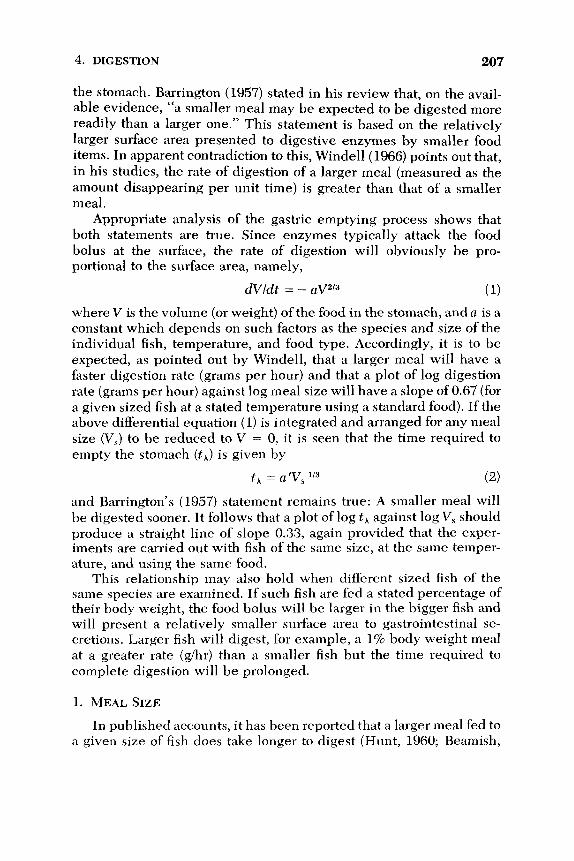
4. DIGESTION 207
the stomach. Barrington (1957) stated in his review that, on the avail-
able evidence, “a smaller meal may be expected to be digested more
readily than a larger one.” This statement is based on the relatively
larger surface area presented to digestive enzymes by smaller food
items. In apparent contradiction to this, Windell (1966) points out that,
in his studies, the rate of digestion of a larger meal (measured as the
amount disappearing per unit time) is greater than that of a smaller
meal.
Appropriate analysis of the gastric emptying process shows that
both statements are true. Since enzymes typically attack the food
bolus at the surface, the rate of digestion will obviously be pro-
portional to the surface area, namely,
( 1)
where V is the volume (or weight) of the food in the stomach, and a is a
constant which depends on such factors as the species and size of the
individual fish, temperature, and food type. Accordingly, it is to be expected, as pointed out by Windell, that a larger meal will have a
faster digestion rate (grams per hour) and that a plot of log digestion
rate (grams per hour) against log meal size will have a slope of 0.67 (for
a given sized fish at a stated temperature using a standard food). If the
above differential equation (1) is integrated and arranged for any meal
size (V,) to be reduced to V = 0, it is seen that the time required to
empty the stomach ( tJ is given by
dvld t = - aV2I3
th = a’Vs */3 (2 )
and Barrington’s (1957) statement remains true: A smaller meal will
be digested sooner. I t follows that a plot of log t k against log V, should
produce a straight line of slope 0.33, again provided that the exper- iments are carried out with fish of the same size, at the same temper-
ature, and using the same food. This relationship may also hold when different sized fish of the
same species are examined. If such fish are fed a stated percentage of their body weight, the food bolus will be larger in the bigger fish and
will present a relatively smaller surface area to gastrointestinal se-
cretions. Larger fish will digest, for example, a 1% body weight meal
at a greater rate (g/hr) than a smaller fish but the time required to
complete digestion will be prolonged.
1. MEAL SIZE
In published accounts, it has been reported that a larger meal fed to
a given size of fish does take longer to digest (Hunt, 1960; Beamish,

208 RAGNAR FANGE AND DAVID GROVE
1972; Elliott, 1972; Swenson and Smith, 1973; Jobling et al., 1977). Beamish (1972) found that a four-fold increase in meal size fed to
Micropterus only doubled the time required to empty the stomach. Using X-radiography, Jobling et al. (1977) found that increasing meal size from 1 to 5% (100-g Limanda at 16°C) leads only to a fourfold increase in stomach emptying time. Clearly both the time to complete the meal and the rate of digestion increased as predicted by the model. Analysis of the published records for various fish shows that digestion rate, expressed in grams per hour, varies as the following exponents of
the wet weight of the meal: 0.46 (gadoids, Jones, 1974), 0.5 (Limanda, Jobling et al., 1977), 0.6 (Prosopium, McKone, 1971; (Megalops, Pan- dian, 1967), 0.7 (Micropterus, Beamish, 1972), and 0.75 (Salmo gairdnerii, Windell et al., 1969). In a few fish digestion rate has been found to increase as (meal size)'.O, such that overall evacuation time in a given fish is the same for different sized meals (Lepomis, Windell, 1966; Oncorhynchus, Brett and Higgs, 1970; BZennius, Crawford and Grove, unpublished observations). The arrival of larger meals in the fish stomach or foregut will initiate or accelerate peristalsis (Section VII) thereby raising the emptying rate above the level predicted by
the simplistic model. Only Steigenberger and Larkin (1974), in a care- ful study of digestion in Ptychocheilus, have found that digestion rate decreases with increased meal size. Clearly more species must be examined before this result can be held anomalous. Elliott (1972) found that the instantaneous rate of gastric evacuation in Salmo trutta is not affected by increases in meal size but, since his evacuation curves are exponential, the early digestion rate (g/hr) increases with meal size. A similar conclusion applies to the data for Oncorhynchus nerka (Brett and Higgs, 1970).
2. FISH SIZE
Several workers have reported the digestion rate of fish of different sizes in a form which can be compared with the predictions of the model for gastric emptying time ( t i ) . Jobling et al. (1977) (Figs. 5 and
6) fed Limanda of different body weights to the same percentage of their weight with a flatfish paste diet. The time required to empty the stomach varied as (fish weight)'."'. Since the stomach volume is pro- portional to body weight in this species (V = 0.081 W - 0.39 where the units are ml and g, respectively) a stated % body weight meal will fill the stomach to the same extent in different sized fish. A similar analysis of Pandian's (1967) data for Megalops fed on prawns shows that t h varies as (fish ~eight)O.~l .
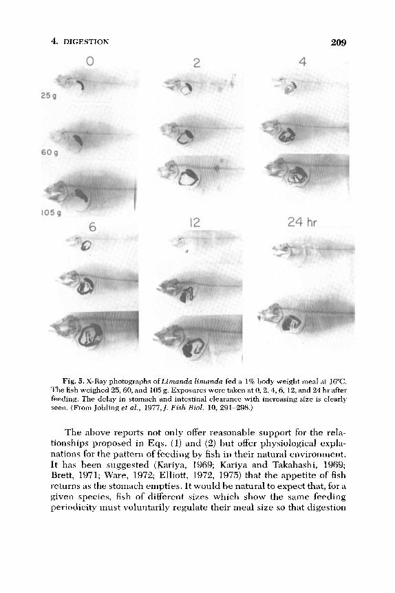
4. DIGESTION
0
I w d y _-
6
2
12
209
4
24 hr
i
Fig. 5. X-Ray photographs ofLirnanda lirnanda fed a 1% body weight meal at 16°C.
The fish weighed 25,60, and 105 g. Exposures were taken at 0 ,2 ,4 ,6 ,12, and 24 hr after
feeding. The delay in stomach and intestinal clearance with increasing size is clearly
seen. (From Jobling et al., 1977,J. Fish B i d . 10, 291-298.)
The above reports not only offer reasonable support for the rela-
tionships proposed in Eqs. (1) and (2) but offer physiological expla-
nations for the pattern of feeding by fish in their natural environment.
It has been suggested (Kariya, 1969; Kariya and Takahashi, 1969;
Brett, 1971; Ware, 1972; Elliott, 1972, 1975) that the appetite of fish
returns as the stomach empties. It would be natural to expect that, for a
given species, fish of different sizes which show the same feeding
periodicity must voluntarily regulate their meal size so that digestion

210 RAGNAR FANGE AND DAVID GROVE
1003
Meal S i z e
1 r n ,
Fig. 6. Relationship between fish weight, meal size (as percentage of body weight)
and gastric evacuation time for Limanda limanda at 16°C. The decrease in relative digestion rate with size shows that, if feeding is to return at regular intervals (e.g., daily)
larger fish must necessarily ingest relatively smaller meals. Voluntary intake in grams
will vary as (body eight)^.^. (From Jobling et al., 1977,J. Fish Biol. 10, 291-298.)
is completed in time for the next feeding bout (Fig. 6). The stomach
contents of freshly captured fish show a relative decrease in amount
for bigger fish (Daan, 1973; Steigenberger and Larkin, 1974) and vol-
untary intake of food has been shown to decrease relative to body
weight by many workers (Table V). It must be recognized, in design-
Table V
Magnitude of the Exponent in the Relationship Meal Size = a(Fish Weight)*
for Voluntarily Feeding Fish
Species b Reference
Petromyzon marinus
Salvelinus fontinalis Salmo trutta Oncorhynchus nerka
Megalops cyprinoides Micropterus salmoides Navodon modestus Ophiocephalus punctatus Blennius pholis
(ammocoete)
Trachurus japonicus
0.72 (summer)
0.84 (winter) 0.91
0.75
0.75 (single meal)
0.65 (multiple meal)
0.71
0.47 0.52
0.76
0.38
1.00
0.56
Moore and Bamish (1973)
Baldwin (1956)
Elliott (1975)
Brett (1971)
Pandian (1967)
Niimi and Beamish (1975)
Suzuki (1976)
Gerald (1973) Wallace (1973)
Crawford and Grove (unpublished observations)
Hotta and Nakashima (1968)

4. DIGESTION 211
ing experiments, that the amount taken at a meal by an individual fish will depend on the previous feeding regime (Kariya and Takahashi, 1969; Ishiwata, 1969; Brett, 1971). For example, young Scomber
japonicus will eat 23% of their body weight of anchovy after several
days of deprivation but, on a regular feeding regime, will ingest only
16%.
3. TYPE OF FOOD AND ITS PRESENTATION
A further variable with significant effects on the gastric emptying rate is the type of food ingested by the fish. Jones (1974) found that gadoids digested the following foods (after an appropriate delay) at the stated rate at 12OC: NereislNephthys, 0.31 glhr; Pollachius muscle, 0.26 glhr; Crangon, 0.19 g/hr. Elliott (1972) found that brown trout completed 90% elimination of gammarids or oligochaetes in 22 hr at 12°C whereas Protonemura (26 hr), Hydropsyche (30 hr), and Tenebrio (49.5 hr) took longer. A similar observation was made by Reimers (1957) on rainbow trout, where Helodrilus (12 hr) was eliminated fas-
ter than Gammarus (13 hr) or Arctopsyche (16 hr) at 10°C when given to 12.4-20 cm fish in 0.5 g meals. Other workers who have detected
decreased evacuation rates with less digestible foodstuffs are Pandian
( 1967) (Megalops fed Gambusia or Metapenaeus), Karpevitch and Bokova (1936, 1937) (Gadus fed Clupea or Gummarus), Western (1971) (Cottus, Enophrys fed on Tubifex, Calliphora or semifluid meals), Reshetnikov et al. (1972) (Lutianus fed onJenkinsia or Haren- gula) , Renade and Kewalramani (1967) (Labeo, Cirrhina or Catla fed
on various algae, plant detritus, and zooplankton), and Kionka and Windell (1972) (Salmo fed various diets). The digestibility of the food will not only affect the emptying rate from the stomach, expressed as
g/hr, but may also determine the time after ingestion before weight decrease of the meal can occur. Jones (1974) found that Merlangus or Melanogrammus start to digest shell-less Mytilus almost immediately but that meals of Ophiopholis, large crustacea or Centronotus require up to 10, 20, and 25 hr, respectively, before weight loss begins. Al-
though in many cases the decrease in digestibility can be attributed to thick or inert casings (e.g., caddis larvae) Windell (1967) suggested that the presence of fat in the food may delay gastric emptying, possi- bly by a release from the intestinal wall of a hormone similar to enterogastrone which in mammals inhibits gastric motility (Hunt and Knox, 1968). Diets with increased fat levels clearly decrease gastric evacuation rate in the rainbow trout (Windell et al., 1969). Finally, Swenson and Smith (1973) found that when Stixostedion were fed the same meal size at the same temperature but with smaller or larger food

212 RAGNAR FANGE AND DAVID GROVE
items, the evacuation rate was faster with the smaller items (they used Pimephales).
A further complication to the measurement of gastric emptying rates in fish are the observations by Windell (1966) and by Swenson and Smith (1973) that force-feeding the test animal usually decreases the rate of evacuation of the meal from the stomach when compared with fish which consume the food voluntarily. Of even greater con- cern, however, are the reports from Kariya and Takahashi (1969), Ishiwata (1969), Tyler (1970), Brett (1971), Jones (1974), and, for larval fish, Laurence (1971), Blaxter and Holliday (1963), Rosenthal and Paf- fenhofer (1972), and Nobel (1973) that fish which have been deprived of food for a time prior to feeding show a slower gastric emptying rate than fish tested under continuous feeding. Digestion rates measured in previously deprived fish are usually only 50-68% of those found in actively-feeding fish. In their study on feeding rate of Tilapia ingest- ing blue-green algae, Moriarty and Moriarty (1973a) found that the rate of stomach emptying during the feeding period was faster than that measured after feeding had ceased. This factor is a clear restric- tion when using laboratory digestion studies to estimate feeding rate in the wild (Healey, 1971).
E. Conclusions
If experiments on gastric evacuation are undertaken (whether to compute daily food intake in nature, to determine the relationship between stomach emptying and the return of appetite for fish culture, or to investigate the physiological control of gastric motility in uiuo) the following procedures must be controlled and reported with the results.
1. The temperature at which observations are made, together with the previous acclimation history of the fish
2. The frequency of feeding, or the previous duration of food de- privation, since the fish will consume a meal size related to this regime if voluntary feeding is allowed
3. The meal size, as absolute wet and dry weight and as percent- age of the body weight
4. The biochemical composition of the food, including the calorific value per unit dry weight of digestible food, both at ingestion and at later stages as stomach emptying proceeds
4. DIGESTION 2 13
5. The method of feeding, especially whether the animals were force fed or ingested voluntarily; mention should be made if the
fish were handled at intervals during the experiments (e.g., if several X-ray photographs were taken)
6. The length and weight of the fish used in the study
It is likely that other factors, not discussed in the preseRt account, might affect gastric emptying. Reproductive cycles, photoperiod of long or short daylength, presence of parasties and/or the stocking den- sity may all modify gastric motility. Many of the results listed in
simplified form in Table I1 are taken from reports where factors 1-6 were not fully controlled. More recent studies (e.g., Elliott, 1972; Steigenberger and Larkin, 1974) have used factorial designs to test some of the variables likely to affect gastric emptying.
It has often been stated that the return of appetite is closely related to the time taken to reduce the stomach contents. Ware (1972), for example, defined hunger in newly caught fish in relation to the degree of stomach fullness. Magnuson (1969), Brett (1971), and Elliott (1975) all report that hunger returns before the stomach is completely empty. However, the observations of Tugendhat (1960) and Beukema (1968) suggest that stomach emptiness alone does not control the amount ingested. Gasterosteus with empty stomachs ingested more food at a faster rate if they had a longer time of deprivation before food was made available. Elliott (1975) found that a trout of stated size ate more in a meal if the temperature is raised. Rozin and Mayer (1961, 1964) found that the goldfish (Carassius) feeds continuously, but consumes a greater amount of food per day if the food is diluted with kaolin. A similar conclusion is reported for S . gairdneri by Lee and Putnam (1973). In an attempt to clarify this latter phenomenon of “calorie counting,” we carried out a study on Salmo gairdneri (Grove et al., 1978). As shown in Fig. 7, rainbow trout kept in continuous light exhibit regular periods of feeding activity similar to those described by Adron et al. (1973). The records from the demand feeders were subjected to periodogram analysis and showed that, as the food is diluted with kaolin, so the period between meals is progressively shortened and, as a consequence, the food intake per day is increased. Adron et al. (1973) suggested that the periodic feeding is determined by the rate of gastric emptying, so X-radiography tests were under- taken to see if dilution of the food with kaolin shortens the time re- quired for the stomach to empty. The gastric evacuation times pre- sented in Table VI clearly show that this is the case. The mechanisms responsible for the increased gastric motility are unknown but may

214 RAGNAR FANGE AND DAVID GROVE
101 A lo-, B
Period h r T i m e hr
Fig. 7. (A) Activation of a “demand’ feeder by a group of 60 g Salmo gairdnerii
during a 24 hr period under continuous illumination at 18°C. (B) Periodogram analysis
of (A) (Williams and Naylor, 1967) shows that appetite returns at 6-hr intervals. (From
Grove et al., 1978,J. Fish Biol. 12, 507-516.)
either depend on a decreased production of enterogastrone from the intestine or a change in activity of extrinsic nerves which supply the
stomach (see Section VII). Digestion Eficiency: Pappas et al. (1973) added 0.37% chromic
oxide to diets fed to channel catfish and observed the disappearance of
nutrients (fat, protein, and carbohydrate) relative to the inert marker
while the food remained in the stomach. Nearly 50% of these dietary
components were removed before the food entered the intestine, al-
Table VI
“Calorie Counting” in Salmo gairdnerii: Control of Appetite by Changes in
Gastric Motilitya
Mean daily Mean feeding Mean gastric
Food intake intervals evacuation time
Test food (cal/g) (ca1/100 g/day) (hr) (hr)
Salmon pellet 4.78 11.3 6 15 75% food/%% kaolin 4.20 15.9 5 14.5 67% food/33% kaolin 3.23 15.0 5 11.8 50% food/80% kaolin 2.18 13.1 4 10
The fish obtained food from a “demand feeder” under continuous illumination at
18°C. Each week the control pellets (Cooper’s salmon starter) were replaced by recon-
stituted pellets diluted with kaolin, and feeding activity was subjected to periodogram
analysis to determine the time for return of appetite during the succeeding 7 days. Separate groups of fish were fed the same diets and X-rayed to determine the time for
complete gastric evacuation. Note that the stomach empties more quickly when the food is diluted, that appetite returns more quickly and, if the evacuation curve is ex-
ponential, this occurs when 70-90% of the previous meal has left the stomach. From Grove et al. (1978,l. Fish BioZ. 12, 507-516).

4. DIGESTION 215
though no conclusion was reached whether they undergo gastric ab- sorption or are passed in solution into the intestine for assimilation.
Bearnish (1972) showed that protein was solubilized more extensively
in Micropterus stomach than fats, which required intestinal digestion
before assimilation occurred. Overall protein digestion is efficient in
fish, more than 85% assimilation being the rule unless oxidation of oils
occurs in the food employed (Nose and Toyama, 1966). Smith and
Lane (1971) reported that protein assimilation of salmonids was im-
paired if a-cellulose was incorporated in the diet. Increase in the
proportion of carbohydrates in the diet, or absolute decrease in the pro-
tein content of the diet, has been reported to decrease protein assimila-
tion without explanation of the mechanism involved (see references in
Nose, 1967). Nose demonstrated that this apparent decrease in protein
assimilation arises from failure of previous workers to allow for the
release of metabolic nitrogen into the feces, which becomes
significant in biochemical determinations only at low leve!s of protein
intake. A true decrease in protein assimilation was detected by Nose
when rainbow trout were given egg albumin, which contains a trypsin inhibitor. The early work of Inaba et al. (1962) clearly demonstrates
that the protein in whalemeat or soya bean residues is much less diges-
tible than that in white fish meal, but the basis for this observation
requires further study. A more extensive account for formulation of fish
diets is given in Chapter 1.
VII. PHYSIOLOGICAL STUDIES ON FISH GASTROINTESTINAL MOTILITY
A. Introduction
1. EXCITATION OF GASTROINTESTINAL SMOOTH MUSCLES
Our understanding of muscular activity of the gastrointestinal tract
in various fish is based on comparisons with higher vertebrates. The major contractile elements in the muscularis are hexagonally packed
groups of smooth muscle cells which act as a contractile unit (Bennett
and Burnstock, 1968). The cells affect each other electrotonically SO
that coordinated myogenic contractions, independent of the nervous
system, can develop in response to stretch as a result of Caz+ flux
across their membrane. In the in situ gut, rhythmic myogenic contrac- tions of the circular muscles temporarily divide the intestine into a
series of segments to facilitate mixing of the luminal contents.

216 RAGNAR FANGE AND DAVID GROVE
Superimposed on this activity is the peristaltic process, dependent on
cell bodies in the nerve plexuses of the gut wall, usually seen as waves
of relaxation and contraction which progress anally along the gut. The combination of these two processes, segmentation and peristalsis, may
cause the segments of the intestine to swing slowly from side to side in
pendular activity.
2. THE SITES OF DRUG ACTION
In view of the difficulty in separating the roles of muscle and nerve
cells, most workers have investigated the neurogenic components by
using drugs which are believed to act at the receptor sites on tissues
with which neurotransmitters are known to act. This approach is not
without problems since few drugs are specific, and may only be con-
sidered selective within certain dose ranges (Daniel, 1968) which
must be determined for each new tissue. It has become general to
refer to nerve cells releasing a stated neurotransmitter (e.g., acetyl-
choline, noradrenaline) as cholinergic, adrenergic, and the receptor
they occupy as cholinoceptor, adrenoceptor. When the transmitter, or a
similar molecule (agonist), occupies the receptors of a tissue, a re-
sponse is evoked in proportion to the agonist concentration. A dose-
response curve can be constructed and the dose producing 50% of
maximum response is a measure of the affinity of the agonist at that site (Ariens and Van Rossum 1957). Similar molecules which occupy the
site without eliciting a response may combine with the agonist rever-
sibly, or irreversibly by binding to the receptor chemically. The block-
ing action of reversible antagonists can be overcome by increasing the
agonist concentration, and may be visualized as a parallel shift of the
agonist dose-response curve to the right. A measure of the affinity of the antagonist is the observed shift of the dose-response curve in the
presence of a stated antagonist concentration. Noncompetitive and ir-
reversible antagonism is usually detected as an insurmountable
blockade, accompanied by a depression of the dose-response curve
(e.g., Guimaraes, 1969). Examples of these relationships are given in
Fig. 8. In Table VII, the drugs which have been commonly used in
studies of fish gastrointestinal motility are shown, together with the
receptors that they occupy.
3. INTRINSIC NEURONS
Two main techniques for activating neurons in the gut wall are
commonly used. Various workers have adapted the technique of Tren-
delenburg (1917) in which distension of the gut wall in vitro initiates

4. DIGESTION
B 1
217
M o l a r Concentration of Agoni st
Fig. 8. Analysis of drug effects on in oitro Magnus preparations of plaice gastric
longitudinal muscle. (A) Dose-related contractions induced by acetylcholine alone.
Control ED% = 2 x 10-* M (and pD, = 7.7). The addition of atropine to the bath in doses
of 3.5 x I&” M causes a parallel shift of the dose-response curve to the right in relation
to the dose: curve a, x = 9; curve b, x = 8; curve c, x = 7. The concentration of atropine
for which a control dose of acetylcholine must be doubled to obtain the control response
is 1.6 x l(r9 M (and PA, = 8.8). (B) A similar study using Shydroxytryptamine as
stimulant gives a control ED,, = 4 x 1e8 M (whence pD, = 7.4). Atropine here acts
noncompetitively when the doses used above are employed. The concentration of at- ropine required to depress the control maximum by half is 1.2 x lo-* M (whence
pD; = 7.9). (From Edwards, 1972b, Comp. Gen. Phamacol. 3,345-358.)
activity. To study stimulant effects on inactive preparations, the gut segment is simply suspended in a bath under minimum stretch (Mag-
nus preparation). The second method, which may be used on Tren-
delenburg or Magnus preparations, is to stiinulate the intrinsic
neurons by electrodes in the bath placed near (field stimulation) or on
either side (transmural stimulation) of the gut wall. The frequency of stimulation may be between 1 and 50 Hz and the pulse duration of 1 insec or less to avoid direct activation of the muscle cells. In addition
to recording the muscle responses mechanically (levers, strain gauges,
pressure transducers), changes in muscle membrane potential can be monitored using the sucrose gap technique (Campbell and Burnstock,
1968; Ito and Kuriyama, 1971). The use of the latter technique has
shown that, in low tone preparations, transmural stimulation may ex-
cite inhibitory neurons in the gut wall which hyperpolarize the mus-
cle cell membranes without a clear relaxation being recorded. After
cessation of the stimulus, the cells repolarize and may overshoot the
resting potential leading to spontaneous electrical activity and “re-
bound contractions.” This phenomenon led Burnstock ( 1969) and Campbell and Burnstock (1968) to reinterpret the conclusions reached
in earlier studies of fish gastrointestinal motility; the vagus nerve was earlier claimed to excite the fish stomach whereas it is now believed to

218 RAGNAR FANGE AND DAVID GROVE
Table VII
Sites of Action and Names and Abbreviations of Drugs Used to Analyze
Gastrointestinal Motility in Fishes
Receptodeffect Agonis t Antagonist
1. Muscarinic; cholino-
ceptor on smooth
muscles
2. “Nicotinic”; cholino-
ceptor on neurons
3. “Nicotinic”; cholino-
ceptor on striated
muscle
4. Adrenoceptors
a
P
5. Tryptaminoceptors
“dibenamine type”
D
6. Purinoceptor
7. Histamine receptor
8. Cholinesterase
9. Neuron blocking
All nerves
(H,)
inhibition
Cholinergic nerves
Adrenergic nerves
Acetylcholine (ACh)
Pilocarpine
Carbachol
Methacol
Acetylcholine
Carbachol
l,l-Dimethyl-4-phenyl-
Acetylcholine
piperazonium (DMPP)
Phenylephrine
Adrenaline (A) Noradrenaline (NA)
Isoprenaline (Iso)
Adrenaline (A) Noradrenaline (NA)
5-H y droxytryptamine
(5-HT)
Adenosine triphosphate
Histamine (ATP)
Eserine
Tetrodotoxin (TTX),
Morphine, hemi-
Reserpine, bretylium,
procaine
cholinium
guanethidine
Atropine (Atr)
Hyoscine
Nicotine
Hexamethonium (C,)
d-Tubocurarine (dTC)
Mecamylamine (Mec)
d-Tubocurarine
Phenoxybenzamine (PBZ)
Phentolamine (Phent)
Dihydroergotamine (DHE)
Piperoxane (Pip)
Yohimbine
Propranolol (Prop)
Butoxamine
Methysergide
2-Bromo-lysergic acid
diethylamide (BOL) Phenoxybenzamine (PBZ)
E rgotamine
Mepyramine
inhibit it in the majority of fishes. I t has been proposed that intramural
neurons exist in vertebrate viscera which excite or inhibit the smooth
muscles by releasing a purinergic transmitter, possibly adenosine
triphosphate (Burnstock, 1972; Burnstock et al., 1970, 1972).

4. DIGESTION 219
4. EXTRINSIC NEURONS
The extrinsic nerves to the fish gut originate from the autonomic
nervous system. The simplistic account of this system in mammals,
attributed to Langley ( 1921), is that “parasympathetic” nerve tracts
from the craniosacral region innervate the intrinsic neurons of the gut
wall by way of vagus and pelvic nerves. A “sympathetic” outflow in
the thoraco-lumbar region innervates ganglion cells in the sympathetic
chain. In both divisions, preganglionic medullated cholinergic nerves
leave the CNS. The postganglionic parasympathetic neurons are
cholinergic. Activity in the parasympathetic division enhances gastro-
intestinal motility whereas sympathetic activity is inhibitory.
It is now acknowledged that adherence to this model is likely to
cause misconceptions, and many mammalian exceptions have been
reported. Vagal fibers may activate either cholinergic or purinergic
intrinsic neurons. The vagus may be joined by postganglionic axons
(adrenergic or cholinergic) from the sympathetic division, or itself
send preganglionic cholinergic fibers along the nearest convenient
sympathetic” tract. The postganglionic adrenergic nerves of the
sympathetic” system may not innervate gut muscle directly, but en-
mesh ganglion cells in the gut wall (Campbell, 1970a,b). These con-
clusions, reached for higher vertebrates, are equally likely to compli- cate studies on the less understood fish groups.
“
“
B. Cyclostomes
Relatively little information is available on the physiology of intes-
tinal motility in hagfish and lampreys. The intestinal muscles of both
groups are poorly developed and food movements may depend on
contractions of the body wall. Fange (1962) and Burnstock (1969) in
their reviews report that the intestine in hagfish is contracted by
acetylcholine (lo-* to 1 0 - ~ g/ml) but relaxed by adrenaline (lop7 to
lop5 g/ml), and by atropine. Peristalsis is induced in Larnpetra rectum
by lop4 g/ml 5-hydroxytryptamine. In the hagfish Myxine glutinosa, the two intestinal rami of the
vagus nerve unite dorsally to the intestine forming an unpaired nerve-
like structure (ramus irnpar) which contains numerous ganglion
cells. Cholinergic and adrenergic neurons are present (Fange and
Johnels, 1958; Hallback, 1973; Fange et al., 1963). Patterson and Fair
(1933) found that vagal stimulation slightly relaxes the hagfish gut. von
Euler and F h g e (1961) detected catecholamines in the gut ofMyxine and Honma (1970) used the fluorescent histological technique ofFalck
220 RAGNAR FANGE AND DAVID GROVE
and Owman (1965) to show that adrenergic neurons and terminals are
present in the enteric plexus of Lampetra japonica and L. planeri, which presumably mediate inhibition. Nonadrenergic nerves in the
gut wall of hagfish and lampreys are likely to be cholinergic and pos-
sibly also purinergic, coordinating peristalsis, and appear to be inner-
vated by fibers of vagal origin.
More recently, Baumgarten et al. (1973) have carried out an exten-
sive study of monoaminergic neurons in the gut wall of Larnpetra f luviati l is using the fluorescent histochemical technique together with
microspectrofluorimetry, chromatography, and electron microscopy.
Green fluorescent nerve cells were detected in the submucosa of the
fore- and midgut which may inhibit the intestine, and which contain
primary catecholamines such as dopamine or noradrenaline. These authors detected an extensive submucosal plexus of tryptaminergic
axons and cell bodies which connected with a subserous plexus con-
taining nerve terminals. They propose that the 5hydroxytryptamine
transmitter released by these endings elicits peristalsis but point out
that similar intramural neurons have not been described in the gut
wall of true vertebrates.
C . Elasmobranchs
In comparison with actinopterygii, relatively little work has been
done on elasmobranch gastrointestinal motility. The early reports of
Lutz (1931), Nicholls (1933), and Dreyer (1949) state that all parts of
the gut are contracted by acetylcholine and pilocarpine. von Euler and
Ostlund (1957) confirmed the observation of Nicholls that atropine
prevents the action of acetylcholine. Adrenaline sometimes causes
contraction of the gut (Babkin et al., 1935a,b; Dreyer, 1949; Young,
1933) but according to Nicholls (1933) this is only at lower doses.
Inhibitory effects of adrenaline have also been recorded on the dogfish
rectum (Lutz, 1931) and ray intestine (von Euler and Ostlund, 1957).
Without more detailed studies it is impossible to conclude whether
adrenaline has different actions in different parts of the gut of different
species. Recently Moore and Hiatt (1967) reinvestigated the problem
of the action of adrenaline in elasmobranchs. They elected to work
with Squalus acanthias, using intragastric balloons in vivo to record
contractions. Injection of adrenaline produced a clear contraction,
which they believed would be due to occupation either of CY- or
P-adrenoceptors. However, they found that neither a (phenoxy-
benzamine) nor p (propranolol) adrenoceptor blocking agents pre-

4. DIGESTION 22 1
vented the action of adrenaline. To extend their study, they tested
other sympathomimetic amines which have differential potencies on
a- and Preceptors in other vertebrates. Surprisingly, isoprenaline,
dopamine, or noradrenaline failed to contract the stomach in control
animals. Phenylephrine was effective, but phenoxybenzamine, which
in rnost vertebrates is a powerful phenylephrine antagonist at a-adrenoceptors, failed to antagonize the response. In further studies,
aminophylline was found to abolish the adrenaline-induced contrac-
tion and the effect of adrenaline decreased rapidly with successive
tests in control animals. They concluded that adrenaline had no inhib- itory effect on Squalus gastrointestinal smooth muscle. The stimula-
tory action of a first test dose of adrenaline depends not on the usual
cell membrane adrenoceptors but on increased glycogenolysis pre-
sumably following an increased intracellular build-up of cyclic AMP.
However, once under way, this intracellular activity hyperpolarizes
the muscle membranes, thus suppressing responses to subsequent
adrenaline treatment. Clearly this abnormal action of adrenaline
should be investigated in other species. It should not be concluded
that adrenoceptors are necessarily aberrant in elasmobranchs since
Nilsson et al. (1975) have shown a-adrenoceptors in spleen and artery
preparations of Squalus and Scyliorhinus with properties similar to
those in teleosts and higher vertebrates.
Gzgzyan and Kuzina ( 1973) investigated spontaneous peristaltic
contractions of the stomach of the ray, Dasyatis pastinaca. This
species does not exhibit alternation of periods of activity and rest
typical of empty mammalian stomachs. The contractions were en-
hanced when cholinomimetics (carbachol, pilocarpine) were injected
but atropine was not a powerful inhibitor of the spontaneous contrac-
tions. There is evidence here that cholinergic nerves may play a coor-
dinating role in gastrointestinal peristalsis but, in common with tele-
osts (see later), myogenic contractions may play a significant part in
rhythmic contractions of the gut wall. Myenteric and submucosal plexuses have been described in the stomach and intestine of elas-
mobranchs (Nicol, 1952).
The digestive canal of elasmobranchs is supplied by vagal and
splanchnic nerves. The vagal fibers are believed to be adrenergic
(Fange and Hanson, 1973). Campbell and Burnstock (1968) examined
the published records from earlier workers of the effect of vagal stimu- lation on stomach activity. They concluded that the influence of the
vagus is limited to the stomach and proximal intestine and that it is
inhibitory. Earlier conclusions that the vagus is excitatory were ex- plained by the long latency of the contraction, reminiscent of the “re-
222 RAGNAR FANGE AND DAVID GROVE
bound contractions” described earlier. Campbell ( 1975) found that the
regular spontaneous activity of Scyliorhinus canicula stomach,
recorded by intragastric balloon, was abolished when the intracranial roots of the vagus nerve were stimulated electrically. He also ob-
served a stimulatory effect of the vagus on esophageal striped muscles.
By inference from other vertebrates, Burnstock (1969) suggests that at
least part of the inhibitory response mediated by the vagus depends on
the presence of inhibitory, nonadrenergic neurons in the stomach wall.
The role of sympathetic nerves on gastrointestinal motility is as yet
unclear. Burnstock (1969) concluded that the records in earlier publi-
cations show evidence of both cholinergic excitatory and adrenergic inhibitory fibers supplying both stomach and intestine in
elasmobranchs.
D. Teleosts
Gastrointestinal motility, and its control, has been more exten-
sively studied in teleosts. The sympathetic system, like that of higher
vertebrates, consists of two ganglionated chains which extend into the
head as far as the trigeminal nerve (V). The esophagus and stomach
have a rich vagal innervation, while in many stomachless fish includ-
ing the cyprinidae the vagal influence extends to the intestine. In the
stomachless pleuronectids Rhombosolea and Ammotretis, however,
the vagal influence does not extend beyond the esophagus (Grove and
Campbell, unpublished observations). The stomach and intestine are
also innervated by fibers from the splanchnic nerves which originate
from sympathetic ganglia in the first few spinal segments. The gut of
the tench (Tinca) contains striated and smooth muscles innervated by
the vagus nerve (Ohnesorge and Rehberg, 1963). Only the smooth
muscle elements participate in the peristaltic movements. Results of pharmacological analysis indicate that the peristalsis is due to a reflex
involving intramural neurons (Ohnesorge and Rauch, 1968). A poste-
rior sympathetic nerve to the rectum has been described in the trout
(Salmo trutta) by Burnstock (1958a,b). The ramus intestinalis of the
vagus nerve probably receives sympathetic fibers from the gan-
glionated chains and may therefore more properly be termed a vago-
sympathetic trunk (Young, 1931).
1. ANALYSIS OF GASTROINTESTINAL RECEPTORS
Few studies have been made to estimate the affinity for specific
receptors on gastrointestinal muscle and nerve cells of agonist

223 4. DIGESTION
molecules by dose-response curve analysis. Table VIII summarizes
much of the recently published data for a number of teleost fish. In
general, cholinesters and Fj-hydroxytryptamine (5-HT) contract the muscle coats whereas sympathomimetic amines relax the gut or in-
hibit spontaneous activity. The problems facing the researcher lie
mainly in the complexity of the gut wall, since the site of action of a
drug must be determined, and the specificity of the drugs, which have
usually been assumed to act in fish as in mammals.
Acetylcholine is probably a direct stimulant of muscarinic recep-
tors on many parts of the gut musculature since atropine is the most
potent inhibitor of its action (Salmo trutta, Burnstock, 1958a,b; Gadus morhua, S. Nilsson and Fange, 1969; Carassius auratus, Ito and
Kuriyama, 1971; Saito, 1973; Pleuronectes platessa, Edwards, 197213;
Goddard, 1975; Blennius, Myoxocephalus, Grove et al., 1974). Acetyl-
choline also exerts effects on nicotinic receptors of a variety of neurons
in the enteric plexus, and the high, potentially nonselective doses of atropine used in the Gadus and Carassius studies may block this ac-
tion as well. Where dose-response analysis has been undertaken, the
PA, of atropine has been found to lie between 8.2 and 9.4. Concen- trations of atropine at ca. lop7 glml or less should be more than
adequate to abolish the muscarinic action of acetylcholine without
significant side effects (Edwards, 1972b; Goddard, 1975; Grove et al., 1974). There is no doubt that acetylcholine effects in Carassius in-
volve the intramural ganglion cells. Saito (1973) believed that all me-
chanical effects exerted by the cholinester ( g/ml) on the intestine
were indirect since tetrodotoxin ( g/ml), which prevents axonal
conduction by blocking sodium channels in the neuronal membrane,
abolished all its actions. He recognized a brief contraction of striated
muscle fibers when the drug was applied, followed by a slower
smooth muscle contraction. After the administration of atropine ( lop6 g/ml) to impair the response of cholinergic excitatory cells, he
recorded relaxation of the preparation. Since this effect is tetro-
dotoxin-sensitive, inhibitory neurons activated by preganglionic
cholinergic fibers are also present in the intestine. Dimethylphenyl-
piperazinium (DMPP) ( g/ml) which also briefly excites ganglion
cells in mammals mimicked acetylcholine. In contrast, the earlier
study by Ito and Kuriyaina (1971) on silver carp showed that acetyl-
choline excitation persisted after tetrodotoxin. They proposed that at
least part of the stimulus must be directly on the muscle cells.
d-Tubocurarine ( g/ml) abolishes the effect of acetylcholine on
striated muscle in Carassius (Saito, 1973) and in Tinca (Mahn, 1898). Hexamethonium, which in mammals is a relatively potent nicotinic

Table VIII
Effects of Agonist Drugs on Resting Gastrointestinal Muscle Preparations of Teleosts"
Species and Cholinergic Sympathomimetic organ stimulants 5-Hydroxytryptamine amines Purines
Salmo trutta
a. Stomach
b. Intestine Pi1 + 1c5 c. Rectum
a,b,c. ACh + 10-8-l@4
p1 to
Carassius auratus Intestine ACh (1st) + 1W6
(Cholinesters ACh (2nd) + cause ACh (3rd) - 1W6
contractions) DMPP (2nd) + multiple DMPP (1st) + 1 0 - 5
Tinca tinca Intestine ACh (1st) + 5 x 10⁻⁸-10-5
ACh(2nd) + 5 x 10⁻⁸-lW5 (Cholinesters
multiple
contractions)
Anguilla anguilla a. Esophagus
b. Stomach c. Intestine
cause DMPP + 10-8-10-6
a,b,c. ACh + l W 5
a,b,c. + lC9 (1) a. NA + (1,2)
A +
b,c. A - lCP-le5 (1)
ISO + 2 x 10⁻⁵ (1,2)
+ 10-8
NA - 10-6-10-5 (3,5) ATP + l C 5 M (5)
A - 10-' (3)
I S 0 - 1 0 - 6 (3)
(8) A - 10-8-10-' (7) NA - 10-8-10-' (7)
a,c. A - l W 5 (9) b. A + then - Tyr + then - l t 5
I S 0 + then - lW5
(9)
(9)
(9)

Gadus morhua Stomach
Pleuronectes platessa
a. Stomach
b. Intestine
ACh + lW8-lW5
CCh + lW7 - l W 5
Pi1 + lW7-10-5
a. ACh + *3 x M MCh + *1W8 M CCh + *2 x M
b. ACh + *3 x lW7 M MCh + * 5 x lo-, M CCh + *7 x M
Blennius pholb Intestine
01
Myorocephalus scorpius
Stomach
ACh + *8 x lW7M
MCh + *5 x 1W8 M
ACh + *5 x lW7M
NA + 10-7-10'5 (10)
Pheny + 10"-10-5 (10)
A + 10-7-10-5 (10)
ISO - 1 0 ' 6 - 1 e 5 (10)
(11,14) a. + 4 x 10-*M (11,12,14) a. NA + lo-'- (11) a. ATP + l t 5 M (14)
(11,14) A 0 ( 1 1 ~ 4 ) 1e5 M
1e3 M (11)
(13) DA - lo-' (13) (13) jso - 10-7 (13)
(13) b. - 10B-lPM (13) b. A - lWLo-lW5 (13) b. ATP - 1W6 (13)
NA - *3 x 1W6 M (16)
a Doses are given as g/ml, except where the author used molar concentrations (M), and ED,, (*) values are given where known. Key:
+, excitation; -, relaxation; 0, no effect. * Abbreviations: A, adrenaline; ACh, acetylcholine; ATP, adenosine triphosphate; CCh; carbachol; DA, dopamine; DMPP, dimethyl-
phenylpiperazinium; dTc, d-tubocurarine; SHT, 5-hydroxytryptamine; ISO, isopropylnoradrenaline; MCh, methacholine; NA, nor-
adrenaline; Pheny, phenylephrine; Pil, pilocarpine; Tyr, tyramine; Vag, vagostigmine.
Numbers in parentheses refer to sources: (1) Burnstock (1958a); (2) Campbell and Cannon (1976); (3) Saito (1973); (4) Ito and
Kuriyama (1971); (5) Burnstock et al. (1972); (6) Mahn (1898); (7) Ohnesorge and Rauch (1968); (8) Baumgarten (1967); (9) Nilsson and
Fange (1967); (10) Nilsson and Fange (1969); (1 1) Edwards (197213); (12) Grove et aE. (1974); (13) Goddard (1975); (14) Stevenson and
Grove (unpublished observations); (15) Gzgzyan et al . (1973); (16) Grove et al. (1976).
226 RAGNAR FANGE AND DAVID GROVE
blocker, has less specificity in fish although its use has been extensive. Edwards ( 1972b) found that it noncompetitively antagonized acetyl- choline and other cholinesters (pDi = 3.1-4.4) and also 5 H T (pDi =
3.9) stimulation of the plaice stomach. Recently Stevenson and Grove (1977) have found that, after tetrodotoxin (lo-’ g/ml) the direct mus- carinic actions of cholinesters are blocked by hexamethonium ( lop3 g/ml) but not by mecamylamine ( g/ml). It is unfortunate that
hexamethonium has been the drug of choice for many analyses of teleost gut function. In some of the studies on peristalsis and extrinsic nerves to be reviewed below, it is not unusual to find doses of hexamethonium as high as g/ml being used as a selective” nicotinic atagonist. At present the bulk of the evidence suggests that exogenous acetylcholine acts at smooth muscle muscarinic sites (Salmo, Gadus, Pleuronectes, Blennius, Myoxocephalus) whereas in the stomachless Tinca and Carassius much of the action involves gan- glion cells of the enteric plexus.
The actions of adrenoceptor stimulants are equally complex. a-Receptors, mediating inhibition of the gut by adrenaline and norad- renaline, have been postulated in Carassius intestine (Saito, 1973) and Salmo intestine (Burnstock, 1958a,b). The inhibition was antagonized by phentolamine, phenoxybenzamine, or dihydroergotamine in rela-
tively high concentrations (ca. g/ml). In teleost and elasmobranch cardiovascular systems (S. Nilsson and Grove, 1974; S. Nilsson et al., 1975) these drugs have pA2 values of the order 6.5-7.5, suggesting that lower doses than used by Saito and Burnstock are sufficient to block a-receptors without side effects. Young (1931) found that ergotoxine
( g/ml) was sufficient to block the inhibitory action of adrenaline. P-Adrenoceptors mediating inhibition have been demonstrated in the intestine of Carassius (Saito, 1973) and Pleuronectes stomach and in- testine (Goddard, 1975; Stevenson and Grove, unpublished observa-
tions). In Pleuronectes the inhibition is reversibly blocked by butoxamine whereas propranolol at doses of g/ml and above had nonspecific depressant effects on spontaneous activity and acetyl- choline responses. In Rhombosolea or Ammotretis, however, the inhibitory actions of adrenaline, which are mediated by
P-adrenoceptors, are competitively antagonized by propranolol (pA2 = 7.9; Grove and Campbell, unpublished observations). Several anomalous effects of adrenergic agonists have been detected. In An- guilla gastric cecum (S. Nilsson and Fange, 1967), Gadus stomach (S. Nilsson and Fange, 1969), Salmo stomach (Burnstock, 195813; Campbell and Gannon, 1976; and occasionally in Pleuronectes stomach (Edwards, 1972b) and intestine (Goddard, 1975) and Blen-
I‘

4. DIGESTION 227
nius intestine (Grove et al., 1976), treatment with catecholamines other than isoprenaline causes excitation. In Gadus these were an-
tagonized by high doses ( g/ml) of a-adrenoceptor antagonists but in Pleuronectes were resistant to both a and &blockade. These effects
recall those described for the stomach of elasmobranchs (Section VI1,C) and remain similarly unexplained.
5Hydroxytryptamine is a powerful stimulant of the gastrointesti- nal tract of various fish (Table VIII) (Fange, 1962). Few studies have
been made to determine the site of action of this amine. Edwards (1972b) showed that atropine (pD; = 7.5) and hexamethonium (pD; =
3.9) were noncompetitive antagonists of 5-HT and suggested that it acted in fish, as in mammals (Gershon, 1967), on cholinergic enteric neurons. Grove et al. (1974) extended the study and found that the
excitatory action persisted after tetrodotoxin, morphine, ergotamine, methysergide, or 2-bromo-lysergic acid diethylamide (each at and
g,'ml). Since hemicholinium impaired the effect, they concluded that much of the action of 5-hydroxytryptamine is indirect, displacing acetylcholine from cholinergic terminals at a site peripheral to that blocked by tetrodotoxin or morphine. In the relaxed Pleuronectes in- testine, 5 H T stimulates the longitudinal, but not the circular, muscle layer (pD2 = 5.2) and is not affected by attempted blockade with morphine or 2-bromo-LSD. In the active isolated preparation how- ever, the amine inhibits peristalsis when applied to the serosal (but not the mucosal) surface (Goddard, 1975).
Burnstock et al. (1970, 1972) have proposed that a new class of neurons releasing a purine as neurotransmitter exists in vertebrates. Adenosine triphosphate has been found to stimulate goldfish intestine (Burnstock et al., 1972) and Pleuronectes stomach (Stevenson and Grove, unpublished observations) but to relax Pleuronectes intestine (Goddard, 1975).
2. EFFECTS OF DRUGS ON ACTIVE PREPARATIONS OF
THE TELEOST GUT
Despite the inadequate knowledge of the sites of action of the agonist and antagonist drugs described above, many workers have used them in attempts to analyze the role of the enteric plexus (Kir- tisinghe, 1940) in coordinating peristalsis in the isolated gut. Further- more, it has become clear that niyogenic contractions as well as
neurogenic peristalsis may develop when an isolated segment is dis- tended. True peristalsis was described by Burnstock (1958a,b) in the isolated trout gut after distension. At 8"-14"C, a powerful longitudinal

228 RAGNAR FANGE AND DAVID GROVE
contraction precedes each peristaltic wave which then moves anally at
ca. 2 cm/min, such waves originating in the cardia, antrum, or about
one-third of the way along the intestine. Similar preparations have
been made in vitro for Tinca intestine (Ohnesorge and Rauch, 1968), Pleuronectes stomach (Stevenson and Grove, unpublished observa-
tions) and intestine (Goddard, 1975), and in vivo using intragastric
balloons for Scorpaena (Gzgzyan et al., 1973), Salmo and Conger (Campbell, 1975). Typically, an increase in intraluminal pressure of 3-5 cm H 2 0 initiates rhythmic contractions of the gut preparation. The effects of drugs and the doses used on this activity in various fish are
listed in Table IX.
There is general agreement that cholinesters stimulate spontane-
ous contractions when the gut is distended whereas atropine inhibits
them. Eserine has been found to increase peristaltic rate in Salmo and
Tinca. On the assumption that these effects depend on the activity of ganglion cells in the enteric nerve plexus, drugs which block the effect
of acetylcholine on ganglion cells (nicotine, DMPP, mecamylamine, or
hexamethonium) or which prevent axonal conduction (tetrodotoxin,
procaine) have been tested and generally found to diminish or abolish
peristalsis. Frequently, the reports have noted that after blockade with
nicotinic and muscarinic blocking drugs, or with tetrodotoxin, the pat-
tern of rhythmic contraction changes but is not abolished. Ito and
Kuriyama (1971) recorded the mechanical activity of Carassius intes-
tine using a strain gauge while simultaneously recording the electrical
activity with the sucrose-gap technique. They detected Contractions
after neuronal blockade which were abolished by exposure to man-
ganous ions, and concluded that these were myogenic contractions
based on calcium fluxes across the muscle membranes. Similar activity
independent of the enteric nervous system has been described for the gut of Salmo (Burnstock, 1958a,b), intestine of Tinca (Ohnesorge and
Rauch, 1968), and stomach and intestine of Pleuronectes (Goddard,
1975; Stevenson and Grove, 1977). The question arises of the role of
myogenic contractions in teleost “peristalsis.” A major difference
exists in the onset of rhythmic activity in isolated, distended fish
gut segments when compared with mammalian preparations. In
Pleuronectes, a delay of several minutes follows distension of the
stomach or intestine before peristalsis begins and rings of contraction
pass anally (Fig. 9A). In Tinca and Pleuronectes the longitudinal and
circular muscle coats contract synchronously, there being little sign of the “preparatory” and “expulsion” phases typical of mammals
(Ohnesorge and Rauch, 1968; Goddard, 1975). The enteric nerve cells
appear essential for the initiation of peristalsis, since pretreatment of

Tahle IX
Effects of Drugs on Active Preparations of Teleost Gastrointestinal Muscles"
Salmo Pleuronectes
tmt ta platessa Stomach and Carassius Gadus Tinca Stomach and Scorpaena
Drug ( g W Source (a.) (b.) intestine Stomach Intestine (a.) (b.) Stomach intestine porcus Dose intestine auratus morhua tinca
ACh
CCH
Eserine or vag
Atropine
dTc
DMPP
Nicotine
Mecamylamine
Hexamethonium
NA
A Pheny
TSO
5 H T
ATP
Tetrodotoxin
Procaine
1 0 - 8 - 1 0 - 4
10-8-10-5
10 -6 -10-5
10-7-10-5
1 0 - 7 - 1 0 - 4
1 0 - 7 - 1 0 - 4
1 0 - 6 - 1 0 - 4
1 0 - 7 - 1 0 - 3
1 0 - 7 - 1 0 - 5
10-~0-10-5
1 0 - 7 - 1 0 - 5
10-8 -10-5
10-6-10-3
10-7 -10-8
1 0 - 6 - 1 0 - 4
10-8 -10 -6
10-0-10-5
+ + +
+
+ -
-
+I-(M)
+/-(M)
+ -(M)
+ + +
a. 0 b. -
a. 0 b. 01- a. +I- b. -
a. + b. -
a. + b. -
-(M)
-(M)
Drug abbreviations and sources (numbers in parentheses) are given in footnote to Table VIII. Key: +, enhancement of peristalsis;
0, no effect; -, inhibition; (M), unmasking of myogenic rhythms; /, followed by (e.g., +I-, excitation followed by inhibition).
230 RAGNAR FANGE AND DAVID GROVE
I
c
NA -L- Fig. 9. Trendelenburg preparations of Pleuronectes platessa intestine. (A) Peristalsis
induced by distension with 3 cm H,O intraluminal pressure. (B) Change from peristaltic
to myogenic activity following 7 x lP7 g/ml tetrodotoxin (TTX). (C) Inhibition of
myogenic activity, in the presence of TTX, by noradrenaline (NA) g/ml). Upper
trace in each figure is longitudinal muscle tension (scale = 1 g) and the lower is in- tramural pressure (scale = 5 cm H,O). Time scale = 1 min. (From Goddard, 1975.)
the stomach or intestine of Pleuronectes with g/ml tetrodotoxin
prevents the onset of activity. Atropine, even in high concentrations
(up to lop5 g/ml) does not prevent the onset, suggesting that non-
cholinergic excitatory neurons are involved. However, once activity
has been generated, attempts to impair the neurogenic control of activ-
ity unmask the myogenic rhythms of many fish preparations as shown
in Fig. 9B and Table IX. The amplitude, but not the frequency, of the
myogenic activity of Pleuronectes and Limanda stomach increases
with distension (Grove et al., 1976) thereby suggesting a mechanism
whereby the digestion rate of larger meals (Section VI) can exceed the
predicted exponent of meal size. Ohnesorge and Rauch (1968) d' lSCUSS

4. DIGESTION 231
the possibility that myogenic activity is controlled by acetylcholine
released from muscle cells themselves when stretched, as described
in mammals (Ikeda et al., 1958). On this basis, the potentiation by
cholinesters or eserine and the depression by atropine, need not in-
volve cholinergic neurons in the maintenance of fish peristalsis.
There is also general agreement that the intestine, and usually the
stomach, is inhibited by sympathomimetic amines (Fig. 9C), although
in Salrno and Gadus a paradoxical stimulation of gastric activity is well
established. In Gadus the excitation is prevented by high doses ( g/ml) of a-adrenoceptor blockers (S. Nilsson and Fange, 1969). Steven-
son and Grove (unpublished observations) examined the inhibition of
gastric peristalsis in Trendelenburg preparations by sympathomimetic
amines. It is likely that the receptors are of the beta variety since the order of potency of agonists is isoprenaline, adrenaline, phenylephrine
(pD, = 7.8, 7.3, and 5, respectively). Of a wide variety of a- and
P-adrenoceptor antagonists tested, only the P2 blocker butoxamine
(lop5 g/ml) clearly antagonized inhibition by these amines. The P re-
ceptors are likely to reside on the smooth muscle cells rather than
enteric neurons, since in tetrodotoxin-treated stomachs exhibiting
myogenic activity adrenaline abolished activity.
Other effective agents on active gut preparations include 5-hy-
droxytryptamine and adenosine triphosphate. 5-HT stimulates
Pleuronectes stomach smooth muscle and the intestinal longitudinal
coat. When applied to a nonactive stomach, whether before or after
tetrodotoxin, activity is initiated apparently by a direct stimulation of
muscle cells. 5-hT also enhances ongoing gastric peristalsis but in-
hibits intestinal peristalsis when presented from the serosal side only
(Goddard, 1975). Adenosine triphosphate excites Pleuronectes stomach (Stevenson and Grove, unpublished observations) and Caras- sius intestine (Burnstock et al., 1972) but inhibits intestinal peristalsis
in Pleuronectes (Goddard, 1975).
3. STUDIES ON INTRINSIC AND EXTRINSIC NERVOUS CONTROL OF
A number of workers have stimulated the gut wall electrically to
analyze the role of enteric neurons in the control of activity. Saito
(1973) and Ito and Kuriyama (1971) described a polyphasic reaction to transmural stimulation in Carassius. Saito observed a preliminary
twitch (blocked by tubocurarine or atropine), a subsequent relaxation
(unaffected by adrenergic or cholinergic blocking agents), and a final
slow contraction (partly blocked by atropine but possibly involving a
THE TELEOST GUT

232 RAGNAR FANGE AND DAVID GROVE
“rebound contraction”). All phases were abolished by tetrodotoxin
( lWS g/ml) and presumably represent the activity of several types of nerve cells. Ito and Kuriyama found that field stimulation induced:
1. A fast twitch at higher frequencies (ca. 30 Hz), blocked by tet-
rodotoxin or d-tubocurarine and apparently caused by striated
muscle fibers
2. A subsequent relaxation (3 Hz and above), blocked by tet-
rodotoxin (lo+ g/ml) but not affected by combined a- and
P-adrenoceptor blockade with phentolamine and propranolol,
each at lW5 g/ml
3. A slow phasic contraction following phase 2
4. A delayed contraction following phase 3 and often lasting sev-
Phases 3 and 4 were prevented by pretreatment with tetrodotoxin
( 1W6 g/ml) or atropine ( g/ml) leading these authors to believe that
they depend on cholinergic neurons and are not “rebound” contrac-
tions, following activity in the nonadrenergic inhibitory fibers respon-
sible for phase 2.
Edwards (1972a) and Grove et al. (1974) found that transmural
excitation of Pleuronectes stomach at 20-30 Hz caused a large contrac-
tion within a few seconds of the onset of stimulation and which is
readily blocked by low doses of atropine (lo+ g/ml). Treatment with
tetrodotoxin ( lop7 g/ml) abolished this action, which is believed to
represent the action of cholinergic motor nerves on longitudinal mus-
cle. In contrast, when the stomach is prepared for recording from the
circular coat as well (Trendelenburg preparation) a triphasic pattern of
mechanical activity is recorded (Fig. 10A). A brief twitch (a) caused by
striated muscle elements is followed by an atropine-sensitive primary
contraction (b, cf. Fig. 10B) and a subsequent rebound contraction (c).
The rebound is closely related to the cessation of stimulation when the
period of excitation is varied between 5 and 60 secs, and is resistant to
cr and P-adrenoceptor blockers or to atropine, but is abolished by tet-
rodotoxin. Stimulation of the plaice vagus nerve (Edwards, 1972a)
causes a powerful contraction of the longitudinal muscles within the
period of stimulation which is strongly antagonized by atropine (
g/ml) and less effectively by hexamethonium g/ml) which has proven muscarinic blocking actions on this organ. A brief relaxation
followed the primary contraction. When responses were recorded in
situ using an intragastric balloon, the biphasic contractions of the
smooth muscle coats (Fig. 11A) persisted when stimulation of the
vagal roots was carried out intracranially. Lesions in the spinal cord
eral minutes

4. DIGESTION 233
Fig. 10. Trendelenberg preparation of Pleuronectes platessa stomach. The fish was
injected with reserpine (2 x 3 mg/kg, 72 and 24 hr previously) to abolish adrenergic
nerve function. (A) Vagal stimulation at 6 V, 50 Hz, 1 msec pulse-width for 10 sec. (B) The same after M atropine added to the bath (final conc.). Muscarinic blockade
abolishes the “primary” cholinergic contraction (b), but leaves the striated muscle
twitch (a) and rebound contraction (c) unimpaired. Vertical scale: 2 cm H,O pressure;
temp., 8°C. (FromStevenson and Grove, 1977,Comp. Biochem. Physiol. C 58, 143- 151.)
and medulla showed that this mixed innervation derives from the medulla, and not by way of a spinal/sympathetic pathway. Injection of
the fish with atropine (1 mg/kg) (Fig. 11A), or bathing the vago-
sympathetic ganglion with mecamylamine or hexamethonium ( lop4 g/ml) abolished the primary contraction often unmasking an inhibitory
phase. The plaice vagus apparently carries both excitatory and inhibi-
tory tracts directly from the medulla. In the intact fish, the nerve exerts
some inhibitory tone since Edwards (1973) found an acceleration in
gastric emptying after vagotomy. Decrease in vagal tone may underly
the change in gastric emptying rate when fish are fed low calorie diets (Section VI).
A separate extrinsic nerve supply has been found controlling
plaice gastric peristalsis (Stevenson and Grove, 1978). In Trendelen-
burg preparations, stimulation of the splanchnic nerve at 15-30 Hz
produces a biphasic response (Fig. 11B). The excitatory phase is

234 RAGNAR FANGE AND DAVID GROVE
R Vagus 3 0 H z 8V A
2 msec
- E r n i n - -+ -- -
t 0 ff
B - 40
SYmP-
1 30Hz Stim. - 50
- 60 I
ag - - 70
- 80
- 90
Fig. 11. Effects of stimulation of the extrinsic nerves on Pleuronectes platessa
stomach activity, recorded by intragastric balloon. (A) Biphasic contraction after stimula-
tion of the vagus nerve. Both primary and secondary (“rebound”) contractions are seen. After atropine, excitation is abolished and vagal stimulation causes inhibition. (B) Biphasic response to stimulation of the splanchnic nerve, which carries both cholinergic
excitatory and adrenergic inhibitory fibers. (From Stevenson and Grove, 1977, 1978.)
abolished by atropine (lop7 g/ml) and the inhibitory phase by reser-
pine pretreatment (5 mg/kg) or in the presence of butoxamine (3 x g/ml). Adrenergic nerve terminals are present in the flatfish enteric
plexus and circular muscle coat (Fig. 12) and extracts of the plaice
stomach contain 0.05 pg/g adrenaline and 0.04 pg/g noradrenaline
when measured by the method of Haggendal (1963) (Grove and S.
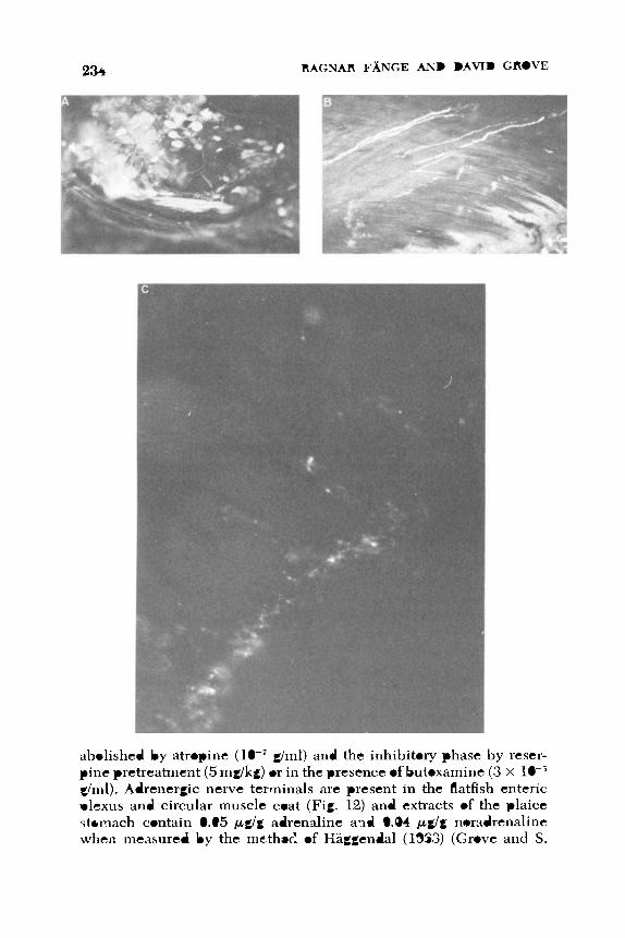
4. DIGESTION 235
Fig. 12. Adrenergic innervation of flatfish gut. Photomicrographs of tissues prepared
using the fluorescent histochemical technique of Falck and Owman (1965). (A) Coeliac ganglion ofAmmotretis containing fluorescent (adrenergic) ganglion cells. (B) Intestine
ofAmmotretis with adrenergic fibers running in the circular muscle layer. (C) Stomach
of Platichthys in which adrenergic terminals envelop nonfluorescent ganglion cells in
Auerbach’s plexus. (From S. Nilsson and Grove, unpublished observations.)
236 RAGNAR FANCE AND DAVID GROVE
Nilsson, unpublished observations). Extensions of the splanchnic
nerves supply the plaice intestine and mediate inhibition (Goddard,
1975). The effect is mimicked by adrenaline (lO-’O g/ml) and other
catecholamines. The intestine contains 0.06 pg/g adrenaline and 0.02 pg/g noradrenaline. However Goddard found a further nonadrenergic
inhibitory system in the wall of the plaice intestine. Transmural stimu-
lation relaxes the intestine (Fig. 13) but atropine, ganglion blockers, or
a- and p-blockers failed to antagonize the response. Adenosine
triphosphate ( lop6 g/ml) both relaxes the intestine and desensitizes the
receptors to transmural stimulation. The response is also blocked by
tetrodotoxin, but not reserpine pretreatment, suggesting that in-
tramural inhibitory purinergic neurons are involved.
As a measure of the complexity of the nervous coordination of
gastrointestinal activity of teleosts, a comparison with Salmo trutta and S. gairdneri is revealing. In this more primitive teleost, Burnstock
(1969) reinterpreted his early results on vagal stimulation, concluding
that the vagus sends cholinergic excitatory nerves to the esophageal
striated muscle but inhibits the stomach. The latter response is often
accompanied by a strong “rebound” contraction, is unaffected by at-
W
A V -
10 Hz ATP
Fig. 13. Magnus preparation of Pleuronectes platessa intestine. The inhibitory ef-
fects of transmural stimulation (10 V, 10 Hz, 1 msec pulses for 60 sec) are compared with
the response to 10-6 g/ml adenosine triphosphate. The time course of each relaxation is
similar and each is followed by spontaneous “rebound” contractions. Vertical scale, 2 g;
horizontal scale, 120 sec. (From Coddard, 1975.)
4. DIGESTION 237
ropint: at high doses ( g/ml) and is only impaired by high doses of nicotine g/ml) and hexamethonium (5 x g/ml) which are
probably nonselective. His claim that the response is dependent on
preganglionic cholinergic fibers depends on the specificity of the
above concentrations of ganglion blocking agents. Campbell (1975) showed that spontaneous stomach contractions in Salmo gairdnerii
were inhibited by intracranial stimulation of the vagal roots (0.5-8 Hz), often accompanied b y a large “rebound” contraction at the cessa-
tion of stimulation. More recently, Campbell and Gannon (1976) have
shown that stimulation of the splanchnic nerve at 5-10 Hz contracts Salmo stomach, confirming the studies of Burnstock (1958a). The re-
sponse was not affected by hyoscine ( g/ml) but was abolished by bretylium (1-3 x 10+ g/ml) and reduced by d-tubocurarine ( 3 x g/ml). Adrenergic fibers were detected histochemically in the splanch-
nic nerve which proceed to form a plexus in the stomach wall and,
in view of the excitatory effect of catecholamines described earlier, are
believed to mediate the excitation. Excitation of the posterior splanch-
nic nerve (Burnstock, 1958a) inhibits or excites the rectum depend-
ing on the frequency of stimulation. He proposed that both cholinergic
and adrenergic nerves are involved, and the latter have been detected
histochemically (Read and Burnstock, 1968a,b, 1969). Clearly the roles of the extrinsic nerves to the stomach and intes-
tine ofPleuronectes and Salmo are very different. It is not yet possible
to distinguish the roles of intramural neurons which may be linked
with, or independent of, extrinsic nerve tracts. The control of peristal-
sis in Tinca is also different in that the vagus in this stomachless fish
extends its influence to the intestine (Mahn, 1898) and the gut wall
consists of the usual smooth muscle coats together with two layers of
striped muscle (circular and longitudinal) with several nerve cell
plexuses (Baumgarten, 1965, 1967). In addition to the left and right
rami vagi intestinales, which exert cholinergic excitatory influences
on all muscle coats, a splanchnic nerve originating from the right sym- pathetic chain innervates the intestine. Baumgarten ( 1967) also
showed that this sympathetic supply carries fluorescent nerves which
innervate the muscle layers and which apparently contain dopamine,
as judged by the thionylchloride test of Corrodi and Jonsson (1967),
and which inhibit the intestine. He also obtained fluorescent his-
tochemical evidence for the presence of 5-hydroxytryptamine in some
nerves and pointed out that this amine contracts smooth muscle of the tench intestine. Saito (1973), using the fluorescent technique in Carus-
s i u s intestine, also detected adrenergic nerves which he proposed
inhibit spontaneous activity.
238 RAGNAR FANGE AND DAVID GROVE
4. In Vivo STUDIES
Very few attempts have been made to analyze the control of peris-
talsis in living, feeding fish. Burnstock (1957) observed in situ peris-
talsis of Salmo trutta by implanting an abdominal window. Edwards
( 1973) observed that carbachol dramatically accelerated the gastric
emptying of a 1% meal ofdrenicola by Pleuronectes whereas atropine
delayed gastric emptying. Goddard ( 1974) found that intestinal transit
time was also shortened by carbachol and extended by atropine when
these were injected at the appropriate time, but that this region of the
gut was less sensitive than the stomach to these drugs. In a separate
study, Goddard (1973) demonstrated similar actions of these agents in
the stomachless Blennius pholis. After 7 days food deprivation,
Pleuronectes (300 g at 15°C) did not show impaired efficiency in
transporting food through the gastrointestinal tract but during the sub-
sequent weeks the transit time slowly increased as the physiological
effects of starvation developed.
Gzgzyan et al. (1973) examined in vivo gastric contractions in
Scorpaena porcus using a balloon to record activity. In addition to
concluding that the stomach was under excitatory cholinergic control,
they showed that extracts of the pituitary gland of Scorpaena con-
tained a powerful gastric inhibitor, reminiscent of the inhibitory action
of oxytocin in mammals. In the dog, it has been found that injected
insulin at first depresses gastric activity but then, as hypoglycemia
develops, vagal centers in the medulla activate gastric activity. In
Scorpaena, insulin injections depressed stomach activity but no com-
pensatory increase accompanied the ensuing hypoglycemia.
E. Summary
Figure 14 represents a generalized account of the mechanisms con-
trolling teleost gastrointestinal motility. Longitudinal and circular
coats of smooth muscle fibers develop spontaneous, myogenic activity
when the gut is distended. In Pleuronectes, noncholinergic excitatory
nerves are necessary to induce this response. Peristalsis depends on
intramural neurons (cholinergic and noncholinergic excitatory; noncholinergichonadrenergic inhibitory) in synaptic connection with
sensory and other neurons in Auerbach’s plexus. In flatfish, but to a
lesser extent in salmonids and cyprinids, peristalsis in isolated prepa-
rations is mainly myogenic, similar to that described by Yung et al. (1965) for the tortoise Amyda japonica. Localized release of
Shydroxytryptamine generally enhances activity. Cranial vagal fibers

4. DIGESTION 239
Fig. 14. Diagram of the intrinsic and extrinsic nerve fibers believed to control the
muscularis of the teleost stomach, together with sites of action of circulating cate-
cholamines. ( 1) Nonadrenergic, noncholinergic inhibitory neurons; (2) cholinergic ex-
citatory neurons; ( 3 ) nonadrenergic, noncholinergic excitatory neurons; (4) stretch re-
ceptors; (5) circulating adrenaline; (6) endogenous Shydroxytryptamine; (7) cholinergic
excitatory neurons of postganglionic, sympathetic origin; (8) adrenergic inhibitory
neurons of postganglionic, sympathetic origin; (9) preganglionic cholinergic neurons
from central nervous system. Notes: (a) striated muscle is not always present in the
stomach and intestine; (b) in Salmo, element 8 to the stomach is excitatory; (c) in
Pleuronectes an additional element 9 in the vagus synapses with excitatory cholinergic
neurons in the jugular ganglion; (d) a vagal element is suggested which controls gastric
gland secretion. The intestine is controlled by elements 1-8 but, in stomachless fish,
vagal influences may extend to this region.
in fish primarily activate noncholinergic intramural inhibitory fibers.
Postganglionic cholinergic excitatory fibers reach the stomach in the
splanchnic nerve and, in some acanthopterygians, in the vagal path-
way to innervate muscle cells directly. Adrenergic neurons reach the

240 RAGNAR FANGE AND DAVID GROVE
stomach and intestine through the splanchnic route and enmesh gan-
glion cells in Auerbach’s plexus as well as entering the muscularis.
Catecholamines from the head kidney (e.g., Grove et al., 1972; Nilsson
et al., 1976) generally inhibit activity, even in denervated prepa-
rations, but the clear gastric excitation which occurs in elasmobranchs
and some teleosts requires further analysis.
I t will be obvious from the account given in this section that the
present knowledge of gastrointestinal motility and its control in fish is
sparse. The onus is on the comparative physiologist to undertake bet- ter designed studies if the complexity of the system is to be unraveled.
The present authors believe that, for a variety of fish species, careful
characterization of tissue receptors and the affinity of agonists and
antagonists is long overdue. The doses of antagonists which are selec- tive against candidate neurotransmitter substances must be deter-
mined. Only then can drugs be used as tools in the study of coordi-
nated peristalsis. The researcher must recognize that both neurogenic
and myogenic rhythmic contractions may be involved in the transport
of ingested food. Mechanical recordings should be accompanied by
electrical measurements of the activity of elements in the gut wall.
Detailed study of the ganglion cells of the enteric plexus, including
their neurotransmitters, and the origin and distribution of extrinsic
nerves should be undertaken. No mention has been made here of the
sensory connections of the gut to the central nervous system, although
it appears from Section VI that such nerves play a significant part in
appetite of fish. Windell and Norris (1969a,b) proposed that the pro-
duction of enterogastrone in response to high energy food may delay the
gastric emptying phase and yet the available data on this and other
local gastrointestinal hormones is almost nonexistent. Although motor
nerves of various origins impinge on the gastrointestinal tract, and
hormones of the adrenal medulla, pituitary, and other endocrines are
also likely to affect motility and secretion, very few attempts to inves- tigate their significance to the fish by in vivo experiments have been
undertaken. Kerkut (1976) has mentioned some of the important con-
tributions to physiology made by comparative pharmacologists; a ful-
ler understanding of the processing of food by the gastrointestinal tract
is likely to contribute significantly to the husbandry of fish.
VIII. PERSPECTIVES
In Section VI it is apparent that digestion rate is related to the
natural diet. The return of appetite following a meal is to a large extent

4. DIGESTION 24 1
controlled by the rate of stomach or foregut emptying (Table V). The
physiological data presented in Section VII suggest that, when a fish in
culture is presented with artificial foodstuffs, the rate of gastro-
intestinal activity can be changed by including a stimulant or inhibit-
ing drug in the diet. In this way a faster or more efficient rate of food
utilization could be obtained, supplemented by further additives
(such as steroids) which promote anabolism. Appropriate balance of nutrients, such as amino acids or oligosaccharides, which compete for
the active transport sites in the gastrointestinal epithelium, may in-
crease assimilation efficiency. Incorporation in the artificial diets of
exogenous enzymes which become active when the gastrointestinal
juices are absorbed may allow the fish to overcome the “surface area”
limitation to digestion rate. Environmental conditions which
minimize the circulation of a,drenaline or discharge of the adrenergic
nerves (“stress”) will allow peristaltic rate and visceral blood flow to
be at a maximum. There is a case for incorporating anti-adrenaline
agents or sedatives to the diet during “thinning-out” operations on the
fish farm, to allow rapid adjustment to the new environment and early
return of feeding. This latter suggestion may be most appropriate for
marine fish which frequently tolerate crowding to a lesser extent than
freshwater fish. These, and similar factors which may emerge, will be
additional to improvements in fish production brought about by ge-
netic selection, control of disease, diet formulation, and control of en-
vironmental variables such as temperature and photoperiod.
ACKNOWLEDGMENTS
We are indebted to our research colleagues with whom we have discussed and tested
our ideas and also to Mr. W. Rowntree for helping prepare the photographs. The British
Council (Academic links scheme) aided our cooperative venture. R. F. wrote mainly
Sections 111, IV, and V and D. C . mainly Sections 11, VI, VII, and VIII. Part of the work was done at the Kristineberg hlarine Biological Station at Fiskebiickskil, Sweden.
REFERENCES
Adam, H. (1960). Uber das Vorkommen eiiier peritrophischen Menibran im Darm von
Myxine glutinosa L. (Cyclostomata). Naturwissenschuften 14, 33Z334.
Adron, J. W., Grant, P. T., and Cowey, C. B. (1973). A system for the quantitative study of
the learning capacity ofrainbow trout and its application to the study of food prefer-
ences and 1lehaviour.J. Fish Biol. 5, 625-636.
Albertini-Berhaut, J., and Vallet, F. (1971). Utilization alimentaire de 1’uri.e chez les
muges. Tethys 3, 677-680.

242 RAGNAR FANGE AND DAVID GROVE
Alexander, R. McN. (1970). Mechanics of the feeding action of various teleost fishes. J . Zool. 162, 145-156.
Al-Hussaini, A. H. (1947). The feeding habits and the morphology of the alimentary tract
of some teleosts living in the neighbourhood of the Marine Biological Station,
Ghardaqa, Red Sea. Puhl. Mar. Biol. St., Ghardaqa, Red Sea 5,4-61.
A]-Hussaini, A. H. (19494. On the functional morphology of the alimentary tract of some
fish in relation to differences in their feeding habits: Anatomy and histology. Q. ]. A4icrosc. Sci. 90, 109-139.
Al-Hussaini, A. H. (1949b). On the functional morphology of the alimentary tract of some
fish in relation to differences in their feeding habits: Cytology and physiology. Q. ]. Microsc. Sci. 90, 323-354.
Al-Hussaini, A. H., and Kholy, A. A. (1953). On the functional morphology of the alimen-
tary tract of some omnivorous teleost fish. Proc. Egypt. Acad. Sci. 4, 17-39.
Alliot, E. (1967). Absorption intestinale de I’N-acetyl-glucosamine chez la petite
Rousette. C. R. SOC. Biol. (Paris) 161, 2544-2546.
Alliot, E., Febvre, A., and Mktailler, R. (1974). Les protkases digestives chez un teleos-
tken carnivore Dicentrus labrax. Ann. Biol. Anim., Biochim., Biophys. 14,229-237.
Angelescu, V., and Gneri, F. S. (1949). Adaptaciones del aparto digestivo a1 regimen
alimenticio en algunos pesces del Rio Uruguay y del Rio de la Plata. Rev. Inst. Nac.
Inuest. Cienc. Nut. (Buenos Aires) 1, 161-272.
Aoyama, T. (1958). On the discharge of stomach contents concerning jack mackerel
(Trachurus japonicus). Bull. Seikai Reg. Fish Res. Lab. (Seikai-Ku Suisan Ken-
kyusho Kenkyu Hokoku) 15,29-32.
Arcangeli, A. (1908). Contributo alla conoscenze della strutture minuta dello stomaco
del Box salpa L. secondo la stato funziale. Arch. Zool. I ta l . 3, 261-346.
Ariens, E. J., and Van Rossum, J. M. (1957). pD,, PA, and pDI values in the analysis of
pharmacodynamics. Arch. Intern. Pharmacodyn. Ther. 110, 275-299.
Armstrong, R. H., and Blackett, R. F. (1966). Digestion rate of the Dolly Varden. Trans. Am. Fish. Soc. 95,429-430.
Arthur, D. K. (1956). Cited in Blaxter (1963).
Babkin, B. P. (1929). Studies on the pancreatic secretion in skates. Biol. Bull. (Woods
Hole, Mass.) 57, 272-291.
Babkin, B. P. (1933). Further studies on the pancreatic secretion in the skate. Contrih. Can. Biol. Fish. 7, 1-9.
Babkin, B. P., and Bowie, D. (1928). The digestive system and its function in Fundulus heteroclitus. Biol. Bull. (Woods Hole, Mass.) 54, 254-277.
Babkin, B. P., Friedman, M. H. F., and MacKay-Sawyer, M. E. (1935a). Vagal and
sympathetic innervation of the skate.]. Biol. Board Can. 1, 239-250.
Babkin, B. P., Chaisson, A. F., and Friedman, ,M. H. F. (19351,). Factors determining the
course of the gastric secretion in elasmobranchs. J. Biol. Board Can. 1, 251- 259.
Backiel, T. (1971). Production and food consumption of predatory fish in the Vistula
River. J. Fish Biol. 3, 369-405.
Baldwin, N. S. (1956). Food consumption and growth of brook trout at different temper-
atures. Trans. Am. Fish. Soc. 86, 323-328.
Ballentijn, C. M., van den Burg, A., and Egberink, B. P. (1972). An electromyographic
study of the adductor mandibulae complex of a free-swimming carp (Cyprinus carpio) during feeding. J . E x p . B i d . 57( I), 261-283.
Barnard, E. A. (1973). Comparative biochemistry and physiology of the digestion. In “Comparative Animal Physiology” (C. L. Prosser, ed.), 3th Ed., pp. 133-152. Saun-
ders, Philadelphia, Pennsylvania.

4. DIGESTION 243
Barrington, E. J. W. (1957). The alimentary canal and digestion. In “The Physiology of
Fishes” (M. E. Brown, ed.), Vol. 1, pp. 109-161. Academic Press, New York.
Barrington, E. J. W. (1962). Digestive enzymes.Adv. Comp. Physiol. Biochern. 1, 1-65.
Barrington, E. J. W. (1972). The pancreas and intestine. In “The Biology of Lampreys”
(M. W. Hardisty and I. Potter, eds.), Vol. 2, pp. 135-169. Academic Press, New York.
Barrington, E. J. W., and Dockray, G. J. (1972). Cholecystokinin-pancreozyminlike ac-
tivity in the eel (Anguilla anguilla L.). Gen. Comp. Endocrinol. 19, 80-87.
Battle, H. I. (1935). Digestion and digestive enzymes in the herring (Clupea harengus L.)J. Biol. Board Can. 1, 145-157.
Baumgarten, H. G. (1965). Uber die Muskulatur und die Nerven in der Darmwand der
Schleie (Tinca vulgaris Cuv.). Z . Zellforsch. Mikrosk. Anat. 68, 116137.
Baumgarten, H. G. (1967). Verkommen und Verteilung adrenerger Nervenfasern in
Darm der Schleie (Tinca vulgaris Cuv.).Z. Zellforsch. Mikrosk. Anat. 76,248-259.
Baumgarten, H. G., Bjorkland, A,, Lachenmayer, L., Nobin, A., and Rosengren, E.
(1973). Evidence for the existence of serotonin-, dopamine- and noradrenaline-
containing neurons in the gut of Lampetra puoiatilis. Z. Zellforsch. Mikrosk. Anat. 141, 3S54.
Bayliss, W. M., and Starling, E. H. (1903). On the uniformity of the pancreatic mecha-
nism in vertebrata. J. Physiol. (London) 29, 174-180.
Beamish, F. W. H. (1972). Ration size and digestion in largemouth bass, Micropterus salmoides Lacepede. Can. J. Zool. 50, 153-164.
Bengelsdorf, H., and Elias, H. (1950). The structure ofthe liver of cyclostomata. Chicago Med. Sch. Q. 12, 7-12.
Bennett, M. R., and Burnstock, G. (1968). Electrophysiology of the innervation of intes-
tinal smooth muscle. In “Handbook of Physiology” (C. F. Code, ed.), Vol. IV, Sect.
6, Alimentary Canal, pp. 1709-1732. Am. Physiol. SOC., Washington, D.C.
Benson, A. A., and Lee, R. F. (1975). The role of wax in oceanic food chains. Sci. Am. 232, 7686.
Bernard, F. (1952). La digestion chez les poissons. Trav. Lab. Hydrobiol. Piscicult. Unio. Grenoble 44, 61-95.
Bertin, L. (1958). Appareil digestif. In “Trait6 de Zoologie” (P. Grasse, ed.), Tome XIII,
Fascicule 2, pp. 1248-1302. Masson, Paris.
Beukema, J. J. (1968). Predation by the three-spined stickleback (Gasterosteus aculeatus): The influence of hunger and experience. Behaviour 31, 1-126.
Biedermann, W. (1911). Die Ernahrung der Fische. In “Handbuch der Vergleichen den
Physiologie” (H. Winterstein, ed.), Vol. 2, Part 1, pp. 1049-1115. Fischer, Jena.
Bilinski, E. (1974). Biochemical aspects of fish swimming. In “Biochemical and Biophysical perspectives in Marine Biology” (D. C. Malins and J. R. Sargent, eds.),
Vol. 1, pp. 239-288. Academic Press, New York.
Bishop, C., and Odense, P. H. (1966). Morphology of the digestive tract of the cod,
Gadus morhua. J . Fish. Res. Board Can. 23, 1607-1615.
Blake, I. H. (1930). Studies on the comparative histology of the digestive tube of certain
fishes. I . A predacious fish, the sea bass Centropristes striatus. J. Morphol. 50,
39-70.
Blaxter, J. H. S. (1963). The feeding of herring larvae and their ecology in relation to
feeding. Cali$ Coop. Oceanogr. Fish Invest. Rep. 10, 79-88. Blaxter, J. H. S., and Holliday, F. G. T. (1963). The behaviour and physiology of herring
and other clupeoids. Adv. Mar. B i d . 1, 261-393.
Bodola, A. (1966). Life history of the gizzard shad, Dorosoma cepedianum (Le Sueur), in
western Lake Erie. U . S . Fish Wildl. Serv., Fish. Bull. 65,391-425. Cited in Schmitz
and Baker (1969).

244 RAGNAR FANGE AND DAVID GROVE
Bokova, E. (1938). Daily food consumption and digestive rate in Rutilus rutilus. Rybn.
Khoz. (Moscow) No. 6.
Borgstrom, B. (1974). Bile salts-their physiological function in the gastrointestinal tract. Acta Med. Scand. 196, 1-10,
Boyer, J. L., Schwarz, J., and Smith, N. (1974). Preliminary studies of bile secretion in
the isolated perfused liver of the small skate, Raja erinacea. Bull. Mt. Desert I s l . Biol. Lab. 14, 12-13.
Bray, R. N., and Ebeling, A. W. (1975). Food, activity and habitat of three “picker” type
microcarnivorous fishes in the kelp forests of Santa Barbara, California. U.S. Fish Wild. Sero., Fish. Bull. 73(4), 815-829.
Brett, J. R. (1971). Starvation time, appetite and maximum food intake of sockeye sal-
mon, Oncorhynchus nerka. J. Fish. Res. Board Can. 28, 409-415.
Brett, J. R., and Higgs, D. A. (1970). Effect of temperature on the rate of gastric digestion
in fingerling sockeye salmon, Oncorhynchus nerka. J. Fish. Res. Board Can. 27,
1767-1779.
Brinn, J. E., Jr. (1973). The pancreatic islets of bony fishes. Am. Zool. 13, 653-665.
Brockerhoff, H. (1966). Digestion of fat by c0d.J. Fish. Res. Board Can. 23, 18.35-1839.
Bryan, P. G. (1975). Food habits, functional digestive morphology, and assimilation
efficiency ofthe rabbitfish Siganus spinus (Pisces, Siganidae) on Guam. Pac. Sci. 29,
269-277.
Bucke, D. (1971). The anatomy and histology of the alimentary tract of the carnivorous
fish the pike Esox lucius L. J . Fish Biol. 3, 421-431.
Bullock, W. L. (1963). Intestinal histology of some salmonid fishes with particular refer-
ence to the histopathology of acanthocephalan infections. J . Morphol. 112, 23-
44.
Bullock, W. L. (1967). The intestinal histology of the mosquito fish Gamhusia afinis
(Baird and Girard). Acta Zool. (Stockholm) 48, 1-17.
Burnstock, G. (1957). A “window” technique for observing fish viscera in cico. Nature
(London) 180, 1491-1492.
Burnstock, G. (195th). The effects of drugs on spontaneous motility and on response to
stimulation of the extrinsic nerves of the gut of a teleostean fish. Br. J. Pharrnacol. 13, 216-226.
Burnstock, G. (195%). Reversible inactivation of nervous activity of fish. J. Physiol.
(London) 141,35-45. Burnstock, G. (195%). The morphology ofthe gut ofthe brown trout (Salmo trutta). Q . J .
Microsc. Sci. 100, 1S3>198.
Burnstock, G. (195913). The innervation ofthe gut ofthe brown trout (Salmo trutta). Q.J. Microsc. Sci. 100, 199-220.
Burnstock, G. (1969). Evolution of the autonomic innervation of visceral and cardio-
vascular systems in vertebrates. Pharmacol. Rev. 21, 247-324.
Burnstock, G. ( 1972). Purinergic nerves. Pharmacol. Rev. 24, 509-581.
Burnstock, G., Campbell, G., Satchell, D., and Smythe, A. (1970). Evidence that
adenosine triphosphate or a related nucleotide is the transmitter substance released
by nonadrenergic inhihitory nerves in the gut. Br. J . Pharmacol. 40,668-689.
Burnstock, G., Satchell, D., and Smythe, A. (1972). A comparison of the excitatory and
inhibitory effects on nonadrenergic, noncholinergic nerve stimulation and
exogenously-applied ATP on a variety of smooth muscle preparations from different
vertebrate species. Br. J. Pharmacol. 46, 234-242.
Cameron, J. N., Kostoris, J., and Penhale, P. A. (1973). Preliminary energy budget of the
ninespine stickleback (Pungitius pungitius) in an Arctic lake. J . Fish. Res. Board
Can. 30, 1179-1189.

4. DIGESTION 245
Campbell, G. (1970a). Autonomic nervous systems. In “Fish Physiology” (W. S. Hoar
and D. J. Randall, eds.), Vol. 4, pp. 109-132. Academic Press, New York.
Campbell, G. (19701,). Autononiic nervous supply to effector tissues. I n “Smooth Mus- cle” (E. Bulbring, A. Brading, A. Jones, and T. Tomita, eds.), pp. 109-132. Arnold,
London.
Campbell, G. (1975). Inhibitory vagal innervation of the stomach in fish. Coinp.
Biochem. Physiol. C 50, 169-170.
Campbell, G., and Burnstock, G. ( 1968). Comparative physiology of gastrointestinal
motility. In “Handbook of Physiology.” Sect. 6: “Alimentary Canal” (C. F. Code,
ed.), Vol. IV, pp. 221-2-2266. Am. Physiol. Soc., Washington, D.C. Campbell, G., and Cannon, B. J. (1976). The splanchnic nerve supply to the stomach of
the trout, Sulmo trutta and S . gairdneri. Comp. Biochem. Physiol. C 55, 51-53.
Cartier, M., and Buclon, M. (1973). Ultrastructure comparke de la bordure en brosse de
I’entkrocyte en fonction de l’ktat nutritionnel, chez trois cyprinides: La tanche
(Tinca tincu L.), la carpe (Cyprinus carpio L.) et le poisson rouge (Curassius au-
rutus L.). Ann. Nutr. Aliment. 27, 181-189.
Chaichara, A., and Bullock, W. L. (1967). The histopathology of acanthocephalan infec-
tions in suckers with observations on the intestinal histology of two species of
catastomid fishes. Acta Zool. (Stockholm) 48, 19-42.
Chakrabarte, J., Saharya, R., and Belsare, D. K. (1973). Structure of the gallbladder in
some freshwater teleosts. Z . Mikrosk.-Anat. Forsch. 87(1), 23-32.
Chao, L. N. (1973). Digestive system and feeding habits of the cunner, Tuutogolabrus
adspersus, a stomachless fish. U.S. Fish Wildl. Serzj., Fish. Bull. 71, 565-586.
Chesley, L. C. (1934). The concentration of proteases, amylase, and lipase in certain
marine fishes. Biol. Bull. (Woods Hole, Mass.) 60, 133-144.
Chiba, K. (1961). The basic study on the production of fish seedling under possible
control. I. The effect of food in quality and quantity on the survival and growth of
common carp fry. Tunsuiku Suisan Kenkyusho Kenkyu Hokoku 11, 105-132.
Chujyo, N. (1953). Site of acetylcholine production in the wall of intestine. Am. J. Physiol. 174, 196-198.
Corner, E. D. S., Denton, E. J., add Fhrster, G. R. (1969). On the buoyancy of some deep
sea sharks. Proc. R. Soc., Ser. B 171, 415-429.
Corrodi, H., and Jonsson, G. (1967). Formaldehyde fluorescence method for the his-
tochemical demonstration of biogenic monoainines. A review on the methodology.
J. Histochem. Cytochem. 15, 6 5 7 8 . Creac’h, P. V. (1963). Les enzymes protkolytiques des poissons. Ann. Nutr. Aliment. 17,
A37SA471. Croston, C. B. (1960). Tryptic enzymes of chinook salmon. Arch. Biochem. Biophys. 89,
202-206.
Croston, C. B. (1965). Endopeptidases of salmon ceca: Chromatographic separation and
some properties. Arch. Biochem. Biophys. 112, 218-223.
Daan, N. (1973). A quantitative analysis of the food intake of North Sea cod, Gadus morhua. Neth. J . Sea Res. 6, 479-517.
Dahlstedt, E., von Euler, U. S., Lishajko, F., and Ostlund, E. (1959). Observations on the
distribution and action of substance P in marine animals. Acta Physiol. Scand. 47,
124- 130. Dandrifosse, G. (1975). Purification of chitinase contained in pancreas of gastric mucosa
of frog. Biochimie 57, 82%831. Daniel, E. E. (1968). Pharmacology ofthe gastrointestinal tract. I n “Handbook of Physi-
ology” (C. F. Code, ed.), Part 6, Vol. 4, pp. 2267-2324. Am. Physiol. Soc., Washing- ton, D.C.

246 RAGNAR FANGE AND DAVID GROVE
Darnell, R. M., and IMeierotto, R. R. (1962). Determination of feeding chronology in
David, H. (1961). Zur submikroskopischen Morphologie intrazellularer Gallen-
Davson, H. (1970). “A Textbook of General Physiology.” Churchill, London.
Dawson, I. (1970). The endocrine cells of the gastrointestinal tract. Histochem. J . 2,
527-549. De Groot, S. J . (1971). On the interrelationship between morphology of the alimentary
tract, food and feeding behavior in flatfishes (Pisces, Pleuronectidae). Neth. J . Sea
Res. 5, 121-196.
De Haen, C., and Gertler, A. (1974). Isolation and amino-terminal sequence analysis of
two dissimilar pancreatic proelastases from the African lungfish. Biochemistry 13,
2673-2677.
Dockray, G. J. (1975). Comparative studies on secretin. Gen. Comp. Endocrinol. 25,
203-210.
Dreyer, N. B. (1949). The action of autonomic drugs on elasmobranch and teleost in-
voluntary muscle. Arch. Int. Pharmacodyn. Ther. 78, 63-66.
Edwards, D. J. (1971). Effect of temperature on rate of passage of food through the
alimentary canal of the plaice, Pleuronectes platessa L.]. Fish Biol. 3,433-439.
Edwards, D. J. (19724. Electrical stimulation of isolated vagus nerve-muscle prepa-
rations of the stomach of the plaice, Pleuronectes platessa L. Comp. Gen. Phar-
macol. 2, 235-242.
Edwards, D. J . (197213). Reactions of the isolated plaice stomach to applied drugs.
Comp. Gen. Pharmacol. 3, 345-358.
Edwards, D. J. (1973). The effects of drugs and nerve section on the rate of passage of
food through the gut ofthe plaice,Pleuronectes platessa L.J. Fish B i d . 5,441-446.
Elias, H., and Sherrick, J. C. (1969). “,Morphology of the Liver.” Academic Press, New
York.
Elliott, J. M. (1972). Rates of gastric evacuation in brown trout, Salmo truttu L. Fresh-
water Biol. 2, 1-18.
Elliott, J. M. (1975). Weight of food and time required to satiate brown trout, Salmo trutta L. Freshwater Biol. 5(1), 51-64.
Epple, A. (1969). The endocrine pancreas. In “Fish Physiology” (W. S. Hoar and D. J.
Randall, eds.), Vol. 2, pp. 275-319. Academic Press, New York.
Eriksen, N. (1945). Purification and properties of halibut pepsin. M.S. Thesis, Univ. of Washington, Seattle. Cited in Jany (1976).
Fibiin, G., MolnLr, G., and Tolg, I. (1963). Comparative data and enzyme kinetic
calculations on changes caused by temperature in the duration of gastric digestion of some predatory fishes. Acta Biol. Acad. Sci. Hung. 14, 123-129.
Falck, B., and Owman, C. H. (1965). Detailed methodological description of the fluores-
cence method for the cellular demonstration of biogenic menoamines. Acta Uniu.
Lund. Sect. 11, No. 7, pp. 1-23.
Falkmer, S., and Olsson, R. (1962). Ultrastructure of the pancreatic islet tissue of normal
and alloxan-treated Cottus scorpius. Acta Endocrinol. (Copenhagen) 39, 32-46. Falkmer, S., Hellman, B., and Voigt, G. E. (1964). On the agranular cells in the pancrea-
tic islet tissue of the marine teleost Cottus scorpius. Acta Pathol. Microhiol. Scund. 60(1), 47-54.
Falkmer, S., Ostberg, Y., and Van Noorden, S. (1978). Entero-insular endocrine systems
of cyclostomes. In “Gut Hormones” (S. R. Bloom, ed.), pp. 57-63. Churchill
Livingstone, New York.
fishes. Trans. Am. Fish. Soc. 91, 313-320.
kapillaren. Acta Anat. 47, 216224.

4. DIGESTION 247
Fange, R. (1962). Pharmacology of poikilothermic vertebrates and invertebrates. Phar-
macol. Reo. 14, 281-316.
Fange, R. (1968). The formation of eosinophilic granulocytes in the oesophageal lym-
phomyeloid tissue of elasmobranchs. Acta Zool. (Stockholm) 49, 155-161.
Fange, R. (1973). The lymphatic system ofMyxine. Acta Regiae Soc. Sci. Litt. Gothob.
Zool. 8, 57-64.
Fange, R., and Hanson, A. (1973). Comparative pharmacology of catecholamines. In
“International Encyclopaedia of Pharmacology” (M. J. Michelson, ed.). Pergamon, New York.
Fange, R., and Johnels, A. G . (1958). An autonomic nerve plexus control of the gall
bladder in Myxine. Acta Zool. (Stockholm) 39, 1-8.
Fange, R., Johnels, A. G., and Enger, P. S. (1963). The autonomic nervous system. In
“The Biology of Myxinc” (A. Brodal and R. Fange, eds.), pp. 124-136. Univer-
sitetsforlaget, Oslo.
Fange, R., Lundblad, G., and Lind, J. (1976). Lysozyme and chitinase in blood and
lymphomyeloid tissues of marine fish. Mar. Biol. 36,277-282. Fange, R., Lundblad, G., Lind, J., and Wikberg, K. (1978). Chitinolytic enzymes in the
digestive system of marine fish. (Submitted for publication.)
Farmanfarmaian, A., Ross, A., and Mazal, D. (1972). In uivo intestinal absorption of the
sugar in the toadfish (marine teleost Opsanus tau) . B i d . Bull. (Woods Hole, Mass.)
142,427-445.
Fish, G. R. (1960). The comparative activity of some digestive enzymes in the alimen-
tary canal of Tilapia and perch. Hydrobiologia 15, 161-179.
Fortunatova, K. R. (1950). Biology of feeding of Scorpaena porcus L. Tr. Seuastop. B i d .
Stn., Akad. Nauk Ukr. SSR 7, 193-235.
Fortunatova, K. R. (1955). Methods of studying the feeding of predatory fishes. Tr. SOG.
Ikhtiol. Kom. Akad. Nauk SSR 6, 62-84. (In Russ.)
Frantsusova, G. P. (1971). Afferent innervation of the digestive tract in Stenodus
Zeucichthys. Arkh. Anat., Gist. Embriol. 60, 39-41. (In Russ.)
Fujita, T. (1962). Uber das Inselsystem des Pancreas von Chimaera monstrosa. Z.
Zellforsch. Mikrosk. Anat. 57, 2272-2277.
Fujita, T. (1974). New aspects on the pancreatic islets. In “Recherches Biologiques
Contemporaines’? (L. Arvy, ed.), pp. 437-440. Imprimerie Wagner, Nancy, France.
Gabe, M., and Martoja, M. (1971). DonnCes histologiques sur les cellules endocrines,
gastriques et pancrdatiques de Mugil auratus (TdlCostCen, Mugilifonne). Arch.
Anat. Microsc. Morphol. E x p . 60, 219-234.
Gammon, R. L., Tiemeier, 0. W., and Gier, H. T. (1972). The gross and microanatomy of
the digestive tract and pancreas of the channel catfish, Zctalurus punctatus. Trans. Kans. Acad. Sci. 75, 141-155.
Gauthier, G. F., and Landis, S. C. (1972). The relationship of ultrastructural and
cytochemical features to absorptive activity in the goldfish intestine. Anat. Rec. 172, 675702.
Gerald, V. M. (1973). Rate of digestion in Ophiocephalus punctatus, Bloch. Cornp.
Biochem. Physiol. A 46, 195205.
Gershon, M. D. (1967). Inhibition of gastrointestinal movement by sympathetic nerve stimulation: The site of action. J. Physiol. (London) 189, 317-328.
Ghazzawi, F. M. (1935). The pharnyx and intestinal tract of the Egyptian mullets Mugil cephalus and Mugil cupito. 11. On the morphology and histology of the alimentary
canal in Mugil capito. Coastal Fish. Serr;., Fish. Res. Dir., Notes Mem. (Cairo) No.
6, pp. 1-31.
248 RAGNAR FANGE AND DAVID GROVE
Girgis, S. (1952). On the anatomy and histology of the alimentary tract of an herbivorous
bottom-feeding cyprinoid fish, Labeo horie (Cuv). J. Morphol. 90, 317-362.
Glaser, G. (1933). Beitrage zur Kenntnis des Lymphgerasssystem der Fische. 2.
Gesamte Anat., Abt. 1 100,433-511.
Goddard, J. S. (1973). The effects of cholinergic drugs on the motility of the alimentary
canal of Blennius pholis L. Experientia 29, 974-975.
Goddard, J. S. (1974). An X-ray investigation of the effects of starvation and drugs on intestinal motility in the plaice, Pleuronectes platessa L. Zchthylolgia (Sarajevo)
6( l), 49-58.
Goddard, J. S. (1975). Studies on intestinal control of the plaice, Pleuronectes platessa
L. Ph.D. Thesis, Univ. of Wales, Cardiff.
Gomazkov, 0. A. (1959). Effect of temperature on digestion of the burbot, Lota Lota.
Byull. Inst. Biol. Vodokhran., Akad. Nauk SSSR 5, 26-28. (In Russ.)
Granick, S. (1953). Investigations in iron metabolism. Am. Nat. 87, 65-75.
Greene, C. W. (1914). The fat-absorbing function of the alimentary tract of the king
salmon. Bull. U.S. Fish. Bur. 33, 14S175. Cited in Schmitz and Baker (1969).
Grodzinski, 2. (1938). Das Blutgefasssystem. I n “Dr. H. G. Bronns Klassen und
Ordnungen des Tierreichs.” Bd. 6, Abt. 1, Buch 2: “Echte Fische,” pp. 1-77. Akad.
Verlagsges., Leipzig.
Grove, D. J., Starr, C. R., Allard, D. R., and Davies, W. (1972). Adrenaline storage in the
pronephros of the plaice, Pleuronectes platessa L. Comp. Gen. Pharmacol. 3,205-
212.
Grove, D. J.. O’Neill, J. G., and Spillett, P. B. (1974). The action of Shydroxytryptamine
on longitudinal gastric smooth muscle of the plaice Pleuronectes platessa. Comp.
Gen. Pharmacol. 5,229-238.
Grove, D. J., Goddard, J. S., Tan, S. P., and Wirtz, P. (1976). Unpublished observations
from M.Sc. and Ph.D. theses, Univ. of Wales, Cardiff.
Grove, D. J . , Lozoides, L., and Nott, J. (1978). Satiation amount, frequency of feeding
and gastric emptying rate in Salmo gairdneri. J . Fish Biol. 12, 507-516.
Guimaraes, S. (1969). Reversal by pronethalol of dibenamine blockade: A study on the
seminal vesicle of the guinea pig. Br. J. Pharmacol. 36, 594-601.
Gzgzyan, D. M., and Kuzina, M. M. (1973). Stomach motor activity in the Black Sea ray,
Dasyatis pastinaca. J. Euol. Biochem. Physiol. (USSR) 9(5), 536-538. (In Russ.)
Gzgzyan, D. M., Zaks, M. G., and Tanasiychuk, 0. F. (1968). Effect of histamine, sub- stance “P” and pituitrin on the gastric secretion of the common European catfish
(Silurus glanis L.). Prohl. Ichthyol. (USSR) 8, 97-100. (Transl. from Vopr. Ikhtiol. 1968, 8, 126-130.)
Gzgzyan, D. M., Kuzina, M. M., and Tanasiichuk, 0. F. (1973). The effects of hypophy-
seal hormones and insulin on motor activity of the stomach in the scorpion fish
Scorpaena porcus. J . Evol. Biochem. Physiol. (USSR) 9(3), 301-302. (In Russ.)
Haggendal, J. (1963). An improved method for fluoriinetric determination of small
amounts of adrenaline and noradrenaline in plasma and tissues. Acta Physiol. Scand. 59,242-254.
Hale, P. (1965). The morphology and histology of the digestive systems of two fresh-
water teleosts, Poecilia retculata and Gasterosteus acu1eatus.J. Zool. 146,132-149. Hallbiick, D. A. (1973). Acetylcholinesterase-containing structures in the intestine of
Myxine glutinosa. Acta Regiae SOC. Sci. Litt. Gothob. Zool. 8, 24-25.
Hansen, D. (1975). Evidence of a gastrin-like substance in Rhinohatus productus.
Comp. Biochem. Physiol. C 52, 61-64.
Haslewood, G. A. D. (1968). Evolution and bile salts. I n “Handbook of Physiology”
(C. F. Code, ed.), Sect. 6, Vol. 5, pp. 2375-2390. Am. Physiol. SOC., Washington, D.C.
4. DIGESTION 249
Hatanaka, id., Kosaka, M., Sato, Y., Yamati, K., and Fukui, K. (1954). Interspecific rela-
tions concerning the predaceous habits among the benthic fish. Tohoku J . Agric. Res. 5, 177-189.
Hay, A. W. M., and Watson, G . (1976). The plasma transport of 25hydroxycalciferol in
fish, amphibians, reGtiles, and birds. Comp. Biochem. Physiol. B 53, 167-172.
Healey, M. C. (1971). The distribution and abundance of sand gobies, Gobius minutus in
the Ythan Estuary.J. Zool. 163, 177-229.
Herting, G . E., and Witt, A. (1968). Rate of digestion in the bowfin. Prog. Fish. Cult. 30( l), 26-28.
Hess, A. D., and Rainwater, J. H. (1939). A method for measuring food preference of
trout. Copeia No. 3, pp. 154-157.
Hickling, C. F. (1966). On the feeding process in the white amur, Ctenopharyngodon idella. J . Zool. 148, 408-418.
Hickling, C. F. (1970). A contribution to the natural history of the English grey mullets
(Pisces, Mugolidae). J . Mar. B i d . Assoc. U . K . 50, 609-633.
Hill, W. C. (1926). A comparative study of the pancreas. Proc. Zool. Soc. London Part 2,
pp. 581-631.
Hinton, D. E., and Pool, C. R. (1976). Ultrastructure of the liver in channel catfish
Ictalurus punctatus (Rafinesque). J . Fish Biol. 8, 209-219.
Hinton, D. E., Snipes, R. L., and Kendall, M. W. (1972). Morphology and enzyme
histochemistry in liver of largemouth bass (Micropterus salmoides). J . Fish. Res. Board Can. 29, 531-534.
Hirao, S., Yamada, J., and Kikuchi, R. (1960). On improving the efficiency of feed for fish
culture. Transit and digestibility of diet in eel and rainbow trout, observed by the
use of
Hirschowitz, B. I. (1975). Regulation of gastric secretion. In “Functions of the Stomach
and Intestine” (M. H. F. Friedman, ed.), pp. 145-165. Univ. Park Press, Baltimore,
Maryland.
Hoagman, W. J. (1974). Vital activity parameters as related to the early life history of
larval and postlarval lake whitefish (Coregonus clupeaformis). In “The Early Life
History of Fish” (J. H. Blaxter, ed.), pp. 547-558. Springer-Verlag, Berlin and Kew
York.
Hohson, E. S. (1974). Feeding relationships of teleostean fishes on coral reefs in Kona,
Hawaii. U.S. Fish Wildl. Sertj., Fish. Bull. 72(4), 915-1031.
Hogben, C. A. M. (1967a). Response of the isolated dogfish gastric mucosa to histamine.
Proc. Soc. E x p . Biol. iMed. 124, 89CL893.
Hogben, C. A. M. (196%). Secretion of acid by the dogfish, Syualus acanthias. In “Sharks, Skates and Rays” (P. W. Gilbert, R. F. Mathewson, and D. P. Rall, eds.), pp.
299-315. Johns Hopkins Press, Baltimore, Maryland.
Holstein, B. (197521). Intestinal diamine oxidase of some teleostean fishes. Comp. Biochem. Physiol. B 50, 291-297.
Holstein, B. (1975b). Gastric secretion in a teleostean fish: A method for the continuous
collection of gastric effluence from a swimming fish and its response to histamine
and pentagastrin. Acta Physiol. Scund. 95, 417-423.
Holstein, B. (1976). Effect of the H2 receptor antagonist metiamide on carbachol- and
histamine-induced gastric acid secretion in the Atlantic cod, Gadus morhua. Actu
Physiol. Scund. 97, 189-195.
Holstein, B. (1977). Effect of atropine and SC-15396 on stimulated gastric acid secretion
in the Atlantic cod, Gadus morhua. Acta Physiol. Scatxd. 101, 185-193. Honma, S . (1970). Presence of monaminergic neurones in the spinal cord and intestine
of the lamprey Lampetra japonica. Arch. Histnl. Jpn. (Niigata, Jpn . ) 32, 383-393.
Tokaiku Suisan Kenkyusho Kenkyu Hokoku 27, 67-72.

250 RAGNAR FANGE AND DAVID GROVE
Hotta, H., and Nakashima, J. (1968). Experimental study on the feeding activity of jack
mackerel Trachurus japonicus. Bull. Sekai Reg. Fish. Res. Lab. (Seikai-Ku Suisan Kenkyusho Kenkyu Hokoku)36,75-83.
House, C. R., and Green, K. (1963). Sodium and water transport across isolated intestine
of a marine teleost. Nature (London) 199, 1293-1294. Hunn, J. B. (1972). Concentrations of some inorganic constituents in gallbladder bile
from some freshwater fishes. Copeia No. 4, pp. 860-861.
Hunt, B. P. (1960). Digestion rate and food consumption of Florida gar, warmouth and
largemouth bass. Trans. Am. Fish. SOC. 89(2), 2OE-210.
Hunt, J. N., and Knox, M. T. (1968). Regulation of gastric emptying. In “Handbook of
Physiology” (C. F. Code, ed.), Sect. 6, Vol. 4, pp. 1917-1935. Am. Physiol. Soc.
Washington, D.C.
Ikeda, M., Hayama, T., Chujyo, N., and Hoshi, A. (1958). Acetylcholine content in the
stretched wall of intestine. Am. J. PhysioZ. 192, 27-29.
Imura, K. (19744. On the alkaline RNase isolated from the pyloric caeca of rainbow
trout. Nippon Suisan Gakkaishi 40, 399-408.
Imura, K. (1974b). Purification and properties of the phosphodiesterase in the pyloric
caeca of rainbow trout. Nippon Suisan Gakkaishi 40,807-814.
Inaba, D., Ogino, C., Takamatsu, C., Sugano, S., and Hata, H. (1962). Digestibility of
dietary components in fishes. I. Digestibility of dietary proteins in rainbow trout.
Nippon Suisan Gakkaishi 28, 367-371.
Iro, S. (1967). Anatomic structure of the gastric mucosa. In “Handbook of Physiology”
(C. F. Code, ed.), Sect. 6, Vol. 2, pp. 705-741. Am. Physiol. SOC., Washington, D.C.
Ishida, J. (1935). Ciliated intestinal epithelium in a teleost. Annot. Zool. Jpn. 15, 158-
160.
Ishiwata, N. (1969). Ecological studies on the feeding of fishes. VII. Frequency of
feeding and satiation amount. Nippon Suisan Gakkaishi 35, 979-984.
Isokawa, S., Kubota, K., Kosakai, T., et al. (1965). Some contributions to study of
esophageal sacs and teeth of fishes./. Nihon Uniu. Sch. Dent. 7, 103-111.
Ito, T., Watanabe, A., and Takahashi, Y. (1962). Histologische und cytologische Unter-
suchungen der Leber bei Fisch und Cyclostomata nebst Bemerkungen uber die
Fettspeicherzellen. Arch. Histol. Jpn. (Niigata, Jpn.) 22, 429-463.
Ito, Y., and Kuriyama, H. (1971). Nervous control of the motility of the alimentary canal
of the silver carp. J. E x p . B i d . 55, 46%487. Jacobshagen, E. (1915). Ueber die Appendices pyloricae, nebst Bemerkungen zur
Anatomie und Morphologie des Rumpfdarmes. Jena Z . Naturwiss. 53, 445-556.
Jacobshagen, E. (1937). Darmsystem. IV. Mittel und Enddarm. Rumpfdarm. In “Hand-
buch der Vergleichenden Anatomie der Wirbeltiere” (L. Bolk, E. Goppert, E. Kal-
lius, and W. Lubosch, eds.), Vol. 3, pp. 563-724. Urban Schwarzenberg, Berlin.
Jansson, B.-O., and Olsson, R. (1960). The cytology ofthe caecal epithelial cells ofPerca.
Acta 2001. (Stockholm) 41, 267-276.
Jany, K.-D. (1976). Studies on the digestive enzymes of the stomachless bonefish Caras-
sius auratus gibelio (Bloch): endopeptidases. Comp. Biochem. Physiol. B 53,31-38. Jobling, M., Gwyther, D., and Grove, D. J. (1977). Some effects of temperature, meal
size and body weight on gastric evacuation time in the dab, Limanda lirnanda (L.).
J. Fish Biol. 10, 291-298.
Jones, R. (1974). The rate of elimination of food from the stomachs of haddock, LMelano-
grammus aeglefinus, cod, Gadus morhua and whiting, Merlangius merlangus. J. Cons., Cons. Znt. Explor. Mer. 35(3), 225-243.

4. DIGESTION 25 1
Kapoor, B. G., Evans, H. E., and Pevzner, R. A. (19754. The gustatory system in fish.
Kapoor, B. G., Smit, H., and Verighina, I. A. (1975b). The alimentary canal and digestion
Kariya, T. (1956). Cited in Kariya (1969).
Kariya, T. (1969). The relationship of food intake to amount of stomach contents in
mebaru Sehastes inermis. Nippon Suisan Gakkaishi 35, 533-536. (Engl. summ.)
Kariya, T., and Takahashi, M. (1969). The relationship of food intake to the stomach contents in the mackerel, Scomber japonicus. Nippon Suisan Gakkaishi 35, 3 8 6
390.
Kariya, T., Hotta, H., and Takahashi, M. (1969). Relation between condition of the
stomach mucous folds and the stomach content in the mackerel. Nippon Suisan Gakkaishi 35, 441-445.
Karpevitch, A. F. (1936). O n changes of the digestive juice reaction in marine fishes. Fiziol. Z . 30, 100-123.
Karpevitch, A. F., and Bokova, E. N. (1936). The rate of digestion in marine fishes. Part
1. Zool. Z. 15, 143-148.
Karpevitch, A. F., and Bokova, E. N. (1937). The rate of digestion in marine fishes. Part
2.Zool. Z. 16, 21-44.
Karzinkin, G. S. (1932). Kizuxheniiu fiziologii pishchevarenia. (Ein Betrag zum studium
der Physiologie der Verdauung bei den Fischen.) Tr. Limnol. S t . Kosina 15,85-123.
Kayanja, F. I . B., Maloity, C. M. O., and Reite, 0. B. (1975). The fine structure of the
intestinal epithelium of Tilapia grahami. Anat. Anz. 138, 451-462.
Keast, A,, and Webb, D. (1966). Mouth and body form relative to feeding ecology in the
fish fauna of a small lake, Lake Opinicon, Ontario. J . Fish. Res. Board Can. 23,
1845-1874.
Kerkut, G. A. (1976). A commentary on comparative pharmacology. Comp. Biochern. Physiol. C 55, 1-3.
Kevern, N. R. (1966). Feeding rate of carp estimated by a radioisotopic method. Trans.
Am. Fish. S O C . 95, 36S371.
Khanna, S. S., and Mehrotra, B. K. (1970). Histomorphology of the buccopharynx in relation to feeding habits in teleosts. Proc. Natl. Acad. Sci., India, Sect. B 40,61-80.
Kilarski, W., and Bigai, J. (1971). The fine structure of striated muscle fibres of tunica muscularis of the intestine in some teleosts. Z . Zellforsch. Mikrosk. Anat. 113,
472-489.
Kionka, B. C., and Windell, J. T. (1972). Differential movement of digestible and undi-
gestible food fractions in rainbow trout Salmo guirdneri. Trans. Am. Fish. SOC.
101( l), 112-115. Kirtisinghe, P. (1940). The myenteric nerve plexus in some lower chordates. Q. J. Mic-
rosc. Sci. 81, 521-529.
Kitamikado, M., and Tachino, S. (1960). Studies on the digestive enzymes of rainbow trout. 11. Proteases. Nippon Suisan Gakkaishi 26, 685690.
Kitchell, J. F., and Windell, J. T. (1968). Rate of gastric digestion in pumpkinseed
sunfish, Lepomis gibbosus. Trans. Am. Fish. SOC, 97, 489-492.
Kjeldson, M. A,, Peters, D. S., Thayer, C. W., and Johnson, C. N. (1975). The general feeding ecology of postlarval fishes in the Newport River estuary. U.S. Fish W i l d .
Krementz, A. B., and Chapman, G. B. (1975). Ultrastructure of the posterior half of the
Adu. Mar. Biol. 13, 5S108.
in teleosts. Adu. Mar. Biol. 13, 109-239.
Sem., Fish. Bull. 73, 137-144.
intestine of the channel catfish, lctalurus punctatus. J. Morphol. 145,441-482.
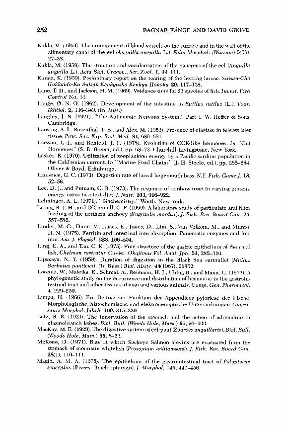
252 RAGNAR F m G E AND DAVID GROVE
Kukla, M. (1954). The arrangement of blood vessels on the surface and in the wall of the
alimentary canal of the eel (Anguilla anguilla L.). Folia Morphol. (Warsaw) 5( 13),
27-38.
Kukla, M. (1958). The structure and vacularisation of the pancreas of the eel (Anguilla
anguilla L.). Acta Biol. Cracoc., Ser. Zool. 1, 99-111. Kurata, K. (1959). Preliminary report on the rearing of the herring larvae. Suisan-Cho
Hokkaido-Ku Suisan Kenkyusho Kenkyu Hokoku 20, 117-138. Lane, T. H., and Jackson, H. M. (1969). Voidance time for 23 species of fish. Incest. Fish
Control No. 33.
Lange, 8. N. 0. (1962). Development of the intestine in Rutilus rutilus (L.). Vopr.
Ikhtiol. 2, 336-349. (In Russ.)
Langley, J . N. (1921). “The Autonomic Nervous System,” Part 1. W. Heffer & Sons,
Cambridge.
Lansing, A. I., Rosenthal, T. B., and Alex, M. (1953). Presence of elastase in teleost islet
tissue. Proc. Soc. E x p . Biol. LMed. 84, 689-691.
Larsson, L.-I., and Rehfeld, J. F. (1978). Evolution of CCK-like hormones. In “Gut
Hormones” ( S . R. Bloom, ed.), pp. 68-73. Churchill Livingstone, New York.
Lasker, R. (1970). Utilization of zooplankton energy by a Pacific sardine popul a t‘ ion in
the Californian current. In “Marine Food Chains” (J. H. Steele, ed.), pp. 265-284.
Oliver & Boyd, Edinburgh.
Laurence, G. C. (1971). Digestion rate of larval largemouth bass. N.Y . Fish. Game J . 18,
52-56.
Lee, D. J., and Putnam, G. B. (1973). The response of rainbow trout to varying protein/
energy ratios in a test diet. J. Nutr. 103, 916-922. Lehninger, A. L. (1971). “Biochemistry.” Worth, New York.
Leong, R. J. H., and O’Connell, C. P. (1969). A laboratory study of particulate and filter
feeding of the northern anchovy (Engraulis mordax). J. Fish. Res. Board Can. 26,
557- 582. Linder, M. C., Dunn, V., Isaacs, E., Jones, D., Lim,,S., Van Volkom, M., and Munro,
H. N. (1975). Ferritin and intestinal iron absorption: Pancreatic enzymes and free
iron. Am. J. Physiol. 228, 196-204.
Ling, E. A., and Tan, C. K. (1975). Fine structure of the gastric epithelium of the coral
fish, Chelmon rostratus Cuvier. Okajimas Fol. Anat. Jpn. 51, 285-310.
Lipskaya, N. Y. (1959). Duration of digestion in the Black Sea surmullet (,Mullus Barbatus ponticus). (In Russ.) B i d . Abstr. 48( 1967), 26852.
Lorentz, W., Matejka, E., Schmal, A., Reiniann, H. J., Uhlig, R., and Mann, G. (1973). A
phylogenetic study on the occurrence and distribution of histamine in the gastroin-
testinal tract and other tissues of man and various animals. Comp. Gen. Pharmacol.
4, 229-250.
Luppa, H. (1966). Ein Beitrag zur Funktion der Appendices pyloricae der Fische.
Morphologische, histochemische und elektronenoptische Untersuchungen. Gagen-
saurs Morphol. Jahrb. 109, 315-339.
Lutz, B. R. (1931). The innervation of the stomach and the action of adrenaline in
elasmobranch fishes. B i d . Bull . (Woods Hole, Mass.) 61,93-100. ,MacKay, M. E. (1929). The digestive system of eel-pout (Zoarces anguillaris). Biol. Bull.
(Woods Hole, Mass.) 56, 8-23. McKone, D. (1971). Rate at which Sockeye Salmon alevins are evacuated from the
stomach of mountain whitefish (Prosopium williamsoni). J. Fish. Res. Board Can.
28(1), 110-111.
Magid, A. M. A. (1975). The epithelium of the gastrointestinal tract of Polypterus senegalus (Pisces: Brachiopterygii). J . Morphol. 146,447-456.
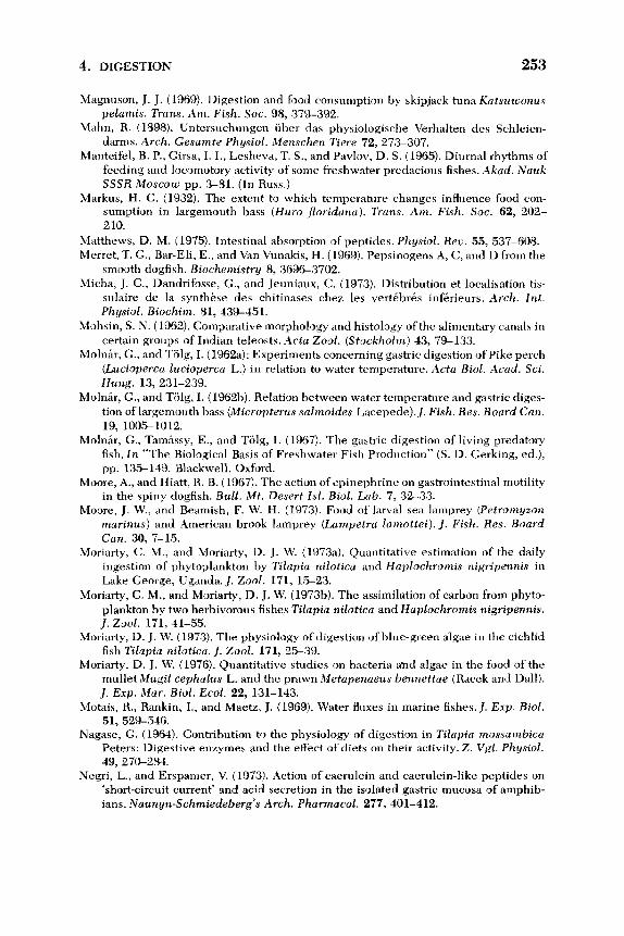
4. DIGESTION 253
Magnuson, J. J. (1969). Digestion and food consumption by skipjack tuna Katsuwonus pelamis. Trans. Am. Fish. Soc. 98, 379-392.
Mahn, R. (1898). Untersuchungen uber das physiologische Verhalten des Schleien-
darnis. Arch. Gesamte Physiol. Menschen Tiere 72, 27X307.
Manteifel, B. P., Girsa, I. I., Lesheva, T. S., and Pavlov, D. S. (1965). Diurnal rhythms of
feeding and locomotory activity of some freshwater predacious fishes. Aknd. Nauk S S S R ~Moscozu pp. 3-81. (In Russ.)
Markus, H. C. (1932). The extent to which temperature changes influence food con-
sumption in largemouth bass (Huro Joridana). Trans. Am. Fish. SOC. 62, 202-
2 10.
Matthews, D. M. (1975). Intestinal absorption of peptides. Physiol. Rea. 55, 537-608.
Merret, T. G., Bar-Eli, E., and Van \7unakis, H. (1969). Pepsinogens A, C, and D from the
smooth dogfish. Biochemistry 8, 3696-3702.
Micha, J. C., Dandrifosse, G., and Jeuniaux, C. (1973). Distribution et localisation tis-
sulaire de la synthtse des chitinases chez les vertibrks infxieurs. Arch. Znt. Physiol. Biochim. 81, 439-451.
Mohsin, S. N. (1962). Comparative morphology and histology of the alimentary canals in
certain groups of Indian teleosts. Acta Zool. (Stockholm) 43, 79-13,
Molnir, G., and Tolg, I. (1962a): Experiments concerning gastric digestion of Pike perch
(Lucioperca lucioperca L.) in relation to water temperature. Acta B i d . Acad. Sci. Hung. 13,231-239.
MolnLr, G., and Tolg, I. (19621)). Relation between water temperature and gastric diges-
tion of largemouth bass (Micropterus salmoides Lacepede).]. Fish. Res. Bourd Can.
19, 1005-1012.
MolnLr, G., Tamassy, E., and Tolg, I. (1967). The gastric digestion of living predatory
fish. In “The Biological Basis of Freshwater Fish Production” (S. D. Gerking, ed.),
pp. 135-149. Blackwell, Oxford.
Moore, A., and Hiatt, R. B. (1967). The action of epinephrine on gastrointestinal motility
in the spiny dogfish. Bull. A4t. Desert Zsl. Biol. Lab. 7, 32-33.
Moore, J. W., and Beamish, F. W. H. (197.3). Food of larval sea lamprey (Petromyzon marinus) and American brook lamprey (Lampetra lnmottei). J. Fish. Res. Board Can. 30, 7-15.
Moriarty, C. M., and Moriarty, D. J. W. (1973a). Quantitative estimation of the daily
ingestion of phytoplankton by Tilapia nilotica and Haplochromis nigripennis in
Lake George, Uganda. J. Zool. 171, 15-23.
Moriarty, C. M., and Moriarty, D. J. W. (1973b). The assimilation of carbon from phyto-
plankton by two herbivorous fishes Tilapia nilotica and Haplochromis nigripennis. J. Zool. 171, 41-55.
Moriarty, D. J. W. (1973). The physiology of digestion of blue-green algae in the cichlid
fish Tilapia nilotica. J. Zool. 171, 25-39.
Moriarty, D. J. W. (1976). Quantitative studies on bacteria ahd algae in the food of the
mullet Mugil cephalus L. and the prawn Metapenaeus hennettae (Racek and Dall).
]. E x p . Mar. Biol. Ecol. 22, 131-143.
Motais, R., Rankin, I., and Maetz, J. (1969). Water fluxes in marine fishes./. E x p . Biol. 51, 529-546.
Nagase, G. (1964). Contribution to the physiology of digestion in Tilapia mossambica Peters: Digestive enzymes and the effect of diets on their activity. Z. Vgl . Physiol. 49,270-284.
Negri, L., and Erspamer, V. (1973). Action of caerulein and caerulein-like peptides on
‘short-circuit current’ and acid secretion in the isolated gastric mucosa of amphib-
ians. Naunyn-Schmiedeberg’s Arch. Pharmacol. 277, 401-412.

254 RAGNAR FhJGE AND DAVID GROVE
Nelson, G. J. (1967). Epibranchial organs in lower teleostean fishes.J. Zool. 153,7139.
Netsch, M., and Witt, A. (1962). Cited in Herting and Witt (1968).
Nicholls, J. V. V. (1931). The influence of temperature on digestion in Fundulus hetero-
clitus. Contrib. Can. Biol. Fish. 7 , 45-55. Nicholls, J. V. V. (1933). The effect of temperature variation and certain drugs on gastric
motility of elasmobranch fishes. Contrib. Can. Biol. Fish. 7 , 449-463.
Nicol, J. A. C. (1952). Autonomic nervous system in lower chordates. Biol. Reo. Cam- bridge Philos. SOC. 27, 1-49.
Niimi, A. J., and Beamish, F. W. H. (1975). Bioenergetic and growth of largemouth bass
(Micropterus salmoides) in relation to body weight and temperature. Can. J . 2001. 52,447-456.
Nilsson, A. (1970). Gastrointestinal hormones in the holocephalian fish Chimaera monstrosa (L.). Comp. Biochem. Physiol. 32, 387-390.
Nilsson, A. (1973). Secretin-like and cholecystokinin-like activity in Myxine glutinosa L. Acta Regiae Soc. Sci. Litt. Gothob. Zool. 8, 30-32.
Nilsson, A., and Fange, R. (1969). Digestive proteases in the holocephalian fish Chima- era monstrosa (L.). Comp. Biochem. Physiol. 31, 147-165.
Nilsson, A., and Fange, R. (1970). Digestive proteases in the cyclostome Myxine glutinosa (L.). Comp. Biochem. Physiol. 32, 237-250.
Nilsson, S. (1971). Adrenergic innervation and drug responses of the oval sphincter in
the swimbladder of the cod (Gadus morhua'). Acta Physiol. Scand. 83,446-453.
Nilsson, S., and Fange, R. (1967). Adrenergic receptors in the swimbladder and gut of a
teleost (Anguilla anguilla). Comp. Biochem. Physiol. 23,661-664.
Nilsson, S., and Fange, R. (1969). Adrenergic and cholinergic vagal effects on the stomach of a teleost (Gadus morhua). Comp. Biochem. Physiol. 30, 691-694.
Nilsson, S., and Grove, D. J. (1974). Adrenergic and cholinergic innervation of the
spleen of the cod, Gadus morhua. Eur. J . Pharmacol. 28,135-143.
Nilsson, S., Holmgren, S., and Grove, D. J. (1975). Effects ofdrugs and nerve stimulation
on the spleen and arteries of two species of dogfish, Scyliorhinus canicula and
Squalus acanthias. Acta Physiol. Scand. 95, 219-230.
Nilsson, S., Abrahamsson, T., and Grove, D. J. (1976). Sympathetic release of adrenaline
from the head kidney of the cod, Gadus morhua. Comp. Biochem. Physiol. C 55,
12.3- 127.
Noaillac-Depeyre, J. , and Gas, N. (1978). Ultrastructural and cytochemical study of the
gastric epithelium in a fresh water teleostean fish (Perca juuiatilis). Tissue G Cell
10, 23-37.
Nobel, R. L. (1973). Evacuation rates of young yellow perch, Perca flawexens (Mitch-
ell). Trans. Am. Fish. Soc. 102(4), 75CL763.
Norris, E. R., and Elam, D. (1940). Preparation and properties and crystallization of tuna pepsin. J . Biol. Chem. 134, 443-454.
Norris, E. R., and Mathies, J. C. (1953). Preparation and properties of tuna pepsin. J . Biol. Chem. 204, 673-686.
Norris, J. S., Norris, D. O., and Windell, J. T. (1973). Effect of simulated meal size on
gastric acid and pepsin secretory rates in bluegill (Lepomis rnacrochirus). J. Fish. Res. Board Can. 30(2), 210-204.
Nose, T. (1967). Recent advance in the study of fish digestion. Tech. Pap. Eur. Inland Fish. Ado. Comm. 3, 83-94.
Nose, T., and Mamiya, H. (1963). Protein digestibility of flatfish meal in rainbow trout.
Tansuiku Suisan Kenkyusho Kenkyu Hokoku 12, 1-4.
Nose, T., and Toyama, K. (1966). Protein digestibility of brown fish meal in rainbow
trout. Tansuiki Suisan Kenkyusho Kenkyu Hokoku 15,213-224.

4. DIGESTION 255
Odense, P. H., and Bishop, C. M. (1966). The ultrastructure of the epithelial border of
the ileum, pyloric caeca and rectum of the cod, Gadus morhua. J . Fish. Res. Board
Can. 23(12), 1841-1843.
Odum, W. E. (1970). Utilization of the direct grazing and detritus food chains by the
striped mullet, Mugil Cechalus. In “Marine Food Chains” ( J . Steele, ed.), pp. 2 2 2
240. Oliver & Boyd, Edinburgh.
Ostberg, Y. (1976). The enter-insular endocrine organ in a cyclostome, Myxine glutinosa. Umea Uniu. Med. Diss. No. 15, pp. 1-41.
Ostberg, Y., Van Noorden, S., Everson Pearse, A. G.., and Thomas, N. W. (1976).
Cytochemical, immunofluorescence, and ultrastructural investigations on
polypeptide hormone containing cells in the intestinal mucosa of a cyclostome,
Myxine glutinosa. Gen. Comp. Endocrinol. 28, 213-227.
Ohnesorge, F. K., and Rauch, R. (1968). Untersuchungen iiber Peristaltik am Darm der
Schleie (Tinca vulgaris). Z. Vgl. Physiol. 58, 153-170.
Ohnesorge, F. K., and Rehberg, M. (1963). Der Darm der Schleie (Tinca uulgaris) als
pharmakologisches Versuchsobject. Naunyn-Schmiedebergs Arch. E x p . Pathol.
Pharmakol. 246, 81-82. Okul, A. V. (1941). The feeding of pelagic fishes in the Azov Sea.Zool. Z h . 20,587-603.
(In Russ.)
Okutani, K. (1966). Studies of chitinolytic systems in the digestive tract of Lateolabrax japonicus. Bull. Misaki Mar. Biol. Inst., Kyoto Uniu. No. 10, pp. 1-47.
Ooshiro, Z. (1962). Biochemical studies on carboxypeptidase contained in the pyloric
caeca of mackerel, Scomber japonicus. Kagoshima Uniu. Fish. Dep. Bull. 1 1 , Suppl., 111-151.
Ooshiro, Z. (1968). Biochemical studies on carboxypeptidase contained in the pyloric
caeca of mackerel, Scomberjaponicus. Nippon Suisan Gakkaishi 11, 111-151.
Ooshiro, Z. (1971). Studies on proteinase in the pyloric caeca of fishes. 11. Some proper-
ties of proteinases purified from the pyloric caeca of mackerel. Nippon Suisan
Gakkaishi 37, 145-158.
Oppel, A. (1896-1900). “Lehrbuch der Vergleichenden Mikroskopischen Anatomie der
Wirbeltiere,” Vols. 1-3. Fischer, Jena.
Osse, J. W. M. (1969). Functional morphology of the head of the perch (Percafluuiatilis
L.): An electromyographic study. Neth. J. Zool. 19, 28%392. Otto, C. (1976). Size, growth, population density and food of brown trout Salmo trutta L.
in two sections of a south Swedish stream. J. Fish Biol. 8, 477-488.
Overnell, J. (1973). Digestive enzymes of the pyloric cacae and of their associated
mesentery in the cod (Gadus morhua). Comp. Biochem. Physiol. B 46, 519-531.
Pandian, T. J. (1967). Intake, digestion, absorption and conversion of food in the fishes Megalops cyprinoides and Ophiocephalus striatus. Mar. Biol. 1, 16-32.
Panov, D. A,, and Sorokin, Y. I . (1962). Speed of digestion in bream larvae. Byull. Inst. Biol. Vodokhran., Akad. Nauk S S S R 13, 24-26. (In Russ.) [Isr. Program Sci. Trans].
Pappas, C. J., Tiemeier, 0. W., and Deyoe, C. W. (1973). Chromic sesquioxide as an
indicator in digestion studies in fish. Progr. Fish Cultunist 35, 97-98.
Patterson, T. L., and Fair, E. (1933). The action of the vagus on the stomach-intestine of
the hagfish. Comparative studies. VIII. J. Cell. Comp. Physiol. 3, 11.3-119. Patton, J . S., Nevenzel, J. C., and Benson, A. A. (1975). Specificity of digestive lipases in
hydrolysis of wax esters and triglycerides studied in anchovy and .other selected
fish. Lipids 10, 575-583.
Payne, A. I. (1975). The reproductive cycle, condition and feeding in Barbus liberiensis, a tropical stream-dwelling cyprinid. 1. Zool. 176, 247-269.
TT-68-50387 (1968).]

256 RAGNAR F-GE AND DAVID GROVE
Pearse, A. G. E. (1977). The diffuse neuroendocrine system and the APUD concept:
Related peptides in brain, intestine, pituitary, placenta and anuran cutaneous
glands. Med. Biol. 55, 115-125.
Pegel, W. A., and Popov, F. G. (1937). The influence of temperature on digestion of
cold-blooded animals. Tomsk. Uniu. Biol. Inst., Tr. 4, 59-79. (In Russ.) Pernkopf, E., and Lehner, J . (1937). Vergleichende Beschreibung des Vorderdarmes bei
den einzelnen Klassen der Kranioten. I n “Handbuch der Vergleichende Anatomie
der Wirbeltiere” (L. Bolk, E. Goppert, E. Kallius, and W. Lubosch, eds.), Vol. 3, pp.
349-398. Urban Schwarzenberg, Berlin.
Peters, D. S., and Hoss, D. E. (1974). A radioisotopic method of measuring food evacua-
tion time in fish. Trans. Am. Fish. SOC. 103, 626629.
Petersen, H. (1908-1909). Beitrage zur kenntnis des Baues und der Entwicklung des
Selachierdarmes. Jena Z. Naturwiss. 43, 619-652; 44, 123-148.
Phillips, A. M., Jr. (1969). Nutrition, digestion and energy. In “Fish Physiology” (W. S.
Hoar and D. J . Randall, eds.), Vol. 1, pp. 391-432. Academic Press, New York.
Piavaux, A. (1973). Origine non bacterienne de la laminarinase intestinale de Tilapia
macrochir Boulenger. Arch. Int. Physiol. Biochim. 81, 737-743.
Pierce, E. L. (1936). Rates of digestion in the yellowtail (Ocyurus chrysurus) and the
white grunt (Haemulon plumieri). Copeia No. 2, pp. 12.3-124.
Prahl, J. W., and Neurath, H. (1966a). Pancreatic enzymes of the spiny pacific dogfish. I.
Cationic chymotrypsinogen and chymotrypsin. Biochemistry 5, 2131-2146.
Prahl, J. W., and Neurath, H. (1966b). Pancreatic enzymes of the spiny pacific dogfish.
11. Procarboxypeptidases B and carboxypeptidase B. Biochemistry 5, 4137-
4145. Prosser, C. L. (1973). “Comparative Animal Physiology,” 3rd ed. Saunders, Philadel-
phia, Pennsylvania.
Rahn, C. H., Sand, D. M., and Schlenk, H. (1973). Wax esters in fish. Metabolism of
dietary palmityl palmitate in the gourami (Trichogaster cosby). /. Nutr. 103, 1441-
1447.
Rauther, M. (1940). Der 1ntestinaltraktus.In “Dr. H. G. Bronns Klassen und Ordnungen
des Tierreichs.” Teil 1: “Echte Fische,” pp. 657-1050. Akad. Verlagsges., Leipzig.
Read, J. B., and Burnstock, G. (1968a). Fluorescent histochemical studies on the mucosa
of the vertebrate gastrointestinaltract. Histochemie 16, 324-332.
Read, J . B., and Burnstock, G. (1968b). Comparative histochemical studies of adrenergic nerves in the enteric plexus of vertebrate large intestine. Comp. Biochem. Physiol. 27, 505-517.
Read, J . B., and Burnstock, G. (1969). Adrenergic innervation of gut musculature in
vertebrates. Histochemie 17, 26S272.
Reeck, G. R., and Neurath, H. (1972). Pancreatic trypsinogen from the African lungfish.
Biochemistry 11, 503-510.
Reeck, G. R., Winter, W. P., and Neurath, H. (1970). Pancreatic enzymes from the African lungfish, Protopterus aethiopicus. Biochemistry 9, 1398-1403.
Reichenbach-Klinke, H.-H., and Reichenbach-Klinke, K.-E. (1970). Enzymunter-
suchungen an Fischen. 11. Trypsin-und Amylase-Inhibitoren. Arch. Fischereiwiss.
21,67-72.
Reif, W. E. (1976). Morphogenesis, pattern formation and function of the dentition of
Heterodontus (Selachii). Zoomorphologie 83( l), 1-47.
Reimers, N. (1957). Some aspects of the relation between stream foods and trout sur-
vival. Calif. Fish Game 43, 43-69.
Reite, 0. B. (1965). A phylogenetical approach to the functional significance of tissue
mast cell histamine. Nature (London) 206, 1334-1336.
4. DIGESTION
Reite, 0. B. (1969). Phylogenetical persistence of the nonmast cell histamine stores of
the digestive tract: A comparison with mast cell histamine. Experientia 25,276-277.
Renade, S. S., and Kewalramani, H. G. (1967). Studies on the rate of food passage in fish
intestines. F.A.O. Fish. Rep. 44, 349-358.
Reshetnikov, Y. S., Claro, R., and Silva, A. (1972). The pattern of feeding and the rate of
digestion in some tropical predatory fish. J. Zchthyol. (USSR) 12(5), 818-824.
Riddle, 0. (1909). The rate of digestion in cold-blooded vertebrates. The influence of season and temperature. Am. J. Physiol. 24, 447-458.
Rosenthal, H., and Hempel, G. (1970). Experimental studies in feeding and food re-
quirements of herring larvae (Clupea harengus L.). In “Marine Food Chains” (J . H. Steele, ed.), pp. 344-364. Oliver & Boyd, Edinburgh.
Rosenthal, H., and Paffenhofer, G. A. (1972). On the digestion rate and calorific content
of food and faeces in young gar fish. Naturwissenschaften 6, 274-275.
Rozin, P., and Mayer, J. (1961). Regulation of food intake in the goldfish. Am. J. Physiol. 201, 968-974.
Rozin, P., and Mayer, J. (1964). Some factors affecting short-term food intake in the
goldfish. Am. J. Physiol. 206, 14304436.
Ryland, J. S. (1964). The feeding ofplaice and sandeel larvae in the southern North Sea.
J. Mar. Biol. Assoc. U.K. 44, 343-364.
Saito, K. (1973). Nervous control of intestinal motility in goldfish. J p n . J. Smooth Muscle
Res. 9, 79-86.
Sarbahi, D. S. (1951). Studies on the digestive tracts and digestive enzymes of the
goldfish Carassius auratus L. and the largemouth black bass Micropterus salmoides (Laceptde). Biol. Bull. (Woods Hole, Mass.) 100, 244-257.
Scarpelli, D. G., Greider, M. H., and Frajola, W. J. (1963). Observations on hepatic cell hyperplasia, adenoma and hepatoma of rainbow trout (Salmo gairdneri). Cancer
Res. 23, 84%857. Scheuring, L. ( 1928). Beziehungen zwischen Temperatur und Verdauungsgeschwin-
digkeit bei Fischen. Z. Fisch. Deren Hilfswiss. 26, 231-235.
Schmitz, E. H., and Baker, C. D. (1969). Digestive anatomy of the gizzard shad Dorosoma Cepedianum and the threadfin shad Dorosoma petenense. Trans. Am.
Microsc. Soc. 88(4), 525546.
Seaburg, K. G. (1956). A stomach sampler for live fish. Prog. Fish Cult . 19, 137-139.
Seaburg, K. G., and Moyle, J. B. (1964). Feeding habits, digestion rates and growth of
some Minnesota warm water fishes. Trans. Am. Fish. SOC. 93, 269-285.
Sharratt, B. M., Bellamy, D., and Chester Jones, I . (1964). Adaptation of the silver eel
(Anguilla anguilla L.) to sea water and to artificial media together with observations on the role of the gut. J. Physiol. (London) 204, 135158.
Shrable, J. B., Tiemeier, 0. W., and Deyoe, C. W. (1969). Effects of temperature on rate
of digestion by channel catfish. Prog. Fish. Cult. 31, 131-138.
Singh, R. P., and Nose, T. (1967). Digestibility of carbohydrates in young rainbow trout. Tansuiku Suisan Kenkyusho Kenkyu Hokoku 17,21-25.
Smit, H. (1968). Gastric secretion in the lower vertebrates and birds. I n “Handbook of
Physiology.” Sect. 6: Alimentary Canal” (C. F. Code, ed.), Vol. V, pp. 2791-2805.
Am. Physiol. SOC., Washington, D.C. Smith, H. W. (1930). The absorption and secretion of water and salts by marine teleosts.
Am. J. Physiol. 93, 480-505.
Smith, M. W. (1966). Sodium-glucose interactions in the goldfish intestine. J. Physiol. (London) 182, 559-572.
Smith, R. L. (1975). Carbonic anhydrase in some coral reef fishes: Adaption to carbonate
ingestion? Comp. Biochem. Physiol. A 50, 131-104.

258 RAGNAR F m G E AND DAVID GROVE
Smith, R. L., and Lane, C. E. (1971). Amino acid transport by the fish intestine. Comp.
Biochem. Physiol. 4, 93-103.
Solcia, E., and Members of the International Committee on Classification of Gut Endo- crine Cells ( 1978). Lausanne 1977 classification of gastroenteropancreatic endo-
crine cells. In “Gut Hormones” (S. R. Bloom, ed.), pp. 40-48. Churchill
Livingstone, New York.
Steigenberger, L. W., and Larkin, P. A. (1974). Feeding activity and rates of digestion of
northern squawfish (Ptychocheilus oregonensis). J . Fish. Res. Board Can. 31(4),
41 1-420.
Stevenson, S. V., and Grove, D. J. (1977). The extrinsic innervation of the stomach of the
plaice, Pleuronectes platessa L. 1. The vagal nerve supply. Comp. Biochem.
Physiol. C 58, 143-151.
Stevenson, S. V., and Grove, D. J. (1978). The extrinsic innervation of the stomach of the
plaice, Pleuronectes platessa L. 2. The splanchnic nerve supply. Comp. Biochem.
Physiol. C 60, 45-50.
Stickney, R. R., and Shumway, S. (1974). Occurrence ofcellulase activity in the stomach
of fishes. J . Fish Biol. 6, 779-790.
Stryer, L. (1974). “Biochemistry.” Freeman, San Francisco, California.
Sumner, J. B., and Somers, G. F. (1947). “Chemistry and Methods of Enzymes,” 2nd Ed.
Academic Press, New York.
Suyehiro, Y. (1942). A study on the digestive system and feeding habits of fish. J p n . J. Zool. 10, 1-303.
Suzuki, T. (1976). Relations between feeding rates and growth rates of filefish Navodon
modestus (Gunther). Bull. Jpn. Sea Reg. Fish. Res. Lab. (Nihonkai-Ku Suisan Ken-
kyusho Kenkyu) 27, 51-57.
Swenson, W. A., and Smith, L. L. (1973). Gastric digestion, food consumption, feeding
periodicity and food conversion efficiency in walleye (Stizostedion vitreum vit-
reum).J. Fish. Res. Board Can. 30(9), 1327-1336.
Tan, C. K., and Teh, S. (1974). The structure of the gut of a coral fish, Chelmon rostratus
Cuvier. Okajimos Folia Anat. Jpn. 51, 63-79.
Tanaka, K. (1955). Observation of the length of the digestive time of feed by the mud
loach Misgurnus anguillicaudatus. Gyorvigaku Zasshi 4 , 3 4 3 9 .
Tanaka, M. (1969). Studies on the structure and function of the digestive system in
teleost larvae. 11. Characteristics of the digestive system of larvae at the stage of first
feeding. Gyoruigaku Zasshi 16(2), 41-49. Tanaka, M. (1973). Studies on the structure and function of the digestive system of
teleost larvae. Ph.D. Thesis, Fac. Agric., Kyoto Univ., Kyoto.
Tomonaga, S., Hirokane, T., Shinohara, H., and Awaya, K. (1973). The primitive spleen
of the hagfish. Dobutsugaku Zasshi 82,215-217.
Track, N. S. (1973). Evolutionary aspects of the gastrointestinal hormones. Comp.
Biochem. Physiol. B 45, 291-301.
Trendelenburg, U. ( 1917). Physiologische und pharmakologische Versuche uber die
Dunndarmperistaltik. Arch. E x p . Pathol. Pharmakol. 81, 5.5129.
Tue, V. T. (1975). Contribution a I’etude histologique des cellules epitheliales glan-
dulaires et endocrines du tube digestif de Chimaera monstrosa L. (Pisces, Holocephali). Vie Milieu, Ser. A 25, 41-58.
Tugendhat, B. (1960). The normal feeding behaviour of the three-spined stickleback
Gasterosteus aculeatus. Behaviour 15, 284-318.
Tyler, A. V. (1970). Rates of gastric emptying in young cod.]. Fish. Res. Board Can. 27,
1177-1189.

4. DIGESTION 259
Uggeri, B. (1938). Ricerche sulle cellule enterchromaffini e sulle cellule argentofile dei
Ugolev, A. M., and Kooshuck, R. I. (1966). Hydrolysis of dipeptides in cells of the small
Ungar, C. (1935). Perfusion de I’estomac des Selaciens; etude pharmacodynamique de
Unger, R. H., Dobbs, R. E., and Orci, L. (1978). Insulin, glucagon, and somatostatin
Vallee, B. L. (1955). Zinc and metalloenzynies. Adu. Protein Chem. 10, 317-384.
van Herwerden, M., and Ringer, W. E. (1911). Die Aciditit des Magensaftes von Scyl- lium stellare. Hoppe-Seyler’s Z . Physiol. Chem. 75, 290.
Van Noorden, S., and Pearse, A. G. E. (1976). The localization of immunoreactivity to insulin, glucagon and gastrin in the gut ofAmphioxus (Branchiostoma) lanceolatus. In “The Evolution of Pancreatic Islets” (T. A. I . Grillo, L. Leibson, and A. Epple,
eds.), pp. 163-178. Pergamon, Oxford.
Van Slyke, D. D., and White, G. F. (1911). Digestion of protein in the stomach and in
intestine of the dogfish. J . B i d . Chem. 9, 209-217.
Vegas-Velez, M. (1972). La structure histologique due tube digestif des poissons tC1Cos-
tbens. Tethys 4, 16S174.
von Euler, U. S., and Fange, R. (1961). Catecholamines in nerves and organs ofMyxine
glutinosa, Squalus acanthias, and Gadus callarias. Gen. Comp. Endocrinol. 1, 191-194.
von Euler, U. S., and Ostlund, E. (1957). Effects of certain biologically occurring sub-
stances in the isolated intestine of fish. Acta Physiol. Scand. 38, 364-372. Wallace, J. C. (1973). Observations on the relationship between food consumption and
metabolic rate of Blennius pholis L. Comp. Biochem. Physiol. A 45, 293-305. Walsh, K. A. (1970). 4. Trypsinogens and trypsins in various species. 5.
Chymotrypsinogens-chymotrypsins. I n “Proteolytic Enzymes” (G. E. Perlmann and L. Lorand, eds.), Vol. 19, pp. 41-63, 64-108. Academic Press, New York.
Ware, D. M. (1972). Predation by rainbow trout (Salmo gairdneri): the influence of
hunger, prey density and prey size. J . Fish. Res. Board Can. 29, 1193-1201.
Weinland, E. (1901). Zur Magenverdauung der Haifische. Z . B i d . 1, 35-68; 27.5294.
Weinreb, E. L., and Bilstad, N. M. (1955). Histology of the digestive tract and adjacent
structures of the rainbow trout, Salmo gairdneri irideus. Copeia 3, 194-204.
Weisel, G. F. (1962). Comparative study of the digestive tract of a sucker Catostomus catastomus and a predaceous minnow, Ptychocheilus oregonense. Am. Midl. Nut. 68, 334-346.
Weisel, G. F. (1973). Anatomy and histology of the digestive system of the paddlefish (Polyodon spathula). J. Morphol. 140, 243-256.
Welsch, U. N., and Storch, V. N. (1973). Enzyme histochemical and ultrastructural ob- servations on the liver of teleost fishes. Arch. Histol. Jpn. (Niigata, Jpn.) 36, 21-
37. Western, J . R. H. (1969). Studies on the diet, feeding mechanism and alimentary tract in
two closely related teleosts, the freshwater Cottus gobio and the marine Parenop- hrys bubalis (Euphrasen). Acta Zool. (Stockholm) 50, 185-205.
Western, J. R. H. (1971). Feeding and digestion in two cottid fishes, the freshwater Cottus gobio and the marine Enophrys bubalis. J . Fish Biol. 3, 225-246.
Western, J. R. H., and Jennings, J. B. (1970). Histochemical demonstration of hy- drochloric acid in gastric tubules of teleosts using an in uiuo Prussian blue tech- nique. Comp. Biochem. Physiol. 35, 879-884.
Pesci. Z . Zellforsch. iMikrosk. Anat. 28, 648.
intestine. Nature (London) 212, 859-860.
la secretion gastrique. C . R . SOC. Biol. 119, 172-173.
secretion in the regulation of metabolism. Annu. Reu. Physiol. 40, 307-343.

260 RAGNAR F W G E AND DAVID GROVE
Williams, B. G. , and Naylor, E. (1967). Spontaneously induced rhythm of tidal periodic-
ity in laboratory-reared Carcinus. J. Exp. Eiol. 47, 229-234.
Windell, J. T. (1966). Rate of digestion in the bluegill sunfish. Invest. Zndiana Lakes Streams 7, 185-214.
Windell, J . T. (1967). Rates of digestion in fishes. In “The Biological Basis of Freshwater
Fish Production” (S. D. Gerking, ed.), pp. 151-173. Blackwell, Oxford.
Windell, J. T., and Norris, D. 0. (1969a). Gastric digestion and evacuation in rainbow
trout. Prog. Fish. Cult . 31, 2&26.
Windell, J. T., and Norris, D. 0. (1969b). Dynamics of gastric evacuation in rainbow
trout, Salmo gairdneri. Am. 2001. 9, 584.
Windell, J. T., Norris, D. O., Kitchell, J. F., and Norris, J. S. (1969). Digestive response
of rainbow trout, Salmo gairdneri, to pellet diets. J. Fish. Res. Board Can. 26,
1801-1812.
Windell, J. T., Hubbard, J. D., and Horak, D. C. (1972). Rate of gastric digestion in
rainbow trout, Salmo gairdneri, fed three pelleted diets. Prog. Fish. Cult . 34(3),
156159.
Yamamoto, H., and Kitamikado, M. (1971). Purification of fish gastric hyaluronidase.
Nippon Suisan Gakkaishi 37, 621-630.
Yamamoto, T. (19654. Some observations on the fine structure of the intrahepatic biliary
passages in goldfish (Carassius auratus).Z. Zellforsch. Mikrosk. Anat. 65,319-330.
Yamamoto, T. (196%). Some observations on the fine structure of the epithelium in the
intestine of the lamprey (Lumpetra japonica). Okajirnas Folia. Anat. J p n . 40, 691-
713.
Yamamoto, T. (1966). An electron microscope study of the columnar epithelial cell in the intestine of freshwater teleosts: Goldfish (Carassius auratus) and rainbow trout
(Salmo irideus). Z. Zellforsch. Mikrosk. Anat. 72, 6 6 8 7 . Yoshida, Y., and Sera, H. (1970). On chitinolytic activities in the digestive tract of several
species of fishes and the mastication and digestion of foods by them. Bull. Nankai
Reg. Fish. Res. Lab. 36, 751-754. (In Jpn.) Cited in Tanaka (1973).
Young, J. 2. (1931). On the autonomic nervous system of the teleostean fish Uranos-
copus scaber. Q . J . Microsc. Sci. 74,492-535.
Young, J. Z. (1933). The autonomic nervous system ofselachians. Q.J. Microsc. Sci. 75,
Yung, E. (1899). Recherches sur la digestion des poissons. Arch. Zool. Erp. Gen. 7,
Yung, I . K., Woo, K. C., and Doo, FV. K. (1965). Peristaltic movement of the tortoise
Zendzian, E., and Barnard, E. A. (1967). Distribution of pancreatic ribonuclease,
57 1-624.
12 1-20 1.
intestine. Experientia 21, 54C5.11.
chymotrypsin, and trypsin in vertebrates. Arch. Biochem. Biophys. 122,699-713.