15 nov 16 chuck's dissertation final - vanderbilt...
TRANSCRIPT

THE ROLE OF FACTOR XI DURING MURINE POLYMICROBIAL SEPSIS
By
Charles Edward Bane, Jr.
Dissertation
Submitted to the Faculty of the
Graduate School of Vanderbilt University
in partial fulfillment of the requirements
for the degree of
DOCTOR OF PHILOSOPHY
in
Pathology
December, 2016
Nashville, Tennessee
Approved:
Richard Hoover, Ph.D.
Stephen Kania, M.S., Ph.D.
Jonathan Schoenecker, M.D., Ph.D.
Edward Sherwood, M.D., Ph.D.
Charles Stratton, M.D.
Keith Wilson, M.D.

ii
ACKNOWLEDGEMENTS
This work was made possible by the financial support of the U.S. Army Medical Department
long-term health education and training program and by National Institutes of Health grants HL81326 and
HL58837. I would like to thank my thesis advisor, Dr. David Gailani for several reasons. First, for
answering an email from an Army veterinarian that asked if he might be willing to take on a Ph.D. student
with no research experience who, by the way, had only three years to complete the work. Second, for
shepherding me through the admissions process and allowing me to be in his lab from the beginning.
Third, and most importantly, for his unending patience, persistence, and professionalism throughout my
graduate student career (which lasted a little longer than three years).
I would also like to thank Dr. Richard Hoover, both for serving as my dissertation committee
chair, and for his assistance during the admissions process. My experience at Vanderbilt was enriched by
Dr. Hoover’s guidance and advocacy, and for that I am extremely grateful. I am also indebted to the
members of my dissertation committee for their direction during this very interesting and rewarding time
in my career.
My lack of research experience at the start of this project must have been immediately evident to
the other members of the Gailani lab, particularly Dr. Qiufang Cheng and Dr. Anton Matafonov.
However, both never let on and were great teachers in their own right. For that I am grateful. Finally, I
would like to thank my family for their love and support during this phase of my Army career. The past
18 years have been an eventful journey that has not always been easy. It is during the more difficult times
that their consistent support is most cherished.

iii
PUBLICATIONS
Bane, C. E., I. Ivanov, A. Matafonov, K. L. Boyd, Q. Cheng, E. R. Sherwood, E. I. Tucker, S. T. Smiley, O. J. McCarty, A. Gruber and D. Gailani (2016). "Factor XI Deficiency Alters the Cytokine Response and Activation of Contact Proteases during Polymicrobial Sepsis in Mice." PLoS One 11(4): e0152968.
Gailani, D., Bane, C.E., and Gruber, A. (2015). "Factor XI and contact activation as targets for antithrombotic therapy." J Thromb Haemost 13(8): 1383-1395.
Bane, C. E., Neff, A. T., and Gailani D. (2014). Factor XI Deficiency (Hemophilia C). Hemostasis and Thrombosis: Practical Guidelines in Clinical Management. H. I. Saba and H. R. Roberts. West Sussex, UK, John Wiley & Sons, Ltd.: 71-81.
Bane, C. E., and D. Gailani (2014). "Factor XI as a target for antithrombotic therapy." Drug Discov Today 19(9); 1454-8.

iv
TABLE OF CONTENTS
Page
ACKNOWLEDGEMENTS .......................................................................................................................... ii
PUBLICATIONS ......................................................................................................................................... iii
LIST OF FIGURES ..................................................................................................................................... vi
LIST OF ABBREVIATIONS .................................................................................................................... viii
Chapter
I. PURPOSE OF STUDIES DESCRIBED IN THIS DISSERTATION ............................................. 1
II. REVIEW OF COAGULATION ...................................................................................................... 2
Introduction to the Thesis Project .................................................................................................... 2 Introduction to Plasma Coagulation ................................................................................................. 3 Models of Plasma Coagulation ........................................................................................................ 4 Cross-Talk Between Coagulation and Inflammation ....................................................................... 9 The Plasma Contact System ........................................................................................................... 10 References ...................................................................................................................................... 13
III. INTRODUCTION TO FACTOR XI ............................................................................................. 16
Factor XI Structure and Function .................................................................................................. 16 Factor XI Deficiency in Humans and Domestic Animals .............................................................. 18 Factor XI and Thrombosis ............................................................................................................. 20 Factor XI in Animal Models of Thrombosis .................................................................................. 21 References ...................................................................................................................................... 23
IV. FACTOR XI AND THE CONTACT SYSTEM IN SEPSIS ......................................................... 27
Introduction .................................................................................................................................... 27 Overview of Sepsis ........................................................................................................................ 28 The Inflammatory Response to Infection ....................................................................................... 29 Disseminated Intravascular Coagulation ....................................................................................... 31 The Contact System and Sepsis ..................................................................................................... 35 Factor XI in Animal Models of Sepsis........................................................................................... 36 References ...................................................................................................................................... 40
V. FACTOR XI DEFICIENCY ALTERS THE CYTOKINE RESPONSE AND ACTIVATION OF CONTACT PROTEASES DURING MURINEPOLYMICROBIAL SEPSIS........................ 44 Introduction .................................................................................................................................... 44

v
Materials and Methods ................................................................................................................... 45 Results ............................................................................................................................................ 50 Discussion ...................................................................................................................................... 69 Additional Experiments ................................................................................................................. 74 References ...................................................................................................................................... 78
VI. CONCLUSIONS AND FUTURE DIRECTIONS ......................................................................... 84
Factor XI is at a Junction Between Coagulation and Inflammation .............................................. 84 Future Studies ................................................................................................................................ 86 Clinical Relevance ......................................................................................................................... 88 References ...................................................................................................................................... 88
APPENDIX
VALIDATION OF THE CECAL LIGATION AND PUNCTURE MODEL FOR EVALUATING THE CONTRIBUTION OF FXI DURING MURINE SEPSIS .......................... 90
Introduction .................................................................................................................................... 90 Cecal Ligation and Puncture .......................................................................................................... 91 Microfil Experiments ..................................................................................................................... 93 Pilot Studies ................................................................................................................................... 96 Discussion .................................................................................................................................... 101 References .................................................................................................................................... 102

vi
LIST OF FIGURES
Figure Page
1. Cascade model of plasma coagulation ............................................................................................. 4
2. Contact Activation ........................................................................................................................... 5
3. Revised model of plasma coagulation ............................................................................................. 6
4. Current theory of coagulant mechanisms......................................................................................... 8
5. Factor XI structure ......................................................................................................................... 17
6. Role of FXI in polymicrobial sepsis .............................................................................................. 39
7. Survival after CLP ......................................................................................................................... 51
8. Pilot studies-plasma cytokine and SAP expression after CLP ....................................................... 53
9. Fold increases in plasma cytokine levels after CLP ...................................................................... 55
10. Absolute increases in plasma cytokine levels after CLP ............................................................... 56
11. SAP levels post-CLP...................................................................................................................... 57
12. Effects of CLP on markers of coagulation ..................................................................................... 59
13. Histology of thymus and spleen ..................................................................................................... 60
14. Histology of liver, brain, and kidney ............................................................................................. 61
15. The effect of CLP on plasma contact proteases ............................................................................. 63
16. FXII activation by α-kal or FXIa in the presence of polyanions ................................................... 65
17. Effect of anti-kallikrein antibody H03 ........................................................................................... 66
18. Effect of HK on FXI activation in the presence of polyanions ...................................................... 67
19. Polyphosphate-induced changes in FXII in mice .......................................................................... 68
20. Complement component C5a and chemokine responses to CLP ................................................... 75
21. Peritoneal leukocyte infiltration after CLP .................................................................................... 76

vii
22. Evaluation of microorganisms during CLP-induced sepsis ........................................................... 77
23. FXI in thrombin generation and contact activation during sepsis .................................................. 85
24. Cecal ligation and puncture procedure........................................................................................... 92
25. Microfil experiments ...................................................................................................................... 95
26. Markers of inflammation after CLP-pilot studies .......................................................................... 96
27. FXI levels and platelet counts after CLP-pilot studies .................................................................. 97
28. Histology of brain tissue-pilot studies ........................................................................................... 98
29. Lymphoid response to CLP in pilot studies ................................................................................... 99
30. Evidence of bacterial dissemination after CLP-pilot studies ....................................................... 100

viii
LIST OF ABBREVIATIONS
Factor XI – FXI
Cecal Ligation and Puncture – CLP
FXI-deficient (mice) – FXI-/-
Factor XII – FXII
Factor IX – FIX
Factor VII – FVII
Factor X – FX
Factor V – FV
Factor VIII - FVIII
Prekallikrein – PK
α-kallikrein – α-kal
High molecular weight kininogen – HK
Prothrombin time – PT
Activated partial thromboplastin time – aPTT
Tissue factor – TF
Disseminated intravascular coagulation – DIC
Nitric oxide - NO
Hereditary angioedema – HAE
C1-inhibitor – C1INH
Polyphosphate – polyP
Ferric chloride – FeCl3
Systemic inflammatory response syndrome – SIRS
Pathogen-associated molecular pattern – PAMP
ix
Damage-associated molecular pattern – DAMP
Pattern recognition receptor – PRR
Lipopolysaccharide – LPS
Toll-like receptor – TLR
Tumor necrosis factor α – TNFα
Interleukin – IL
Macrophage inflammatory protein-2 – MIP-2
Keratinocyte chemoattractant – KC
Monocyte chemotactic protein-1 – MCP-1
Thrombin-antithrombin – TAT
Serum amyloid P – SAP

1
CHAPTER I
PURPOSE OF STUDIES DESCRIBED IN THIS DISSERTATION
The work described in this dissertation focuses on the plasma protein factor XI
(FXI), which is the zymogen of a protease, called factor XIa (FXIa), that contributes to
blood coagulation. For this project, we investigated the role of FXI in the mortality and
pathophysiology of murine sepsis. We hypothesized that FXI contributes to mortality during
sepsis, and that it influences both the coagulopathy, as well as the inflammatory response,
that occurs during sepsis. To test our hypotheses, we induced polymicrobial sepsis in mice
using the cecal ligation and puncture (CLP) model to fulfill three Specific Aims. For Aim 1,
we evaluated the contribution of FXI to survival during sepsis by comparing FXI deficient
(FXI-/-) mice to their wild-type (FXI+/+) and FXI+/- littermates. We determined survival for
each genotype over a period of 7 days after CLP. To fulfill Aim 2, we explored the role of
FXI on the activation of coagulation in mice after CLP. We collected blood and tissue from
FXI-/- and FXI+/+ littermates sacrificed at several time points after CLP to evaluate systemic
and histologic markers of coagulopathy over time. For Aim 3, we assessed the role of FXI
on the inflammatory response to CLP by looking at markers of inflammation in blood and
tissue from FXI-/- and FXI+/+ mice. Chapter V describes our findings for these experiments,
as well as the results of additional in vitro and in vivo experiments that further investigated
the influence of FXI on the pro-inflammatory contact activation system. Our data provide
novel insight into the influence of FXI on coagulation and inflammation, and suggest there
are therapeutic advantages to targeting FXI for the treatment of patients who suffer from
pathologies arising from each system.

2
CHAPTER II
REVIEW OF COAGULATION
Introduction to the Thesis Project
Formation of the plasma enzyme thrombin is critical for normal formation of a
blood clot (hemostasis). Thrombin generation is beneficial when generated locally to seal
damaged blood vessels during hemostasis, but may contribute to pathology when generated
inappropriately during thrombosis. Thrombin is the product of a series of reactions
involving several plasma serine proteases and their cofactors. One of these proteases, factor
FXIa, is generated from its precursor FXI by the protease factor XIIa (FXIIa), or by
thrombin itself. FXIa contributes to thrombin generation by cleaving factor IX (FIX) to
form the protease factor IXa (FIXa). Epidemiologic data indicate that FXI makes a
disproportionately greater contribution to thrombosis than to hemostasis in humans. These
data, bolstered by studies with animal models that utilize FXI deficient mice, have led to the
development of FXI inhibitors, which may be safer alternatives for anticoagulant therapy. In
addition to being a valuable tool for determining the role of FXI in hemostasis and
thrombosis, studies with FXI-/- mice are also providing clues about the role of FXI during the
host response to invading micro-organisms. These studies, combined with in vitro findings,
highlight several unique features of FXI that place it at a junction between coagulation and
inflammation. In this project, I evaluated the effect of FXI on the coagulopathy and
inflammatory response during the crucial early stages of murine sepsis.

3
Introduction to Plasma Coagulation
The plasma coagulation system is a tightly regulated host response mechanism that
minimizes blood loss after vascular injury through formation of a fibrin and platelet-rich clot
(Dahlback 2000). Blood clots result from an interaction between subendothelial collagen,
platelets, and numerous proteins within blood plasma. Clot formation begins when
circulating platelets near a vascular lesion attach to exposed subendothelial collagen. This
attachment is mediated by von Willebrand factor, a multimeric glycoprotein that tethers
platelets to collagen by binding to glycoprotein (GP) Ib of the platelet GPIb/IX/V receptor
complex (Ruggeri and Mendolicchio 2015). Platelets are then activated through several
mechanisms, and bind to each other through a process called aggregation. Platelet adherence
and subsequent aggregation on transected vessels occurs within 30 seconds after injury
(Weiss and Lages 1988). In addition to forming a vital platelet plug to staunch bleeding, this
initial stage of hemostasis sets the conditions for effective fibrin formation, which is
essential for clot integrity and stability. Phospholipids, which are expressed by activated
platelets and damaged endothelial cells, provide a scaffold for the cell surface reactions that
result in fibrin formation (Davie, Fujikawa et al. 1991), while activated platelets secrete
factors that support the process (Monroe and Hoffman 2014). Vascular damage also exposes
molecules that trigger the fibrin-forming plasma coagulation system, a set of enzymatic
reactions that result in generation of thrombin. Thrombin converts soluble fibrinogen into
insoluble fibrin and contributes to platelet activation. One of the plasma enzymes that
contribute to thrombin generation is the protease FXIa, the subject of my thesis work.
4
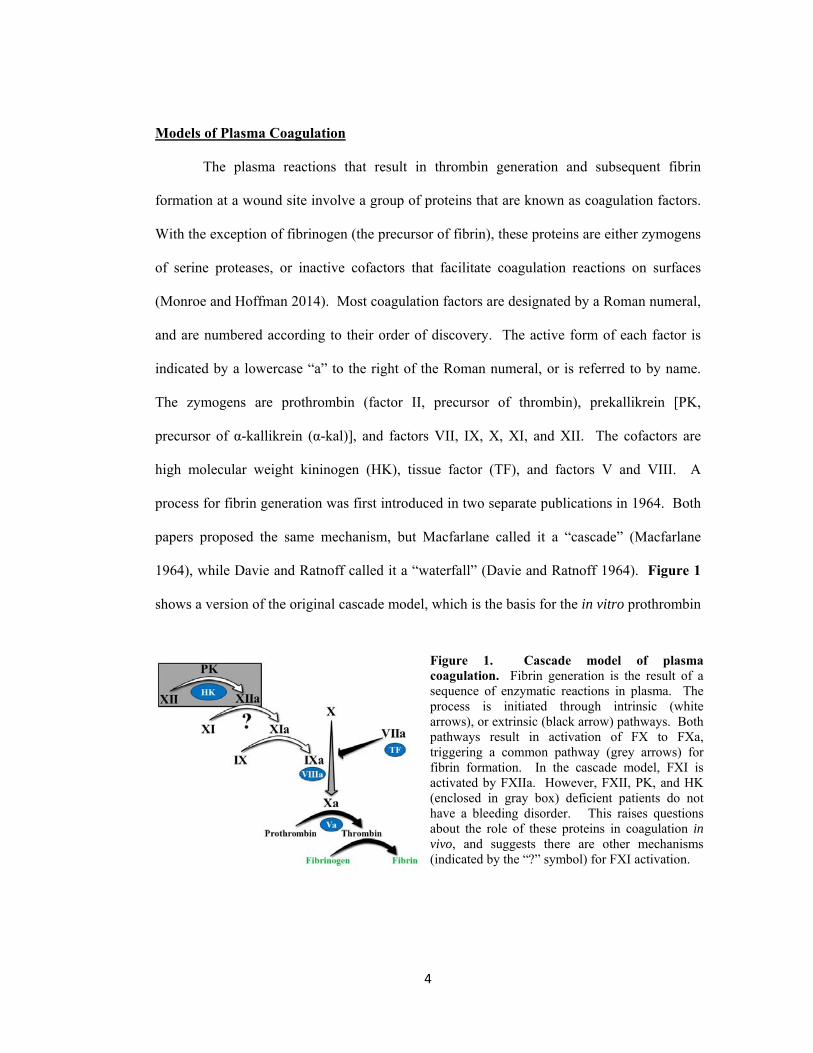
Models of Plasma Coagulation
The plasma reactions that result in thrombin generation and subsequent fibrin
formation at a wound site involve a group of proteins that are known as coagulation factors.
With the exception of fibrinogen (the precursor of fibrin), these proteins are either zymogens
of serine proteases, or inactive cofactors that facilitate coagulation reactions on surfaces
(Monroe and Hoffman 2014). Most coagulation factors are designated by a Roman numeral,
and are numbered according to their order of discovery. The active form of each factor is
indicated by a lowercase “a” to the right of the Roman numeral, or is referred to by name.
The zymogens are prothrombin (factor II, precursor of thrombin), prekallikrein [PK,
precursor of α-kallikrein (α-kal)], and factors VII, IX, X, XI, and XII. The cofactors are
high molecular weight kininogen (HK), tissue factor (TF), and factors V and VIII. A
process for fibrin generation was first introduced in two separate publications in 1964. Both
papers proposed the same mechanism, but Macfarlane called it a “cascade” (Macfarlane
1964), while Davie and Ratnoff called it a “waterfall” (Davie and Ratnoff 1964). Figure 1
shows a version of the original cascade model, which is the basis for the in vitro prothrombin
Figure 1. Cascade model of plasma coagulation. Fibrin generation is the result of a sequence of enzymatic reactions in plasma. The process is initiated through intrinsic (white arrows), or extrinsic (black arrow) pathways. Both pathways result in activation of FX to FXa, triggering a common pathway (grey arrows) for fibrin formation. In the cascade model, FXI is activated by FXIIa. However, FXII, PK, and HK (enclosed in gray box) deficient patients do not have a bleeding disorder. This raises questions about the role of these proteins in coagulation in vivo, and suggests there are other mechanisms (indicated by the “?” symbol) for FXI activation.

5
time (PT) and activated partial thromboplastin time (aPTT) clotting assays. The model
illustrates a series of reactions wherein a zymogen is converted to an active protease, which
then acts upon the next zymogen in the cascade until prothrombin is converted to thrombin
by the protease FXa. According to the cascade model, there are two distinct biochemical
pathways for thrombin generation. In the intrinsic pathway, the basis for the aPTT assay,
FXII is activated on a surface in a process that also involves PK and HK. The resulting
FXIIa then activates FXI to complete a set of reactions that are collectively referred to as
contact activation (Figure 2). Contact activation triggers the sequential activation of FIX,
FX, and prothrombin. In the extrinsic pathway, the basis for the PT assay, tissue factor (TF)
binds to factor VII (FVII), forming a TF/FVII(a) complex that subsequently activates factor
X (FX). The aPTT and PT clotting assays are useful for detecting deficiencies of clotting
factors in plasma because factor-deficient plasmas clot more slowly than normal plasma in
vitro. In many cases, this in vitro phenomenon is translated to the clinical setting, wherein
Figure 2. Contact Activation. On a surface (gray disk) FXII is auto-activated to form FXIIa (1). FXIIa activates PK to generate -kal (2), which reciprocally activates additional FXII (3). -kal also cleaves HK to liberate BK (4). FXIIa activates FXI (5) to propagate coagulation. The cofactor HK (shown in red) facilitates FXI and PK binding to the surface.

6
patients with a deficiency of a clotting factor will exhibit a clotting defect and therefore tend
to bleed. However, this is not always the case. For example, patients with a hereditary
deficiency of FXII, PK, or HK have a markedly prolonged aPTT, but no bleeding disorder,
while patients with a deficiency of FXI, the substrate for FXIIa in the cascade model, have a
prolonged aPTT and may bleed excessively, but only in certain instances. This places the
role of contact activation in hemostasis in doubt, and suggests there are other mechanisms
for FXI activation in vivo.
Advances in coagulation research have resulted in a revised model of thrombin
generation (Figure 3) that excludes proteins that are not required for hemostasis (FXII, PK,
and HK). This model proposes that tissue factor is the initiator of thrombin generation in
vivo, based upon clinical observations and in vitro work showing that (a) in addition to
activating FX, the TF/FVIIa complex also activates FIX (Osterud and Rapaport 1977), and
(b) thrombin activates FXI (Gailani and Broze 1991, Naito and Fujikawa 1991). According
to the revised model, thrombin generation is initiated after vascular injury exposes tissue
X
IX
IXaVIIIa
VIIa
VesselInjury
TF
Thrombin
X
Xa
Prothrombin
VaXI
IX
XIa
TFPI
Fibrinogen Fibrin
Figure 3. Revised model of plasma coagulation. Vascular injury exposes TF to FVII, and the resulting TF/FVIIa complex initiates thrombin generation by activating FX to FXa. The activation of FX by TF/FVIIa is limited by Tissue Factor Pathway Inhibitor (TFPI) (red arrow), so that sustained thrombin generation depends upon activation of FIX by TF/FVIIa. FXIa activates FIX (grey arrows) to provide additional thrombin to offset TF/FVIIa inhibition. In the revised model, FXI is activated by thrombin. For both figures, zymogens and proteases are shown in black. When Roman numerals are used, the active protease is designated by the letter “a.” Cofactors are shown in blue.

7
factor (TF), an integral membrane protein on the surface of a variety of non-vascular cells, to
the circulation (Dahlback 2000, Morrissey 2004, Gailani and Renne 2007). TF quickly
binds to zymogen FVII within the circulation forming a 1:1 complex. This interaction on the
surface of cells allows a variety of plasma proteases to convert zymogen FVII to its active
form, FVIIa (Davie, Fujikawa et al. 1991, Furie and Furie 1992, Monroe and Key 2007).
The TF/FVIIa enzyme complex then binds and activates FX to FXa, which, in a reaction that
is accelerated by the cofactor FVa, converts the plasma zymogen prothrombin to active
thrombin. FX is also activated by FIXa, which is primarily produced by cleavage of FIX by
the TF/FVIIa complex. Efficient activation of FX by FIXa requires the cofactor factor VIIIa
(FVIIIa). The activation of FIX by TF/FVIIa is an important mechanism for supporting
thrombin generation because the initiating pathway (activation of FXI by TF/FVIIa) is
inhibited by the Kunitz-type protease inhibitor tissue factor pathway inhibitor (TFPI) (Broze
and Girard 2012, Monroe and Hoffman 2014). In the revised model, the protease FXIa,
generated by the cleavage of zymogen FXI by thrombin (and FXIa), completes a positive
feedback loop for thrombin generation by activating FIX (Gailani and Broze 1991,
Borissoff, Spronk et al. 2009). Thus, FXIa functions in clot maintenance, after thrombin
generation is first initiated via the “tissue factor pathway”. This current understanding of the
role of FXI, which is illustrated by the revised model (Figure 3), helps explain why FXI
deficiency contributes to abnormal bleeding in some patients (described in Chapter III), and
why FXII and HK-deficient individuals appear clinically normal. It also illustrates why the
revised model currently is the more clinically relevant representation of hemostasis.
8
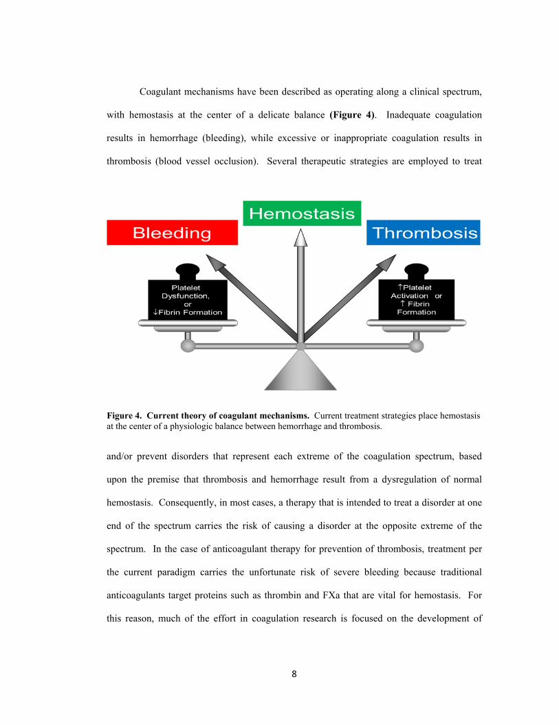
Coagulant mechanisms have been described as operating along a clinical spectrum,
with hemostasis at the center of a delicate balance (Figure 4). Inadequate coagulation
results in hemorrhage (bleeding), while excessive or inappropriate coagulation results in
thrombosis (blood vessel occlusion). Several therapeutic strategies are employed to treat
and/or prevent disorders that represent each extreme of the coagulation spectrum, based
upon the premise that thrombosis and hemorrhage result from a dysregulation of normal
hemostasis. Consequently, in most cases, a therapy that is intended to treat a disorder at one
end of the spectrum carries the risk of causing a disorder at the opposite extreme of the
spectrum. In the case of anticoagulant therapy for prevention of thrombosis, treatment per
the current paradigm carries the unfortunate risk of severe bleeding because traditional
anticoagulants target proteins such as thrombin and FXa that are vital for hemostasis. For
this reason, much of the effort in coagulation research is focused on the development of
Figure 4. Current theory of coagulant mechanisms. Current treatment strategies place hemostasis at the center of a physiologic balance between hemorrhage and thrombosis.

9
novel anticoagulants that have reduced bleeding side effects. As will be discussed in
Chapter 2, FXI has emerged as a target for such therapies because of its relatively minor role
in hemostasis.
Cross-Talk Between Coagulation and Inflammation
The coagulation system functions alongside other host defense systems, including
inflammation. Inflammation is a host response to injury that, when directed against
microorganisms, is a key component of innate immunity (Tizard 2008). Many (perhaps
most) clinical situations involve “cross-talk” between coagulation and inflammation (Levi,
Keller et al. 2003, Levi and van der Poll 2010, van der Poll, de Boer et al. 2011, van der Poll
and Levi 2012). The two systems are often activated by a common stimulus (Markiewski,
Nilsson et al. 2007), and each pathway modulates the other through a variety of mechanisms
to restore normal physiologic function after an insult. Like coagulation, inflammation
operates along a clinical spectrum. Localized inflammation of an appropriate intensity is at
the center of the balance, and results in microbial elimination and tissue repair. At the
extremes are inadequate inflammation, which may contribute to microbial dissemination,
and overwhelming inflammation, which manifests as an infection-induced systemic
inflammatory response syndrome, or sepsis. Thus, the interaction between coagulation and
inflammation, which is normally beneficial, contributes to increased morbidity and mortality
during sepsis. This interaction presents many challenges for sepsis therapy. For example,
systemic inflammation may trigger a coagulopathy [disseminated intravascular coagulation
(DIC)] that is characterized by enhanced systemic thrombin generation (Levi and Ten Cate
1999). Thrombin can then propagate a vicious cycle by amplifying inflammation through
various mechanisms (Borissoff, Spronk et al. 2009). Although anticoagulant therapy is a

10
tempting strategy for limiting thrombin generation and breaking this vicious cycle, the
bleeding risk of currently available anticoagulants outweighs any potential benefit during
sepsis. Individuals with sepsis therefore represent an additional patient population that may
benefit from treatment with anticoagulant therapies that do not compromise normal
hemostasis.
The Plasma Contact System
FXI is traditionally considered to be a component of the contact activation pathway,
or contact system (Figure 2). The contact system consists of FXII, PK, FXI, and the
cofactor HK. Contact activation is initiated when FXII binds to negatively-charged surfaces.
This binding causes a conformational change in zymogen FXII, resulting in the generation of
a small amount of FXIIa. FXIIa then cleaves PK to generate α-kal, which then reciprocally
cleaves FXII to generate more FXIIa (Bjorkqvist, Nickel et al. 2014). In addition to its role
in FXII activation, α-kal also cleaves HK to liberate BK, a pro-inflammatory peptide that
stimulates prostaglandin and nitric oxide (NO) production, resulting in fever, pain, and tissue
swelling (Hall 1992).
Contact activation was initially assumed to be important for coagulation because of
the observation that blood clots rapidly when exposed to negatively charged (anionic)
artificial surfaces such as glass or kaolin. However, current discussions exclude all but one
contact factor, FXI, when describing hemostasis. Although the clinical data do not support a
role for contact activation in hemostasis, there is evidence that contact activation contributes
to clinically relevant inflammation. For example, hereditary angioedema (HAE), is a rare,
life-threatening disorder that is characterized by acute episodes of tissue swelling,
particularly in the skin, gastrointestinal mucosa, and oropharynx (Bjorkqvist, Nickel et al.

11
2014). HAE results from a functional or quantitative deficiency of C1-inhibitor (C1INH), a
serine protease inhibitor. A consistent clinical finding in HAE patients is the presence of
high levels of BK. Because C1INH regulates the contact factors FXIIa and α-kal (Gailani
2010), BK release during HAE episodes is consistent with unchecked contact activation.
Patients with HAE do not appear to have an increased risk of thrombosis (Bjorkqvist, Nickel
et al. 2014), suggesting, consistent with the current model described in Figure 3, that contact
activation does not contribute to coagulation in this setting.
Contact activation is likely to be an important host defense mechanism against
microorganisms. Similar to components of innate immunity, such as the complement
system, the contact factors can assemble and become activated on the surfaces of bacteria,
including the human pathogens Streptococcus pyogenes, Staphylococcus aureus,
Escherichia coli, and Salmonella (Mattsson, Herwald et al. 2001, Herwald, Morgelin et al.
2003, Frick, Akesson et al. 2006). Contact activation is triggered through recognition of
molecular patterns on bacterial surfaces in a manner that is strikingly similar to that observed
with the complement system (Frick, Bjorck et al. 2007). Furthermore, consistent with in
vitro observations of contact activation on negatively-charged surfaces, bacteria and
damaged cells provide a ready supply of polyanions in the form of phosphate polymers
[polyphosphate (polyP)], DNA, and RNA (Gailani, Bane et al. 2015). In addition to
enhancing the innate immune response to microorganisms through BK release, contact
activation-induced HK cleavage also generates antimicrobial peptides that directly target
bacterial pathogens (Frick, Akesson et al. 2006). The importance of contact activation
during infection was displayed in a mouse model of S. pyogenes infection using a synthetic
peptide (H-D-Pro-Phe-Arg-CMK) that specifically inhibits FXIIa and α-kal. Inhibitor-
treated mice experienced increased bacterial dissemination, suggesting that a functional
12
contact system is instrumental in the host response to bacterial infection (Frick, Akesson et
al. 2006). The contact system, like the coagulation system, can also be harmful if activated
inappropriately. Because bacteria and damaged cells can trigger contact activation, it stands
to reason that bacterial infection and cellular injury above a certain threshold may activate
the contact system to a level that is detrimental to the host. This is especially true in cases
where bacteria disseminate and reach the bloodstream (bacteremia), allowing products of
contact activation to reach the systemic circulation. BK is a potent vasodilator that may
cause severe hypotension during sepsis. Septic patients with low blood pressure display a
higher level of contact activation than septic patients with normal blood pressure (Mason,
Kleeberg et al. 1970), while contact system inhibition corrects hypotension in animal models
of lethal bacteremia (Pixley, De La Cadena et al. 1993).
While in vitro studies suggest an important role for contact activation in host
defense, the in vivo significance of this process remains uncertain (Gailani 2010). Many
studies have investigated possible mechanisms for FXII activation on a surface. A notable
finding is that FXIa was shown to activate FXII in plasma and purified systems (Griffin
1978, Matafonov, Sarilla et al. 2011). This is perhaps not surprising, given that (a) FXI is
structurally similar to PK, the zymogen for a protease (α-kal) that activates FXII, and (b)
FXI and PK arose from a common gene during the course of vertebrate evolution (Ponczek,
Gailani et al. 2008) and may therefore display similar features. This raises the possibility
that FXIa is an effector, rather than simply a product, of contact activation, and that FXI
links and influences two complex systems: one for thrombin generation, and the other for
inflammation.

13
References
Bjorkqvist, J., K. F. Nickel, E. Stavrou and T. Renne (2014). "In vivo activation and functions of the protease factor XII." Thromb Haemost 112(5): 868-875.
Borissoff, J. I., H. M. Spronk, S. Heeneman and H. ten Cate (2009). "Is thrombin a key player in the 'coagulation-atherogenesis' maze?" Cardiovasc Res 82(3): 392-403.
Broze, G. J., Jr. and T. J. Girard (2012). "Tissue factor pathway inhibitor: structure-function." Front Biosci (Landmark Ed) 17: 262-280.
Dahlback, B. (2000). "Blood coagulation." Lancet 355(9215): 1627-1632.
Davie, E. W., K. Fujikawa and W. Kisiel (1991). "The coagulation cascade: initiation, maintenance, and regulation." Biochemistry 30(43): 10363-10370.
Davie, E. W. and O. D. Ratnoff (1964). "Waterfall Sequence for Intrinsic Blood Clotting." Science 145(3638): 1310-1312.
Frick, I. M., P. Akesson, H. Herwald, M. Morgelin, M. Malmsten, D. K. Nagler and L. Bjorck (2006). "The contact system--a novel branch of innate immunity generating antibacterial peptides." EMBO J 25(23): 5569-5578.
Frick, I. M., L. Bjorck and H. Herwald (2007). "The dual role of the contact system in bacterial infectious disease." Thrombosis and haemostasis 98(3): 497-502.
Furie, B. and B. C. Furie (1992). "Molecular and cellular biology of blood coagulation." N Engl J Med 326(12): 800-806.
Gailani, D., C. E. Bane and A. Gruber (2015). "Factor XI and contact activation as targets for antithrombotic therapy." J Thromb Haemost 13(8): 1383-1395.
Gailani, D. and G. J. Broze, Jr. (1991). "Factor XI activation in a revised model of blood coagulation." Science 253(5022): 909-912.
Gailani, D. and T. Renne (2007). "Intrinsic pathway of coagulation and arterial thrombosis." Arteriosclerosis, thrombosis, and vascular biology 27(12): 2507-2513.
Gailani, D., Renne, T., Emsley, J. (2010). Factor XI and the contact system. The Online Metabolic and Molecular Basis of Inherited Disease. B. A. e. a. Valle, Lippincott, Williams and Wilkins.
Griffin, J. H. (1978). "Role of surface in surface-dependent activation of Hageman factor (blood coagulation factor XII)." Proc Natl Acad Sci U S A 75(4): 1998-2002.
Hall, J. M. (1992). "Bradykinin receptors: pharmacological properties and biological roles." Pharmacol Ther 56(2): 131-190.

14
Herwald, H., M. Morgelin and L. Bjorck (2003). "Contact activation by pathogenic bacteria: a virulence mechanism contributing to the pathophysiology of sepsis." Scand J Infect Dis 35(9): 604-607.
Levi, M., T. T. Keller, E. van Gorp and H. ten Cate (2003). "Infection and inflammation and the coagulation system." Cardiovasc Res 60(1): 26-39.
Levi, M. and H. Ten Cate (1999). "Disseminated intravascular coagulation." The New England journal of medicine 341(8): 586-592.
Levi, M. and T. van der Poll (2010). "Inflammation and coagulation." Crit Care Med 38(2 Suppl): S26-34.
Macfarlane, R. G. (1964). "An Enzyme Cascade in the Blood Clotting Mechanism, and Its Function as a Biochemical Amplifier." Nature 202: 498-499.
Markiewski, M. M., B. Nilsson, K. N. Ekdahl, T. E. Mollnes and J. D. Lambris (2007). "Complement and coagulation: strangers or partners in crime?" Trends Immunol 28(4): 184-192.
Mason, J. W., U. Kleeberg, P. Dolan and R. W. Colman (1970). "Plasma kallikrein and Hageman factor in Gram-negative bacteremia." Ann Intern Med 73(4): 545-551.
Matafonov, A., S. Sarilla, M. F. Sun, J. P. Sheehan, V. Serebrov, I. M. Verhamme and D. Gailani (2011). "Activation of factor XI by products of prothrombin activation." Blood 118(2): 437-445.
Mattsson, E., H. Herwald, H. Cramer, K. Persson, U. Sjobring and L. Bjorck (2001). "Staphylococcus aureus induces release of bradykinin in human plasma." Infect Immun 69(6): 3877-3882.
Monroe, D. M. and M. Hoffman (2014). Theories of Blood Coagulation: Basic Concepts and Recent Updates. Hemostasis and Thrombosis: Practical Guidelines in Clinical Management. H. I. Saba and H. R. Roberts. West Sussex, UK, John Wiley & Sons, Ltd.: 1-13.
Monroe, D. M. and N. S. Key (2007). "The tissue factor-factor VIIa complex: procoagulant activity, regulation, and multitasking." Journal of thrombosis and haemostasis : JTH 5(6): 1097-1105.
Morrissey, J. H. (2004). "Tissue factor: a key molecule in hemostatic and nonhemostatic systems." Int J Hematol 79(2): 103-108.
Naito, K. and K. Fujikawa (1991). "Activation of human blood coagulation factor XI independent of factor XII. Factor XI is activated by thrombin and factor XIa in the presence of negatively charged surfaces." J Biol Chem 266(12): 7353-7358.
15
Osterud, B. and S. I. Rapaport (1977). "Activation of factor IX by the reaction product of tissue factor and factor VII: additional pathway for initiating blood coagulation." Proc Natl Acad Sci U S A 74(12): 5260-5264.
Pixley, R. A., R. De La Cadena, J. D. Page, N. Kaufman, E. G. Wyshock, A. Chang, F. B. Taylor, Jr. and R. W. Colman (1993). "The contact system contributes to hypotension but not disseminated intravascular coagulation in lethal bacteremia. In vivo use of a monoclonal anti-factor XII antibody to block contact activation in baboons." J Clin Invest 91(1): 61-68.
Ponczek, M. B., D. Gailani and R. F. Doolittle (2008). "Evolution of the contact phase of vertebrate blood coagulation." Journal of thrombosis and haemostasis : JTH 6(11): 1876-1883.
Ruggeri, Z. M. and G. L. Mendolicchio (2015). "Interaction of von Willebrand factor with platelets and the vessel wall." Hamostaseologie 35(3): 211-224.
Tizard, I. R. (2008). Veterinary immunology : an introduction. St. Louis, MO, Saunders, an imprint of Elsevier Inc.
van der Poll, T., J. D. de Boer and M. Levi (2011). "The effect of inflammation on coagulation and vice versa." Curr Opin Infect Dis 24(3): 273-278.
van der Poll, T. and M. Levi (2012). "Crosstalk between inflammation and coagulation: the lessons of sepsis." Curr Vasc Pharmacol 10(5): 632-638.
Weiss, H. J. and B. Lages (1988). "Evidence for tissue factor-dependent activation of the classic extrinsic coagulation mechanism in blood obtained from bleeding time wounds." Blood 71(3): 629-635.

16
CHAPTER III
INTRODUCTION TO FACTOR XI
Factor XI Structure and Function
FXI, the zymogen of the trypsin-like protease FXIa, is a 160kd protein that is made
primarily in the liver. The protein circulates in a complex with the glycoprotein HK at a
plasma concentration of approximately 30 nM (range of 15-45 nM) (Smith and Gailani
2008). FXI is the most recent member of the plasma coagulation system, having arisen from
a duplication of the gene for the protease zymogen PK (Ponczek, Gailani et al. 2008, Bane
and Gailani D. 2014). In humans, FXI has a plasma half-life of approximately 48 hours
(range of 45-52 hours). Unlike the vitamin K [“Koagulations-Vitamin”(Kresage N 2005)]
dependent coagulation proteins (prothrombin, FVII, FIX, and FX), FXI lacks a calcium-
binding γ-carboxyglutamic acid (Gla) domain (Gailani and Smith 2009). Consequently, FXI
synthesis does not require vitamin K, and treatment with vitamin K antagonists such as
warfarin does not affect plasma FXI levels. Each FXI molecule circulates as a dimer of two
identical 80kd subunits, which is a unique feature among the coagulation proteins. The
functional significance of the dimeric structure is unclear, but may be related to activation of
zymogen FXI by FXIIa (Geng, Verhamme et al. 2013), or to an interaction between platelets
and FXI during hemostasis (Gailani and Smith 2009). Each 607-amino acid FXI subunit
contains an N-terminal heavy chain consisting of four 90 to 91 amino acid repeats termed
“apple” domains (numbered A1-A4), and a C-terminal trypsin-like catalytic domain (Figure
5A) (Emsley, McEwan et al. 2010). An inter-chain disulfide bond at Cys321 in the A4
domain (Figures 5A & 5B) links each subunit within the dimer. The apple domains form a

17
disc-like structure that serves as a platform for binding of FXI to other molecules (Figure
5C) (Bane and Gailani D. 2014). For example, HK binds to the FXI monomer within a
groove formed by A1, A2, and A4 on the side of the platform opposite the catalytic domain,
while the FXIa substrate FIX binds to the A3 domain (Emsley, McEwan et al. 2010, Bane
and Gailani 2014). FXI activation occurs through cleavage of the peptide bond between the
Arg369 and Ile370 residues, which are located between the A4 and catalytic domains (indicated
by the black arrowhead on Figure 5A) of each FXI subunit (Gailani and Smith 2009). The
proteases thrombin, FXIIa, and FXIa (autoactivation) convert FXI to FXIa in vitro. The
Figure 5. Factor XI structure. (A) Diagram indicating the domain organization of a factor XI (FXI) subunit. Each circle represents one amino acid. Cysteine residues that are associated with disulfide bonds are shown in black. FXI is activated through the cleavage of the peptide bond (indicated by the black arrowhead) between Arg369 and Ile370. The amino acids of the catalytic triad (described in text) are shown in red. A disulfide bond between Cys321 residues on the A4 domains link FXI subunits to form (B) a FXI dimer. (C) The four “apple” domains form a platform upon which the catalytic domain rests. Panels B and C are models based on the crystal structure of FXI.

18
process involves an intermediate, ½-FXIa (only one of the two monomers is activated) that
may be a major form of activated FXI (Emsley, McEwan et al. 2010). FXI activation
induces a conformational change that facilitates FXIa activation of substrate FIX by
exposing a binding site for FIX on the A3 domain of FXIa (Gailani and Smith 2009).
Proteolysis of FXIa substrates is mediated by a “catalytic triad” (indicated by the solid red
circles in Figure 5A) of serine (Ser557), aspartic acid (Asp462), and histidine (His413). As
discussed in chapter I, FXIa does not appear to be instrumental in initiating thrombin
generation. Rather, it supports the formation of additional thrombin to bolster existing fibrin
clots that are constantly subjected to numerous endogenous anticoagulant and fibrinolytic
processes. The bleeding pattern of FXI deficient patients (described below) supports this
interpretation of FXI’s role in coagulation.
Factor XI Deficiency in Humans and Domestic Animals
This dissertation describes studies that compared mice with normal levels of FXI
(FXI+/+ mice) to those that possessed approximately 50% and 0% of normal levels (FXI+/-
and FXI-/- mice, respectively) of this protein. As will be described later in this chapter, FXI
deficiency is not associated with abnormal hemostasis in laboratory mice. This section is
included to briefly summarize the clinical presentation of FXI deficiency in humans (and
domestic animals) so that the reader is more equipped to relate FXI deficiency in humans to
FXI deficiency in laboratory mice.
Although rare in the general population [approximately 1 in 1 million (Asakai,
Chung et al. 1991)], severe (FXI activity <20% of normal) FXI deficiency is common (1 in
450) in people of Ashkenazi Jewish descent (Seligsohn 2009). The true incidence of FXI
deficiency may be higher, as some patients may not experience bleeding that is of sufficient

19
intensity to require medical attention. The disorder may remain undiscovered until a
prolonged aPTT is revealed during routine pre-surgical screening. In contrast to the X-
linked hemophilias, people with FXI deficiency do not, with the exception of menorrhagia
(Seligsohn 2009), bleed spontaneously. For example, hemarthrosis, a hallmark of the
classical hemophilias, rarely occurs in FXI-deficient patients (Bolton-Maggs 2009).
Significant hemorrhage, if observed, is usually related to trauma or surgery, particularly in
tissues where fibrinolysis is most active, such as the oropharynx or genitourinary tract. For
instance, approximately 20% of women with FXI deficiency experience excessive bleeding
related to childbirth (Seligsohn 2009, Santoro, Prejano et al. 2011). It is postulated that FXIa
is more important for hemostasis after trauma to these tissues, both for sustaining thrombin
generation and for providing resistance to fibrinolysis by promoting activation of thrombin
activatable fibrinolysis inhibitor (Von dem Borne, Bajzar et al. 1997, Bolton-Maggs 2009,
Bane and Gailani D. 2014). Bleeding in FXI-deficient patients may occur immediately after
injury, or may be delayed for several hours (Seligsohn 2009). Interestingly, there is a poor
correlation between FXI levels and bleeding tendency. Some individuals with severe FXI
deficiency do not bleed at all after trauma (Rapaport, Proctor et al. 1961), while others may
experience bleeding that varies over time, even after re-exposure to a similar type of injury
(Asakai, Chung et al. 1991, Bolton-Maggs, Patterson et al. 1995). The complex clinical
presentation of FXI deficiency, combined with the potentially serious side effects of FXI
replacement therapy, provide a difficult challenge to clinicians who manage these patients
(Bolton-Maggs 2009).
Congenital FXI deficiency has also been reported in several species of domestic
animals, where multiple modes of inheritance are described (Marron, Robinson et al. 2004,
Ohba, Takasu et al. 2008). The disorder is most widely reported in cattle (Marron, Robinson

20
et al. 2004, Ohba, Takasu et al. 2008), but has also been described in dogs (Knowler, Giger
et al. 1994) and in a domestic cat (Troxel, Brooks et al. 2002). Some reports describe injury-
related bleeding in FXI-deficient animals (Knowler, Giger et al. 1994, Troxel, Brooks et al.
2002, Marron, Robinson et al. 2004), while others indicate no apparent bleeding phenotype
(Ohba, Takasu et al. 2008).
Factor XI and Thrombosis
In healthy individuals, the mechanisms that promote coagulation are balanced by
numerous anticoagulant mechanisms that localize clots to the area of tissue injury, where
they are eventually broken down through fibrinolysis. A disruption to the procoagulant-
anticoagulant balance may result in excessive bleeding (hemorrhage) or pathologic clot
formation within blood vessels (thrombosis). The pathophysiology of thrombosis is
complex, but has been attributed to increased thrombin generation and/or decreases in
intrinsic fibrinolytic mechanisms. In keeping with this hypothesis, most antithrombotic
therapies are directed at minimizing thrombin generation through inhibition of proteins that
are vital for normal coagulation. Such therapies are very effective at preventing thrombosis,
but, predictably, carry a significant bleeding risk. This fact is perhaps most vividly
illustrated by the anticoagulant drug warfarin, a vitamin K-antagonist that is the drug of
choice for treatment of venous thrombosis and prevention of stroke in patients with non-
valvular atrial fibrillation. This anticoagulant has been in use since the early 1950’s, but was
first registered as a rodenticide in 1948 (van Montfoort and Meijers 2013). Interestingly,
warfarin is a synthetic analogue of dicoumarol, a compound within spoiled sweet clover that
was found to be the causative agent of an outbreak of hemorrhagic cattle disease in the
1920’s (Kresage N 2005).

21
Consistent with traditional reasoning, proteins that are not vital for normal
hemostasis would be predicted to play a proportionally smaller role in thrombosis. In the
case of FXI, studies in humans have challenged this reasoning. In the Leiden Thrombophilia
study, patients with plasma FXI levels in the top 10% of the normal distribution were nearly
twice as likely to develop venous thromboembolism compared to the rest of the study
population (Meijers, Tekelenburg et al. 2000). In the same study, men with higher levels of
FXI were at a 2-fold greater risk for myocardial infarction. Another study showed a
correlation between elevated FXI levels and stroke (Yang, Flanders et al. 2006).
Conversely, population studies of individuals with severe FXI deficiency reveal a lower risk
for stroke and deep-vein thrombosis (Salomon, Steinberg et al. 2008, Salomon, Steinberg et
al. 2011). Severe FXI deficiency has not been shown to be protective against myocardial
infarction (Salomon, Steinberg et al. 2003), indicating that FXI deficiency may not
uniformly influence thrombus formation in all tissues.
Factor XI in Animal Models of Thrombosis
The observation that humans with FXI deficiency appear to be protected against
certain thrombotic events has important therapeutic implications, and has supported an
ongoing interest in research that investigates the role of FXI in experimental thrombosis. A
mouse model of FXI deficiency was developed on the C57Bl/6 background by Gailani, et
al., using homologous recombination in embryonic stem cells. The mice are fertile, and
offspring from heterozygous matings follow the expected Mendelian ratio of approximately
25% wild type (WT), 50% heterozygous null, 25% homozygous null, indicating that partial
or complete FXI deficiency is not embryonic lethal (Gailani, Lasky et al. 1997). The aPTT
in plasma from FXI-deficient (FXI-/-) mice is prolonged compared to WT mice, while

22
clotting times in plasma from mice that are homozygous for the FXI null allele (FXI+/-) are
intermediate between FXI-/- and WT (FXI+/+) mice. Under normal conditions, these mice
have no discernable phenotype and do not bleed excessively when subjected to amputation
of the tail tip or other invasive procedures (Gailani, Lasky et al. 1997, Renne, Oschatz et al.
2009). This is in contrast, for example, to FIX-deficient mice, which experience injury-
related (tail transection) hemorrhage that is comparable to animals given high doses of the
anticoagulant heparin (Wang, Cheng et al. 2005).
Despite the lack of a bleeding disorder, FXI-/- mice are remarkably resistant to
experimentally-induced arterial and venous thrombosis (Bane and Gailani 2014). In an
important proof of concept study, Rosen, et al. discovered that, in contrast to WT mice, FXI -
/- mice failed to form occlusive thrombi after application of a 20% solution of ferric chloride
(FeCl3) to the external surface of the carotid artery (Rosen, Gailani et al. 2002). The ability
of FXI-/- mice to generate thrombi in response to FeCl3 was restored after infusion of human
FXI. Wang and colleagues (Wang, Cheng et al. 2005) used a modified version of this model
to show that FXI-/- mice were protected from arterial thrombosis to a degree that was
comparable to FIX deficiency. Importantly, the Wang study also reported that FXI
deficiency was superior to aspirin therapy, and conferred a level of thrombosis resistance
that was similar to treatment with doses of heparin that was well above the therapeutic range.
Further work has confirmed that FXI is also instrumental in formation of venous thrombi
(Wang, Smith et al. 2006), and that it contributes to experimental thrombus formation in
numerous species, including rabbits and baboons (Minnema, Friederich et al. 1998, Gruber
and Hanson 2003, Yamashita, Nishihira et al. 2006, Tucker, Marzec et al. 2009).
Interestingly, the animal data also implicate FXII in thrombosis. For instance, WT mice that
received a monoclonal antibody that blocks activation of FXI by FXIIa (but not thrombin or

23
FXIa) displayed a resistance to FeCl3 thrombosis that was similar to FXI-deficient mice
(Cheng, Tucker et al. 2010). These findings raise the profile of the contact system in
thrombosis, and support the proposition that thrombus-generating mechanisms may be
different from hemostatic mechanisms in some instances. Additional animal models,
described in the following chapter, indicate that FXI and the contact system also participate
in inflammation-induced pathology.
References
Asakai, R., D. W. Chung, E. W. Davie and U. Seligsohn (1991). "Factor XI deficiency in Ashkenazi Jews in Israel." The New England journal of medicine 325(3): 153-158.
Bane, C. E. and N. A. T. Gailani D. (2014). Factor XI Deficiency (Hemophilia C). Hemostasis and Thrombosis: Practical Guidelines in Clinical Management. H. I. Saba and H. R. Roberts. West Sussex, UK, John Wiley & Sons, Ltd.: 71-81.
Bane, C. E., Jr. and D. Gailani (2014). "Factor XI as a target for antithrombotic therapy." Drug Discov Today 19(9); 1454-8.
Bolton-Maggs, P. H. (2009). "Factor XI deficiency--resolving the enigma?" Hematology Am Soc Hematol Educ Program: 97-105.
Bolton-Maggs, P. H., D. A. Patterson, R. T. Wensley and E. G. Tuddenham (1995). "Definition of the bleeding tendency in factor XI-deficient kindreds--a clinical and laboratory study." Thromb Haemost 73(2): 194-202.
Cheng, Q., E. I. Tucker, M. S. Pine, I. Sisler, A. Matafonov, M. F. Sun, T. C. White-Adams, S. A. Smith, S. R. Hanson, O. J. McCarty, T. Renne, A. Gruber and D. Gailani (2010). "A role for factor XIIa-mediated factor XI activation in thrombus formation in vivo." Blood 116(19): 3981-3989.
Emsley, J., P. A. McEwan and D. Gailani (2010). "Structure and function of factor XI." Blood 115(13): 2569-2577.
Gailani, D., N. M. Lasky and G. J. Broze, Jr. (1997). "A murine model of factor XI deficiency." Blood coagulation & fibrinolysis : an international journal in haemostasis and thrombosis 8(2): 134-144.
Gailani, D. and S. B. Smith (2009). "Structural and functional features of factor XI." Journal of thrombosis and haemostasis : JTH 7 Suppl 1: 75-78.

24
Geng, Y., I. M. Verhamme, S. B. Smith, M. F. Sun, A. Matafonov, Q. Cheng, S. A. Smith, J. H. Morrissey and D. Gailani (2013). "The dimeric structure of factor XI and zymogen activation." Blood 121(19): 3962-3969.
Gruber, A. and S. R. Hanson (2003). "Factor XI-dependence of surface- and tissue factor-initiated thrombus propagation in primates." Blood 102(3): 953-955.
Knowler, C., U. Giger, W. J. Dodds and M. Brooks (1994). "Factor XI deficiency in Kerry Blue Terriers." J Am Vet Med Assoc 205(11): 1557-1561.
Kresage N, S. R., Hill RL (2005). "Hemorrhagic Sweet Clover Disease, Dicumarol, and Warfarin: the Work of Karl Paul Link." J Biol Chem 280(February 25): e5-e6.
Marron, B. M., J. L. Robinson, P. A. Gentry and J. E. Beever (2004). "Identification of a mutation associated with factor XI deficiency in Holstein cattle." Anim Genet 35(6): 454-456.
Meijers, J. C., W. L. Tekelenburg, B. N. Bouma, R. M. Bertina and F. R. Rosendaal (2000). "High levels of coagulation factor XI as a risk factor for venous thrombosis." The New England journal of medicine 342(10): 696-701.
Minnema, M. C., P. W. Friederich, M. Levi, P. A. von dem Borne, L. O. Mosnier, J. C. Meijers, B. J. Biemond, C. E. Hack, B. N. Bouma and H. ten Cate (1998). "Enhancement of rabbit jugular vein thrombolysis by neutralization of factor XI. In vivo evidence for a role of factor XI as an anti-fibrinolytic factor." J Clin Invest 101(1): 10-14.
Ohba, Y., M. Takasu, N. Nishii, E. Takeda, S. Maeda, T. Kunieda and H. Kitagawa (2008). "Pedigree analysis of factor XI deficiency in Japanese black cattle." J Vet Med Sci 70(3): 297-299.
Ponczek, M. B., D. Gailani and R. F. Doolittle (2008). "Evolution of the contact phase of vertebrate blood coagulation." Journal of thrombosis and haemostasis : JTH 6(11): 1876-1883.
Rapaport, S. I., R. R. Proctor, M. J. Patch and M. Yettra (1961). "The mode of inheritance of PTA deficiency: evidence for the existence of major PTA deficiency and minor PTA deficiency." Blood 18: 149-165.
Renne, T., C. Oschatz, S. Seifert, F. Muller, J. Antovic, M. Karlman and P. M. Benz (2009). "Factor XI deficiency in animal models." J Thromb Haemost 7 Suppl 1: 79-83.
Rosen, E. D., D. Gailani and F. J. Castellino (2002). "FXI is essential for thrombus formation following FeCl3-induced injury of the carotid artery in the mouse." Thromb Haemost 87(4): 774-776.
Salomon, O., D. M. Steinberg, R. Dardik, N. Rosenberg, A. Zivelin, I. Tamarin, B. Ravid, S. Berliner and U. Seligsohn (2003). "Inherited factor XI deficiency confers no protection

25
against acute myocardial infarction." Journal of thrombosis and haemostasis : JTH 1(4): 658-661.
Salomon, O., D. M. Steinberg, N. Koren-Morag, D. Tanne and U. Seligsohn (2008). "Reduced incidence of ischemic stroke in patients with severe factor XI deficiency." Blood 111(8): 4113-4117.
Salomon, O., D. M. Steinberg, M. Zucker, D. Varon, A. Zivelin and U. Seligsohn (2011). "Patients with severe factor XI deficiency have a reduced incidence of deep-vein thrombosis." Thrombosis and haemostasis 105(2): 269-273.
Santoro, R., S. Prejano and P. Iannaccaro (2011). "Factor XI deficiency: a description of 34 cases and literature review." Blood coagulation & fibrinolysis : an international journal in haemostasis and thrombosis 22(5): 431-435.
Seligsohn, U. (2009). "Factor XI deficiency in humans." J Thromb Haemost 7 Suppl 1: 84-87.
Smith, S. B. and D. Gailani (2008). "Update on the physiology and pathology of factor IX activation by factor XIa." Expert review of hematology 1(1): 87-98.
Troxel, M. T., M. B. Brooks and M. L. Esterline (2002). "Congenital factor XI deficiency in a domestic shorthair cat." J Am Anim Hosp Assoc 38(6): 549-553.
Tucker, E. I., U. M. Marzec, T. C. White, S. Hurst, S. Rugonyi, O. J. McCarty, D. Gailani, A. Gruber and S. R. Hanson (2009). "Prevention of vascular graft occlusion and thrombus-associated thrombin generation by inhibition of factor XI." Blood 113(4): 936-944.
van Montfoort, M. L. and J. C. Meijers (2013). "Anticoagulation beyond direct thrombin and factor Xa inhibitors: indications for targeting the intrinsic pathway?" Thromb Haemost 110(2): 223-232.
Von dem Borne, P. A., L. Bajzar, J. C. Meijers, M. E. Nesheim and B. N. Bouma (1997). "Thrombin-mediated activation of factor XI results in a thrombin-activatable fibrinolysis inhibitor-dependent inhibition of fibrinolysis." J Clin Invest 99(10): 2323-2327.
Wang, X., Q. Cheng, L. Xu, G. Z. Feuerstein, M. Y. Hsu, P. L. Smith, D. A. Seiffert, W. A. Schumacher, M. L. Ogletree and D. Gailani (2005). "Effects of factor IX or factor XI deficiency on ferric chloride-induced carotid artery occlusion in mice." J Thromb Haemost 3(4): 695-702.
Wang, X., P. L. Smith, M. Y. Hsu, D. Gailani, W. A. Schumacher, M. L. Ogletree and D. A. Seiffert (2006). "Effects of factor XI deficiency on ferric chloride-induced vena cava thrombosis in mice." J Thromb Haemost 4(9): 1982-1988.
Yamashita, A., K. Nishihira, T. Kitazawa, K. Yoshihashi, T. Soeda, K. Esaki, T. Imamura, K. Hattori and Y. Asada (2006). "Factor XI contributes to thrombus propagation on injured neointima of the rabbit iliac artery." J Thromb Haemost 4(7): 1496-1501.

26
Yang, D. T., M. M. Flanders, H. Kim and G. M. Rodgers (2006). "Elevated factor XI activity levels are associated with an increased odds ratio for cerebrovascular events." Am J Clin Pathol 126(3): 411-415.

27
CHAPTER IV
FACTOR XI AND THE CONTACT SYSTEM IN SEPSIS
Introduction
As discussed in Chapter II, numerous lines of evidence point to cross-talk between
coagulation and inflammation. This chapter describes how both systems are triggered after
infection, and provides examples of how coagulation and inflammation are intricately linked
during sepsis. It is intended to provide a framework to help explain how FXI may contribute
to morbidity and mortality during sepsis, thus setting the stage for a more complete
understanding of the data reported in the next chapter.
An inflammatory syndrome known as sepsis is almost always accompanied by some
degree of activation of coagulation (Levi 2010, Stearns-Kurosawa, Osuchowski et al. 2011).
An extreme example of this relationship is DIC, a complication of sepsis that is associated
with higher mortality (Hook and Abrams 2012). Contact activation is another complication
of sepsis. Septic patients undergo systemic contact activation, as evidenced by consumption
of contact factors and release of BK (Saugstad, Buo et al. 1992, Mattsson, Herwald et al.
2001). Notably, low levels of contact factors are associated with death among patients with
sepsis (Oehmcke and Herwald 2010), suggesting that contact activation contributes to
mortality. The contact activation and coagulation systems contribute to the pathology of
sepsis through distinct mechanisms. Although it is extremely difficult to determine the
precise mechanism of mortality during sepsis, inflammation and contact activation appear to
figure more prominently than a coagulopathy in the mortality of sepsis in some animal
models (Pixley, De La Cadena et al. 1993, Jansen, Pixley et al. 1996, Ganopolsky and

28
Castellino 2004, Corral, Yelamos et al. 2005, Iwaki, Cruz-Topete et al. 2008). FXI, which
may participate in both coagulation and inflammation, may primarily influence mortality in
some animal sepsis models through a pro-inflammatory effect.
Overview of Sepsis
Sepsis is defined as a systemic inflammatory response to infection (Bone, Balk et al.
1992). Clinically, the disorder is the result of a bacterial (gram-positive, gram-negative, or
mixed) or fungal infection that originates primarily within the lungs, peritoneum, or urinary
tract (Angus and van der Poll 2013). Sepsis is diagnosed when there is evidence of a
systemic inflammatory response syndrome (SIRS) in the face of a confirmed or suspected
infection (Bone, Balk et al. 1992). Criteria for the diagnosis of SIRS include an elevated
heart rate, increased respiratory rate, and body temperature and white blood cell counts that
are either above or below the normal range (Stearns-Kurosawa, Osuchowski et al. 2011).
Sepsis may progress to severe sepsis, which is defined as SIRS complicated by organ
dysfunction, and finally to septic shock, which is noted by the development of persistent
hypotension in addition to the features described for less severe sepsis (Levy, Fink et al.
2003). Mortality from sepsis is thought to be a result of either an exaggerated inflammatory
response, which typically occurs early in the course of the disease, or to prolonged
immunosuppression, where patients succumb to secondary bacterial infections (Rittirsch,
Flierl et al. 2008, Stearns-Kurosawa, Osuchowski et al. 2011). Additional complications,
such as systemic activation of the coagulation and complement systems, may also contribute
to mortality, particularly in the later stages of sepsis (Russell 2006, Rittirsch, Flierl et al.
2008, Amara, Flierl et al. 2010). Following is a discussion of the acute inflammatory
response during the initial stages of sepsis, which is of relevance to this research project.

29
The Inflammatory Response to Infection
Inflammation is a feature of innate immunity, which is a nonspecific host response
to infection and tissue injury. At its core, acute inflammation is a process of transporting
cellular and soluble inflammatory factors from the bloodstream to damaged tissues. This
occurs through a standard sequence of events: A local increase in blood flow is followed by
increased vascular permeability, which facilitates entry of protein-rich edema fluid and
leukocytes into extravascular sites of injury (Slauson and Cooper 1990). Edema fluid
contains nutrients and beneficial proteins, such as coagulation factors and complement
components, while leukocytes, predominantly neutrophils, augment the local response of
resident macrophages to engulf, kill, and remove invading pathogens. The process is
orchestrated by several types of inflammatory mediators, including cytokines, chemokines,
and numerous vasoactive molecules. These mediators are protective when resolving
infections that are contained locally. However, they contribute to sepsis morbidity and
mortality when infections become more widespread.
Inflammation is triggered during infections when pathogens and damaged host
cells express molecules called pathogen-associated molecular patterns (PAMPs) and
damage- associated molecular patterns (DAMPs), respectively. These molecules are
recognized by pattern recognition receptors (PRRs) on resident “sentinel” cells, such as
macrophages and dendritic cells (Rittirsch, Flierl et al. 2008, Tizard 2008, Angus and van
der Poll 2013). The classical PAMPs are lipopolysaccharide (LPS) and peptidoglycan within
the cell walls of gram-negative and gram-positive bacteria, respectively, but PAMPs are also
expressed by yeasts and viruses (Tizard 2008). DAMPs, also called alarmins, include high
mobility group box protein-1 (Tizard 2008), and extracellular DNA, RNA, and histones
(Chan, Roth et al. 2012). Toll-like receptors (TLRs) are the most prominent class of PRRs

30
(Tizard 2008). PAMP/DAMP recognition by PRRs results in the activation of receptor
complexes on sentinel cells. This triggers intracellular signaling pathways, resulting in the
activation of transcription factors, such as NF-κB. NF-κB and related proteins then regulate
transcription of the cytokines, chemokines, and vasoactive molecules that contribute to acute
inflammation.
Cytokines are polypeptides that mediate cellular interactions (Tizard 2008). They
exert a variety of effects, including activation of immune cells, induction of fever, and
initiation of the acute phase response. The major cytokines of acute inflammation are tumor
necrosis factor α (TNFα), interleukin-1 (IL-1) and interleukin-6 (IL-6). TNFα and IL-1 have
nearly identical effects (Sherwood and Toliver-Kinsky 2004) and play a central role in the
vascular changes that exemplify the acute inflammatory response (Tizard 2008). IL-6 is a
major mediator of the acute phase response of the liver (Sherwood and Toliver-Kinsky 2004,
Tizard 2008), and also stimulates production of additional cytokines, including the anti-
inflammatory cytokine IL-10 (Tizard 2008). Chemokines are a subset of cytokines that
regulate the emigration of leukocytes from the bloodstream to the tissues (Tizard 2008).
Among these are macrophage inflammatory protein-2 (MIP-2) and keratinocyte
chemoattractant (KC), which attract neutrophils, and monocyte chemotactic protein-1 (MCP-
1), which exerts chemotactic activity on monocytes.
Vasodilation is a critical step in acute inflammation. It facilitates the opening of
gaps between adjacent vascular endothelial cells to allow passage of edema fluid and
leukocytes from the intravascular to the extravascular space (Slauson and Cooper 1990,
Sherwood and Toliver-Kinsky 2004). Vasoactive molecules, NO, BK, histamine, and
vasoactive lipids, such as phospholipase, mediate this process (Sherwood and Toliver-
Kinsky 2004, Tizard 2008). NO is the most important contributor to vasodilation during

31
sepsis (Vallance and Chan 2001, Sherwood and Toliver-Kinsky 2004). It is derived from the
amino acid L-arginine by the enzyme nitric oxide synthase, and acts through cyclic
guanosine monophosphate-dependent mechanisms to cause vascular smooth muscle
relaxation. There are three forms of NOS: endothelial, neuronal, and inducible (Vallance
and Chan 2001). The inducible form is released by numerous cell types, primarily
endothelial cells and macrophages (Moncada, Palmer et al. 1991) in response to
inflammatory cytokines and BK (Vallance and Chan 2001, Zhao, Qiu et al. 2001). In
addition to its role in vasodilation, NO is also thought to enhance the microbial killing
efficiency of macrophages (Moncada, Palmer et al. 1991).
Severe bacterial infection and/or massive trauma lead to excessive, systemic release
of the inflammatory mediators described above. If left unchecked, this systemic
inflammatory response ultimately results in septic shock, which is characterized by
irreversible hypotension, organ injury, and death. The coagulation and contact systems
(described below) are also excessively activated during sepsis. Dysregulation of each
pathway presents unique challenges that may compromise the host’s ability to survive.
Disseminated Intravascular Coagulation
DIC is a dysregulated coagulation response that accompanies a number of primary
conditions, including sepsis, neoplasia, and trauma (Levi, de Jonge et al. 1999). The
syndrome is characterized by intravascular activation of the coagulation system, with
simultaneous down-regulation of intrinsic anticoagulant and fibrinolytic mechanisms
(Russell 2006, Hook and Abrams 2012). This condition tips the coagulant-anticoagulant
balance in favor of coagulation, resulting in ischemic organ damage secondary to widespread
microvascular thrombosis. Eventually, DIC causes depletion of platelets and plasma

32
coagulation proteins, resulting in diffuse hemorrhage. The impact of DIC during sepsis
depends on several factors, including the intensity of the coagulopathy and any existing
disease process that may complicate the management of septic patients. Although
individuals that develop DIC subsequent to sepsis or severe trauma are twice as likely to die,
it is unclear if DIC is a definitive mechanism of mortality, or simply an indicator of disease
severity, in such patients (Levi, de Jonge et al. 1999).
The coagulation system is activated during sepsis in a number of ways. Gram-
negative infections trigger coagulation when the LPS component of these organisms
associates with extracellular LPS-binding protein. This initiates toll-like receptor 4 (TLR4)-
mediated intracellular signaling cascades, resulting in TF expression by circulating
mononuclear cells (Guha, O'Connell et al. 2001). Sepsis-induced TF expression is also
mediated by pro-inflammatory cytokines, particularly IL-6 (Levi 2010). The consequence of
inflammation-induced TF expression is the intravascular formation of thrombin, the central
enzyme in coagulation. In addition to driving DIC, thrombin also perpetuates the
inflammation-coagulation cycle by stimulating cytokine production in mononuclear cells
through protease activated receptor (PAR) cleavage (Borissoff, Spronk et al. 2009).
Gram-negative sepsis is the best understood cause of DIC (Hook and Abrams 2012),
and most animal models of DIC employ LPS as the inciting agent (Berthelsen, Kristensen et
al. 2011). The role of gram-positive organisms in coagulation is less clear, but is probably
also related to toll receptor signaling upon recognition of gram-positive associated
peptidoglycans by TLR2 (Russell 2006, Hook and Abrams 2012). Patel and co-workers
used a “two hit” model to explore the influence of gram-negative (LPS) and polymicrobial
(CLP) sepsis on TLR-mediated thrombosis. In this study, groups of mice were given IP
injections of LPS or saline, or were subjected to CLP or sham surgery. Four to six hours

33
later, a light-dye laser injury was applied to a section of the cremaster vasculature of each
(living) animal. Time to thrombosis at this discrete site was then evaluated using intravital
microscopy. Thrombosis was enhanced (compared to controls) in laser-injured WT animals
after CLP and LPS infusion. LPS infusion enhanced thrombosis in TLR2 deficient mice, but
did not enhance thrombosis in TLR4 deficient mice. Conversely, CLP enhanced thrombosis
in TLR4, but not TLR2, deficient mice. The authors concluded that CLP promoted laser-
induced thrombosis through a mechanism that required TLR2 signaling, while the pro-
thrombotic manifestations of E. coli endotoxemia required TLR4. This is consistent with
our understanding of TLR4 and TLR2 signaling in gram-negative and gram-positive sepsis,
respectively, since LPS is a component of gram-negative bacteria, while bacteria that enter
the bloodstream of mice during the initial stages of CLP-induced sepsis are reported to be
predominantly gram-positive (Hyde, Stith et al. 1990). Apart from the TLR data, the Patel
study highlighted additional key differences in the coagulation response elicited by LPS and
CLP. Notably, the pro-thrombotic phenotype of the CLP-operated mice disappeared when
the mice were given intravenous fluid replacement with isotonic saline post-operatively,
while fluid replacement had no effect on thrombosis in LPS-treated mice. This is telling,
considering that they also showed that, in contrast to LPS-treated mice, thrombin-
antithrombin (TAT) complex levels did not rise in mice after CLP (Patel, Soubra et al.
2010). Antithrombin is the major plasma inhibitor of thrombin (Egberg and Blomback
2014), and TAT complex levels are a surrogate marker of thrombin levels (Teitel, Bauer et
al. 1982). The authors provided several hypotheses that could explain their observations,
speculating, for example, that fluid replacement improves microvascular hemodynamics,
leading to enhanced shear-dependent release of the antiplatelet molecules NO and
prostacyclin (Saba and Saba 2014). While these data indicate sepsis promotes a pro-

34
coagulant state in vivo, the influence of the coagulopathy of sepsis on mortality is
controversial (Stearns-Kurosawa, Osuchowski et al. 2011). Corral et al (Corral, Yelamos et
al. 2005) inhibited the nuclear enzyme PARP-1 in mice and observed reduced inflammation
and improved survival after LPS infusion or CLP. Notably, they saw comparable evidence
of DIC in both PARP-1 deficient and WT mice, leading them to conclude that inflammation,
not DIC, influenced mortality in their studies. Conversely, Opal and colleagues (Opal,
Palardy et al. 2001), observed improved survival in recombinant human tissue factor
pathway inhibitor (rhTFPI)-treated mice after CLP. Unfortunately, the authors did not
evaluate DIC in this study, but TFPI inhibits the TF (extrinsic) pathway of coagulation
(Lwaleed and Bass 2006), which is the central mechanism for intravascular coagulation
during sepsis (Opal and Esmon 2003). Accordingly, it is reasonable to assume that rhTFPI
treatment would reduce DIC if it were present. Interestingly, rhTFPI anticoagulant treatment
improved survival when the cecum was punctured with a small-bore needle (defined as 23
gauge), but not when injury was inflicted with a large bore needle (defined as 21 gauge).
This suggests that higher levels of injury may trigger or enhance additional pathways, such
as contact activation, that exacerbate sepsis pathology.
In theory, anticoagulant therapy is a rational approach for treating sepsis because it
addresses both the coagulopathy and the inflammatory response of the disorder by inhibiting
thrombin. In practice, such therapies have met with disappointing results, probably because
anticoagulants increase the bleeding risk in septic patients. This is illustrated by studies that
describe hemorrhage in fibrinogen-deficient mice infected with the protozoal parasite
Toxoplasma gondii and the gram-positive bacteria Listeria monocytogenes (Johnson,
Berggren et al. 2003, Mullarky, Szaba et al. 2005). Another hypothesis for treatment failures
is that antithrombotic therapy actually contributes to sepsis pathology by dampening the

35
protective effects of coagulation during infections. Many authors speculate the coagulation
system limits infection through various mechanisms, including the formation of a protective
fibrin barrier around invading microbes (Ahrenholz and Simmons 1980, Echtenacher, Weigl
et al. 2001) and up-regulation of inflammation (Szaba and Smiley 2002, Flick, Du et al.
2004, Adams, Schachtrup et al. 2007). These effects undoubtedly depend on the degree of
infection, type of organism, and numerous host factors.
The Contact System and Sepsis
In contrast to DIC, which some consider to be a “bystander” in the pathology of
sepsis (Stearns-Kurosawa, Osuchowski et al. 2011), vasodilation secondary to excessive NO
production is the primary feature of septic shock (Vallance and Chan 2001). The contact
activation product BK contributes to vasodilation through the canonical pathway by
stimulating endothelial NO production (Vallance and Chan 2001, Zhao, Qiu et al. 2001).
BK also activates phospholipase (Sriskandan and Cohen 2000), which stimulates the release
of prostaglandins and other vasoactive lipids from cell membranes (Tizard 2008). Despite
the traditional association between contact activation and coagulation, animal models have
demonstrated that hypotension, not coagulopathy, is the primary consequence of infection-
induced contact activation. Furthermore, there is strong evidence that hypotension is the
mechanism through which the contact system contributes to sepsis mortality. Using a
baboon model of lethal Escherichia coli sepsis, Pixley and colleagues (Pixley, De La Cadena
et al. 1993) found that an anti-FXII monoclonal antibody reduced contact activation and
hypotension, and prolonged survival. Importantly, the antibody did not influence the
development of DIC. Consistent with the baboon work, Iwaki, et al(Iwaki, Cruz-Topete et
al. 2008) found that FXII deficiency improved hypotension but not coagulopathy in

36
endotoxemic mice. A later study showed that anti-FXII antibody treatment lowered IL-6
levels in septic baboons (Jansen, Pixley et al. 1996), indicating that contact activation also
influences cytokine release. The mechanism of contact activation-induced cytokine release
during sepsis is not known, but may be related to monocyte stimulation by FXIIa and HK
fragments (Toossi, Sedor et al. 1992, Khan, Bradford et al. 2006). These animal studies
provide evidence that the contact system operates independently of the coagulation system in
some cases of sepsis. This proposition is supported by a retrospective analysis of
streptococcal toxic shock syndrome (STSS) in humans, which noted the absence of DIC in
patients with an “isolated prolongation of aPTT” (prolonged aPTT with normal PT)
(Sriskandan and Cohen 2000). Recall that the aPTT evaluates the intrinsic (contact
activation) pathway, while the PT evaluates the extrinsic (TF) pathway. If activation of one
or both pathways in vivo results in consumption (depletion) of the corresponding coagulation
factors, the ex vivo observation of isolated prolongation of aPTT suggests that contact
activation predominates in this syndrome. Apart from reinforcing the role of the contact
system in sepsis, the animal and STSS studies suggest contact activation-induced
hypotension has a relatively greater role than coagulopathy in the morbidity and mortality
that accompanies some infections.
Factor XI in Animal Models of Sepsis
The in vitro data, discussed in Chapter II, that indicate FXI influences both contact
activation/inflammation and coagulation are bolstered by recent findings that point to a role
for this protein in the host response to infection. In a murine study of peritonitis induced by
intraperitoneal injection of the gram-positive intracellular bacteria Listeria monocytogenes
(Luo, Szaba et al. 2012), FXI-/- mice had improved survival (67% FXI-/- alive at 10 days

37
compared to 30% for WT, p= 0.04) when challenged with a high dose of L. monocytogenes,
and displayed reduced levels of plasma IL-6 (p=0.03), and hepatic fibrin (p=0.02). Markers
of DIC indicated a less intense coagulopathy in FXI-/- mice compared to WT mice (higher
fibrinogen [p=0.0005], lower TAT levels [p=0.008], and increased platelet numbers
[p=0.009]). Dissemination of the organism to liver and spleen was markedly reduced in
FXI-/- mice (p=0.007 and 0.0006, respectively). An interesting finding from this study was
the discovery that hepatic FXI mRNA levels were elevated in WT mice infected with lethal
doses of L. monocytogenes. This suggests FXI synthesis is increased in response to infection
with this organism. It should be noted that FXI does not appear to contribute to the
pathophysiology of all types of infection. For example, FXI deficiency was not protective in
mice infected with the gram-negative bacteria Yersinia enterocolitica (Luo, Szaba et al.
2011). Furthermore, FXI-/- mice were not resistant to the detrimental effects of injections of
Escherichia coli endotoxin (Tucker, Gailani et al. 2008).
In a study of sepsis accompanying bacterial peritonitis induced by CLP, Tucker and
co-workers showed that FXI-/- mice had a survival advantage over WT mice (Tucker, Gailani
et al. 2008). One week after CLP, 46% of FXI-/- mice were alive, while only 13% of WT
mice were alive (P=0.0001). Markers of DIC [platelet counts, fibrinogen levels, TAT
complex levels] in blood drawn 24 hours after CLP indicated a less severe coagulopathy in
FXI-/- mice compared to WT mice. FXI-/- mice also had lower peritoneal leukocyte counts
24 hours post CLP compared to WT littermates, suggesting that FXI influenced leukocyte
trafficking. In contrast to the Listeria study, the authors found no differences in peritoneal or
blood levels of bacteria between FXI-/- and WT mice, suggesting there are fundamental
differences in the disease processes that follow Listeria infection and CLP, respectively.
More recently, Tucker et al tested an anti-FXI antibody that blocks FXI activation by FXIIa

38
(14E11) in vitro in the CLP model (Tucker, Verbout et al. 2012). Treatment with 14E11
improved survival by 30% (p=0.001) in WT mice, when administered up to 12 hours after
CLP. 14E11-treated mice also had significant attenuation of the inflammatory markers
TNFα and IL-6 in plasma at 12 hours after CLP. Surprisingly, WT mice given 14E11 after
CLP had peritoneal leukocyte counts that were comparable to vehicle (PBS) treated animals
at 24 hours. This contrasts with the study using FXI-/- mice, where peritoneal leukocyte
counts were noted to be lower in the knockout animals, and suggests that FXI-associated
leukocyte migration is not dependent on the contact system.
The earlier CLP data indicate FXI is associated with enhanced thrombin generation
and consumption of platelets and fibrinogen during CLP-induced sepsis. However, this is
insufficient evidence to conclude that the mice were in fulminant DIC after CLP.
Hemorrhage and microvascular thrombosis are hallmarks of DIC in laboratory animals
(Berthelsen, Kristensen et al. 2011), yet histology of tissues taken 24 hours after CLP was
unremarkable in the study that compared FXI-/- to WT mice. These histologic findings were
similar to those of a prior study by Ganopolsky and colleagues (Ganopolsky and Castellino
2004) that failed to show severe pathology (including evidence for DIC) in mice 24 hours
after CLP. Ganopolsky, et al. concluded that severe DIC is not a feature of CLP-induced
sepsis, and attributed the unremarkable histology to the acute nature of septic shock after
CLP. Considering that plasma inflammatory cytokines correlate with death after CLP in
mice (Remick, Bolgos et al. 2002, Osuchowski, Welch et al. 2006), the finding that plasma
TNFα and IL-6 was lower in 14E11-treated mice after CLP is perhaps a better explanation
for the difference in mortality observed between treatment groups in both Tucker studies.
Taken together, the earlier studies suggest a role for FXI in the host response to CLP
injury (summarized in Figure 6). However, our knowledge of the course of this response in

39
Figure 6. Role of FXI in polymicrobial sepsis. Coagulation and inflammation are both triggered because of the tissue damage and bacterial release that occurs after CLP. Both systems work synergistically and lead to resolution of the infection when contained at the locus of infection (green arrow and box). However, systemic activation of coagulation and inflammation may contribute to morbidity through DIC, endothelial damage, and multiple system organ failure (red arrow and box). Using FXI deficient mice, we attempted to define the role of FXI in polymicrobial sepsis by evaluating the effects of this protein on survival, and on the coagulopathy and inflammatory response induced by CLP.
WT and FXI-/- mice during polymicrobial sepsis over time was limited to a single time point
(24 hours). Studies with the 14E11 antibody were informative, but administration of this
antibody did not result in FXI deficiency, and thus to blockage of all FXI-related activities.
The goal of my research project was to address important gaps in our current understanding
of the mechanisms by which FXI contributes to the pathophysiology of sepsis. Chapter V
describes the findings of my project.
References
Adams, R. A., C. Schachtrup, D. Davalos, I. Tsigelny and K. Akassoglou (2007). "Fibrinogen signal transduction as a mediator and therapeutic target in inflammation: lessons from multiple sclerosis." Curr Med Chem 14(27): 2925-2936.
Ahrenholz, D. H. and R. L. Simmons (1980). "Fibrin in peritonitis. I. Beneficial and adverse effects of fibrin in experimental E. coli peritonitis." Surgery 88(1): 41-47.
24 h

40
Amara, U., M. A. Flierl, D. Rittirsch, A. Klos, H. Chen, B. Acker, U. B. Bruckner, B. Nilsson, F. Gebhard, J. D. Lambris and M. Huber-Lang (2010). "Molecular intercommunication between the complement and coagulation systems." J Immunol 185(9): 5628-5636.
Angus, D. C. and T. van der Poll (2013). "Severe sepsis and septic shock." N Engl J Med 369(21): 2063.
Berthelsen, L. O., A. T. Kristensen and M. Tranholm (2011). "Animal models of DIC and their relevance to human DIC: a systematic review." Thromb Res 128(2): 103-116.
Bone, R. C., R. A. Balk, F. B. Cerra and R. P. Dellinger (1992). "American College of Chest Physicians/Society of Critical Care Medicine Consensus Conference: definitions for sepsis and organ failure and guidelines for the use of innovative therapies in sepsis." Crit Care Med 20(6): 864-874.
Borissoff, J. I., H. M. Spronk, S. Heeneman and H. ten Cate (2009). "Is thrombin a key player in the 'coagulation-atherogenesis' maze?" Cardiovasc Res 82(3): 392-403.
Chan, J. K., J. Roth, J. J. Oppenheim, K. J. Tracey, T. Vogl, M. Feldmann, N. Horwood and J. Nanchahal (2012). "Alarmins: awaiting a clinical response." J Clin Invest 122(8): 2711-2719.
Corral, J., J. Yelamos, D. Hernandez-Espinosa, Y. Monreal, R. Mota, I. Arcas, A. Minano, P. Parrilla and V. Vicente (2005). "Role of lipopolysaccharide and cecal ligation and puncture on blood coagulation and inflammation in sensitive and resistant mice models." Am J Pathol 166(4): 1089-1098.
Echtenacher, B., K. Weigl, N. Lehn and D. N. Mannel (2001). "Tumor necrosis factor-dependent adhesions as a major protective mechanism early in septic peritonitis in mice." Infect Immun 69(6): 3550-3555.
Egberg, N. and M. Blomback (2014). Coagulation Testing: Basic and Advanced Clinical Laboratory Tests. Hemostasis and Thrombosis: Practical Guidelines in Clinical Management. H. I. Saba and H. R. Roberts. West Sussex, UK, John Wiley & Sons, Ltd.: 30-44.
Flick, M. J., X. Du, D. P. Witte, M. Jirouskova, D. A. Soloviev, S. J. Busuttil, E. F. Plow and J. L. Degen (2004). "Leukocyte engagement of fibrin(ogen) via the integrin receptor alphaMbeta2/Mac-1 is critical for host inflammatory response in vivo." J Clin Invest 113(11): 1596-1606.
Ganopolsky, J. G. and F. J. Castellino (2004). "A protein C deficiency exacerbates inflammatory and hypotensive responses in mice during polymicrobial sepsis in a cecal ligation and puncture model." Am J Pathol 165(4): 1433-1446.
Guha, M., M. A. O'Connell, R. Pawlinski, A. Hollis, P. McGovern, S. F. Yan, D. Stern and N. Mackman (2001). "Lipopolysaccharide activation of the MEK-ERK1/2 pathway in

41
human monocytic cells mediates tissue factor and tumor necrosis factor alpha expression by inducing Elk-1 phosphorylation and Egr-1 expression." Blood 98(5): 1429-1439.
Hook, K. M. and C. S. Abrams (2012). "The loss of homeostasis in hemostasis: new approaches in treating and understanding acute disseminated intravascular coagulation in critically ill patients." Clin Transl Sci 5(1): 85-92.
Hyde, S. R., R. D. Stith and R. E. McCallum (1990). "Mortality and bacteriology of sepsis following cecal ligation and puncture in aged mice." Infect Immun 58(3): 619-624.
Iwaki, T., D. Cruz-Topete and F. J. Castellino (2008). "A complete factor XII deficiency does not affect coagulopathy, inflammatory responses, and lethality, but attenuates early hypotension in endotoxemic mice." J Thromb Haemost 6(11): 1993-1995.
Jansen, P. M., R. A. Pixley, M. Brouwer, I. W. de Jong, A. C. Chang, C. E. Hack, F. B. Taylor, Jr. and R. W. Colman (1996). "Inhibition of factor XII in septic baboons attenuates the activation of complement and fibrinolytic systems and reduces the release of interleukin-6 and neutrophil elastase." Blood 87(6): 2337-2344.
Johnson, L. L., K. N. Berggren, F. M. Szaba, W. Chen and S. T. Smiley (2003). "Fibrin-mediated protection against infection-stimulated immunopathology." J Exp Med 197(6): 801-806.
Khan, M. M., H. N. Bradford, I. Isordia-Salas, Y. Liu, Y. Wu, R. G. Espinola, B. Ghebrehiwet and R. W. Colman (2006). "High-molecular-weight kininogen fragments stimulate the secretion of cytokines and chemokines through uPAR, Mac-1, and gC1qR in monocytes." Arterioscler Thromb Vasc Biol 26(10): 2260-2266.
Levi, M. (2010). "The coagulant response in sepsis and inflammation." Hamostaseologie 30(1): 10-12, 14-16.
Levi, M., E. de Jonge, T. van der Poll and H. ten Cate (1999). "Disseminated intravascular coagulation." Thrombosis and haemostasis 82(2): 695-705.
Levy, M. M., M. P. Fink, J. C. Marshall, E. Abraham, D. Angus, D. Cook, J. Cohen, S. M. Opal, J. L. Vincent, G. Ramsay (2003). "2001 SCCM/ESICM/ACCP/ATS/SIS International Sepsis Definitions Conference." Intensive Care Med 29(4): 530-538.
Luo, D., F. M. Szaba, L. W. Kummer, L. L. Johnson, E. I. Tucker, A. Gruber, D. Gailani and S. T. Smiley (2012). "Factor XI-Deficient Mice Display Reduced Inflammation, Coagulopathy, and Bacterial Growth during Listeriosis." Infect Immun 80(1): 91-99.
Luo, D., F. M. Szaba, L. W. Kummer, E. F. Plow, N. Mackman, D. Gailani and S. T. Smiley (2011). "Protective roles for fibrin, tissue factor, plasminogen activator inhibitor-1, and thrombin activatable fibrinolysis inhibitor, but not factor XI, during defense against the gram-negative bacterium Yersinia enterocolitica." J Immunol 187(4): 1866-1876.

42
Lwaleed, B. A. and P. S. Bass (2006). "Tissue factor pathway inhibitor: structure, biology and involvement in disease." J Pathol 208(3): 327-339.
Mattsson, E., H. Herwald, H. Cramer, K. Persson, U. Sjobring and L. Bjorck (2001). "Staphylococcus aureus induces release of bradykinin in human plasma." Infect Immun 69(6): 3877-3882.
Moncada, S., R. M. Palmer and E. A. Higgs (1991). "Nitric oxide: physiology, pathophysiology, and pharmacology." Pharmacol Rev 43(2): 109-142.
Mullarky, I. K., F. M. Szaba, K. N. Berggren, M. A. Parent, L. W. Kummer, W. Chen, L. L. Johnson and S. T. Smiley (2005). "Infection-stimulated fibrin deposition controls hemorrhage and limits hepatic bacterial growth during listeriosis." Infect Immun 73(7): 3888-3895.
Oehmcke, S. and H. Herwald (2010). "Contact system activation in severe infectious diseases." J Mol Med (Berl) 88(2): 121-126.
Opal, S. M. and C. T. Esmon (2003). "Bench-to-bedside review: functional relationships between coagulation and the innate immune response and their respective roles in the pathogenesis of sepsis." Crit Care 7(1): 23-38.
Opal, S. M., J. E. Palardy, N. A. Parejo and A. A. Creasey (2001). "The activity of tissue factor pathway inhibitor in experimental models of superantigen-induced shock and polymicrobial intra-abdominal sepsis." Crit Care Med 29(1): 13-17.
Osuchowski, M. F., K. Welch, J. Siddiqui and D. G. Remick (2006). "Circulating cytokine/inhibitor profiles reshape the understanding of the SIRS/CARS continuum in sepsis and predict mortality." J Immunol 177(3): 1967-1974.
Patel, K. N., S. H. Soubra, F. W. Lam, M. A. Rodriguez and R. E. Rumbaut (2010). "Polymicrobial sepsis and endotoxemia promote microvascular thrombosis via distinct mechanisms." J Thromb Haemost 8(6): 1403-1409.
Pixley, R. A., R. De La Cadena, J. D. Page, N. Kaufman, E. G. Wyshock, A. Chang, F. B. Taylor, Jr. and R. W. Colman (1993). "The contact system contributes to hypotension but not disseminated intravascular coagulation in lethal bacteremia. In vivo use of a monoclonal anti-factor XII antibody to block contact activation in baboons." J Clin Invest 91(1): 61-68.
Remick, D. G., G. R. Bolgos, J. Siddiqui, J. Shin and J. A. Nemzek (2002). "Six at six: interleukin-6 measured 6 h after the initiation of sepsis predicts mortality over 3 days." Shock 17(6): 463-467.
Rittirsch, D., M. A. Flierl and P. A. Ward (2008). "Harmful molecular mechanisms in sepsis." Nat Rev Immunol 8(10): 776-787.
Russell, J. A. (2006). "Management of sepsis." N Engl J Med 355(16): 1699-1713.

43
Saba, H. I. and S. R. Saba (2014). Vascular Endothelium, Influence on Hemostasis: Past and Present. Hemostasis and Thrombosis: Practical Guidelines in Clinical Management. H. I. Saba and H. R. Roberts. West Sussex, UK, John Wiley & Sons, Ltd.: 14-29.
Saugstad, O. D., L. Buo, H. T. Johansen, O. Roise and A. O. Aasen (1992). "Activation of the plasma kallikrein-kinin system in respiratory distress syndrome." Pediatr Res 32(4): 431-435.
Sherwood, E. R. and T. Toliver-Kinsky (2004). "Mechanisms of the inflammatory response." Best Pract Res Clin Anaesthesiol 18(3): 385-405.
Slauson, D. O. and B. J. Cooper (1990). "Mechanisms of disease: a textbook of comparative general pathology." 1990.
Sriskandan, S. and J. Cohen (2000). "Kallikrein-kinin system activation in streptococcal toxic shock syndrome." Clin Infect Dis 30(6): 961-962.
Stearns-Kurosawa, D. J., M. F. Osuchowski, C. Valentine, S. Kurosawa and D. G. Remick (2011). "The pathogenesis of sepsis." Annu Rev Pathol 6: 19-48.
Szaba, F. M. and S. T. Smiley (2002). "Roles for thrombin and fibrin(ogen) in cytokine/chemokine production and macrophage adhesion in vivo." Blood 99(3): 1053-1059.
Teitel, J. M., K. A. Bauer, H. K. Lau and R. D. Rosenberg (1982). "Studies of the prothrombin activation pathway utilizing radioimmunoassays for the F2/F1 + 2 fragment and thrombin--antithrombin complex." Blood 59(5): 1086-1097.
Tizard, I. R. (2008). Veterinary immunology : an introduction. St. Louis, MO, Saunders, an imprint of Elsevier Inc.
Toossi, Z., J. R. Sedor, M. A. Mettler, B. Everson, T. Young and O. D. Ratnoff (1992). "Induction of expression of monocyte interleukin 1 by Hageman factor (factor XII)." Proc Natl Acad Sci U S A 89(24): 11969-11972.
Tucker, E. I., D. Gailani, S. Hurst, Q. Cheng, S. R. Hanson and A. Gruber (2008). "Survival advantage of coagulation factor XI-deficient mice during peritoneal sepsis." J Infect Dis 198(2): 271-274.
Tucker, E. I., N. G. Verbout, P. Y. Leung, S. Hurst, O. J. McCarty, D. Gailani and A. Gruber (2012). "Inhibition of factor XI activation attenuates inflammation and coagulopathy while improving the survival of mouse polymicrobial sepsis." Blood.
Vallance, P. and N. Chan (2001). "Endothelial function and nitric oxide: clinical relevance." Heart 85(3): 342-350.
Zhao, Y., Q. Qiu, F. Mahdi, Z. Shariat-Madar, R. Rojkjaer and A. H. Schmaier (2001). "Assembly and activation of HK-PK complex on endothelial cells results in bradykinin liberation and NO formation." Am J Physiol Heart Circ Physiol 280(4): H1821-1829.

44
CHAPTER V
FACTOR XI DEFICIENCY ALTERS THE CYTOKINE RESPONSE AND
ACTIVATION OF CONTACT PROTEASES DURING MURINE POLYMICROBIAL SEPSIS
Introduction
Sepsis, a systemic inflammatory response that may develop with severe bacterial,
fungal, or viral infection (Levy, Fink et al. 2003), is a major cause of mortality (Dellinger,
Levy et al. 2013). It is estimated that nearly twenty million cases of sepsis occur each year
worldwide, with death occurring in more than 25% of those with severe sepsis (Angus,
Linde-Zwirble et al. 2001). Inflammation and coagulation are key players in the initial host
response to infection, and while both are beneficial when localized to an injury site, they can
be destructive when activated systemically. The hyper-inflammatory response during sepsis
causes tissue damage and organ dysfunction, resulting in morbidity and mortality, while
perturbations in coagulation (both thrombotic and hemorrhagic) signal a worsening
prognosis, further complicating the management of septic patients.
FXIa contributes to thrombin generation through proteolytic activation of several
plasma coagulation factors, most prominently FIX (Emsley, McEwan et al. 2010,
Matafonov, Cheng et al. 2013). While FXIa serves a relatively modest role hemostasis, it
makes major contributions to pathologic thrombus formation in a variety of animal models
(Tucker, Marzec et al. 2009, Cheng, Tucker et al. 2010, Yau, Liao et al. 2014), and appears
to contribute to thromboembolic disease in humans (Meijers, Tekelenburg et al. 2000,
Doggen, Rosendaal et al. 2006, Suri, Yamagishi et al. 2010). Previously, we observed that
complete deficiency of FXI, the zymogen of FXIa, improved survival in a mouse
polymicrobial sepsis model induced by ligation and puncture of the cecum (CLP) (Tucker,

45
Gailani et al. 2008). There was evidence of a consumptive coagulopathy in WT C57Bl/6
mice 24 hours post-CLP that was not evident in FXI-deficient mice. Subsequent work
showed that a monoclonal anti-mouse FXI IgG (14E11) improved survival in WT mice
subjected to CLP, with blunting of the coagulopathy (Tucker, Verbout et al. 2012). In that
study, mice treated with 14E11 immediately after CLP had ~50% reductions in the levels of
the cytokines IL-6 and TNF- 12 hours after CLP, suggesting FXI inhibition produced an
anti-inflammatory effect in addition to an anti-thrombotic effect. As inflammatory and
hemodynamic changes characteristic of septic shock become evident within the first few
hours after CLP in mice (Walley, Lukacs et al. 1996, Ganopolsky and Castellino 2004,
Murphy, Morgelin et al. 2011), we investigated the importance of FXI to inflammation and
coagulation early in the course of CLP-induced polymicrobial sepsis. We observed
improved survival in FXI-/- mice, and found significant differences in cytokine responses
between WT and FXI-/- mice as early as four hours post-CLP. In contrast to our earlier
work, which employed a less severe level of injury, we did not find histologic or
hematologic evidence of consumptive coagulopathy. Interestingly, while there was evidence
of contact activation in the plasma of WT mice after CLP, the process was blunted in FXI-
deficient mice. A mechanism that might explain this observation was investigated.
Materials and Methods
Mice
For CLP survival studies, C57Bl/6 mice heterozygous for a null FXI allele (FXI+/-)
(Gailani, Lasky et al. 1997) were crossed to generate WT (FXI+/+), heterozygous null (FXI+/-
), and homozygous null (FXI-/-) mice. FXI genotypes for study mice were determined by

46
PCR. Age-matched 3 to 6-month-old littermates were subjected to CLP. For time course
studies, FXI+/+ and FXI-/- littermates were used. Animals were housed under a 12-hour
day/night cycle with unrestricted access to food and water. Experiments were approved by
the Institutional Animal Care and Use Committee at Vanderbilt University.
Cecal Ligation and Puncture (CLP) model of polymicrobial sepsis
CLP was carried out as described (Tucker, Gailani et al. 2008, Rittirsch, Huber-Lang
et al. 2009, Tucker, Verbout et al. 2012). Briefly, mice were anesthetized with isoflurane and
aseptically prepared for surgery. A 1 cm midline celiotomy was performed, and the cecum
was exteriorized. The cecum was ligated using 4-0 coated Vicryl™ (Ethicon, Inc.,
Somerville, NJ) either 1 cm from the distal end (low-grade injury) or at the base just distal to
the ileocecal valve (high-grade injury). Care was taken to avoid mechanical obstruction of
the bowel. The cecum was punctured twice using a sterile 21-gauge hypodermic needle, and
a drop of cecal contents was expressed. The cecum was returned to the abdominal cavity,
the abdominal wall was closed with 4-0 Vicryl™, and the skin was closed with wound clips
(Roboz Surgical, Gaithersburg, MD). Mice received a 1 mL subcutaneous (SC) injection of
warmed 0.9% NaCl immediately after surgery, and were monitored on a warming pad until
recovery. After consultation with a veterinarian, it was decided not to use analgesics
because they alter the process being studied. After CLP, mice were evaluated every 6 hours,
and received 1 mL warm 0.9% NaCl SC daily. At each inspection, animals that (1) were
moribund (no response to stimulation), (2) in respiratory distress, (3) were cool to touch, (4)
had dusky discoloration of the tail or ears, or (5) had lost >30% body weight were
euthanized (CO2 inhalation, followed by cervical dislocation). For pilot studies, WT mice
underwent low-grade CLP. At 2, 4, 8, 12, and 24 hours after surgery, mice were

47
anesthetized with isoflurane, blood was collected by cardiac puncture, and the animals were
sacrificed by cervical dislocation while under isoflurane anesthesia. For later time course
studies that compared FXI+/+ and FXI-/- littermates, mice underwent high-grade CLP or sham
(cecum manipulated without ligation or puncture) surgery, and samples were collected at 4,
8, and 24 hours post-CLP. No animals met criteria for euthanasia prior to reaching
designated time points.
Sample collection and cell counts
Blood was drawn from control and post-surgical animals by cardiac puncture into a
1/10th volume of 100 mM EDTA. Complete blood counts were measured using a
Hemavet® 950 FS analyzer (Drew Scientific, Waterbury, CT). Blood samples were then
immediately centrifuged to obtain platelet poor plasma. Plasma was stored at -80°C.
Measurements of plasma proteins
Plasma cytokine (IL-1β, IL-6, IL-10, TNFα) levels were measured with a multi-plex
assay on a Luminex 100 system (Luminex Corporation, Austin, TX). Mouse specific ELISA
kits were used to measure plasma levels of TAT complex, fibrinogen, and serum amyloid P
(SAP) (Innovative Research, Novi, MI). Changes in plasma FXII, PK and FXI concentration
were determined by densitometry of western immunoblots. One microliter samples of mouse
plasma were size fractionated under non-reducing conditions on 10% polyacrylamide-SDS
gels, and transferred to nitrocellulose paper. FXII and PK were detected by HRP-conjugated
goat anti-human FXII and sheep anti-human PK (Affinity Biologicals, Ancaster, Ontario),
respectively, while FXI was detected with a biotinylated monoclonal antibody to murine FXI
(14E11) (Cheng, Tucker et al. 2010), followed by streptavidin-HRP. Detection was by

48
chemiluminescence. Films were evaluated with BioRad Quantity 1 software (BioRad,
Richmond, CA). Mean signals for samples obtained from mice that did not undergo surgery
were arbitrarily assigned a value of 100%, and served as a baseline reference for signals at
various time points
Histology
Liver, spleen, thymus, kidney, and brain samples were collected from controls and
from mice sacrificed at 4, 8, and 24 hours post-CLP or sham surgery, fixed in 10% buffered
neutral formalin for 24 hours, then processed routinely, embedded in paraffin, longitudinally
sectioned at 4 microns, and stained with hematoxylin-eosin. Light microscopic evaluation
was performed by an experienced veterinary pathologist. For each sample, at least 20 high-
power fields of view were evaluated for evidence of fibrin formation, platelet clumping,
inflammation, lymphoid apoptosis, hemorrhage, and necrosis. Samples were submitted to
the pathologist with a numeric code to facilitate blinding.
Chromogenic assay for FXII activation
Human FXII, FXIa, α-kal and HK were purchased from Enzyme Research
Laboratory (South Bend, IN). Genomic DNA was isolated from human leukocytes, and total
RNA from mouse liver. Polyphosphate (60-100 phosphate units) was a gift from Thomas
Renné (Karolinska Institute). Activation of FXII (200 nM) was studied in 20 mM HEPES,
pH 7.4, 100 mM NaCl, 0.1% PEG-8000, ZnCl2 (10 µM) at 37 ºC. Reactions included DNA
(5.0 µg/ml), RNA (5.0 µg/ml), polyphosphate (20 µg/ml), HK (20 nM), α-kal (2.0 nM), or
FXIa (1.0 nM). At various times, 20 µl of reactions were mixed with 10 µl Polybrene (0.4
mg/ml) and 40 nM of an anti-kallikrein IgG H03 (Kenniston, Faucette et al. 2014) (for

49
reactions with α-kal) or 40 μM aprotinin (for reactions with FXIa). Ten microliters S-2302 (2
mM in H2O) was added and ΔOD 405 nm was monitored on a microplate reader. Results
were compared to a standard curve prepared with pure FXIIa.
Polyphosphate-induced FXII activation in mice
WT C57Bl/6 mice, and C57Bl/6 lacking FXII (Pauer, Renne et al. 2004), PK
(Stavrou, Fang et al. 2015), or FXI (Gailani, Lasky et al. 1997) underwent general anesthesia
with sodium pentobarbital. The abdomen was opened, and polyphosphate (60 mg/kg) in 100
µL of PBS was infused into the inferior vena cava over 30 seconds. Five minutes later, blood
was collected from the inferior vena cava into 1/10th volumes of 3.2% sodium citrate, and
plasma was prepared by centrifugation. Mice were sacrificed by cervical dislocation after
phlebotomy while still under anesthesia. Plasma samples (1 L) were size fractionated under
non-reducing conditions on 12% polyacrylamide-SDS-gels, transferred to nitrocellulose
paper, and developed with HRP-conjugated goat anti-human FXII (Affinity Biologicals).
Detection was by chemiluminescence.
Statistical analysis
Data were analyzed using GraphPad Prism® 5.0 software (San Diego, CA).
Survival differences between groups were evaluated by log-rank test. Differences in
continuous variables (cytokines, plasma proteins, cell counts) were assessed with a non-
parametric Mann-Whitney test. For densitometric measurements of Western blots, the one-
sample t-test was used to differentiate between mean intensity for each group from signal
intensity at the pre-surgical baseline (assigned a value of 100%). Data are presented as mean

50
± standard error of the mean (SEM). A p value of < 0.05 was considered significant for all
analyses.
Results
FXI deficiency confers a survival advantage after high-grade CLP injury
FXI-/- and FXI+/- mice had significantly better survival than WT mice (p=0.002 and
0.003, respectively) after CLP using a high-grade injury level (Figure 7A). Here, in contrast
to our previous work (Tucker, Gailani et al. 2008), study animals of all genotypes were
littermates, to address a concern that our earlier observations of a survival difference
between unrelated FXI-/- and WT mice may have been due to differences in gut flora or
background genetics. Overall survival at 7 days post-CLP was 3% in WT mice (n=36), 17%
in FXI+/- mice (n=41), and 30% in FXI-/- mice (n=33). Hazard ratios were 2.4 (95%
confidence interval 1.4-4.1) for WT compared to FXI-/- mice, and 2.1 (95% confidence
interval 1.3-3.6) for WT compared to FXI+/- mice. Median survival times for WT, FXI+/-,
and FXI-/- mice were 42, 61, and 54 hours, respectively, while median time to death for mice
dying after CLP was 42, 52, and 44 hours, respectively. Survival curves between FXI-/- and
FXI+/- mice were not significantly different (p=0.3). FXI deficiency conferred a survival
advantage in male (Figure 7B) and female (Figure 7C) mice, with no differences between
genders. Due to concerns regarding effects of the female estrus cycle on the CLP model
(Zellweger, Wichmann et al. 1997), the remainder of the study was performed using male
mice.
Survival was similar between genotypes subjected to CLP with a low-grade injury
(Figure 7D, p=0.3 for WT vs FXI-/-, p=1.0 for WT vs FXI+/-). In fact, survival was unrelated

51
to injury grade in FXI-/- mice (Figure 7E), while it was influenced by injury grade for FXI+/-
(Figure 7F) and WT (Figure 7G) mice. Survival rates for WT and FXI+/- mice, which have
~100% and ~50% of the normal plasma levels of FXI, respectively, were significantly
different at low- and high-grade injuries (p=0.0002 for WT, p=0.001 for FXI+/-), while there
Figure 7. Survival after CLP. (A) Survival of WT (----, n=36), FXI+/- (---, n=41), or FXI-/- (, n=33) mice after high-grade CLP injury. *p= 0.002 for WT vs FXI-/- and **p= 0.003 for WT vs FXI+/- mice. Survival in FXI-/- mice was higher than FXI+/- mice (30% vs. 17%), but the difference was not significantly different (p= 0.3). (B) Survival for male mice in panel A (n=17, 24, and 18 for WT, FXI+/-, and FXI-/- mice, respectively). Survival was significantly higher (*p= 0.03) in FXI-
/-, but not FXI+/- (p= 0.09) compared to WT littermates. (C) Survival for female mice in panel A (n=19, 17, and 15 for WT, FXI+/-, and FXI-/- mice, respectively). Survival was significantly higher (*p= 0.04) in FXI-/- and FXI+/- (**p= 0.01) compared to WT littermates. (D) Survival of WT (n=17), FXI+/- (n=28), or FXI-/- (n=19) male mice after low-grade CLP injury. No difference in survival was noted between genotypes (p=0.4 for WT vs FXI-/-, p=1.0 for WT vs FXI+/-, p=0.5 for FXI-/- vs FXI+/-). (E-G) Survival curves for male mice from panels B and D, comparing high-grade injury () to low-grade injury (----) for (E) FXI-/-, (F) FXI+/-, and (G) WT mice. For FXI-/- mice, there was no difference in survival after low- or high- grade injury (p= 0.5), while survival was significantly different in FXI+/- (p=0.001) and WT (p= 0.0002) mice between the two levels of injury. Curves were compared using the log-rank test.

52
was no difference in survival in FXI-/- mice at the two levels of injury. Taken together, these
data suggest that FXI contributes to mortality during more severe sepsis, that reduction of
plasma FXI levels to as much as ̴ 50% of normal may be protective, and that differences in
gut flora do not explain differences in survival. Because the high-grade injury model
revealed differences in survival between genotypes, we used this model to assess the effects
of FXI deficiency on inflammation and coagulation in subsequent studies.
FXI contributes to inflammation during the early stages of sepsis
We measured plasma levels of cytokines in response to CLP or sham injury,
concentrating on responses within the first 24 hours to avoid losing study subjects, as deaths
started occurring within 24 hours of high-grade CLP. Studies focused on cytokines that
increase early in sepsis, as determined in pilot studies using WT C57Bl/6 mice. In our pilot
studies, CLP resulted in marked elevations of the pro-inflammatory cytokines TNF-α, IL-1β,
and IL-6, and the anti-inflammatory cytokine IL-10 (Figures 8A and 8B). For TNF-α, IL-
1β, and IL-6 there was a peak of activity four hours post-CLP, and for IL-10, 2 hrs. post-
CLP. A biphasic pattern was observed for some cytokine responses, which is consistent
with reports from previous investigators (Walley, Lukacs et al. 1996, Torres and De Maio
2005). By 8 hours, TNF-α and IL-10 levels were decreased from the initial peak, but were
again increased (compared to 8 hour levels) at 24 hours (Figures 8B and 8A, respectively).
Notably, sham surgery did not elicit the profound cytokine response that was observed after
CLP. Figures 8C and 8D compare plasma levels of TNF-α, IL-1β, IL-6, and IL-10 four
hours after CLP in pilot studies to levels measured at four hours after sham surgery. The
plasma level of serum SAP, a major acute phase protein in rodents that is considered
comparable to C-reactive protein as an indicator of inflammation in humans (Tizard 2008),

53
was significantly elevated by 24 hours in our pilot studies of WT mice undergoing CLP
(Figure 8E), consistent with an intense inflammatory response.
Figure 8. Pilot studies-plasma cytokine and SAP expression after CLP. Plasma levels of (A) IL-6 () and IL-10 () and (B) IL-1 () and TNFα () at various time points after CLP in WT mice. Plasma levels of (C) IL-6 () and IL-10 () and (D) IL-1 () and TNFα () in WT mice 4 hours after CLP or sham surgery (sham levels are from a separate study). Note that the IL-6 levels at 4 hr. in CLP mice exceeded the upper limit of the assay (20 ng/ml). (E) Plasma levels of serum amyloid P (SAP) at various time points after CLP in WT mice. Error bars represent SEM. N=3-4 mice per group.

54
Cytokine data for time course studies comparing FXI+/+ mice with FXI-/- littermates
are displayed in Figure 9, and are expressed as fold-increase compared to sham surgery to
illustrate the impact of high-grade CLP over and above anesthesia and celiotomy alone.
Increases in plasma cytokine levels were significantly greater in FXI+/+ mice than in FXI-/-
mice 4 hours post-CLP for TNF-α (Figure 9A, p=0.009), IL-1β (Figure 9B, p=0.0009), IL-6
(Figure 9C, p=0.003), and IL-10 (Figure 9D, p=0.0003). For TNF-α (Figure 9E) and IL-6
(Figure 9G) the second increase in cytokine levels was present at 24 hours in both
genotypes. Indeed, for IL-6, the increases at 8 and 24 hours were significantly greater in
FXI-/- mice than WT mice (p=0.04). The levels of IL-1β increased more in FXI-/- mice than
WT mice 8 hours post-CLP (Figure 9F, p=0.08), suggesting a delay in the early increase at
4 hours observed in WT mice. For IL-10, both early (4 hr.) and late (24 hr.) changes were
reduced in FXI-/- mice (Figure 9H), but only the early reduction reached significance (4 hrs.
p=0.0003, 24 hrs. p=0.9) compared to WT mice. Analysis of absolute cytokine levels
(Figure 10) revealed that Plasma TNF-α (Figure 10A) and IL-10 (Figure 10D) levels were
significantly greater in WT mice than in FXI-/- mice 4 hours post-CLP (p= 0.006 and 0.0003,
respectively), while there was a trend toward higher IL-1β and IL-6 levels four hours post-
CLP in WT animals (p= 0.16 and 0.06, respectively). Later increases out to 24 hours for
TNF-α, IL-6, and IL-10 were comparable between genotypes. Peak IL-1β levels were similar
between WT and FXI-/- mice, however, they occurred later (8 hrs.) in FXI-/- mice than in WT
mice (4 hrs.). The data demonstrate that absence of FXI alters the normal (WT) cytokine
response pattern induced by CLP, with prominent blunting of the early response within the
first few hours after injury.

55
A
B
C
D
E
F
G
H
Figure 9. Fold increases in plasma cytokine levels after CLP. Plasma cytokine concentrations were measured by multiplex (Luminex 100) assay, and are displayed as fold-increases for TNFα, IL-1, IL-6 and IL-10 in WT (black bars) and FXI-/- (white bars) littermates after high-grade CLP, compared to sham treated animals. Panels A to D are comparisons 4 hr. post-CLP, and panels E to H are comparisons 0, 4, 8, and 24 hr. post-CLP. Eight or nine mice were tested at each time point for each genotype for CLP, and three mice were used at each time point for each genotype for sham surgery. Increases in plasma levels of TNFα, (p=0.009), IL-1, (p=0.009), IL-6 (p=0.003) and IL-10 (p=0.0003) were significantly greater in WT mice than in FXI-/- mice 4 hr. post-CLP. For IL-6, FXI-/- mice had significantly greater increases in plasma levels 8 (**p=0.005) and 24 hr. (**p=0.04) post-CLP than did WT mice. Error bars represent SEM.

56
Figure 10. Absolute increases in plasma cytokine levels after CLP. Shown are plasma concentrations of (A) TNFα, (B) IL-1, (C) IL-6 and (D) IL-10 in WT (black bars) and FXI-/- (white bars) littermates at various times after high-grade CLP. Eight or nine mice were tested at each time point for each genotype for CLP; n=8-9 for non-operated controls. Plasma TNFα (A, p=0.006) and IL-10 (C, p=0.0003) levels were significantly greater in WT mice than in FXI-/- mice 4 hr. post-CLP. Error bars represent SEM.
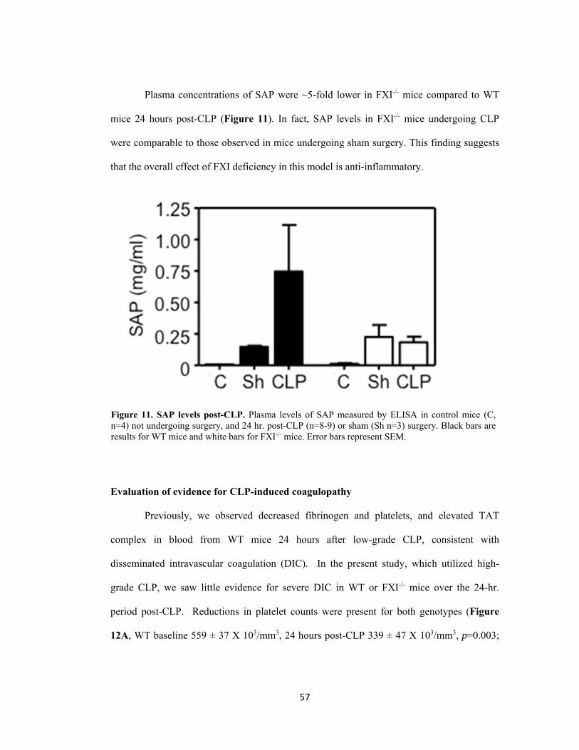
57
Plasma concentrations of SAP were ~5-fold lower in FXI-/- mice compared to WT
mice 24 hours post-CLP (Figure 11). In fact, SAP levels in FXI-/- mice undergoing CLP
were comparable to those observed in mice undergoing sham surgery. This finding suggests
that the overall effect of FXI deficiency in this model is anti-inflammatory.
Evaluation of evidence for CLP-induced coagulopathy
Previously, we observed decreased fibrinogen and platelets, and elevated TAT
complex in blood from WT mice 24 hours after low-grade CLP, consistent with
disseminated intravascular coagulation (DIC). In the present study, which utilized high-
grade CLP, we saw little evidence for severe DIC in WT or FXI-/- mice over the 24-hr.
period post-CLP. Reductions in platelet counts were present for both genotypes (Figure
12A, WT baseline 559 ± 37 X 103/mm3, 24 hours post-CLP 339 ± 47 X 103/mm3, p=0.003;
Figure 11. SAP levels post-CLP. Plasma levels of SAP measured by ELISA in control mice (C, n=4) not undergoing surgery, and 24 hr. post-CLP (n=8-9) or sham (Sh n=3) surgery. Black bars are results for WT mice and white bars for FXI-/- mice. Error bars represent SEM.

58
FXI-/- baseline 664 ± 34 X 103/mm3, 24 hours post-CLP 362 ± 37 X 103/mm3, p=0.001),
although the mean platelet counts remained above 300,000 cells/μL in both groups (Figure
12A). Platelet count reductions were significantly greater with CLP than with sham surgery
(Figure 12B). For both genotypes, plasma TAT levels did not increase significantly above
baseline (Figure 12C), and were not different from animals undergoing sham operations
(Figure 12D). Levels of fibrinogen decreased modestly over the first 4 to 8 hours post-CLP,
then increased in both genotypes (Figure 12E, WT: baseline 1.7 ± 0.2 mg/mL, 24 hrs. 3.3 ±
1.0 mg/mL; FXI-/-: baseline 1.7 ± 0.2, 24 hrs. 2.1 ± 0.4). The results at 24 hours for CLP-
treated mice and sham-treatment did not reach statistical significance for either genotype
(Figure 12F). These results are consistent with work from Patel et al. that showed large
increases in TAT in C57Bl6 mice after LPS administration, but not after CLP (Patel, Soubra
et al. 2010). While changes in fibrinogen were not significant for either genotype, there is an
impression that the level at 24 hrs. was somewhat greater in WT mice than in FXI-/- mice.
Fibrinogen levels rise with inflammation, and the modest difference could reflect the greater
inflammatory response induced by CLP in WT mice.

59
Figure 12. Effects of CLP on markers of coagulation. (A, B) Whole blood platelet counts, (C, D) plasma thrombin-antithrombin complex (TAT) levels, and (E, F) plasma fibrinogen levels in WT (black bars) and FXI-/- (white bars) mice after CLP or sham surgery. Panels A, C and E show values various times post-CLP. Panels B, D and F compare values 24 hour post-CLP or sham (Sh) surgery to 0 hr. controls. CLP induced significant platelet reductions in WT (*p=0.003) and FXI-/- (**p=0.001) mice. For panels A to F, error bars represent SEM.

60
At necropsy 24 hours post-CLP, lymphocyte apoptosis, a common finding in murine
sepsis (Lang and Matute-Bello 2009), was noted in spleen and thymus for both genotypes
(Figure 13). There was no evidence of hemorrhage. Histologic analysis of multiple organs
including liver, kidney, brain, spleen, and thymus (Figures 13 and 14) did not reveal
microvascular thrombus accumulation, which is considered essential for diagnosing DIC in
laboratory animals (Berthelsen, Kristensen et al. 2011). Cumulatively, these data are
consistent with published studies indicating that DIC is not a prominent feature of sepsis in
mice during the first 24 hours after high-grade CLP (Ganopolsky and Castellino 2004, Patel,
Soubra et al. 2010).
Figure 13. Histology of thymus and spleen. Photomicrographs (400X magnification) of paraffin embedded sections of spleen and thymus stained with hematoxylin and eosin before, and 24 hours after, CLP in WT and FXI-/- mice. Evidence of lymphocyte apoptosis (indicated by white arrows) was present in both organs for mice of both genotypes 24 hrs. post-CLP.

61
62 Figure 14. Histology of liver, brain, and kidney. Photomicrographs (400X magnification) of paraffin embedded sections of mouse liver, kidney and brain stained with hematoxylin and eosin before, and 24 hours after, CLP in WT and FXI-/- mice. There was no evidence of disseminated intravascular coagulation in animals of either genotype following cecal ligation and puncture.

62
Effects of CLP on plasma levels of contact protease zymogens
During contact activation-initiated coagulation in vitro, the plasma zymogen FXII is
activated on a surface to generate the protease FXIIa, which cleaves PK and FXI to release
-kal and FXIa, respectively (Bjorkqvist, Nickel et al. 2014, Schmaier 2014, Wu 2015).
According to current models of contact activation, α-kal amplifies the process by
reciprocally activating FXII and also contributes to inflammation by cleaving HK to release
the pro-inflammatory peptide BK. FXIa augments thrombin generation by activating FIX,
but is not considered important for FXII activation.
During sepsis, blood may be exposed to a variety of substances that induce contact
activation, including polyphosphate (polyP) released from platelets (Muller, Mutch et al.
2009) or microorganisms (Herwald, Morgelin et al. 2003, Frick, Bjorck et al. 2007, Yun and
Morrissey 2009), and extracellular nucleic acids including DNA and RNA from damaged
tissue and chromatin extruded from activated neutrophils (neutrophil extracellular
traps)(Esmon 2013, Gould, Lysov et al. 2015). Noting that in vivo activation of contact
system proteins has been assessed by measuring reductions in plasma levels of their
respective zymogens (Oschatz, Maas et al. 2011), we measured changes in plasma FXII, PK,
and FXI levels to assess contact system activation. In WT mice, FXI decreased by ~50% by
8 hours after CLP (Figures 15A and 15B), and PK was comparably reduced by 24 hours
(Figures 15B and 15C). While FXII levels remained similar to baseline in WT mice post-
CLP, we observed a modest increase in plasma FXII levels in FXI-/- mice (Figure 15D).
FXII levels were also moderately increased in sham-treated animals (not shown), suggesting
a reaction to surgery. The data, therefore, indicate that there is activation and consumption of
FXII in WT mice after CLP that was not as prominent in FXI-/- mice. Interestingly, PK
levels were not reduced 24 hours post-CLP in FXI-/- mice (Figure 15D). Taken as a whole,

63
the data are consistent with activation of the contact proteases during CLP-induced sepsis in
WT mice, but suggest that FXI deficiency blunts this process.
Figure 15. The effect of CLP on plasma contact proteases. Plasma FXI, PK and FXII levels at various times after high-grade CLP were determined using densitometry of western blots and are reported as percent of baseline (0 hr.) control. (A) Plasma FXI levels after CLP in WT mice (B) Examples of western blots for FXI and PK for WT mouse plasma at various times after CLP (upper panels of each pair) or sham surgery (lower panels of each pair). (C) Plasma PK levels after high-grade CLP in WT (black bars) or FXI-/- (white bars) mice. The asterisk above the 24-hr. bar for PK in WT animals indicates the value is significantly different than 0 hr. control (p<0.05). (D) Plasma FXII levels after high-grade CLP in WT (black bars) or FXI-/- (white bars) mice. In panels A, C, and D, each bar represents data for eight mice. For all panels, error bars represent SEM.

64
FXII activation by α-kal and FXIa
To explore the possibility that FXI may have contributed to contact activation in our
study, we first evaluated FXII activation by α-kal or FXIa in vitro in the presence of the
polyanions DNA, RNA, and polyphosphate. FXII was converted to FXIIa at comparable
relatively low rates by -kal and FXIa in the absence of a polyanion (Figure 16A). The
FXIa-mediated reaction was enhanced 56-fold by DNA (Figure 16B) and 90-fold by RNA
(Figure 16C). With -kal, DNA and RNA enhanced FXII activation modestly (2.8 and 5.6-
fold, respectively). In reactions with polyP, contributions of -kal and FXIa to FXII
activation appeared comparable, but were difficult to assess because FXII autoactivation was
prominent (Figure 16D). FXI and PK are homologs (Emsley, McEwan et al. 2010), and can
be difficult to separate chromatographically when purified from plasma. To address the
concern that FXIa was contaminated with -kal, we ran reactions in the presence of an anti-
kallikrein IgG (H03) that binds to the protease active site (Kenniston, Faucette et al. 2014).
H03 blocked cleavage of chromogenic substrate S-2302 by α-kal, but not FXIa or FXIIa
(Figure 17A). H03 blocked FXII activation by -kal (Figure 17B) but did not affect FXII
activation by FXIa (Figure 17C), thus illustrating that FXIa can cleave FXII under
conditions that block the enzymatic activity of α-kal.
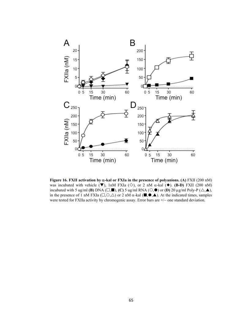
65
Figure 16. FXII activation by -kal or FXIa in the presence of polyanions. (A) FXII (200 nM) was incubated with vehicle (), 1nM FXIa (), or 2 nM -kal (). (B-D) FXII (200 nM) incubated with 5 ug/ml (B) DNA (,), (C) 5 ug/ml RNA (,) or (D) 20 g/ml Poly-P (,), in the presence of 1 nM FXIa (,,) or 2 nM -kal (,,). At the indicated times, samples were tested for FXIIa activity by chromogenic assay. Error bars are +/ one standard deviation.

66
Figure 17. Effect of anti-kallikrein antibody H03. (A) FXIIa (100 nM), -kal (5 nM) or FXIa (5 nM) were incubated at room temperature for 3 min with 10-fold molar excess of anti-kal antibody (H03) respectively or control vehicle (C) in a buffer with 10 M ZnCl2, and residual activity was determined by chromogenic substrate. Error bars are +/ one standard deviation. (B) -al (5 nM) and 10 M ZnCl2 were incubated with the chromogenic substrate S-2302 (200 uM) in the presence of vehicle (), 100 nM H03 () or 100 nM H03 and 10 ug/ml DNA () and changes in OD 405 nm were monitored. (C) FXII (200 nM) and 10 M ZnCl2 were incubated with vehicle (), 5 nM -kal (), 5 nM -kal and 100 nM H03 (), or 5 nM -kal with 100 nM H03 and 10 ug/ml DNA () in the presence of S-2302 (200 uM). Changes in OD 405 nm were monitored. (D) FXII (200 nM) and 10 M ZnCl2 were incubated with vehicle (), 5 nM FXI (), 5 nM FXI and 10 ug/ml DNA (), or 5 nM FXI and 10 ug/ml DNA with 100 nM H03 () in the presence of S-2302 (200 uM). Changes in OD 405 nm were monitored

67
Three-quarters of PK and nearly all FXI in plasma circulates as a complex with the
glycoprotein HK (Mandle, Colman et al. 1976, Thompson, Mandle et al. 1977, Thompson,
Mandle et al. 1979). HK is thought to be required for proper binding of PK and FXI to the
contact surface during contact activation. Consistent with the importance of HK during
contact activation on non-biologic surfaces such as kaolin (Meier, Pierce et al. 1977,
Wiggins, Bouma et al. 1977), HK enhanced FXII activation by -kal with polyP (Figure
18A), DNA (Figure 18B) or RNA (Figure 18C). In contrast, HK had no appreciable effect
on FXII activation by FXIa in the presence of polyP (Figure 18D), and a modest inhibitory
Figure 18. Effect of HK on FXII activation in the presence of polyanions. Plasma FXII (200 nM) was incubated with (A-C) 2 nM -kal or (D-F) 1 nM FXIa and (A, D) 20 g/ml poly-P, (B, E) 5 g/ml DNA or (C, F) 5 g/ml RNA in the absence (,,) or presence (,,) of 20 nM HK. At the indicated times, samples were tested for FXIIa activity by chromogenic assay. Error bars are +/ one standard deviation.
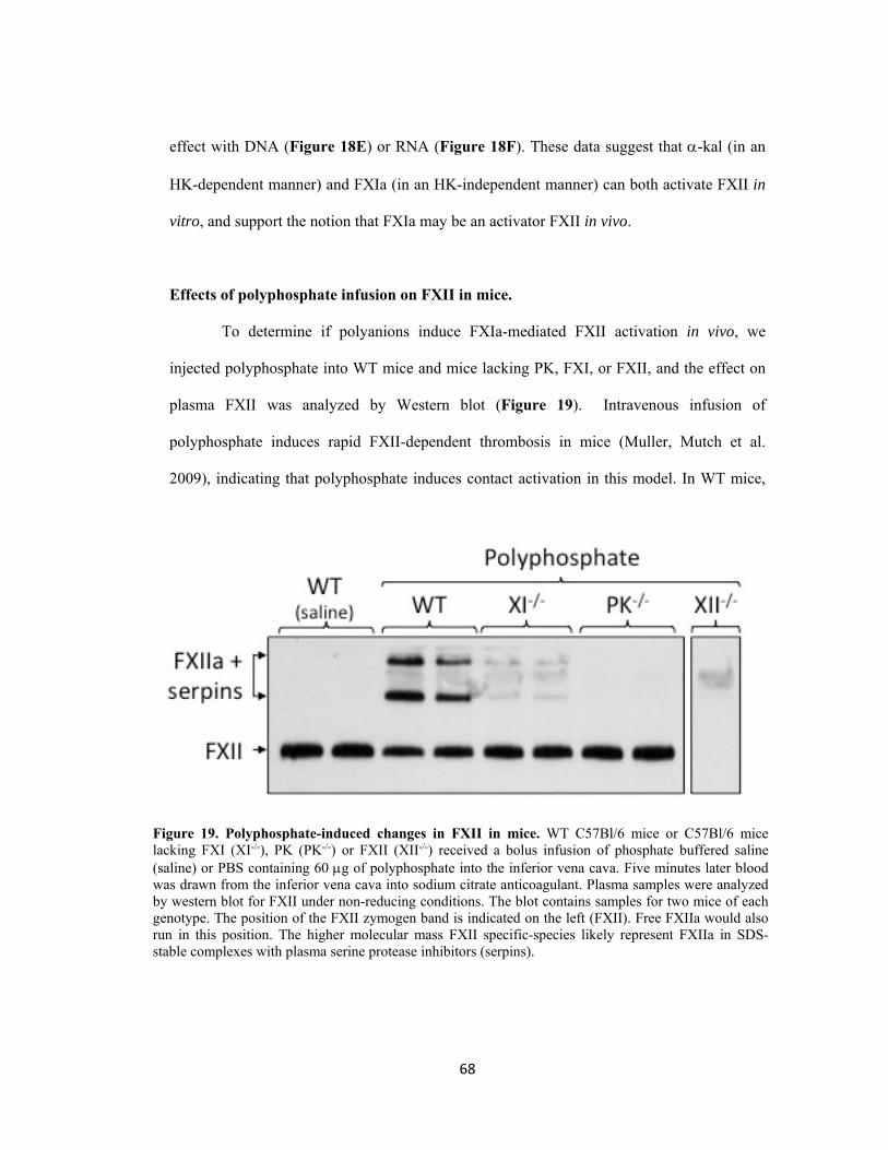
68
effect with DNA (Figure 18E) or RNA (Figure 18F). These data suggest that -kal (in an
HK-dependent manner) and FXIa (in an HK-independent manner) can both activate FXII in
vitro, and support the notion that FXIa may be an activator FXII in vivo.
Effects of polyphosphate infusion on FXII in mice.
To determine if polyanions induce FXIa-mediated FXII activation in vivo, we
injected polyphosphate into WT mice and mice lacking PK, FXI, or FXII, and the effect on
plasma FXII was analyzed by Western blot (Figure 19). Intravenous infusion of
polyphosphate induces rapid FXII-dependent thrombosis in mice (Muller, Mutch et al.
2009), indicating that polyphosphate induces contact activation in this model. In WT mice,
Figure 19. Polyphosphate-induced changes in FXII in mice. WT C57Bl/6 mice or C57Bl/6 mice lacking FXI (XI-/-), PK (PK-/-) or FXII (XII-/-) received a bolus infusion of phosphate buffered saline (saline) or PBS containing 60 g of polyphosphate into the inferior vena cava. Five minutes later blood was drawn from the inferior vena cava into sodium citrate anticoagulant. Plasma samples were analyzed by western blot for FXII under non-reducing conditions. The blot contains samples for two mice of each genotype. The position of the FXII zymogen band is indicated on the left (FXII). Free FXIIa would also run in this position. The higher molecular mass FXII specific-species likely represent FXIIa in SDS-stable complexes with plasma serine protease inhibitors (serpins).

69
infusion of polyP, but not saline, caused a modest decrease in intensity of the band
representing FXII, and produced higher molecular weight species that likely represent FXIIa
in SDS-stable complexes with serpins such as C1-inhibitor and antithrombin. There were no
bands on blots with plasma from polyP treated FXII-deficient mice, indicating the high
molecular mass bands on blots for WT mice are specific FXII/XIIa signals. No changes were
apparent in the FXII band in PK-deficient mice after polyP infusion, and no higher
molecular mass species appeared. This is consistent with the impression that PK is required
for normal FXII turnover in healthy mice (Revenko, Gao et al. 2011). Surprisingly, FXI
deficiency also affected polyphosphate-induced changes in FXII, although perhaps not quite
as profoundly as did PK deficiency. Bands for the higher molecular weight species were
markedly less intense in blots for FXI-/- mice than for WT mice. These data support the
hypothesis that -kallikrein and FXIa are required for optimal FXII activation after
polyphosphate infusion in mice, and are consistent with the notion that FXI deficiency may
interfere with activation of contact proteases in the CLP model.
Discussion
The protease FXIa, which plays a relatively modest role in hemostasis, has been
identified as contributing to pathologic coagulation in humans (Meijers, Tekelenburg et al.
2000, Doggen, Rosendaal et al. 2006, Suri, Yamagishi et al. 2010, Bane and Gailani 2014,
Key 2014). Consistent with this, we previously reported evidence for DIC in WT mice after
CLP but not in similarly treated FXI-/- mice, or WT mice receiving the anti-FXI IgG 14E11
(Tucker, Gailani et al. 2008, Tucker, Verbout et al. 2012). We concluded that the survival
advantage after CLP conferred by absence of FXI was primarily due to blunting of sepsis-
induced coagulation. However, WT mice treated with 14E11 also had lower plasma levels of

70
TNFα and IL-6 twelve hours post-CLP than vehicle-treated mice (Tucker, Verbout et al.
2012), suggesting FXI inhibition affected cytokine responses. The study presented here
explored the importance of FXI in early cytokine and coagulation responses after CLP.
The survival data from WT mice show that mortality increased as injury severity
increased. This is in keeping with the literature, which indicates that mortality after CLP
correlates with the proportion of the cecum that is ligated (Rittirsch, Huber-Lang et al. 2009,
Toscano, Ganea et al. 2011). In contrast to this paradigm, the data show that FXI deficiency
confers a survival advantage after high-grade, but not low grade, CLP. This suggests the
contribution of FXI is more prominent in severe sepsis. The findings are consistent with a
previous observation that FXI-/- mice had better survival when exposed to a dose of Listeria
monocytogenes that was uniformly lethal in WT mice, but not after inoculation with a lower
concentration of organisms (Luo, Szaba et al. 2012). It is unclear why FXI deficiency did
not reduce mortality with low-grade injury in the current study. In contrast to the earlier
study, WT and FXI-/- mice were littermates in the work presented here. It is plausible that
differences in gut flora between separate inbred lines of WT and FXI-/- affected earlier
results. In septic patients, death is likely due to a constellation of factors, including
hypotension, the systemic inflammatory response, DIC, and the specific microorganism
involved (Russell 2006). We did not determine causes of death post-CLP, but previous work
provides some insight. Death in our study occurred during the “early” or “acute” phase
(within the first 4 or 5 days) of sepsis (Osuchowski, Welch et al. 2006, Xiao, Siddiqui et al.
2006). Deaths during this phase correlate with elevations in pro- and anti-inflammatory
cytokines (Osuchowski, Welch et al. 2006). The altered cytokine pattern after high-grade
CLP in FXI-/- mice suggests that a FXI contribution to systemic inflammation figures
prominently in mortality in WT animals.

71
The difference in levels of the acute phase protein SAP in WT and FXI-/- mice 24
hours post-CLP suggests that the altered cytokine response in FXI-/- mice was associated
with a less robust inflammatory response compared to WT mice. Previous work with mouse
models indirectly suggested that FXI contributions to inflammation. Mice lacking the key
coagulation regulatory protease protein C typically die in utero from extensive thrombosis.
Chan and coworkers (Chan, Ganopolsky et al. 2001) reported that superimposing total FXI
deficiency on protein C deficiency partially overcomes this phenotype, with some animals
living to adulthood. Interestingly, despite the well described anti-inflammatory effect of
protein C, mice lacking both protein C and FXI had similar plasma and tissue TNF and IL-
6 levels to littermates with normal protein C levels. Kleinschnitz and co-workers
(Kleinschnitz, Stoll et al. 2006) showed that mice lacking either FXII or FXI display a
marked reduction in cerebral ischemia-reperfusion injury compared to WT mice after
temporary occlusion of the middle cerebral artery. The results were attributed to reduced
fibrin deposition in the null animals, but injury in this model is known to have a significant
inflammatory component (Carmichael 2005). The mechanisms by which FXI contributes to
inflammation in the CLP-model are not clear. In earlier work with a low-grade injury CLP
model, we noted evidence of DIC in WT mice that was not present in FXI-deficient animals,
raising the possibility that the pro-inflammatory effects of FXI are related to its well-
characterized role in generating thrombin. Surprisingly, we did not observe evidence for
consumptive coagulation during the 24 hours post-CLP in WT or FXI-/- mice; findings that
are discrepant with our prior experience. Several factors may have contributed to the
different results. First, CLP, unlike LPS, does not consistently induce DIC in mice
(Ganopolsky and Castellino 2004, Patel, Soubra et al. 2010). It is also possible that
differences in gut flora between inbred lines were a factor in our original studies.

72
Alternatively, our earlier studies used a lower grade injury than was used in the present
work, and differences in injury level may create models with different features (Rittirsch,
Huber-Lang et al. 2009). These issues aside, an important point that can be taken from the
cumulative experience is that FXI deficiency confers a survival advantage after CLP,
regardless of the presence or absence of DIC. This in turn suggests that the detrimental
effects of FXI in this model are not primarily tied to consumptive coagulation.
Our findings indicate that it is likely that FXI makes thrombin-independent
contributions to inflammation. For example, while FXIa is classically considered a product
of contact activation, and not a contributor to the process itself, our results raise the
possibility that FXIa may contribute to the turnover of the two main zymogens (FXII and
PK) involved in contact activation. During contact activation, FXIIa converts PK to -kal,
which then cleaves HK, liberating the nonapeptide BK, and forming cleaved kininogen
(HKa) and antimicrobial peptides. BK is a potent vasodilator and inducer of vascular
permeability (Couture, Blaes et al. 2014, Sharma 2014), and contact activation-induced
hypotension may be an important contributor to mortality in sepsis. Contact activation is
probably triggered by traces of FXIIa and/or -kal present in plasma, and are enhanced by
substances such as the polyanions used in our experiments. HK is an essential co-factor for
the process, facilitating PK binding to the surface (Meier, Pierce et al. 1977, Wiggins,
Bouma et al. 1977). Our data indicate FXIa activates FXII in the presence of certain types of
surfaces in an HK-independent manner. In 1978 Griffin reported that FXIa activates FXII on
celite (diatomaceous earth), although significantly more slowly than the -kal-mediated
reaction (Griffin 1978). In our experiments with polyP or nucleic acids, FXIa was a
significantly better activator of FXII than -kal in the absence of HK, with the difference
largely disappearing when HK was included in reactions. The reason for this is not certain,

73
but FXI does contain anion-binding sites not found on PK that may allow it to interact
properly with polyanions in the absence of HK (Geng, Verhamme et al. 2013). In addition
to the effects mentioned above, products of contact activation may also contribute to
increased cytokine levels. FXIIa (Toossi, Sedor et al. 1992) and peptides derived from HK
cleavage (Khan, Bradford et al. 2006) can elicit cytokine release in human mononuclear
cells. FXIa may indirectly contribute to a cytokine response by affecting cleavage of these
contact proteins.
Targeting thrombin generation with anticoagulants such as heparin appears to be of
some benefit in patients with severe sepsis (Wang, Chi et al. 2014), probably by limiting
tissue damage from thrombosis, DIC, and perhaps inflammation. However, such therapy
increases bleeding in patients who are already coagulopathic, limiting its applicability. The
anti-inflammatory effects of activated protein C (rhAPC, a.k.a., Xigris®) in sepsis were
probably partially offset by increased bleeding due to proteolysis of factors Va and VIIIa
(Warren, Suffredini et al. 2002). Our work suggests that FXI inhibition may provide a
benefit early in sepsis by altering the inflammatory response. While the CLP-model used in
the current study did not include a major coagulopathic component, earlier work suggests
that FXI inhibition may also blunt the DIC that frequently accompanies severe sepsis in
humans. The importance of FXI to hemostasis in the setting of sepsis in humans remains to
be established, but based on clinical observations of patients with congenital FXI deficiency,
FXI inhibition would likely be safer than anticoagulation with heparin.

74
Additional Experiments
Effect of CLP on plasma C5a and chemokine levels.
The blunted early cytokine response in FXI-/- mice undergoing CLP raises the
possibility of a suboptimal initial immune response to infection. With this in mind, we
measured factors known to affect leukocyte migration in response to CLP. C5a is a
proteolytic fragment of the complement protein C5 that enhances neutrophil migration and
adherence. It is also an anaphylatoxin that has been linked to deleterious outcomes in
experimental sepsis (Ward 2010). As FXIa cleaves C5 to form C5a in vitro (Amara, Flierl et
al. 2010), we measured plasma C5a levels by ELISA (R & D Systems, Minneapolis, MN) 24
hours after CLP or sham surgery in WT and FXI-/- mice. Both types of surgery induced
comparable elevations of plasma C5a 24 hours post-procedure in WT and FXI-/- mice
(Figure 20A). Levels of the murine neutrophil chemoattractants KC (Figure 20B) and MIP-
2 (Figure 20C) were elevated in WT and FXI-/- mice within four hours of CLP (Figures 20E
& 20G), with levels reaching comparable levels by 24 hours. We also measured levels of the
monocyte chemoattractant protein MCP-1, which is reported to affect leukocyte recruitment
through a mechanism that is at least partly independent of MIP-2 and KC (Matsukawa,
Hogaboam et al. 1999). While MCP-1 levels were somewhat lower in FXI-/- mice compared
to WT animals at 4 and 24 hours after CLP (Figure 20D) the difference was not statistically
significant. Cumulatively, the data indicate that processes that stimulate leukocyte migration
are largely intact in FXI-/- mice, despite the blunted initial cytokine response.

75
Leukocyte migration into the peritoneal cavity
Since the chemokine response appeared to be intact, we assessed leukocyte
infiltration into the peritoneum after high-grade CLP (Figure 21). We noted significant
increases in total leukocytes in the peritoneal cavity in both WT (baseline 9 ± 0.7 X 10³ cells,
24 hours post-CLP 50 ± 12 X 103, p=0.0007) and FXI-/- (baseline 9 ±1.1 X 10³ cells, 24
hours post-CLP 29 ± 4 X 10³ p=0.0002) mice 24 hours post-CLP, (Figure 21A). While the
increase appeared to be somewhat greater in WT mice (50 ± 12 X 103 for WT vs. 29 ± 4 X
103 for FXI-/-), the difference was not significant (p=0.4), a trend that was repeated in the
Figure 20. Complement component C5a and chemokine responses to CLP. For all panels, results for WT mice are represented by black bars and for FXI-/- mice by white bars. C5a levels were measured by ELISA, while chemokine levels were measured by Luminex technology. (A) Plasma levels of C5a in control mice (C) not undergoing surgery, and 24 hr. post-CLP or sham (Sh) surgery. For both genotypes, sham surgery and CLP both caused significant increases in C5a compared to untreated control (C) mice (p<0.05); however, differences in C5a after CLP or sham surgery were not significant for either genotype, nor between genotypes. Plasma levels of KC (B), MCP-1 (C), and MIP-2 (D) at various time points after CLP.

76
various leukocyte subsets [neutrophils (Figure 21B), lymphocytes (Figure 21C), and
monocytes (Figure 21D]. Notably, there was a significant increase in lymphocyte infiltration
above baseline in WT mice (baseline 6 0.5 X 10³ cells, 24 hrs. post-CLP 14 4 X 10³
cells, p=0.008) that was not observed in FXI-/- mice (baseline 6 ± 0.7 X 10³ cells, 24 hrs.
post-CLP 8 ± 1 X 10³ cells, p=0.2) (Figure 21C). Taken as a whole, the data do not support
a clear influence of FXI on leukocyte migration after CLP, but do introduce the possibility
that lymphocyte response may be more robust in WT mice.
Figure 21. Peritoneal leukocyte infiltration after CLP. Leukocyte counts in fluid aspirated after peritoneal wash with 5ml NaCl, 2mM EDTA, as measured by an automated hematology analyzer (Hemavet 950FS). Data are from 24 hours after CLP in WT (black bars) and FXI-/-
(white bars) mice compared to untreated controls (noted by the letter “C”) and to samples taken 24 hours after sham (Sh) surgery. (A) Total leukocyte counts were significantly greater than control for both WT (p=0.0007) and FXI-/- (p=0.0002) mice. This was also true for (B) neutrophils and (D) monocytes. However, only WT mice had a significant elevation in lymphocyte counts (*p=0.0008) compared to controls. For all panels, error bars represent SEM.

77
Evaluation of microorganisms during CLP-induced sepsis.
As mortality after CLP has been associated with bacteremia (Cuenca, Delano et al.
2010), we performed aerobic and anaerobic CFU counts on blood collected 24 hours after
CLP or sham surgery in groups of 6 mice to see if differences in the level of bacteremia
might explain the survival difference between WT and FXI-/- mice. No bacteria were
cultured from blood obtained from either group after sham surgery, while 4 of 6 WT and 6
of 6 FXI-/- mice had positive cultures for either aerobic or anaerobic bacteria 24 hours after
CLP. There was no difference in CFU counts between WT and FXI-/- mice in this study
(Figure 22).
Figure 22. Evaluation of microorganisms during CLP-induced sepsis. Whole blood obtained 24 hours post-CLP or sham surgery was diluted 1:10 in pre-reduced anaerobically sterilized (PRAS) media (Anaerobe Systems, Morgan Hill, CA) (Hyde, Stith et al. 1990) for aerobic and anaerobic bacteria colony forming unit (CFU) counts. Samples were processed by 10-fold serial dilution in PBS, pH 7.00. Aliquots (100 l) of each dilution were plated on BBL™ Columbia Sheep Blood Agar (SBA), BBL™ CDC Anaerobic SBA, and BBL™ CDC Anaerobic SBA with kanamycin and vancomycin. Columbia SBA was incubated aerobically at 35.0 C, while other plates were incubated anaerobically at 35.0 C for 48 hours. CFU counts were recorded for plates containing 30-300 CFUs. Data are presented as combined aerobic and anaerobic CFU counts in WT and FXI-/- mice. There were
diff i C b b d d d i i

78
References Amara, U., M. A. Flierl, D. Rittirsch, A. Klos, H. Chen, B. Acker, U. B. Bruckner, B. Nilsson, F. Gebhard, J. D. Lambris and M. Huber-Lang (2010). "Molecular intercommunication between the complement and coagulation systems." J Immunol 185(9): 5628-5636.
Angus, D. C., W. T. Linde-Zwirble, J. Lidicker, G. Clermont, J. Carcillo and M. R. Pinsky (2001). "Epidemiology of severe sepsis in the United States: analysis of incidence, outcome, and associated costs of care." Crit Care Med 29(7): 1303-1310.
Bane, C. E., Jr. and D. Gailani (2014). "Factor XI as a target for antithrombotic therapy." Drug Discov Today 19(9); 1454-8.
Berthelsen, L. O., A. T. Kristensen and M. Tranholm (2011). "Animal models of DIC and their relevance to human DIC: a systematic review." Thromb Res 128(2): 103-116.
Bjorkqvist, J., K. F. Nickel, E. Stavrou and T. Renne (2014). "In vivo activation and functions of the protease factor XII." Thromb Haemost 112(5): 868-875.
Carmichael, S. T. (2005). "Rodent models of focal stroke: size, mechanism, and purpose." NeuroRx 2(3): 396-409.
Chan, J. C., J. G. Ganopolsky, I. Cornelissen, M. A. Suckow, M. J. Sandoval-Cooper, E. C. Brown, F. Noria, D. Gailani, E. D. Rosen, V. A. Ploplis and F. J. Castellino (2001). "The characterization of mice with a targeted combined deficiency of protein c and factor XI." Am J Pathol 158(2): 469-479.
Cheng, Q., E. I. Tucker, M. S. Pine, I. Sisler, A. Matafonov, M. F. Sun, T. C. White-Adams, S. A. Smith, S. R. Hanson, O. J. McCarty, T. Renne, A. Gruber and D. Gailani (2010). "A role for factor XIIa-mediated factor XI activation in thrombus formation in vivo." Blood 116(19): 3981-3989.
Couture, R., N. Blaes and J. P. Girolami (2014). "Kinin receptors in vascular biology and pathology." Curr Vasc Pharmacol 12(2): 223-248.
Cuenca, A. G., M. J. Delano, K. M. Kelly-Scumpia, L. L. Moldawer and P. A. Efron (2010). "Cecal ligation and puncture." Curr Protoc Immunol Chapter 19: Unit 19 13.
Dellinger, R. P., M. M. Levy, A. Rhodes, D. Annane, H. Gerlach, S. M. Opal, J. E. Sevransky, C. L. Sprung, I. S. Douglas, R. Jaeschke, T. M. Osborn, M. E. Nunnally, S. R. Townsend, K. Reinhart, R. M. Kleinpell, D. C. Angus, C. S. Deutschman, F. R. Machado, G. D. Rubenfeld, S. Webb, R. J. Beale, J. L. Vincent, R. Moreno and S. Surviving Sepsis Campaign Guidelines Committee including The Pediatric (2013). "Surviving Sepsis Campaign: international guidelines for management of severe sepsis and septic shock, 2012." Intensive Care Med 39(2): 165-228.

79
Doggen, C. J., F. R. Rosendaal and J. C. Meijers (2006). "Levels of intrinsic coagulation factors and the risk of myocardial infarction among men: Opposite and synergistic effects of factors XI and XII." Blood 108(13): 4045-4051.
Emsley, J., P. A. McEwan and D. Gailani (2010). "Structure and function of factor XI." Blood 115(13): 2569-2577.
Esmon, C. T. (2013). "Molecular circuits in thrombosis and inflammation." Thromb Haemost 109(3): 416-420.
Frick, I. M., L. Bjorck and H. Herwald (2007). "The dual role of the contact system in bacterial infectious disease." Thrombosis and haemostasis 98(3): 497-502.
Gailani, D., N. M. Lasky and G. J. Broze, Jr. (1997). "A murine model of factor XI deficiency." Blood coagulation & fibrinolysis : an international journal in haemostasis and thrombosis 8(2): 134-144.
Ganopolsky, J. G. and F. J. Castellino (2004). "A protein C deficiency exacerbates inflammatory and hypotensive responses in mice during polymicrobial sepsis in a cecal ligation and puncture model." Am J Pathol 165(4): 1433-1446.
Geng, Y., I. M. Verhamme, S. A. Smith, Q. Cheng, M. Sun, J. P. Sheehan, J. H. Morrissey and D. Gailani (2013). "Factor XI anion-binding sites are required for productive interactions with polyphosphate." J Thromb Haemost 11(11): 2020-2028.
Gould, T. J., Z. Lysov and P. C. Liaw (2015). "Extracellular DNA and histones: double-edged swords in immunothrombosis." J Thromb Haemost 13 Suppl 1: S82-91.
Griffin, J. H. (1978). "Role of surface in surface-dependent activation of Hageman factor (blood coagulation factor XII)." Proc Natl Acad Sci U S A 75(4): 1998-2002.
Herwald, H., M. Morgelin and L. Bjorck (2003). "Contact activation by pathogenic bacteria: a virulence mechanism contributing to the pathophysiology of sepsis." Scand J Infect Dis 35(9): 604-607.
Hyde, S. R., R. D. Stith and R. E. McCallum (1990). "Mortality and bacteriology of sepsis following cecal ligation and puncture in aged mice." Infect Immun 58(3): 619-624.
Kenniston, J. A., R. R. Faucette, D. Martik, S. R. Comeau, A. P. Lindberg, K. J. Kopacz, G. P. Conley, J. Chen, M. Viswanathan, N. Kastrapeli, J. Cosic, S. Mason, M. DiLeo, J. Abendroth, P. Kuzmic, R. C. Ladner, T. E. Edwards, C. TenHoor, B. A. Adelman, A. E. Nixon and D. J. Sexton (2014). "Inhibition of plasma kallikrein by a highly specific active site blocking antibody." J Biol Chem 289(34): 23596-23608.
Key, N. S. (2014). "Epidemiologic and clinical data linking factors XI and XII to thrombosis." Hematology Am Soc Hematol Educ Program 2014(1): 66-70.

80
Khan, M. M., H. N. Bradford, I. Isordia-Salas, Y. Liu, Y. Wu, R. G. Espinola, B. Ghebrehiwet and R. W. Colman (2006). "High-molecular-weight kininogen fragments stimulate the secretion of cytokines and chemokines through uPAR, Mac-1, and gC1qR in monocytes." Arterioscler Thromb Vasc Biol 26(10): 2260-2266.
Kleinschnitz, C., G. Stoll, M. Bendszus, K. Schuh, H. U. Pauer, P. Burfeind, C. Renne, D. Gailani, B. Nieswandt and T. Renne (2006). "Targeting coagulation factor XII provides protection from pathological thrombosis in cerebral ischemia without interfering with hemostasis." J Exp Med 203(3): 513-518.
Lang, J. D. and G. Matute-Bello (2009). "Lymphocytes, apoptosis and sepsis: making the jump from mice to humans." Crit Care 13(1): 109.
Levy, M. M., M. P. Fink, J. C. Marshall, E. Abraham, D. Angus, D. Cook, J. Cohen, S. M. Opal, J. L. Vincent, G. Ramsay and Sccm/Esicm/Accp/Ats/Sis (2003). "2001 SCCM/ESICM/ACCP/ATS/SIS International Sepsis Definitions Conference." Crit Care Med 31(4): 1250-1256.
Luo, D., F. M. Szaba, L. W. Kummer, L. L. Johnson, E. I. Tucker, A. Gruber, D. Gailani and S. T. Smiley (2012). "Factor XI-Deficient Mice Display Reduced Inflammation, Coagulopathy, and Bacterial Growth during Listeriosis." Infect Immun 80(1): 91-99.
Mandle, R. J., R. W. Colman and A. P. Kaplan (1976). "Identification of prekallikrein and high-molecular-weight kininogen as a complex in human plasma." Proc Natl Acad Sci U S A 73(11): 4179-4183.
Matafonov, A., Q. Cheng, Y. Geng, I. M. Verhamme, O. Umunakwe, E. I. Tucker, M. F. Sun, V. Serebrov, A. Gruber and D. Gailani (2013). "Evidence for factor IX-independent roles for factor XIa in blood coagulation." J Thromb Haemost 11(12): 2118-2127.
Matsukawa, A., C. M. Hogaboam, N. W. Lukacs, P. M. Lincoln, R. M. Strieter and S. L. Kunkel (1999). "Endogenous monocyte chemoattractant protein-1 (MCP-1) protects mice in a model of acute septic peritonitis: cross-talk between MCP-1 and leukotriene B4." J Immunol 163(11): 6148-6154.
Meier, H. L., J. V. Pierce, R. W. Colman and A. P. Kaplan (1977). "Activation and function of human Hageman factor. The role of high molecular weight kininogen and prekallikrein." J Clin Invest 60(1): 18-31.
Meijers, J. C., W. L. Tekelenburg, B. N. Bouma, R. M. Bertina and F. R. Rosendaal (2000). "High levels of coagulation factor XI as a risk factor for venous thrombosis." N Engl J Med 342(10): 696-701.
Muller, F., N. J. Mutch, W. A. Schenk, S. A. Smith, L. Esterl, H. M. Spronk, S. Schmidbauer, W. A. Gahl, J. H. Morrissey and T. Renne (2009). "Platelet polyphosphates are proinflammatory and procoagulant mediators in vivo." Cell 139(6): 1143-1156.

81
Murphy, E. C., M. Morgelin, J. C. Cooney and I. M. Frick (2011). "Interaction of Bacteroides fragilis and Bacteroides thetaiotaomicron with the kallikrein-kinin system." Microbiology 157(Pt 7): 2094-2105.
Oschatz, C., C. Maas, B. Lecher, T. Jansen, J. Bjorkqvist, T. Tradler, R. Sedlmeier, P. Burfeind, S. Cichon, S. Hammerschmidt, W. Muller-Esterl, W. A. Wuillemin, G. Nilsson and T. Renne (2011). "Mast cells increase vascular permeability by heparin-initiated bradykinin formation in vivo." Immunity 34(2): 258-268.
Osuchowski, M. F., K. Welch, J. Siddiqui and D. G. Remick (2006). "Circulating cytokine/inhibitor profiles reshape the understanding of the SIRS/CARS continuum in sepsis and predict mortality." J Immunol 177(3): 1967-1974.
Patel, K. N., S. H. Soubra, F. W. Lam, M. A. Rodriguez and R. E. Rumbaut (2010). "Polymicrobial sepsis and endotoxemia promote microvascular thrombosis via distinct mechanisms." J Thromb Haemost 8(6): 1403-1409.
Pauer, H. U., T. Renne, B. Hemmerlein, T. Legler, S. Fritzlar, I. Adham, W. Muller-Esterl, G. Emons, U. Sancken, W. Engel and P. Burfeind (2004). "Targeted deletion of murine coagulation factor XII gene-a model for contact phase activation in vivo." Thromb Haemost 92(3): 503-508.
Revenko, A. S., D. Gao, J. R. Crosby, G. Bhattacharjee, C. Zhao, C. May, D. Gailani, B. P. Monia and A. R. MacLeod (2011). "Selective depletion of plasma prekallikrein or coagulation factor XII inhibits thrombosis in mice without increased risk of bleeding." Blood 118(19): 5302-5311.
Rittirsch, D., M. S. Huber-Lang, M. A. Flierl and P. A. Ward (2009). "Immunodesign of experimental sepsis by cecal ligation and puncture." Nat Protoc 4(1): 31-36.
Russell, J. A. (2006). "Management of sepsis." N Engl J Med 355(16): 1699-1713.
Schmaier, A. H. (2014). "Physiologic activities of the contact activation system." Thromb Res 133 Suppl 1: S41-44.
Sharma, J. N. (2014). "Basic and clinical aspects of bradykinin receptor antagonists." Prog Drug Res 69: 1-14.
Stavrou, E. X., C. Fang, A. Merkulova, O. Alhalabi, N. Grobe, S. Antoniak, N. Mackman and A. H. Schmaier (2015). "Reduced thrombosis in Klkb1-/- mice is mediated by increased Mas receptor, prostacyclin, Sirt1, and KLF4 and decreased tissue factor." Blood 125(4): 710-719.
Suri, M. F., K. Yamagishi, N. Aleksic, P. J. Hannan and A. R. Folsom (2010). "Novel hemostatic factor levels and risk of ischemic stroke: the Atherosclerosis Risk in Communities (ARIC) Study." Cerebrovasc Dis 29(5): 497-502.

82
Thompson, R. E., R. Mandle, Jr. and A. P. Kaplan (1977). "Association of factor XI and high molecular weight kininogen in human plasma." The Journal of clinical investigation 60(6): 1376-1380.
Thompson, R. E., R. Mandle, Jr. and A. P. Kaplan (1979). "Studies of binding of prekallikrein and Factor XI to high molecular weight kininogen and its light chain." Proc Natl Acad Sci U S A 76(10): 4862-4866.
Tizard, I. R. (2008). Veterinary immunology : an introduction. St. Louis, MO, Saunders, an imprint of Elsevier Inc.
Toossi, Z., J. R. Sedor, M. A. Mettler, B. Everson, T. Young and O. D. Ratnoff (1992). "Induction of expression of monocyte interleukin 1 by Hageman factor (factor XII)." Proc Natl Acad Sci U S A 89(24): 11969-11972.
Torres, M. B. and A. De Maio (2005). "An exaggerated inflammatory response after CLP correlates with a negative outcome." J Surg Res 125(1): 88-93.
Toscano, M. G., D. Ganea and A. M. Gamero (2011). "Cecal ligation puncture procedure." J Vis Exp(51).
Tucker, E. I., D. Gailani, S. Hurst, Q. Cheng, S. R. Hanson and A. Gruber (2008). "Survival advantage of coagulation factor XI-deficient mice during peritoneal sepsis." J Infect Dis 198(2): 271-274.
Tucker, E. I., U. M. Marzec, T. C. White, S. Hurst, S. Rugonyi, O. J. McCarty, D. Gailani, A. Gruber and S. R. Hanson (2009). "Prevention of vascular graft occlusion and thrombus-associated thrombin generation by inhibition of factor XI." Blood 113(4): 936-944.
Tucker, E. I., N. G. Verbout, P. Y. Leung, S. Hurst, O. J. McCarty, D. Gailani and A. Gruber (2012). "Inhibition of factor XI activation attenuates inflammation and coagulopathy while improving the survival of mouse polymicrobial sepsis." Blood.
Walley, K. R., N. W. Lukacs, T. J. Standiford, R. M. Strieter and S. L. Kunkel (1996). "Balance of inflammatory cytokines related to severity and mortality of murine sepsis." Infect Immun 64(11): 4733-4738.
Wang, C., C. Chi, L. Guo, X. Wang, L. Guo, J. Sun, B. Sun, S. Liu, X. Chang and E. Li (2014). "Heparin therapy reduces 28-day mortality in adult severe sepsis patients: a systematic review and meta-analysis." Crit Care 18(5): 563.
Ward, P. A. (2010). "Role of C5 activation products in sepsis." ScientificWorldJournal 10: 2395-2402.
Warren, H. S., A. F. Suffredini, P. Q. Eichacker and R. S. Munford (2002). "Risks and benefits of activated protein C treatment for severe sepsis." N Engl J Med 347(13): 1027-1030.

83
Wiggins, R. C., B. N. Bouma, C. G. Cochrane and J. H. Griffin (1977). "Role of high-molecular-weight kininogen in surface-binding and activation of coagulation Factor XI and prekallikrein." Proc Natl Acad Sci U S A 74(10): 4636-4640.
Wu, Y. (2015). "Contact pathway of coagulation and inflammation." Thromb J 13: 17.
Xiao, H., J. Siddiqui and D. G. Remick (2006). "Mechanisms of mortality in early and late sepsis." Infect Immun 74(9): 5227-5235.
Yau, J. W., P. Liao, J. C. Fredenburgh, A. R. Stafford, A. S. Revenko, B. P. Monia and J. I. Weitz (2014). "Selective depletion of factor XI or factor XII with antisense oligonucleotides attenuates catheter thrombosis in rabbits." Blood 123(13): 2102-2107.
Yun, T. H. and J. H. Morrissey (2009). "Polyphosphate and omptins: novel bacterial procoagulant agents." J Cell Mol Med 13(10): 4146-4153.
Zellweger, R., M. W. Wichmann, A. Ayala, S. Stein, C. M. DeMaso and I. H. Chaudry (1997). "Females in proestrus state maintain splenic immune functions and tolerate sepsis better than males." Crit Care Med 25(1): 106-110.

84
CHAPTER VI
CONCLUSIONS AND FUTURE DIRECTIONS
Factor XI is at a Junction Between Coagulation and Inflammation
The primary finding of this work is that FXI influences systemic inflammation
during murine polymicrobial sepsis through mechanisms that are not exclusively related to
thrombin generation, a finding that has not been reported to our knowledge. We present
compelling evidence that suggests this influence is at least partly due to an effect on contact
activation. Taken together with our earlier findings that assigned a more “traditional” role
for FXI in sepsis-induced DIC, the accumulated data place FXI at a junction between pro-
inflammatory and pro-coagulant mechanisms, where it is positioned to contribute to either or
both, depending on the nature of the insult. Figure 23 summarizes the various mechanisms
by which FXI may influence the two pathways. First, FXI is a component of the thrombin
generating mechanism that is triggered during hemostasis (Figure 23, panel 1). Here, FXI
is activated by thrombin, and the resulting FXIa cleaves FIX to sustain thrombin production
by participating in a feedback loop. Thrombin generation may ultimately amplify the
inflammatory response by, for example, enhancing cytokine production through PAR
cleavage on vascular endothelial cells and leukocytes (Figure 23, panel 3). FXIa could also
serve as a bridge between contact activation and thrombin generation (Figure 23, panel 2).
In this case, FXIa is generated during contact activation on polyanions (such as platelet or
bacterial polyphosphates, and nucleic acids) and bacterial surfaces, where it then exerts a
procoagulant effect by cleaving FIX. Our results indicate that FXIa also indirectly
contributes to inflammation by promoting the activation of the contact proteins FXII and PK,

85
Figure 23. FXI in thrombin generation and contact activation during sepsis. Thrombin Generation (Panel 1). Depicted are proteolytic reactions that generate thrombin at a site of vascular injury. The process is initiated by activation of factors X and IX by the factor VIIa/tissue factor (TF) complex. Vitamin-K dependent protease zymogens are shown in black type with the active protease forms indicated by a lower case “a”. The blue ovals represent cofactors. During hemostasis, FXI is converted to FXIa by thrombin (white arrow). FXIa then activates FIX (yellow arrow). Contact activation (Panel 2). On a surface, FXII and PK undergo reciprocal activation to FXIIa and -kal. HK serves as a cofactor for this reaction. FXIIa can promote thrombin generation by activating FXI (green arrow). -kal cleaves HK liberating bradykinin (BK) and antimicrobial peptides (AMPs). FXIa, can also contribute to contact activation through activation of FXII (gray arrow). Panels A and B list factors that could trigger or enhance thrombin generation (A) or contact activation (B). Panels 3, 4, and 5 list processes mediated by thrombin (3), FXIIa and HKa (4) or -kal (5) that could contribute to sepsis. Panel 6 lists some of the consequences of those processes.

86
perhaps resulting in BK-mediated vasodilation and tissue edema, as well as cytokine release
(Figure 23, panels 4 and 5).
Future Studies
More work is needed to dissect the precise role of FXIa in cytokine release and
inflammation, particularly considering earlier data, which suggest there are FXIIa-
independent mechanisms through which FXI/FXIa operates. Host responses in WT mice
treated with the anti-FXI monoclonal antibody 14E11, which blocks activation of FXI by
FXIIa (Kravtsov, Matafonov et al. 2009), were not identical to those of FXI deficient mice
after CLP or infection with lethal doses of L. monocytogenes (Tucker, Gailani et al. 2008,
Luo, Szaba et al. 2012, Tucker, Verbout et al. 2012). FXI deficient mice had a survival
advantage compared to WT mice after inoculation with high concentrations of L.
monocytogenes, but pre-treatment of WT mice with 14E11 was only protective in similarly-
infected mice when sub-therapeutic doses of ampicillin were given (Luo, Szaba et al. 2012).
One explanation for this is that FXI participates in host defense through mechanisms that do
not involve contact activation. One mechanism may be through a direct effect on leukocyte
trafficking, especially considering that peritoneal leukocyte counts were significantly lower
in FXI-/- mice compared to WT mice 24 hours after low-grade CLP (Tucker, Gailani et al.
2008), but not in CLP-operated WT mice after pre-treatment with 14E11 (Tucker, Verbout et
al. 2012). This introduces the possibility that FXI alters neutrophil migration in a FXIIa-
independent manner. FXI binds to neutrophils, and may affect neutrophil chemotaxis
(Itakura, Verbout et al. 2011) and activation (Coomber, Galligan et al. 1997). Although our
current study does not indicate a clear influence of FXI on leukocyte migrate into the
peritoneum, they do show a non-significant increase in the various leukocyte subsets in WT

87
mice compared to their FXI-/- littermates. We used an automated cell counter to determine
peritoneal leukocyte counts and obtained results that, although inconclusive, do not rule out
a role for FXI in leukocyte migration. With this in mind, future studies should employ flow
cytometry to more accurately determine peritoneal levels of both the myeloid (monocytes,
macrophages, and neutrophils) and lymphoid (CD4+ T cells, CD8+ T cells, NK cells, etc.)
leukocyte populations.
Our work does not exclude the possibility that FXI activation through the extrinsic
(TF) pathway contributes to pathology during sepsis. The CLP model does not cause overt
DIC in mice in our hands, but it is still possible that pathology from thrombin-mediated FXI
activation occurs. To further investigate this possibility, WT and FXI-/- mice should be
compared with FXII-/- and PK-/- mice in the CLP model to determine the influence of these
proteins on survival, thrombin generation, inflammation (measurement of plasma cytokines
and SAP, and peritoneal leukocyte counts), and bacterial dissemination. If survival rates
after CLP are comparable among FXI-/-, FXII-/-, and PK-/- animals, we may conclude that
contact activation-induced FXI activation is largely responsible for the mortality observed in
WT mice. An observation of improved survival in FXI-/- mice relative to FXII-/- and PK-/-
mice would suggest a more complex pathophysiology. It could indicate that FXI activation
is mediated through additional mechanisms, such as the TF pathway, and/or, that FXII and
PK are more important, relative to FXI, for the mounting of an effective host defense against
infection. Time course studies should follow the survival experiments so that the complex
host responses that contribute to morbidity and mortality during CLP-induced sepsis may be
further understood. For example, if mortality in FXII-/- or PK-/- results from increased
susceptibility to infection, we may expect to see elevated peritoneal and blood levels of
bacteria in these animals.

88
Clinical relevance
The clinical relevance of this work remains to be determined. Most sepsis therapies
that have shown promise in animal models do not advance to clinical trials, and those that do
often fail to show a benefit in humans (Deitch 1998). Anticoagulants are certainly no
exception to this trend. In the most recent update to the International Guidelines for the
Management of Severe Sepsis and Septic Shock (Dellinger, Levy et al. 2013), heparin
therapy for thrombus prophylaxis is the only recommended anticoagulant strategy, but this
therapy can only be used in septic patients that are not experiencing a coagulopathy. The
present study did not reproduce the apparent coagulopathy that was observed in previous
work with the CLP model (Tucker, Gailani et al. 2008, Tucker, Verbout et al. 2012).
However, the earlier studies, along with studies with mouse thrombosis models, do suggest
that targeting FXI may be safer than using heparin when treating patients that are in DIC.
Considering this, the major advantage of FXI as a target for sepsis therapy over other
anticoagulant strategies may be its ability to “first, do no harm.”
References
Coomber, B. L., C. L. Galligan and P. A. Gentry (1997). "Comparison of in vitro function of neutrophils from cattle deficient in plasma factor XI activity and from normal animals." Vet Immunol Immunopathol 58(2): 121-131.
Deitch, E. A. (1998). "Animal models of sepsis and shock: a review and lessons learned." Shock 9(1): 1-11.
Dellinger, R. P., M. M. Levy, A. Rhodes, D. Annane, H. Gerlach, S. M. Opal, J. E. Sevransky, C. L. Sprung, I. S. Douglas, R. Jaeschke, T. M. Osborn, M. E. Nunnally, S. R. Townsend, K. Reinhart, R. M. Kleinpell, D. C. Angus, C. S. Deutschman, F. R. Machado, G. D. Rubenfeld, S. A. Webb, R. J. Beale, J. L. Vincent, R. Moreno and S. Surviving Sepsis Campaign Guidelines Committee including the Pediatric (2013). "Surviving sepsis campaign: international guidelines for management of severe sepsis and septic shock: 2012." Crit Care Med 41(2): 580-637.

89
Itakura, A., N. G. Verbout, K. G. Phillips, R. H. Insall, D. Gailani, E. I. Tucker, A. Gruber and O. J. McCarty (2011). "Activated factor XI inhibits chemotaxis of polymorphonuclear leukocytes." J Leukoc Biol.
Kravtsov, D. V., A. Matafonov, E. I. Tucker, M. F. Sun, P. N. Walsh, A. Gruber and D. Gailani (2009). "Factor XI contributes to thrombin generation in the absence of factor XII." Blood 114(2): 452-458.
Luo, D., F. M. Szaba, L. W. Kummer, L. L. Johnson, E. I. Tucker, A. Gruber, D. Gailani and S. T. Smiley (2012). "Factor XI-Deficient Mice Display Reduced Inflammation, Coagulopathy, and Bacterial Growth during Listeriosis." Infect Immun 80(1): 91-99.
Tucker, E. I., D. Gailani, S. Hurst, Q. Cheng, S. R. Hanson and A. Gruber (2008). "Survival advantage of coagulation factor XI-deficient mice during peritoneal sepsis." J Infect Dis 198(2): 271-274.
Tucker, E. I., N. G. Verbout, P. Y. Leung, S. Hurst, O. J. McCarty, D. Gailani and A. Gruber (2012). "Inhibition of factor XI activation attenuates inflammation and coagulopathy while improving the survival of mouse polymicrobial sepsis." Blood.

90
APPENDIX
VALIDATION OF THE CECAL LIGATION AND PUNCTURE MODEL FOR EVALUATING THE CONTRIBUTION OF FXI DURING MURINE SEPSIS
Introduction
A multitude of animal sepsis models are described in the literature. These models
have been divided into three categories: exogenous administration of a viable pathogen (e.g.,
a bacteria), exogenous administration of a toxin (e.g. LPS), or techniques that alter the
permeability of the bowel (e.g., CLP) (Buras, Holzmann et al. 2005). Of these, CLP most
closely mimics the hemodynamic and cytokine response observed in humans with sepsis,
(Wichterman, Baue et al. 1980, Deitch 1998, Buras, Holzmann et al. 2005, Hubbard,
Choudhry et al. 2005, Dyson and Singer 2009, Dejager, Pinheiro et al. 2011), probably
because it provides a continuous source of endogenous gut flora instead of a massive bolus
of laboratory strains of bacteria or toxin (Deitch 1998). Additionally, because sepsis in
humans typically arises from a local infection that subsequently becomes systemic (Freise,
Bruckner et al. 2001), the infection that is induced by CLP is also considered to be more
clinically relevant. Considering this, we chose the CLP model to evaluate the role of FXI
during sepsis. This model was new to our laboratory, requiring us to perform several
preliminary studies. These studies in WT C57Bl/6 mice allowed us to refine our technique
(and thus improve reproducibility), evaluate survival after CLP, and identify which
inflammatory mediators change reliably during sepsis. The pilot studies also helped us to
gain insight into any histologic changes that are apparent during sepsis.
Our pilot studies of WT C57Bl/6 mice featured an in-depth examination of the
cytokine response, along with a cursory examination of the impact of CLP on the
coagulation system. Of note is that we examined a greater range of time points (2, 4, 8, 12,

91
24, and 48 hours after CLP) in the pilot study, while we limited our investigations to 4, 8,
and 24 hours after CLP in the full time course study. Pilot studies were conducted using
low-grade CLP as, at this stage in our investigation, we had yet to discover that FXI-/- mice
only exhibited a survival advantage after high-grade CLP in our hands. Indeed, this
observation is what led us to use high-grade CLP in follow-on time course studies, and
contributed to our decision to limit the latter studies to the first 24 hours of CLP-induced
sepsis. Our survival studies predicted that a large proportion of animals would succumb to
sepsis within 48 hours after high-grade CLP, so that too few animals would be available for
investigating the inflammatory and coagulation system response at this stage of sepsis. The
lower grade of injury in the pilot study may have resulted in a disease process that was
different than what we observed with high grade injury (Rittirsch, Huber-Lang et al. 2009),
but it did allow us to look at changes at a later time point than in the full study. Histology
findings at 24 hours after high-grade CLP in the full study were largely unremarkable, but
we were able to see significant changes in tissue histology at 48 hours after low-grade CLP
in the pilot study. Of note is that we saw no clear evidence of DIC, even at 48 hours, in this
model. This appendix includes a brief review of CLP, followed by some interesting findings
from our preliminary work.
Cecal Ligation and Puncture
CLP was introduced by Wichterman and colleagues, who first used the technique to
induce polymicrobial sepsis in rats (Wichterman, Baue et al. 1980). The procedure, which is
described in Figure 24 and in the Materials and Methods section of Chapter IV, involves
ligation of the cecum below the ileocecal valve after midline celiotomy, followed by
puncture of the organ with a hypodermic needle (Rittirsch, Huber-Lang et al. 2009). It is a

92
modification of a procedure, cecal ligation, that was originally used to study sepsis in dogs
(Clowes, Zuschneid et al. 1968) and pigs (Imamura and Clowes 1975). Although Ryan et
al., who adapted the model for use in laboratory rodents, observed peritonitis in rats after
cecal ligation (Ryan, Blackburn et al. 1974), Wichterman added the “P” (puncture) to “CL”
(cecal ligation) after failing to induce peritonitis and death (no deaths among 71 rats) with
cecal ligation alone. The procedure is now in common use in mice as well as rats, and has
also been described in neonatal piglets (Kato, Hussein et al. 2004) and sheep (Richmond,
Walker et al. 1985).
Figure 24. Cecal ligation and puncture procedure. (A). Photograph of the murine distal ileum, cecum, and proximal ascending colon. The yellow line indicates a ligature location that induces a high grade of sepsis; the white line indicates a ligature location that induces a low-grade sepsis. Surgery is initiated through a midline celiotomy (B). The cecum is located and exteriorized (C), then ligated according to the desired level of severity and punctured (D). The abdomen is closed with a simple continuous suture pattern, and the skin is closed with wound clips (E). The surgery takes approximately 10-15 minutes. The image shown in panel F is a necropsy photograph showing the cecum after high-grade CLP.

93
Microfil Experiments
There are many factors that affect morbidity and mortality in the CLP model of
experimental sepsis, such as amount of cecum ligated, needle gauge used for cecal
perforations, and number of perforations (Singleton and Wischmeyer 2003, Rittirsch, Huber-
Lang et al. 2009). It is important to conduct the CLP surgeries in a systematic manner to
reduce variability. To accomplish this, we used the same surgeon (the author) for all CLP
procedures, and refined our surgical technique, specifically the cecal ligation portion of CLP,
using experiments with Microfil (MV-122 Flow Tech Inc., Carver, MA). This allowed us to
evaluate the cecal microcirculation of mice subjected to cecal ligation (no puncture) to assess
the effectiveness of our ligation. Microfil is a compound that fills and opacifies blood
vessels, allowing the user to visualize the microcirculation (http://www.flowtech-
inc.com/microfil.htm). Because the goal of the procedure is to cut off circulation to the
affected portion of the organ, we reasoned that an effective ligation would result in no
Microfil infiltration distal to the ligation. We conducted a small study (Figure 25) where we
ligated two mice with the “traditional” method (suture), and two mice with metallic vascular
clips. One control mouse, in which no ligation was performed, was used for comparison.
After euthanasia, mice were perfused with Microfil as previously described (Yuasa,
Mignemi et al. 2014) (methods summarized in the figure legend). Microvascular infiltration
of Microfil was evident in the control mouse (Figure 25A). The vascular clips (Figure 25B)
did not prevent infiltration of Microfil, indicating that this method is not an effective means
of achieving tissue ischemia. Interestingly, we noted a difference in the degree of blood
vessel occlusion between the two suture-ligated mice. One mouse (Figure 25C) displayed
complete tissue ischemia, with no Microfil infiltration noted distal to the ligature. This
indicated that our ligature successfully occluded both arteries and veins. In the other mouse,

94
however, it was evident that the suture had been place inappropriately. In this mouse
(Figure 25D), the cecum was engorged with blood distal to the ligation and a small amount
of Microfil infiltration was observed, indicating that we had left the arterial circulation intact
while rendering the veins unable to drain blood out of the organ. We determined that this
difference was likely due to variability in the method we used to tighten our ligatures. We
had previously experienced premature breakage of the suture if we grasped one end of the
knot with a needle holder (surgical instrument) while tightening the ligature. This likely led
to differences in the tightening pressure applied to avoid breakage. The results of the
Microfil experiments led us to tighten our ligatures by hand, which did not result in ligature
breakage, even when sutures were applied very firmly. We feel that this modification to our
technique further reduced variability in our model.

95
Figure 25. Microfil experiments. Two hours after cecal ligation, mice were euthanized and a ventral midline incision extending from the thoracic inlet to the caudal abdomen was made. The thorax and abdomen were opened and a 25G butterfly catheter was placed in the left ventricle of the heart and glued in place. The caudal vena cava was transected proximal to the liver and approximately 9ml of warm heparinized saline was flushed through the butterfly catheter and vasculature. The vasculature was then perfused with a 10% solution of buffered neutral formalin, then injected with 3ml of Microfil contrast polymer. The control mouse exhibited complete perfusion of contrast polymer throughout the cecal microvasculature (A), while the metallic vascular clips failed to prevent Microfil infiltration (B). The suture ligation in one mouse (C) effectively prevented all Microfil infiltration. In contrast, some Microfil infiltration was noted in the other suture ligated mouse (D). Additionally, the cecum of this mouse was engorged with blood, indicating that the cecal ligation had left the arterial circulation intact while blocking venous drainage, thereby allowing blood to pool within the organ before euthanasia. Microfil experiments were performed by Dr. Nicholas Mignemi, who also provided the images shown.

96
Figure 26. Markers of inflammation after CLP-pilot studies. Blood was collected from WT C57BL/6 mice at the indicated times after CLP. Plasma cytokine and chemokine (A & B) and SAP (D) levels were measured with a Luminex assay. Leukocyte counts in peritoneal wash fluid were measured with an automated hematology analyzer (C). There were significant increases in peritoneal leukocyte (p= 0.02) and SAP (p=0.03) levels starting at 24 hours post-CLP.
Pilot studies
We conducted a pilot time course study of WT mice to evaluate the feasibility of our
sample collection strategy and experimental technique for follow-on work, and to determine
the optimum time points for collection. We collected samples from groups of mice at 2, 4, 8,
12, 24, and 48 hours after CLP to evaluate markers of inflammation and coagulopathy. For
the cytokine and chemokine markers, we constructed simple XY plots with each point
representing the mean levels from 3 or 4 mice (no error bars shown), and lines drawn

97
Figure 27. FXI levels and platelet counts after CLP-pilot studies. CLP was conducted on groups of 4 male, 4-month-old C57BL/6 mice at the indicated time points, and blood was collected by cardiac puncture. (A) Plasma factor XI levels. (B) Platelet counts, measured with an automated hematology analyzer. * p=0.03.
between points to more easily identify the time points that would yield the most information
in further studies. Figure 26A shows mean levels of IL-6, IL-10, MIP-2, KC, and MCP-1.
(Note: levels of these markers exceeded the maximum limit of detection (20,000 pg/ml) for
the assay). Figure 26B shows mean levels of IL-1 beta and TNFα. We also measured
peritoneal leukocyte counts (Figure 26C) and plasma SAP (Figure 26D) in these mice, and
found significant increases beginning at 24 hours after CLP. These data confirmed that the
markers we chose were useful for gauging the inflammatory response after CLP.
We then evaluated FXI levels by ELISA because of an earlier finding that showed
elevated FXI mRNA after infection (Luo, Szaba et al. 2011). In contrast to the earlier study,
FXI levels decreased modestly, though not significantly, in the 48 hours after CLP (Figure
27A). Mean platelet counts were moderately decreased from baseline (not significantly)
between 4 and 24 hours, and were significantly lower than baseline at 48 hours (p=0.03)
(Figure 27B).

98
We evaluated hematoxylin and eosin stained sections of several organs (liver,
kidney, spleen, heart, and brain) taken from WT mice at 48 hours after CLP surgery to look
for evidence of inflammation, bacterial dissemination, or DIC. We observed a number of
histologic lesions, but, notably, we did not see obvious evidence of thrombosis.
Microhemorrhage with perivascular inflammation and edema was commonly observed
within brain tissue collected from WT mice (Figures 28A and 28B). A photograph of brain
section from a control (no surgery) mouse is provided in figure 28C for comparison.
Histologic evaluation of spleen provided additional information on the inflammatory
course of disease. In samples taken from WT mice 48 hours post-CLP, we observed severe
congestion and white pulp depletion of the spleen with lymphoid apoptosis (Figures 29A
and 29B), suggestive of acute bacteremia (Carlton, McGavin et al. 1995). This contrasts
with the marked hyperplasia of splenic white pulp that was observed in a mouse that
survived the 7-day study period of an earlier survival pilot experiment (Figure 29E).
Figure 28. Histology of brain tissue-pilot studies. Photomicrographs (400X magnification) of paraffin embedded sections of brain stained with hematoxylin and eosin. (A&B) mice 48 hours after CLP and control mouse that did not undergo surgery (C). (A) Microhemorrhages are commonly observed throughout the brain(arrows). Perivascular inflammation and edema are also present (B, arrows) This is not observed in the control brain (C).

99
Figure 29. Lymphoid response to CLP in pilot studies. Photomicrographs of paraffin embedded sections of spleen stained with hematoxylin and eosin. Images A, C, & E are 40X; images B, D, & F are 400X. 48 hours after CLP, splenic sections from a WT mouse show depletion of the white pulp, while the red pulp is expanded by congestion (A). Apoptosis of lymphocytes is prominent in the white pulp (arrows) (B). Images C and E are of a non-operated WT control mouse. Images E and F are from a FXI-/- mouse that was still alive at the end of a 7-day pilot survival study. There is hyperplasia of the white pulp (E), and the red pulp is expanded by extramedullary hematopoiesis.

100
Perhaps the most striking finding from the histological evaluation of mouse organs
48 hours after CLP was the clear evidence of bacterial dissemination. Figures 30A and 30B
show heavy bacterial colonization of the surface of the liver, along with areas of hepatic
necrosis, while the photograph in Figure 30F shows bacterial dissemination to a thoracic
lymph node with concurrent apoptosis of lymphocytes. Figures 30C and 30D are
photographs of a liver section from a mouse that survived to 7 days, and are similar to
images from a control mouse (not shown).
Figure 30. Evidence of bacterial dissemination after CLP-pilot studies. H&E stained sections of paraffin-embedded liver (A-D) and thoracic lymph node (E & F). Images A, C, and E are 40X, while images B, D, and F are 400X. 48 hours after CLP, the surface of the liver is heavily colonized with bacteria (arrows) and there are areas of hepatic necrosis (asterisks) (A & B). This is not observed in a mouse that survived to 7 days after CLP (C & D). Images E and F show bacterial dissemination to thoracic lymph node (arrows in image F) and apoptotic bodies (asterisks in image F) that are indicative of apoptosis of lymphocytes.

101
Discussion
Given the results from earlier work with the CLP model (Tucker, Gailani et al.
2008), as well as our understanding of the role of FXI in thrombosis, we were somewhat
surprised to find no evidence of thrombosis in our pilot studies of WT mice. We initially
postulated that the microhemorrhage observed in some brain sections of WT mice 48 hours
after CLP could indicate that mice were entering the latter stages of DIC, wherein
hemorrhage predominates. In retrospect, this now seems unlikely. First, we did not see
evidence of hemorrhage at necropsy or during the histologic evaluation of other organs.
Second, mean platelet counts remained above 200,000 cells/µl, with a count of 172,000
cells/µl being the lowest observed. Using a tail bleeding time assay, Morowski and
colleagues recently demonstrated that platelet counts as low as 25,000 cells/µl are adequate
for supporting normal hemostasis in C56BL/6 mice (Morowski, Vogtle et al. 2013). The
levels we observed were at least 8-fold higher than this, suggesting that the observed
hemorrhage in mouse brains was not a result of platelet consumption secondary to DIC.
This notion is reinforced by the findings from our published study of WT and FXI-/- mice
(Bane, Ivanov et al. 2016), which utilized a broader array of markers of DIC (TAT and
fibrinogen levels, platelet counts, and histology) and showed no evidence of DIC in mice of
either genotype at 24 hours after CLP. Considering this, we are inclined to conclude that
CLP, at least in our hands, is an inappropriate model for assessing DIC in mice.
In contrast to markers of coagulopathy, we saw ample evidence of inflammation
after CLP in our pilot studies, with elevations in numerous plasma cytokines and
chemokines. SAP levels rose appreciably after CLP, as did peritoneal leukocyte counts.
The histologic findings provide compelling evidence that CLP triggered a profound
lymphoid response, resulting in marked lymphocyte apoptosis in spleen and lymph node. In

102
the tissue sections that we examined, the lymphoid depletion was accompanied by
widespread bacterial dissemination, with areas of hepatocyte necrosis at 48 hours after CLP,
suggesting that immunosuppression was the predominant feature of the disease process at
this point. We have no way of knowing if the animals sacrificed at 48 hours would have
ultimately survived this insult. We did note splenic white pulp hyperplasia and no evidence
of bacterial dissemination or hepatocyte necrosis in a mouse that survived to 7 days after
CLP. Two possible scenarios for mice that survived to 7 days in pilot studies are that some
mice (1) mounted a robust immune response that prevented bacterial dissemination
altogether, or (2) mounted an insufficient response early on, but then recovered to clear the
bacterial infections.
In summary, the pilot experiments were instructive and guided many of our
decisions for follow-on work. Both the pilot and follow-on studies appeared to implicate the
inflammatory response (whether it be overwhelming or insufficient) in the morbidity and
mortality of sepsis. However, neither study ruled out a role for FXI in the coagulopathy of
sepsis because CLP did not result in DIC in our hands. A different model is required to
make this determination.
References
Bane, C. E., Jr., I. Ivanov, A. Matafonov, K. L. Boyd, Q. Cheng, E. R. Sherwood, E. I. Tucker, S. T. Smiley, O. J. McCarty, A. Gruber and D. Gailani (2016). "Factor XI Deficiency Alters the Cytokine Response and Activation of Contact Proteases during Polymicrobial Sepsis in Mice." PLoS One 11(4): e0152968.
Buras, J. A., B. Holzmann and M. Sitkovsky (2005). "Animal models of sepsis: setting the stage." Nat Rev Drug Discov 4(10): 854-865.
Carlton, W., M. D. McGavin and R. G. Thomson (1995). Thomson's special veterinary pathology. St. Louis, Mosby.

103
Clowes, G. H., Jr., W. Zuschneid, M. Turner, G. Blackburn, J. Rubin, P. Toala and G. Green (1968). "Observations on the pathogenesis of the pneumonitis associated with severe infections in other parts of the body." Ann Surg 167(5): 630-650.
Deitch, E. A. (1998). "Animal models of sepsis and shock: a review and lessons learned." Shock 9(1): 1-11.
Dejager, L., I. Pinheiro, E. Dejonckheere and C. Libert (2011). "Cecal ligation and puncture: the gold standard model for polymicrobial sepsis?" Trends Microbiol 19(4): 198-208.
Dyson, A. and M. Singer (2009). "Animal models of sepsis: why does preclinical efficacy fail to translate to the clinical setting?" Crit Care Med 37(1 Suppl): S30-37.
Freise, H., U. B. Bruckner and H. U. Spiegel (2001). "Animal models of sepsis." J Invest Surg 14(4): 195-212.
Hubbard, W. J., M. Choudhry, M. G. Schwacha, J. D. Kerby, L. W. Rue, 3rd, K. I. Bland and I. H. Chaudry (2005). "Cecal ligation and puncture." Shock 24 Suppl 1: 52-57.
Imamura, M. and G. H. Clowes, Jr. (1975). "Hepatic blood flow and oxygen consumption in starvation, sepsis and septic shock." Surg Gynecol Obstet 141(1): 27-34.
Kato, T., M. H. Hussein, T. Sugiura, S. Suzuki, S. Fukuda, T. Tanaka, I. Kato and H. Togari (2004). "Development and characterization of a novel porcine model of neonatal sepsis." Shock 21(4): 329-335.
Luo, D., F. M. Szaba, L. W. Kummer, L. L. Johnson, E. I. Tucker, A. Gruber, D. Gailani and S. T. Smiley (2011). "Factor XI-deficient mice display reduced inflammation, coagulopathy, and bacterial growth during listeriosis." Infect Immun.
Morowski, M., T. Vogtle, P. Kraft, C. Kleinschnitz, G. Stoll and B. Nieswandt (2013). "Only severe thrombocytopenia results in bleeding and defective thrombus formation in mice." Blood 121(24): 4938-4947.
Richmond, J. M., J. F. Walker, A. Avila, A. Petrakis, R. J. Finley, W. J. Sibbald and A. L. Linton (1985). "Renal and cardiovascular response to nonhypotensive sepsis in a large animal model with peritonitis." Surgery 97(2): 205-214.
Rittirsch, D., M. S. Huber-Lang, M. A. Flierl and P. A. Ward (2009). "Immunodesign of experimental sepsis by cecal ligation and puncture." Nat Protoc 4(1): 31-36.
Ryan, N. T., G. L. Blackburn and H. A. Clowes, Jr. (1974). "Differential tissue sensitivity to elevated endogenous insulin levels during experimental peritonitis in rats." Metabolism 23(11): 1081-1089.

104
Singleton, K. D. and P. E. Wischmeyer (2003). "Distance of cecum ligated influences mortality, tumor necrosis factor-alpha and interleukin-6 expression following cecal ligation and puncture in the rat." Eur Surg Res 35(6): 486-491.
Tucker, E. I., D. Gailani, S. Hurst, Q. Cheng, S. R. Hanson and A. Gruber (2008). "Survival advantage of coagulation factor XI-deficient mice during peritoneal sepsis." J Infect Dis 198(2): 271-274.
Wichterman, K. A., A. E. Baue and I. H. Chaudry (1980). "Sepsis and septic shock--a review of laboratory models and a proposal." J Surg Res 29(2): 189-201.
Yuasa, M., N. A. Mignemi, J. V. Barnett, J. M. Cates, J. S. Nyman, A. Okawa, T. Yoshii, H. S. Schwartz, C. M. Stutz and J. G. Schoenecker (2014). "The temporal and spatial development of vascularity in a healing displaced fracture." Bone 67: 208-221.