14 from dna to protein: gene expression. 14 from dna to protein: gene expression 14.1 what is the...
TRANSCRIPT
14From DNA to Protein:
Gene Expression
14 From DNA to Protein: Gene Expression
14.1 What Is the Evidence that Genes Code for Proteins?
14.2 How Does Information Flow from Genes to Proteins?
14.3 How Is the Information Content in DNA Transcribed to Produce RNA?

14 From DNA to Protein: Gene Expression
14.4 How Is Eukaryotic DNA Transcribed and the RNA Processed?
14.5 How Is RNA Translated into Proteins?
14.6 What Happens to Polypeptides after Translation?
14 From DNA to Protein: Gene Expression
Methicillin-resistant Staphylococcus aureus (MRSA) is now a major cause of serious illness and death.
It is treated with antibiotics such as tetracycline that target its gene expression, but many strains are now becoming resistant to tetracycline.
Opening Question:
Can new treatments focused on gene expression control MRSA?
14.1 What Is the Evidence that Genes Code for Proteins?
The molecular basis of phenotypes was discovered before it was known that DNA is the genetic material.
Studies of many different organisms showed that major phenotypic differences were due to differences in specific proteins.
14.1 What Is the Evidence that Genes Code for Proteins?
Identification of gene products as proteins began with studies of alkaptonuria, a disease in children.
It was more common in children of first cousins. A recessive mutant allele was inherited from both parents.
The mutation produced homogentisic acid, which accumulated in blood, joints, and urine, and turned the urine dark brown.
14.1 What Is the Evidence that Genes Code for Proteins?
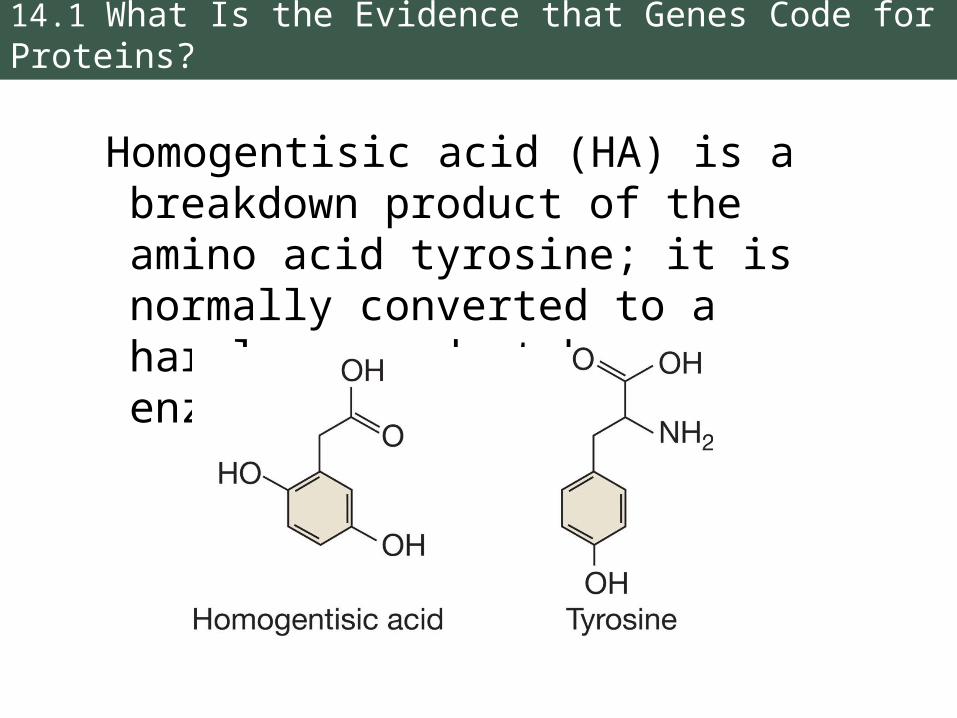
Homogentisic acid (HA) is a breakdown product of the amino acid tyrosine; it is normally converted to a harmless product by an enzyme.
14.1 What Is the Evidence that Genes Code for Proteins?
When the allele is mutated, the enzyme is inactive, and HA accumulates.
Thus, the researchers correlated one gene to one enzyme.
Confirmation required identification of the specific enzyme and gene mutation, which occurred much later.
Biologists turned to model organisms to understand gene expression.
14.1 What Is the Evidence that Genes Code for Proteins?
Model organisms:
• Easy to grow in the laboratory
• Short generation times
• Easy to manipulate genetically
• Produce large numbers of progeny
Examples: Pea plants, Drosophila, E. coli, and common bread mold—Neurospora crassa.

14.1 What Is the Evidence that Genes Code for Proteins?
Neurospora is haploid for most of its life cycle, so there are no dominant or recessive alleles.
Beadle and Tatum used Neurospora to test the one-gene, one-enzyme hypothesis.
Wild-type Neurospora strains have enzymes to catalyze all the reactions needed for growth.
14.1 What Is the Evidence that Genes Code for Proteins?
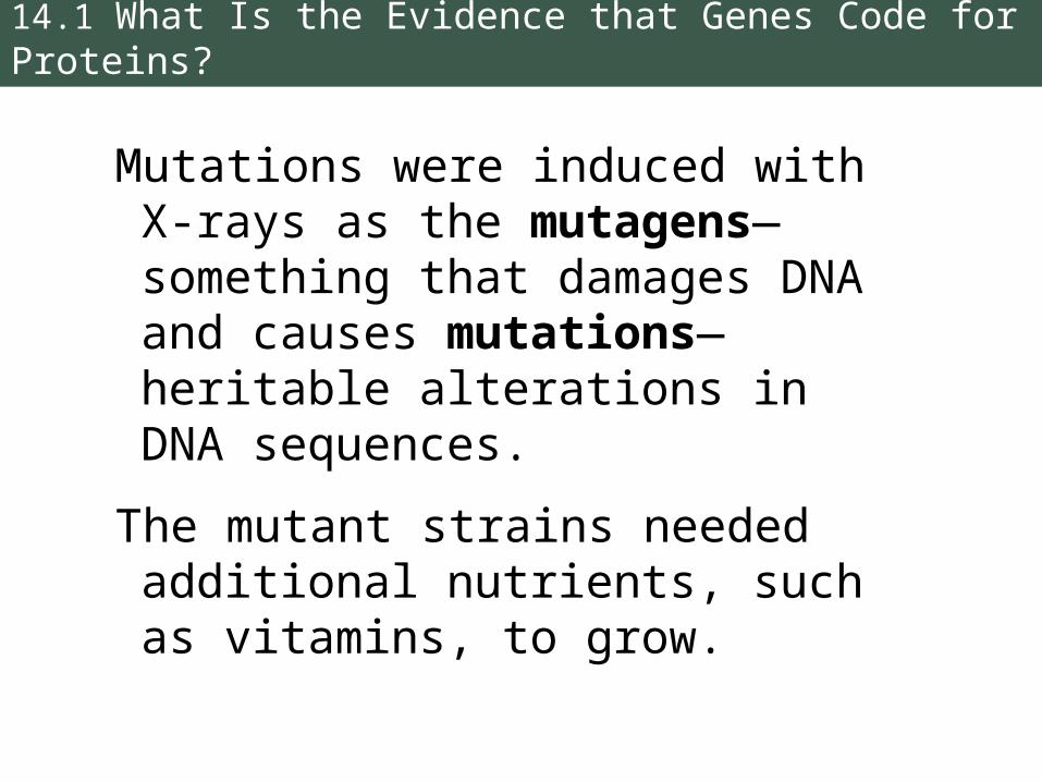
Mutations were induced with X-rays as the mutagens—something that damages DNA and causes mutations—heritable alterations in DNA sequences.
The mutant strains needed additional nutrients, such as vitamins, to grow.

14.1 What Is the Evidence that Genes Code for Proteins?
Each mutant strain required only one additional nutrient.
Results suggested that each mutation caused a defect in only one enzyme in a metabolic pathway, confirming the one-gene, one-enzyme hypothesis.
14.1 What Is the Evidence that Genes Code for Proteins?
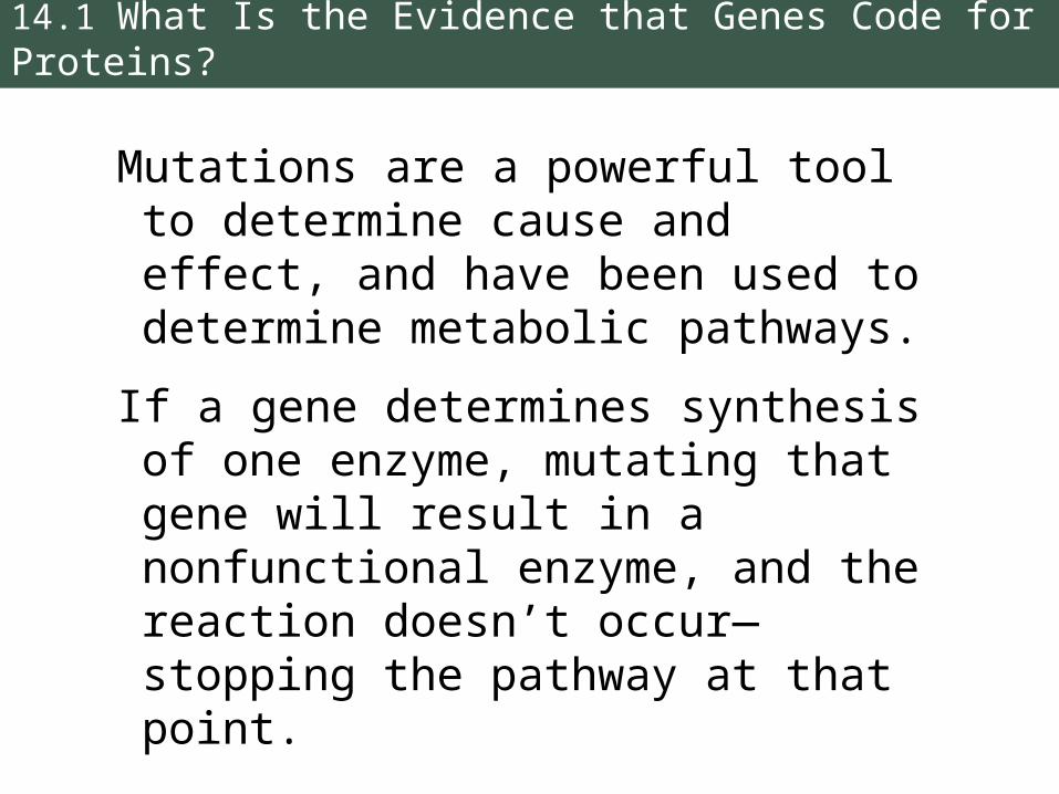
Mutations are a powerful tool to determine cause and effect, and have been used to determine metabolic pathways.
If a gene determines synthesis of one enzyme, mutating that gene will result in a nonfunctional enzyme, and the reaction doesn’t occur—stopping the pathway at that point.
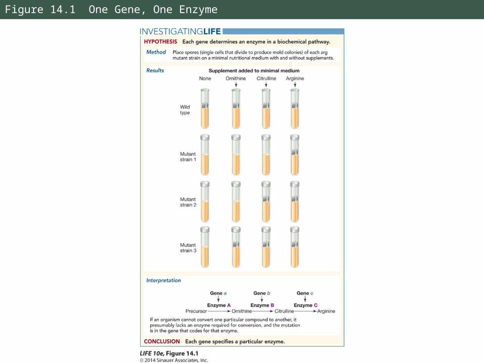
Figure 14.1 One Gene, One Enzyme
14.1 What Is the Evidence that Genes Code for Proteins?
One-gene, one-enzyme has since been revised to the one-gene, one-polypeptide relationship.
Many proteins have several polypeptides chains, or subunits.
Example: Hemoglobin has four subunits, each specified by a separate gene.
Not all genes code for polypeptides.
Working with Data 14.1: One Gene, One Enzyme
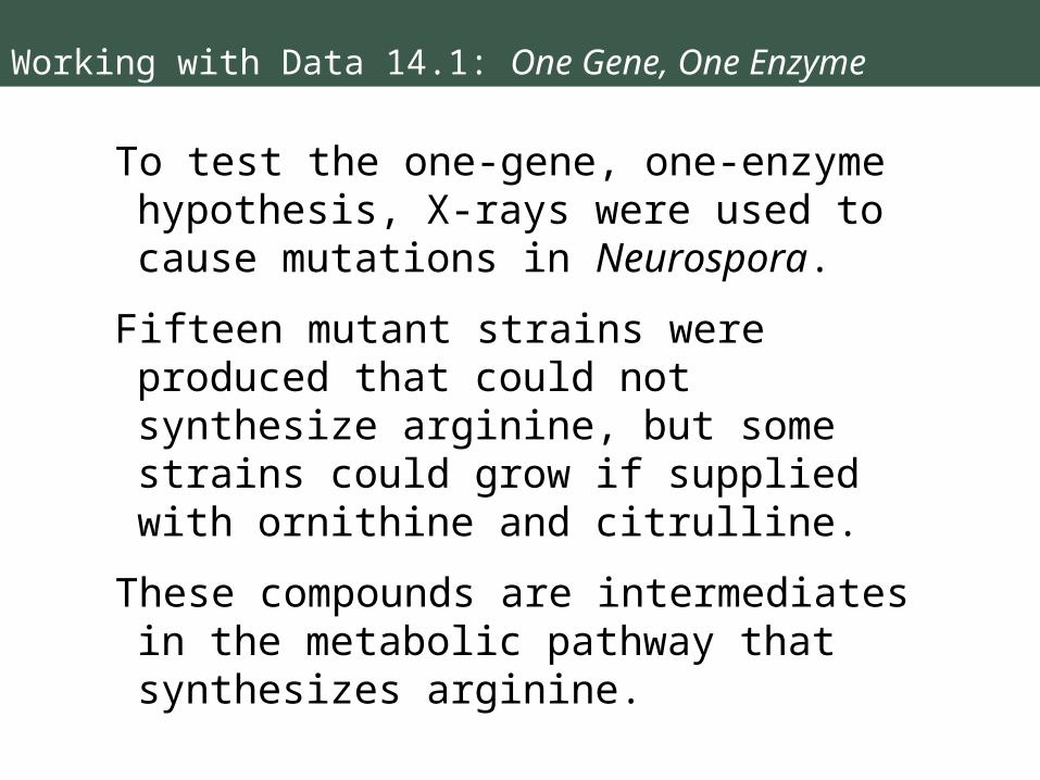
To test the one-gene, one-enzyme hypothesis, X-rays were used to cause mutations in Neurospora.
Fifteen mutant strains were produced that could not synthesize arginine, but some strains could grow if supplied with ornithine and citrulline.
These compounds are intermediates in the metabolic pathway that synthesizes arginine.
Working with Data 14.1: One Gene, One Enzyme
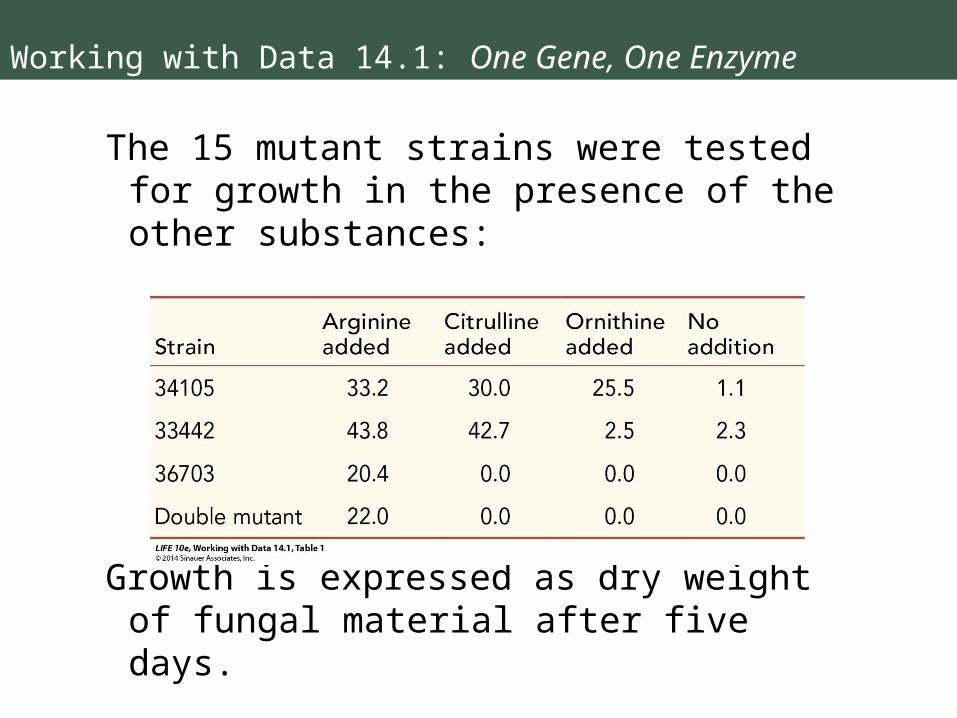
The 15 mutant strains were tested for growth in the presence of the other substances:
Growth is expressed as dry weight of fungal material after five days.

Working with Data 14.1: One Gene, One Enzyme
Question 1:
Based on the biochemical pathway for arginine synthesis shown in Figure 14.1, which enzyme (A, B, or C) was mutated in each strain?
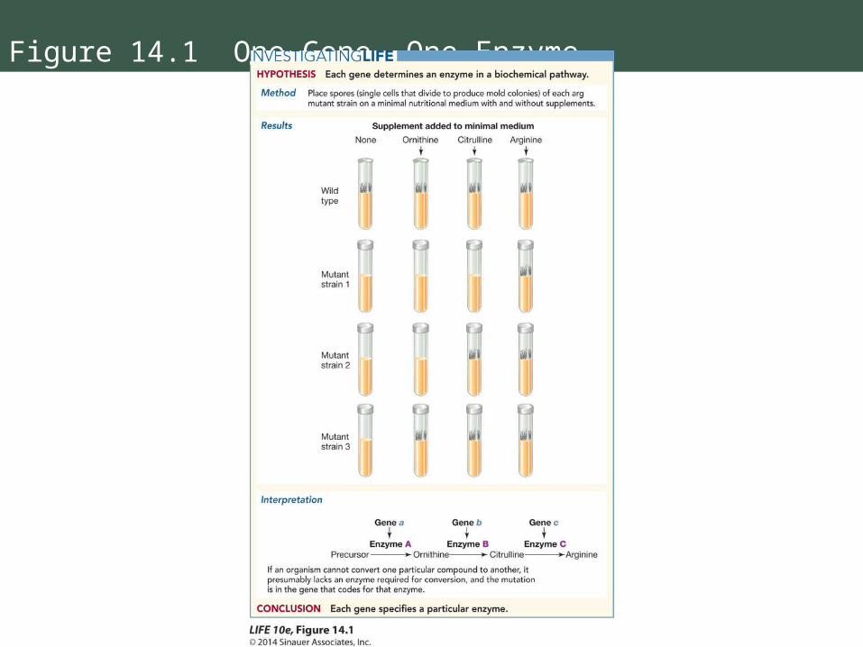
Figure 14.1 One Gene, One Enzyme

Working with Data 14.1: One Gene, One Enzyme
Question 2:
Why was there some growth in strains 34105 and 33442 even when there were no additions to the growth medium?
Working with Data 14.1: One Gene, One Enzyme
Question 3:
Nineteen other amino acids were tested as substitutes for arginine in the three strains. In all cases, there was no growth.
Explain these results.
Working with Data 14.1: One Gene, One Enzyme
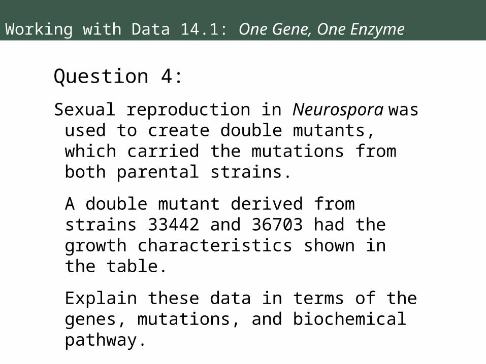
Question 4:
Sexual reproduction in Neurospora was used to create double mutants, which carried the mutations from both parental strains.
A double mutant derived from strains 33442 and 36703 had the growth characteristics shown in the table.
Explain these data in terms of the genes, mutations, and biochemical pathway.
14.2 How Does Information Flow from Genes to Proteins?
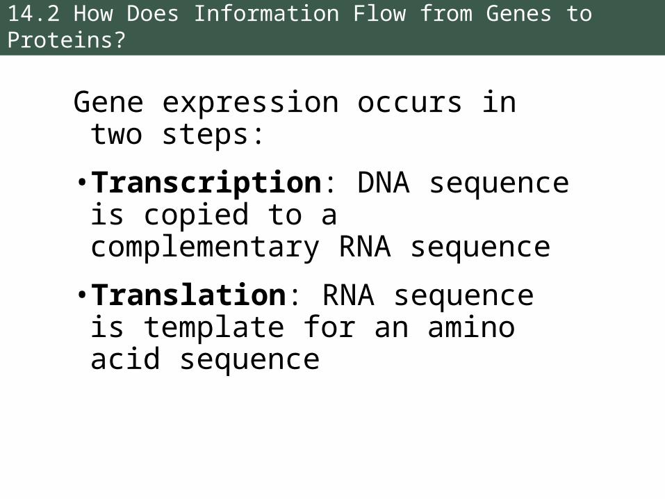
Gene expression occurs in two steps:
• Transcription: DNA sequence is copied to a complementary RNA sequence
• Translation: RNA sequence is template for an amino acid sequence
14.2 How Does Information Flow from Genes to Proteins?
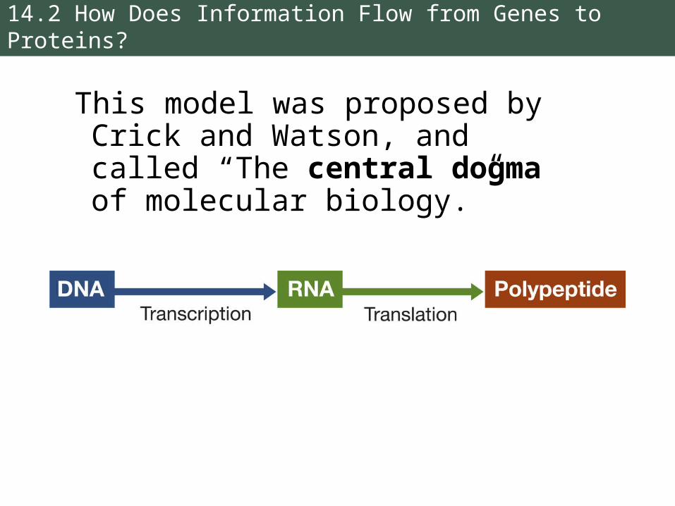
This model was proposed by Crick and Watson, and called “The central dogma of molecular biology.”
14.2 How Does Information Flow from Genes to Proteins?
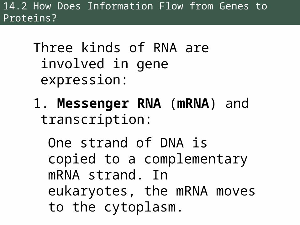
Three kinds of RNA are involved in gene expression:
1. Messenger RNA (mRNA) and transcription:
One strand of DNA is copied to a complementary mRNA strand. In eukaryotes, the mRNA moves to the cytoplasm.
14.2 How Does Information Flow from Genes to Proteins?
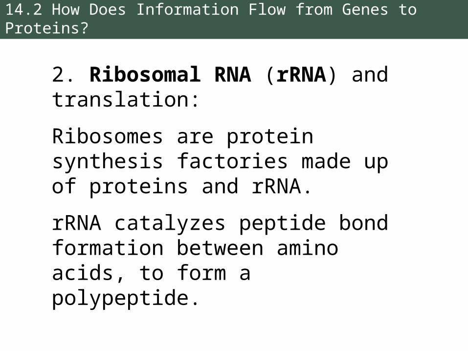
2. Ribosomal RNA (rRNA) and translation:
Ribosomes are protein synthesis factories made up of proteins and rRNA.
rRNA catalyzes peptide bond formation between amino acids, to form a polypeptide.
14.2 How Does Information Flow from Genes to Proteins?
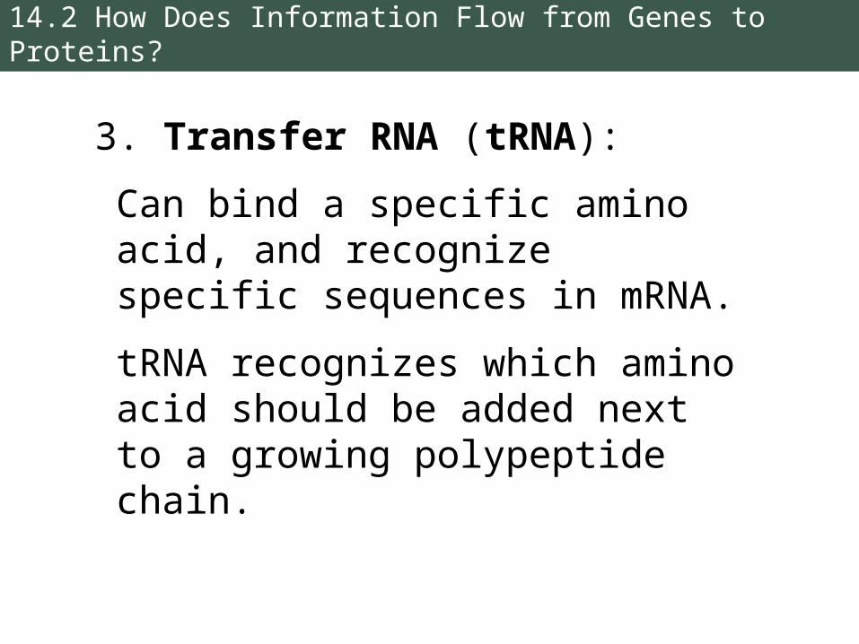
3. Transfer RNA (tRNA):
Can bind a specific amino acid, and recognize specific sequences in mRNA.
tRNA recognizes which amino acid should be added next to a growing polypeptide chain.
14.2 How Does Information Flow from Genes to Proteins?
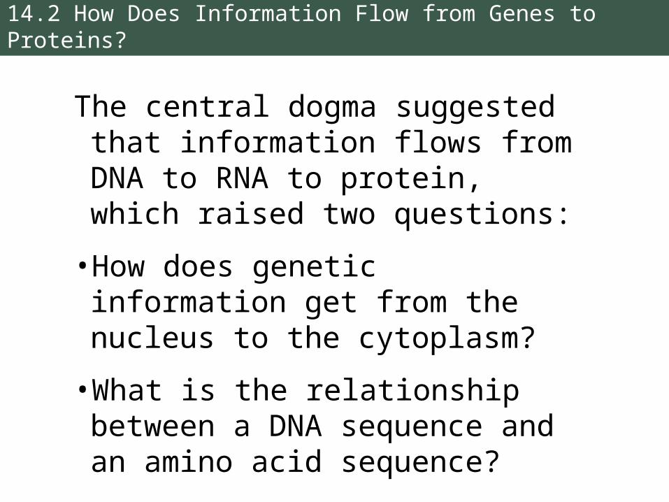
The central dogma suggested that information flows from DNA to RNA to protein, which raised two questions:
• How does genetic information get from the nucleus to the cytoplasm?
• What is the relationship between a DNA sequence and an amino acid sequence?
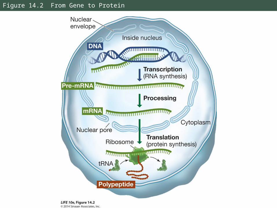
Figure 14.2 From Gene to Protein
14.2 How Does Information Flow from Genes to Proteins?
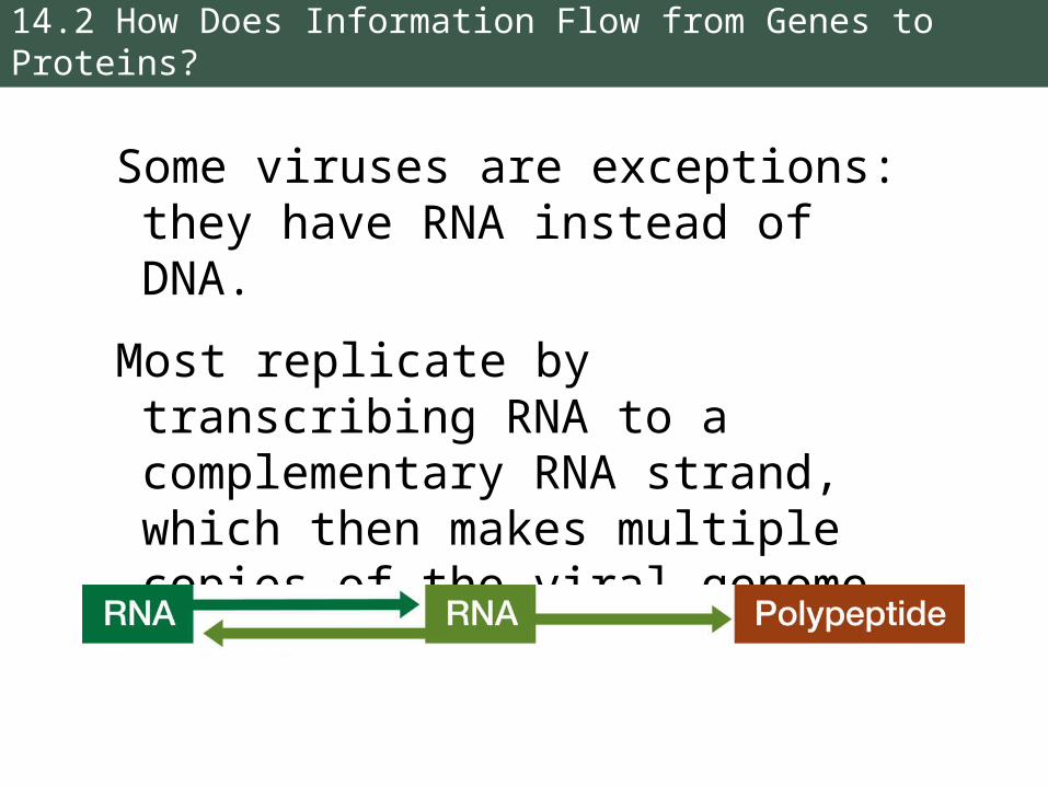
Some viruses are exceptions: they have RNA instead of DNA.
Most replicate by transcribing RNA to a complementary RNA strand, which then makes multiple copies of the viral genome.
14.2 How Does Information Flow from Genes to Proteins?
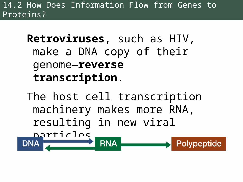
Retroviruses, such as HIV, make a DNA copy of their genome—reverse transcription.
The host cell transcription machinery makes more RNA, resulting in new viral particles.

14.3 How Is the Information Content in DNA Transcribed to Produce RNA?
Transcription requires:
• A DNA template for base pairings
• The four ribonucleoside triphosphates (ATP,GTP,CTP,UTP)
• An RNA polymerase
• Salts and pH buffer, if done in a test tube
14.3 How Is the Information Content in DNA Transcribed to Produce RNA?
Transcription produces mRNA, tRNA, and rRNA.
These RNAs are encoded by specific genes.
Eukaryotes also make several small RNAs, including small nuclear RNA (snRNA), microRNA (miRNA), and small interfering RNA (siRNA).
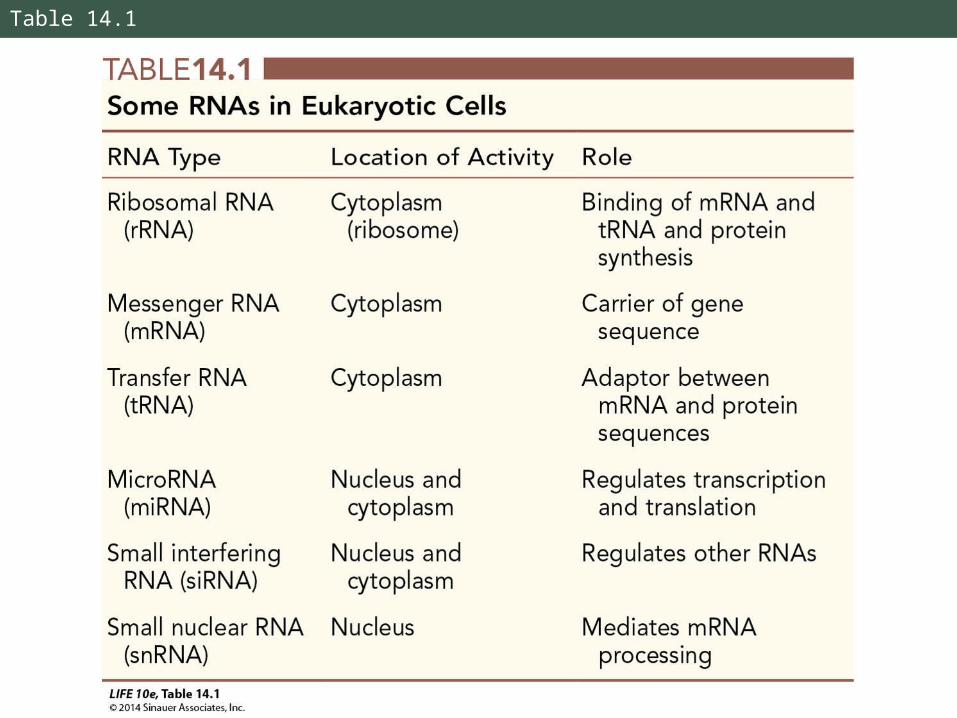
Table 14.1

14.3 How Is the Information Content in DNA Transcribed to Produce RNA?
RNA polymerases catalyze synthesis of RNA:
• Catalyze addition of nucleotides in a 5′-to-3′ direction
• Processive—one enzyme-template binding results in polymerization of hundreds of RNA bases
• They do not need primers
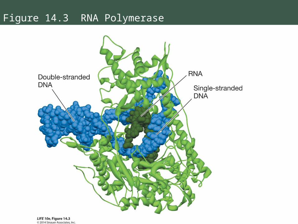
Figure 14.3 RNA Polymerase
14.3 How Is the Information Content in DNA Transcribed to Produce RNA?
Transcription occurs in three phases:
1. Initiation:
RNA polymerase binds to a DNA sequence called a promoter.
Promoters tell the enzyme where to start and which strand of DNA to transcribe.
The promoter has an initiation site where transcription begins.
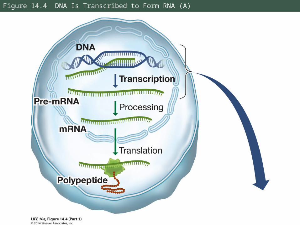
Figure 14.4 DNA Is Transcribed to Form RNA (A)
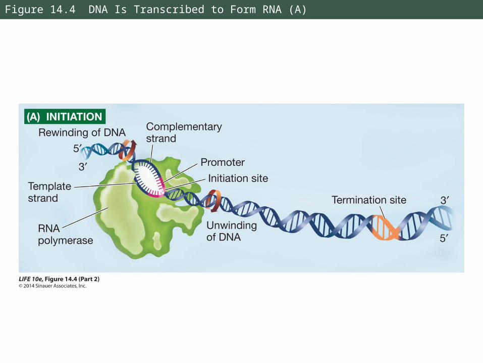
Figure 14.4 DNA Is Transcribed to Form RNA (A)
14.3 How Is the Information Content in DNA Transcribed to Produce RNA?
Sigma factors and transcription factors are proteins that bind to DNA sequences and to RNA polymerase.
They help direct the polymerase onto the promoter, and help determine which genes are expressed at particular times.
14.3 How Is the Information Content in DNA Transcribed to Produce RNA?
2. Elongation:
RNA polymerase unwinds DNA about 10 base pairs at a time; reads template in 3′ to 5′ direction.
The transcript is antiparallel to the DNA template strand.
RNA polymerases do not proofread and correct mistakes.

Figure 14.4 DNA Is Transcribed to Form RNA (B)
14.3 How Is the Information Content in DNA Transcribed to Produce RNA?
RNA polymerase uses (ribo)nucleoside triphosphates (NTPs) as substrates.
Two phosphate groups are removed from each substrate molecule; the energy released is used to drive the polymerization reaction.
14.3 How Is the Information Content in DNA Transcribed to Produce RNA?
3. Termination:
Specified by a specific DNA sequence.
Mechanism in eukaryotes is not well understood.
In bacteria, the transcript forms a loop and falls away from the DNA; or a helper protein binds to the transcript and causes it to detach from the DNA.
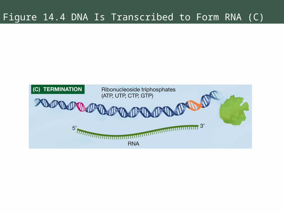
Figure 14.4 DNA Is Transcribed to Form RNA (C)
14.3 How Is the Information Content in DNA Transcribed to Produce RNA?
The genetic code specifies which amino acids will be used to build a protein.
Codon: a sequence of three bases, something like a three-letter “word.”
Each codon specifies a particular amino acid.

14.3 How Is the Information Content in DNA Transcribed to Produce RNA?
How was the code deciphered?
How could 20 “code words” (amino acids) be written with only four “letters” (the four bases)?
A triplet code seemed likely; it could result in 4 × 4 × 4 = 64 codons.
14.3 How Is the Information Content in DNA Transcribed to Produce RNA?
Nirenberg and Matthaei used simple artificial mRNAs of known composition to identify the polypeptide that resulted.
This led to the identification of the first three codons.

Figure 14.5 Deciphering the Genetic Code
14.3 How Is the Information Content in DNA Transcribed to Produce RNA?
Later, scientists used artificial mRNAs only three nucleotides long (one codon).
These would bind to a ribosome and a corresponding tRNA carrying an amino acid.
Thus the codes for all the amino acids were determined.

Figure 14.6 The Genetic Code
14.3 How Is the Information Content in DNA Transcribed to Produce RNA?
There are more codons than amino acids.
AUG is the start codon—initiation signal for translation.
Stop codons—termination signals, include UAA, UAG, and UGA.
14.3 How Is the Information Content in DNA Transcribed to Produce RNA?
For most amino acids, there is more than one codon: the genetic code is redundant.
The genetic code is not ambiguous—each codon specifies only one amino acid.
14.3 How Is the Information Content in DNA Transcribed to Produce RNA?
The genetic code is nearly universal:
The codons that specify amino acids are the same in all organisms.
A few exceptions: Within mitochondria and chloroplasts, and in one group of protists, there are codon differences.
14.3 How Is the Information Content in DNA Transcribed to Produce RNA?
This common genetic code is also a common language for evolution.
The code is ancient and has remained intact throughout evolution.
The common code also allows genetic engineering—human genes can be expressed in E. coli, for example.

14.3 How Is the Information Content in DNA Transcribed to Produce RNA?
The base sequence of the DNA strand that is transcribed is complementary and antiparallel to the mRNA codons.
The non-template DNA strand has the same sequence as the mRNA and is called the “coding strand.”
By convention, DNA sequences are shown beginning with the 5′ end of the coding sequence.
14.4 How Is Eukaryotic DNA Transcribed and the RNA Processed?
Gene expression is basically the same in prokaryotes and eukaryotes.
But there are differences in gene structure:
• In eukaryotes, the nucleus separates transcription and translation.
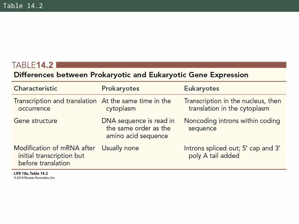
Table 14.2
14.4 How Is Eukaryotic DNA Transcribed and the RNA Processed?
mRNA sequences are complementary to gene sequences. This can be shown by nucleic acid hybridization:
DNA is denatured to separate strands.
An mRNA strand called a probe is incubated with the denatured DNA.
If the probe has a complementary sequence, base pairing forms a hybrid.

Figure 14.7 Nucleic Acid Hybridization and Introns (Part 1)
14.4 How Is Eukaryotic DNA Transcribed and the RNA Processed?
Hybridization experiments can be performed with various combinations of DNA and RNA.
The probe may be labeled in some way to detect binding to a specific target sequence.
The double-stranded hybrids can be viewed by electron microscopy.
14.4 How Is Eukaryotic DNA Transcribed and the RNA Processed?
When mRNA probes from prokaryotes are incubated with their DNAs and viewed under an electron microscope, there is a 1:1 complementarity.
In eukaryotes, loops of DNA are often observed—indicating stretches of DNA that don’t have a complementary mRNA sequence.

14.4 How Is Eukaryotic DNA Transcribed and the RNA Processed?
If the initial mRNA transcript (precursor or pre-mRNA) is hybridized with DNA, there is full, linear, loop-free hybridization.
The intervening regions (introns) get transcribed then sliced out of pre-mRNA in the nucleus.
Only expressed sequences (exons) reach the ribosome.
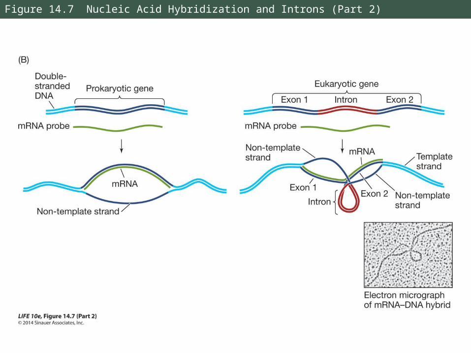
Figure 14.7 Nucleic Acid Hybridization and Introns (Part 2)

Figure 14.8 Transcription of a Eukaryotic Gene
14.4 How Is Eukaryotic DNA Transcribed and the RNA Processed?
Introns interrupt but do not scramble the DNA sequence that encodes a polypeptide.
Sometimes the separated exons code for different domains (functional regions) of the protein.
14.4 How Is Eukaryotic DNA Transcribed and the RNA Processed?
In the nucleus, pre-mRNA is modified:
• A 5′ cap is added at the 5′ end. The cap is a modified GTP which facilitates mRNA binding to a ribosome.
Also protects mRNA from being digested by ribonucleases.

14.4 How Is Eukaryotic DNA Transcribed and the RNA Processed?
• A poly A tail is added at 3′ end.
AAUAAA sequence after last codon is a signal for an enzyme to cut the pre-mRNA; then another enzyme adds 100 to 300 adenines—the “tail.”
The tail assists in export from the nucleus and is important for stability of mRNA.

Figure 14.9 Processing the Ends of Eukaryotic Pre-mRNA
14.4 How Is Eukaryotic DNA Transcribed and the RNA Processed?
• Introns are then removed.
RNA splicing removes introns and splices exons together.
snRNPs (small nuclear ribonucleoprotein particles) bind to ends of introns at consensus sequences—short DNA stretches that appear in many genes.
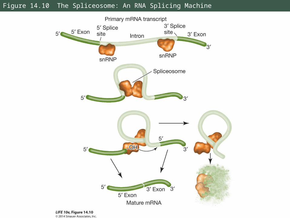
Figure 14.10 The Spliceosome: An RNA Splicing Machine
14.4 How Is Eukaryotic DNA Transcribed and the RNA Processed?
With energy from ATP, proteins are added to form an RNA-protein complex, the spliceosome.
The complex cuts pre-mRNA, releases introns, and splices exons together to produce mature mRNA.
14.4 How Is Eukaryotic DNA Transcribed and the RNA Processed?
In the disease β-thalassemia, a mutation may occur at an intron consensus sequence in the β-globin gene—the pre-mRNA cannot be spliced correctly.
Non-functional β-globin mRNA is produced.

14.4 How Is Eukaryotic DNA Transcribed and the RNA Processed?
Mature mRNA leaves the nucleus through nuclear pores.
A cap-binding protein complex binds to the 5′ cap and to other proteins that are recognized by receptors at the nuclear pore.
These proteins lead the mRNA through the pore; unused pre-mRNAs stay in the nucleus.
14.5 How Is RNA Translated into Proteins?
Transfer RNA (tRNA) links mRNA codons with specific amino acids.
There is at least one specific tRNA molecule for each of the 20 amino acids.
14.5 How Is RNA Translated into Proteins?
Each tRNA has three functions:
• It binds to a specific enzyme that attaches it to only one amino acid: it is then “charged”
• Binds to mRNA at a triplet called the anticodon, which is complementary to an mRNA codon
• Interacts with ribosomes
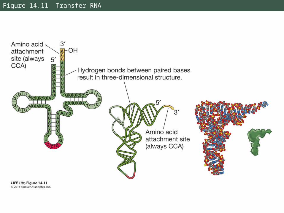
Figure 14.11 Transfer RNA

14.5 How Is RNA Translated into Proteins?
Wobble: Specificity for the base at the 3′ end of the codon is not always observed.
Example: Codons for alanine—GCA, GCC, and GCU—are recognized by the same tRNA.
Wobble allows cells to produce fewer tRNA species, but does not allow the genetic code to be ambiguous.

14.5 How Is RNA Translated into Proteins?
tRNAs are charged by aminoacyl-tRNA synthetases.
Each enzyme is specific for one amino acid and its corresponding tRNA.
Charging requires ATP; a high-energy bond forms between the amino acid and the tRNA—it is later used to form the peptide bond.

Figure 14.12 Charging a tRNA Molecule

14.5 How Is RNA Translated into Proteins?
Specificity between the tRNA and its amino acid is extremely important.
Experiments showed that specificity is at the anticodon:
Cysteine already bound to tRNA was chemically changed to alanine. Everywhere cysteine was supposed to be in the polypeptide, alanine appeared instead.
14.5 How Is RNA Translated into Proteins?
Translation occurs at a ribosome.
It holds mRNA and charged tRNAs in the correct position to allow assembly of the polypeptide.
Ribosomes can make any type of protein, they can be used over and over. Most cells have thousands of them.

14.5 How Is RNA Translated into Proteins?
Ribosomes have two subunits, large and small, held together non-covalently.
In eukaryotes, the large subunit has three different molecules of ribosomal RNA (rRNA) and 49 different proteins in a precise pattern.
The small subunit has one rRNA and 33 proteins.

Figure 14.13 Ribosome Structure
14.5 How Is RNA Translated into Proteins?
Ribosomal proteins and RNAs are different in prokaryotes.
This explains why antibiotics that target prokaryotic ribosomes (such as tetracycline) kill bacteria without harming the patient’s cells.
Mitochondria and chloroplasts also have ribosomes, similar to those of prokaryotes.

14.5 How Is RNA Translated into Proteins?
A large subunit has three tRNA binding sites:
• A (aminoacyl tRNA) site binds with anticodon of charged tRNA
• P (peptidyl tRNA) site where tRNA adds its amino acid to the growing chain
• E (exit) site where tRNA sits before being released from the ribosome
14.5 How Is RNA Translated into Proteins?
Ribosomes have a fidelity function: When proper binding occurs, hydrogen bonds form between the base pairs of the anticodon and the mRNA codon.
Small subunit rRNA validates the match—if hydrogen bonds have not formed between all three base pairs, the tRNA must be an incorrect match for that codon and the tRNA is rejected.
14.5 How Is RNA Translated into Proteins?
Translation occurs in three steps:
1. Initiation
An initiation complex forms—a charged tRNA and small ribosomal subunit, both bound to mRNA.
In prokaryotes rRNA binds to the Shine-Dalgarno sequence on the mRNA.
In eukaryotes it binds to the 5′ cap.
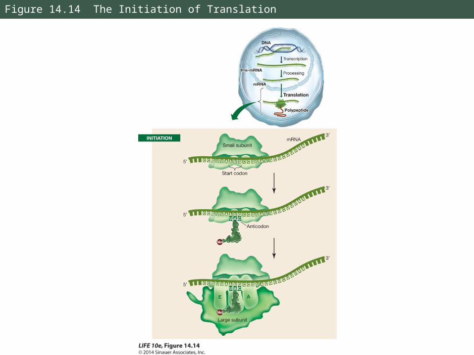
Figure 14.14 The Initiation of Translation
14.5 How Is RNA Translated into Proteins?
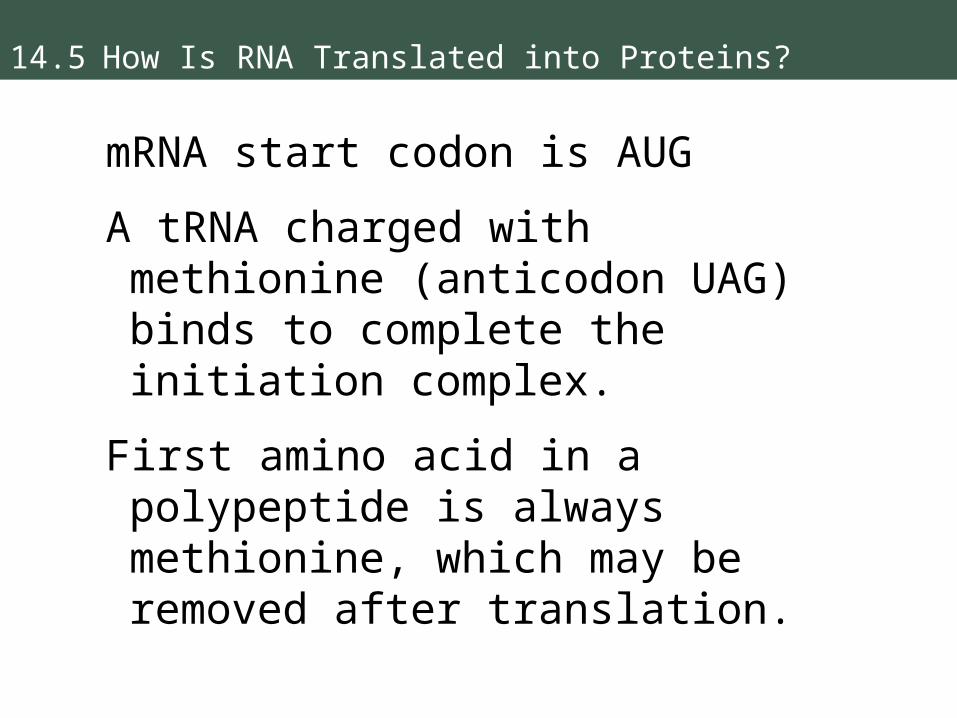
mRNA start codon is AUG
A tRNA charged with methionine (anticodon UAG) binds to complete the initiation complex.
First amino acid in a polypeptide is always methionine, which may be removed after translation.
14.5 How Is RNA Translated into Proteins?
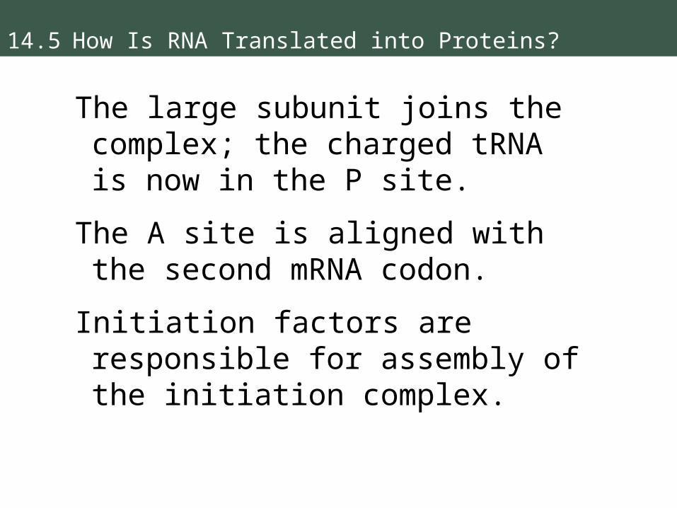
The large subunit joins the complex; the charged tRNA is now in the P site.
The A site is aligned with the second mRNA codon.
Initiation factors are responsible for assembly of the initiation complex.

14.5 How Is RNA Translated into Proteins?
2. Elongation
Another charged tRNA enters A site and the large subunit catalyzes two reactions:
• Bond between tRNA in P site and its amino acid is broken
• Peptide bond forms between that amino acid and the amino acid on tRNA in the A site
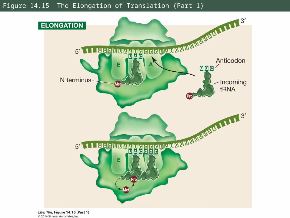
Figure 14.15 The Elongation of Translation (Part 1)

14.5 How Is RNA Translated into Proteins?
The large subunit has peptidyl transferase activity.
RNA was shown to be the catalyst by:
• Removing the proteins from the large subunit – it still catalyzed peptide bonds
• Modifying rRNA, which destroyed peptidyl transferase activity
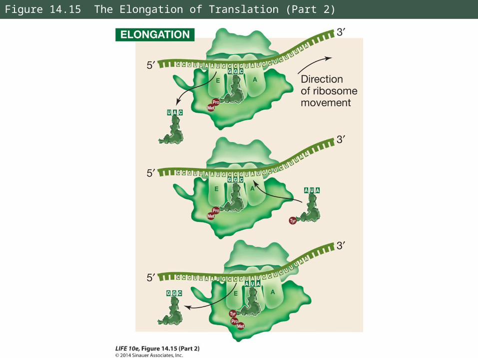
Figure 14.15 The Elongation of Translation (Part 2)
14.5 How Is RNA Translated into Proteins?
Purification and crystallization of ribosomes has allowed scientists to confirm the catalytic role of rRNA.
This supports the hypothesis that RNA, and catalytic RNA in particular, evolved before DNA.
14.5 How Is RNA Translated into Proteins?
When the first tRNA has released its methionine, it moves to the E site and dissociates from the ribosome.
The tRNA can be charged again.
Elongation occurs as the steps are repeated, assisted by proteins called elongation factors.
14.5 How Is RNA Translated into Proteins?
3. Termination
Translation ends when a stop codon enters the A site.
Stop codons bind a protein release factor which hydrolyzes bond between the polypeptide and the tRNA in the P site.
The polypeptide then separates from the ribosome.
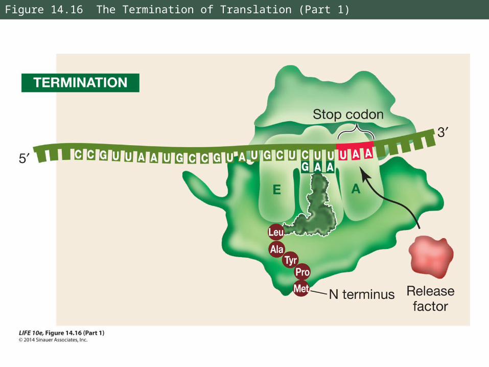
Figure 14.16 The Termination of Translation (Part 1)

Figure 14.16 The Termination of Translation (Part 2)
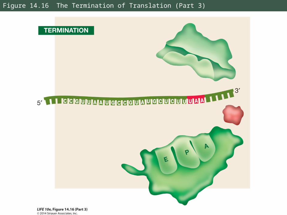
Figure 14.16 The Termination of Translation (Part 3)
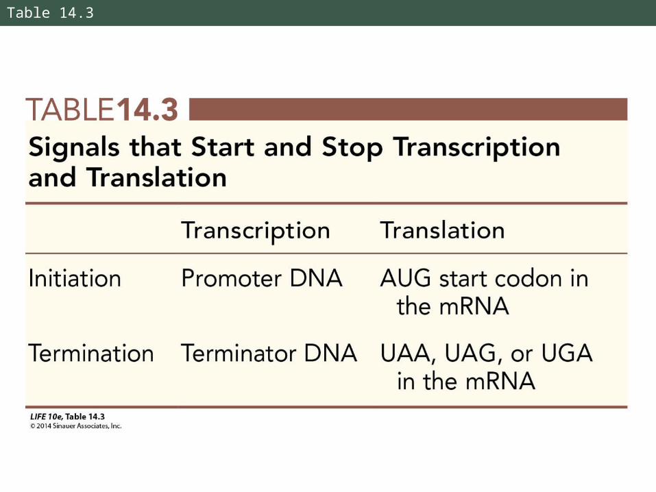
Table 14.3
14.5 How Is RNA Translated into Proteins?
Several ribosomes can work together to translate the same mRNA, producing multiple copies of the polypeptide.
A strand of mRNA with associated ribosomes is called a polyribosome, or polysome.
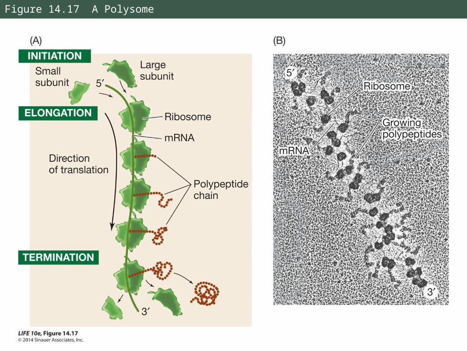
Figure 14.17 A Polysome
14.6 What Happens to Polypeptides after Translation?
After translation, polypeptides may move into an organelle, or out of the cell.
They are often modified by the addition of new chemical groups that affect their function.
14.6 What Happens to Polypeptides after Translation?
Polypeptide emerges from the ribosome and folds into its 3-D shape.
It may contain a signal sequence indicating where in the cell it belongs.
If there is no signal sequence, it remains where it was synthesized.
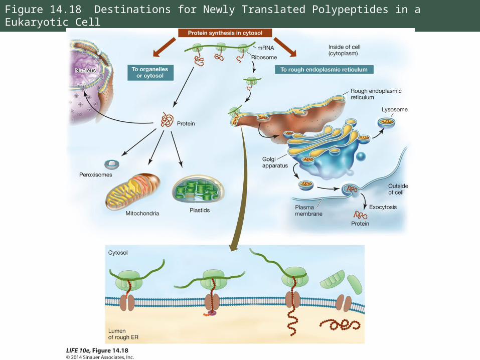
Figure 14.18 Destinations for Newly Translated Polypeptides in a Eukaryotic Cell

14.6 What Happens to Polypeptides after Translation?
A signal sequence binds to a receptor protein on the surface of an organelle.
A channel forms in the organelle membrane and the protein enters.
Example: a nuclear localization signal (NLS)—
-Pro-Pro-Lys-Lys-Lys-Arg-Lys-Val-

14.6 What Happens to Polypeptides after Translation?
The nuclear localization signal was determined by making proteins with and without the signal sequence, then injecting them into cells.
Only proteins with the signal ended up in the nucleus.

Figure 14.19 Testing the Signal
14.6 What Happens to Polypeptides after Translation?
Some polypeptides have a signal sequence that stops translation and sends the ribosome to the endoplasmic reticulum.
Ribosome binds to the ER and translation resumes. The polypeptide will move into the ER lumen.
14.6 What Happens to Polypeptides after Translation?
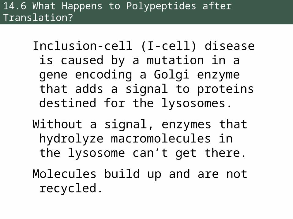
Inclusion-cell (I-cell) disease is caused by a mutation in a gene encoding a Golgi enzyme that adds a signal to proteins destined for the lysosomes.
Without a signal, enzymes that hydrolyze macromolecules in the lysosome can’t get there.
Molecules build up and are not recycled.
14.6 What Happens to Polypeptides after Translation?
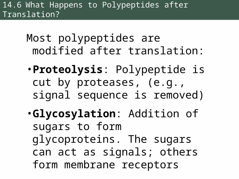
Most polypeptides are modified after translation:
• Proteolysis: Polypeptide is cut by proteases, (e.g., signal sequence is removed)
• Glycosylation: Addition of sugars to form glycoproteins. The sugars can act as signals; others form membrane receptors

14.6 What Happens to Polypeptides after Translation?
• Phosphorylation: Addition of phosphate groups catalyzed by protein kinases
The charged phosphate groups change the conformation and may expose active sites or binding sites.
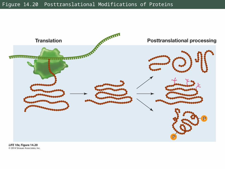
Figure 14.20 Posttranslational Modifications of Proteins
14 Answer to Opening Question
A different type of antibiotic is being developed that targets mRNA breakdown.
mRNA is broken down after use and quickly recycled, so that bacteria can adapt rapidly to changing environments.
The antibiotic targets a protein that is part of the breakdown machinery—without it, the cell can’t recycle mRNA.
MRSA is particularly sensitive to this molecule.