14 - congress of apvrs 20172017.apvrs.org/wp-content/uploads/2017/11/apvrs_showdaily-day2-low...14 1...
TRANSCRIPT

SHOWDAILYThe Official Conference News of APVRS 2017APVRS
December 8-10, 2017Kuala Lumpur, Malaysia Day 2
by
PhotooftheDay
135
14
1
Highlights
Published by:
Media MICE Pte. Ltd. 6001 Beach Road, #19-06
Golden Mile Tower, Singapore 199589
Tel: +65 8186 7677 Fax: +65 6298 6316
Email: [email protected]
Of Bionic Eyes and Robot Surgeries
What’s New in AMD?
The Latest in Laser Retina
CEO & PublisherMatt Young
CFOHannah Nguyen
Chief EditorGloria D. Gamat
Associate EditorBrooke Herron
Project ManagerRuchi Mahajan Ranga
Publications & Digital ManagerTimmo Gunst
Graphic DesignersWinson ChuaEdison Tan
PhotographerDwayne Foong
WritersHazlin HassanKhor Hui Min
Collins Santhanasamy
www.piemagazine.orgMatt Young, CEO of PIE Magazine and Dr. Kenneth Fong, APVRS 2017 Congress President, share a selfie.
by Collins Santhanasamy
The banquet hall of the world renowned Kuala Lumpur Convention Centre (KLCC),
conveniently located in the heart of the city and adjacent to the iconic twin towers, was abuzz with energy and excitement as experts in the vitreo-retina community of the Asia-Pacific region gathered to discuss and exchange knowledge, experiences and opinions in the development of ophthalmology.
The opening ceremony began with the welcoming of honored guests with a traditional Malaysian procession followed by the singing of the national anthem. Dr. Kenneth Fong, President of the 11th APVRS Congress and President of the Malaysian Society of Ophthalmology (MSO), delivered the welcome address in front of some 1800 delegates from over 50 countries across the globe who arrived for the 11th Asia-Pacific Vitreo-retina Society Congress (APVRS 2017).
Dr. Fong welcomed the delegates with a brief introduction on the cultural diversity that Malaysia has to offer. He highlighted that even though the Asia-Pacific region is currently the largest and fastest growing region in the world, it is still a region that has one of the highest rates of blindness. He stressed that this congress
would help to improve the skills and knowledge of ophthalmologists in treating these blinding diseases with intensive lectures, masterclasses and discussions.
Dr. Fong also announced that today, the morning of the 9th of December would be the much anticipated day for the release of the two-year results of two very important clinical trials for the management of polypoidal choroidal vasculopathy (PCV): The EVEREST 2 and the PLANET studies, which would lead to interesting and informative discussions.
Cont. on Page 12 >>
thAPVRS Congress Opens in
Vibrant ColorsKL with

Bayer Co. (Malaysia) Sdn. Bhd.B-19-1 & B-19-2, The Ascent Paradigm,No. 1, Jalan SS 7/26A, Kelana Jaya, 47301 Petaling Jaya, Selangor, Malaysia.Tel: +603 7801 3088 I Fax: +603 7886 3338 I Website: www.bayer.com
ABBREVIATED PRESCRIBING INFORMATIONBrand name of product EYLEA 40mg/ml solution for injection. Approved name of the active ingredient Aflibercept. Indication: Treatment of neovascular (wet) age-related macular degeneration (wet AMD), visual impairment due to macular edema secondary to retinal vein occlusion (branch RVO or central RVO), visual impairment due to diabetic macular edema (DME) and visual impairment due to myopic choroidal neovascularization (myopic CNV). Dosage and method of administration The recommended dose for Eylea is 2 mg aflibercept, equivalent to 0.05mL (50 μL); Neovascular (wet) age-related macular degeneration (wet AMD): Eylea treatment is initiated with one injection per month for three consecutive doses, followed by one injection every two months. After the first 12 months of treatment with Eylea, based on visual and/or anatomic outcomes, the treatment interval may be extended, such as with a treat-and- extend dosing regimen; Visual impairment due to macular edema secondary to retinal vein occlusion (branch RVO or central RVO): After the initial injection, treatment is given monthly. Monthly treatment continues until maximal visual acuity is achieved and/or there are no signs of disease activity. Three or more consecutive, monthly injections may be needed. Treatment may then be continued with a treat and extend regimen with gradually increased treatment intervals to maintain stable visual and/or anatomic outcomes, however there are insufficient data to conclude on the length of these intervals; Diabetic macular edema (DME): Eylea treatment is initiated with one injection per month for five consecutive doses, followed by one injection every two months. After the first 12 months of treatment with Eylea, and based on visual and/or anatomic outcomes, the treatment interval may be extended, such as with a treat- and-extend dosing regimen; Myopic choroidal neovascularization (myopic CNV): Single intravitreal injection is recommended. Additional doses may be administered if visual and/or anatomic outcomes indicate that the disease persists. Recurrences are treated like a new manifestation of the disease. The interval between two doses should not be shorter than one month. Contraindications Eylea is contraindicated in patients: with ocular or periocular infection; with Active severe intraocular inflammation; with Known hypersensitivity to aflibercept or to any of the excipients. Special warnings and special precautions for use Endophthalmitis: Proper aseptic injection technique must always be used when administering EYLEA. Patients should be instructed to report any symptoms suggestive of endophthalmitis without delay and should be managed appropriately. Increase in intraocular pressure: Increases in intraocular pressure have been seen within 60 minutes of an intravitreal injection, including with EYLEA. Special precaution is needed in patients with poorly controlled glaucoma. Other: The safety and efficacy of Eylea therapy administered to both eyes concurrently have not been systematically studied; In the event of a retinal break the dose should be withheld and treatment should not be resumed until the break is adequately repaired; The dose should be withheld based on the clinical judgement of the treating physician, in the event of a performed or planned intraocular surgery; EYLEA should not be used during pregnancy unless the potential benefit outweighs the potential risk to the fetus. EYLEA is not recommended during breast-feeding. Undesirable effects The most frequently observed adverse reactions (in at least 5% of patients treated with EYLEA) were conjunctival hemorrhage, eye pain, cataract, intraocular pressure increased, vitreous detachment and vitreous floaters. For further prescribing information, please contact Bayer Co. (M) Sdn Bhd, B-19- 1 & B-19- 2, The Ascent Paradigm, No. 1, Jalan SS 7/26A, Kelana Jaya, 47301 Petaling Jaya, Selangor. Subject to medical prescription. Date of text revision 18.05.2017.
REFERENCES1. Ikuno Y, Ohno-Matsui K, Wong TY, et al. Intravitreal aflibercept injection in patients with myopic choroidal neovascularization: the MYRROR Study. Ophthalmology. 2015;122(6):1220-1227. doi:10.1016/ j.ophtha.2015.01.025. 2. Heier JS, Brown DM, Chong V, et al. Intravitreal aflibercept (VEGF Trap-Eye) in wet age-related macular degeneration. Ophthalmology. 2012;119(12):2537-2548. doi:10.1016/ j.ophtha.2012.09.006. 3. Campochiaro PA, Clark WL, Boyer DS, et al. Intravitreal aflibercept for macular edema following branch retinal vein occlusion: the 24-week results of the VIBRANT study. Ophthalmology. 2015;122(3):538-544. doi:10.1016/j. ophtha.2014.08.031. 4. Korobelnik J-F, Holz FG, Roider J, et al. Intravitreal aflibercept injection for macular edema resulting from central retinal vein occlusion: one-year results of the phase 3 GALILEO study. Ophthalmology. 2014;121:202-208. doi:10.1016/j. ophtha.2013.08.012. 5. Korobelnik J-F, Do DV, Schmidt-Erfurth U, et al. Intravitreal aflibercept for diabetic macular edema. Ophthalmology. 2014;121(11):2247-2254.doi:10.1016/j.ophtha.2014.05.006. 6. EYLEA 40mg/L solution for injection Prescribing Information, Malaysia, 13 January 2017.
1-5
L.MY.MKT.10.2017.0568
3| December 8-10, 2017 | Kuala Lumpur, MalaysiaSHOWDAILYAPVRS
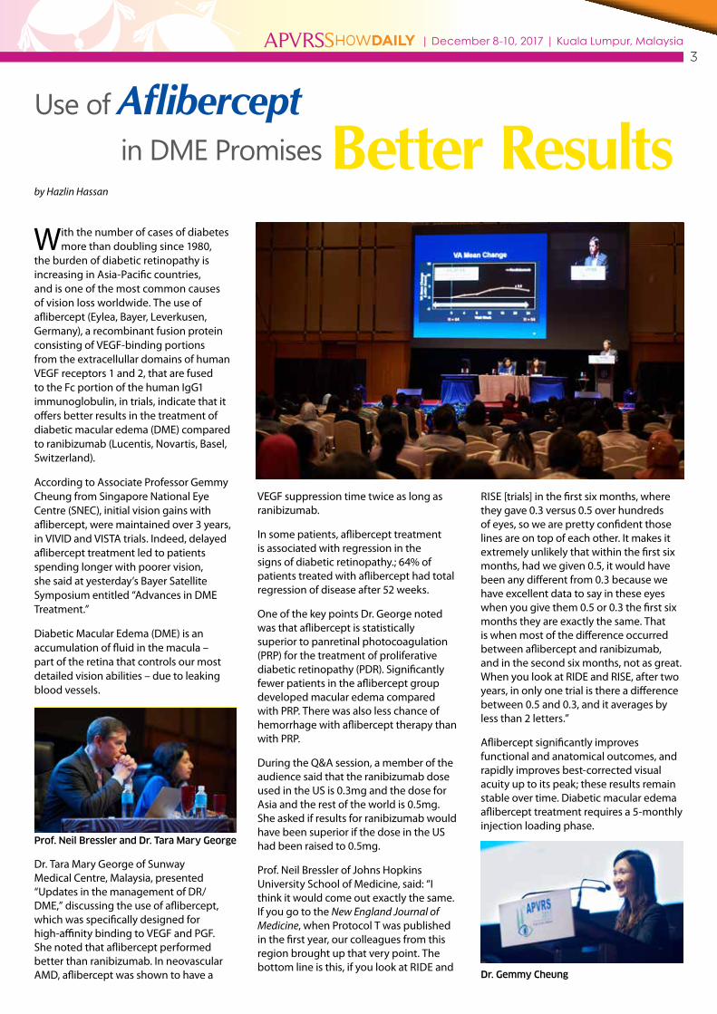
Use of Aflibercept in DME Promises by Hazlin Hassan
With the number of cases of diabetes more than doubling since 1980,
the burden of diabetic retinopathy is increasing in Asia-Pacific countries, and is one of the most common causes of vision loss worldwide. The use of aflibercept (Eylea, Bayer, Leverkusen, Germany), a recombinant fusion protein consisting of VEGF-binding portions from the extracellullar domains of human VEGF receptors 1 and 2, that are fused to the Fc portion of the human IgG1 immunoglobulin, in trials, indicate that it offers better results in the treatment of diabetic macular edema (DME) compared to ranibizumab (Lucentis, Novartis, Basel, Switzerland).
According to Associate Professor Gemmy Cheung from Singapore National Eye Centre (SNEC), initial vision gains with aflibercept, were maintained over 3 years, in VIVID and VISTA trials. Indeed, delayed aflibercept treatment led to patients spending longer with poorer vision, she said at yesterday’s Bayer Satellite Symposium entitled “Advances in DME Treatment.”
Diabetic Macular Edema (DME) is an accumulation of fluid in the macula – part of the retina that controls our most detailed vision abilities – due to leaking blood vessels.
Dr. Tara Mary George of Sunway Medical Centre, Malaysia, presented “Updates in the management of DR/DME,” discussing the use of aflibercept, which was specifically designed for high-affinity binding to VEGF and PGF. She noted that aflibercept performed better than ranibizumab. In neovascular AMD, aflibercept was shown to have a
VEGF suppression time twice as long as ranibizumab.
In some patients, aflibercept treatment is associated with regression in the signs of diabetic retinopathy.; 64% of patients treated with aflibercept had total regression of disease after 52 weeks.
One of the key points Dr. George noted was that aflibercept is statistically superior to panretinal photocoagulation (PRP) for the treatment of proliferative diabetic retinopathy (PDR). Significantly fewer patients in the aflibercept group developed macular edema compared with PRP. There was also less chance of hemorrhage with aflibercept therapy than with PRP.
During the Q&A session, a member of the audience said that the ranibizumab dose used in the US is 0.3mg and the dose for Asia and the rest of the world is 0.5mg. She asked if results for ranibizumab would have been superior if the dose in the US had been raised to 0.5mg.
Prof. Neil Bressler of Johns Hopkins University School of Medicine, said: “I think it would come out exactly the same. If you go to the New England Journal of Medicine, when Protocol T was published in the first year, our colleagues from this region brought up that very point. The bottom line is this, if you look at RIDE and
RISE [trials] in the first six months, where they gave 0.3 versus 0.5 over hundreds of eyes, so we are pretty confident those lines are on top of each other. It makes it extremely unlikely that within the first six months, had we given 0.5, it would have been any different from 0.3 because we have excellent data to say in these eyes when you give them 0.5 or 0.3 the first six months they are exactly the same. That is when most of the difference occurred between aflibercept and ranibizumab, and in the second six months, not as great. When you look at RIDE and RISE, after two years, in only one trial is there a difference between 0.5 and 0.3, and it averages by less than 2 letters.”
Aflibercept significantly improves functional and anatomical outcomes, and rapidly improves best-corrected visual acuity up to its peak; these results remain stable over time. Diabetic macular edema aflibercept treatment requires a 5-monthly injection loading phase.
Better Results
Dr. Gemmy Cheung
Prof. Neil Bressler and Dr. Tara Mary George

5| December 8-10, 2017 | Kuala Lumpur, MalaysiaSHOWDAILYAPVRS
Advances inby Khor Hui Min
Opening this session, Dr. Kenneth Fong, Congress President of the
11th APVRS, talked about ‘How to Laser Better in 2018’. He discussed SubLiminal (Quantel Medical, France) and MicroPulse (Iridex, California), which are similar technology, but have different trademark names. They are based on a train of microsecond pulses, according to Dr. Fong. He explained that in 2008, the 532 nm and 577 nm sub-threshold lasers were introduced. However, in 2011, randomized controlled trials (RCTs) showed that sub-threshold laser was more effective than conventional laser for diabetic macular edema (DME). The laser beam is deflected by two motorized mirrors to produce a pattern, and the multi-
spot photocoagulation delivers many impacts in the shortest time possible. Customized patterns are delivered at a fixed size of 400 µm, at fixed short pulse durations of 10 to 20ms. The power level can be customized in the range of 250 mW to 1 W. The further you move into the wavelength part of the spectrum, the better the laser will pass easily through a cataract. The low-intensity treatment effect is maximized by the placement of many laser spots (high-intensity treatment) over the entire area of retina pathology.
Dr. Xiaoling Liu from Wenzhou University Eye Hospital in China, talked about the ‘Concept and Treatment Guidelines in Subliminal Laser Therapy’. She compared micropulse laser to conventional laser, whereby micropulse was the better option, because it produced invisible burns, did not have heat diffusion and the spot size was the same as the laser. She shared that titration guidelines were very important to prevent burns. “Start from 500 mW on a healthy area, and increase or decrease the power to barely visible burn (threshold),” she explained. “Then reduce this power to 50 percent.” Dr. Liu then continued with another presentation on ‘577 nm Subliminal Laser in Central Serous Chorioretinopathy (CSCR)’. In her study, she found that there were no laser-
induced scars at week 12 after treatment. She concluded that subliminal laser with 5% duty circle was efficacious and safe for CSCR.
The symposium continued with a presentation on ‘Subliminal Laser Treatment for DME’ by Dr. Kenneth Foong. The subliminal treatment can induce biological changes in the retinal pigment epithelium (RPE) microenvironment, closing the micro-aneurysms and drying the edema. In foveal involved DME, in general, for thicker retina, anti-vascular endothelial growth factor (anti-VEGF) treatment will likely be the first line of treatment. Once the edema is settled, the subliminal laser can be used to reduce the number of injections. In patients without visual loss, subliminal laser can be used as the first line of treatment. If the edema deteriorates, the anti-VEGF treatment can be added. Currently, multiple anti-VEGF injections for DME are a huge burden for patients and the healthcare system. The use of subthreshold laser with 577 nm wavelength is a cost-effective option for DME and CSCR, and can reduce the burden of anti-VEGF injections.
The final presentation on ‘Photodynamic Therapy (PDT)’ was delivered by Dr. Colin Tan from Tan Tock Seng Hospital (TTSH), Singapore. PDT was previously used to treat age-related macular degeneration and myopic choroidal neovascularization (CNV). Now, it is used to treat polypoidal choroidal vasculopathy (PCV) and CSCR. Dr. Colin gave step-by-step explanations on how to conduct the PDT procedure.
Retina Laser Therapy

6| December 8-10, 2017 | Kuala Lumpur, MalaysiaSHOWDAILYAPVRS
Pathological Myopia: Dealing with the Epidemicby Collins Santhanasamy
At the symposium on pathological myopia, eight speakers delivered
presentations on topics ranging from myopia control to management of macular hole (MH).
Dr. Gemmy Cheung, FRCOphth, of Singapore National Eye Centre (SNEC), opened the session. She spoke about ‘Epidemiology and Low-Dose Atropine for Myopia Control’. She highlighted that the prevalence of myopia is highest in Asians, but also rising worldwide with a projected 2.5 billion people who will be affected by year 2020. There is now a substantial body of evidence to support the use of topical atropine in the reduction of the myopia progression rate with multiple studies showing promising results in childhood myopia.
Dr. Pei-chang Wu, Director of the Department of Ophthalmology, Kaohsiung Chang Gung Memorial
Hospital in Taiwan presented findings on ‘Natural Light Exposure for Myopia Control’. With high myopia maculopathy being the number one cause of irreversible blindness In Taiwan, focus on strengthening the protective factors and weakening the risk factors in the prevention of myopia in children is crucial to the reduction of the number of adult cases of high myopia. It was found that outdoor activity was the most significant protective factor against myopia.
In 2013, a study was published as an interventional policy for schools to prevent myopia. During this program, schools increased outdoor time to 80 minutes per day in several short breaks ranging between 10 to 20 minutes each. After one year it was found that in the interventional group there was a significant decrease in the incidence of myopia, almost approaching 50%.
Teachers are encouraged to find creative ways to give students outdoor homework and assignments with rewards like “outdoor passport” and “outdoor learning sheet” as a form of positive reinforcement.
Dr. Salomon Cohen from France and Dr. Hideharu Ohsugi from Japan both spoke on the topic of dome-shaped macula (DSM), about its relevance, and imaging in pathological myopia, respectively. The key conclusions of both presentations was the need for further studies in order to increase the understanding of the pathological cause of the condition and the treatment modalities of serous retinal detachment (SRD), which is the main complication of DSM.
Advanced Vitrectomy – Optimizing Outcomesby Collins Santhanasamy
The surgical retinal symposium on vitrectomy was chaired by Associate
Professor Ian Yeo from the Singapore National Eye Center. In this session, many participants came to view videos that featured tips and tricks from 6 surgeons who are experts on the topic.
Dr. Gavin Tan was the first surgeon to present his take on macular hole (MH) surgery and internal limiting membrane (ILM) peeling. ILM peeling is the standard for care of MH surgery with an increased closure rate and ILM flaps are useful in difficult cases such as chronic and large macular holes. Essentially the aim is to relieve traction and to provide a form of tamponade which would allow the migration of newer cells to fill the defect. A gas tamponade is important for closure and adjuvants such as brilliant blue improves surgery.
Dr. Lee Mun Wai from the Lee Eye Center in Ipoh, Malaysia, shared tips on step-
by-step guide for successful phaco-vitrectomy. They included keeping the eye firm by starting with an infusion port first in a combined situation in the inferior temporal area with capsular staining and a large capsulorhexis with the use of iris hooks where necessary.
Associate Professor I-Van Ho, a vitreo-retinal surgeon and a macular disease specialist from the University of Sydney, Australia, said that the 2-entry ports in a patient with cataract requiring phacoemulsification could be placed
slightly closer to the horizontal which would allow better access to the superior and inferior retina.
Dr. Unnikrishnan Nair from the Chaithanya Eye Hospital and Research Institute in India, highlighted that that the advantages of the use of a smaller gauge (27G) is not limited to only the smaller port size as commonly thought but rather includes a smaller sphere of influence which allows the surgeon to approach closer to the retina with more focused suction and less tissue damage to the underlying retina. This in turn allows easy peeling of membranes as compared to using a larger gauge or forceps. Unlike forceps, if it were to attract the retina the pull or resistance would increase and it would slip off. However if forceps were used, the retina would end up being torn. Another key advantage of this smaller sphere of influence is that a single instrument can have multiple uses.

Introducing the new CR4 Premium upgrade for the CONSTELLATION® Vision SystemDesigned to enhance both anterior and posterior capabilities with: • Higher vitrectomy cut speeds • INTREPID® AutoSert® IOL Delivery System functionality • OZil® IP intelligent energy management • Multiple usability and maintenance improvements
ELEVATE YOUR PERFORMANCE
Constellation®
VISION SYSTEMConstellation®
VISION SYSTEM
© 2015 Novartis 5/15 CON15004SAi
AdvancingVITREORETINAL SURGERY
Constellation®
VISION SYSTEM

Dr. Wong Jun ShyanInternational Specialist Eye Centre, Malaysia
Dr. Wong Jun ShyanInternational Specialist Eye Centre, Malaysia

Dr. Wong Jun ShyanInternational Specialist Eye Centre, Malaysia
Dr. Wong Jun ShyanInternational Specialist Eye Centre, Malaysia
by Collins Santhanasamy
Findings of the ongoing RIVAL study, the first randomized clinical trial (RCT) to compare ranibizumab (RBZ) and
aflibercept (AFL) as a treatment option for neovascular age related macular degeneration (nAMD) using an identical treat-and-extend dosing regimen were presented yesterday morning at the rapid fire session of the 11th Asia-Pacific Vitreo-retina Society Congress (APVRS 2017).
Prof. Ian L. McAllister, MBBS (WA), DM (PhD), FRANZCO, FRACS, Professor of Ophthalmology at University of Western Australia, Director of Clinical Services at Lions Eye Institute and Consultant Ophthalmologist at the Royal Perth and Sir Charles Gairdner hospitals, presented the pre-specified 12-month interim analysis of the RIVAL study which focused on 2 key secondary endpoints: the mean number of injections and the mean change in best corrected visual acuity (BCVA) (logMAR) from baseline to Month 12 in these 2 groups.
The RIVAL study was designed as a phase IV, randomized, partially masked study in patients with one treatment-naïve eye presenting with subfoveal choroidal neovascularization (CNV) secondary to nAMD. Two hundred seventy-eight (278) treatment-naïve patients were subjected to an identical treat-and-extend program up to 2 years.
The final analysis of the study would be to see the mean change in area of geographic atrophy from baseline to Month 24 as measured by multimodal imaging assessed by an independent, masked, central reading center.
All participants of the study received 3 monthly loading doses of ranibizumab 0.5 mg or aflibercept 2.0 mg respectively before entering the treat-and-extend phase for a period of 24 months.
Three markers of disease activity were used: a loss of VA of ≥5 letters than the best visual acuity (VA) recorded since treatment started (where VA loss is considered, by the investigator, to be due to disease activity), new retinal hemorrhage or the presence of any intraretinal fluid or subretinal fluid on optical coherence tomography (OCT). The dose adjustment for 1 criteria present was the reduction of interval by 2 weeks and if 2 or more criteria were present, the interval was brought back to 4-weekly.
Significant visual acuity improvements were achieved by Month 12 by both RBZ (+7.1 letters) and AFL (+4.9 letters) (p=0.063) and with the same mean number of injections (9.7). Treatment effect assessed by the random effects mixed model was found to be approximately 2.3 letters in favour of RBZ which is non-significant.
These results add to the body of evidence that RBZ and AFL are both equally effective treatments for nAMD in terms of VA gain and in their duration of action when used in a treat-and-extend regimen.
Prof. McAllister, who is actively involved in research for cures for vitreoretinal disorders, especially retinal vascular disorders, stated that the full analysis on primary endpoint and other secondary endpoints (safety and efficacy) will be carried out at the end of the study (24 months).
When asked if the research team expected to find such differences in efficacy between the 2 drugs used before beginning the study, he stated that the interesting difference found was unexpected but non-significant and could not be explained at the moment.
Ranibizumab and Aflibercept in Patients with nAMD Treated following a ‘Treat and Extend’ Protocol
10| December 8-10, 2017 | Kuala Lumpur, MalaysiaSHOWDAILYAPVRS
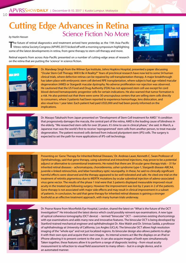
by Hazlin Hassan
The future of retinal diagnostics and treatment arrived here yesterday as the 11th Asia-Pacific Vitreo-retina Society Congress (APVRS 2017) kicked off with a morning symposium highlighting
some of the latest developments in retina, from gene therapy to stem cell therapy and more.
Retinal experts from across Asia-Pacific presented on a number of cutting-edge areas of research on the retina that are putting the ‘science’ in science fiction.
Dr. Mandeep Singh from the Wilmer Eye Institute, Johns Hopkins Hospital, presented a paper discussing “Ocular Stem Cell Therapy: Will It Be A Reality?” Years of preclinical research have now led to some 54 human clinical trials, where defective retinas can be repaired by cell transplantation therapy. A major breakthrough has taken place with embryonic stem cell derived RPE transplantation, where subjects had age-related macular degeneration (AMD) or Stargardt macular dystrophy. No adverse proliferation nor rejection was observed. He cautioned that the US Food and Drug Authority (FDA) has not approved stem cell use except for cord blood-derived hematopoietic progenitor cells for certain indications. He also warned that tumor formation is a risk. He also pointed out that there were some 30 unscrupulous centers that are selling stem cells directly to consumers, where 3 patients had been reported to experience hemorrhage, lens dislocation, and also visual loss 1 year later. Each patient had paid US$5,000 and had been poorly informed on the implications.
Dr. Masayo Takahashi from Japan presented on “Development of Stem Cell treatment for AMD.” A condition that progressively damages the macula, the central part of the retina, AMD is the leading cause of blindness in the elderly. “We researched stem cells for over 20 years. It’s time to use in clinical phase,” she said. In March, a Japanese man was the world’s first to receive ‘reprogrammed’ stem cells from another person, to treat macular degeneration. The patient received cells derived from induced pluripotent stem (iPS) cells. The surgery is expected to set the path for more applications of iPS-cell technology.
Dr. Pearse Keane from Moorfields Eye Hospital, London, shared the latest on “What is the future of the OCT Investigation?” He introduced the latest device which could possibly reinvent eye examinations. A new type of optical coherence tomography (OCT device) – termed “binocular” OCT – overcomes existing shortcomings with eye examinations and adds many new and innovative features. The binocular OCT is being developed by Stanford-trained mechanical engineer and ophthalmologist Dr. Alex Walsh and Dr. Srinivas Sadda, a professor of ophthalmology at University of California, Los Angles (UCLA). The binocular OCT allows high resolution imaging of the “whole eye” and not just localized regions. Its binocular design also allows patients to align it with their own eyes and acquire their own images. Its internal screens act like the displays on an iPad or iPhone allowing it to present content to the user. It can also acquire images of both eyes simultaneously. Taken together, these features allow it to perform a range of diagnostic testing – from visual acuity measurement to refraction to visual field assessment to many others – but in a single device, and in an automated manner.
Presenting on ‘Gene Therapy for Inherited Retinal Diseases,’ Dr. Andreas Lauer, Kenneth C. Swan Professor of Ophthalmology, said that gene therapy, using subretinal and intravitreal injections, may prove to be a potential adjunct or alternative to conventional treatments. He noted that there are 39 ocular gene therapy trials – 31 for inherited retinal diseases – achromatopsia, choroideremia, usher syndrome type-1, Stargardt disease-ABCA4, juvenile x-linked retinoschisis, and leber hereditary optic neuropathy. In these, he said no clinically significant harmful effects were observed and the therapy appeared to be well-tolerated and safe. He cited one trial on the treatment of retinitis pigmentosa due to MERTK mutations by ocular subretinal injection of adeno-associated virus gene vector. The results of the phase 1 trial were that 3 patients displayed measurable improved visual acuity in the treated eye following surgery. However the improvement was lost by 2 years in 2 of the patients. Gene therapy is not associated with major side effects and may result in clinical improvement in a subset of patients. In conclusion, he said that gene therapy for inherited retinal diseases is gaining a stronger foothold as an effective treatment approach, with many human trials underway.
Cutting Edge Advances in Retina Science Fiction No More

11| December 8-10, 2017 | Kuala Lumpur, MalaysiaSHOWDAILYAPVRS
What’s Next in
Retinal Imaging?by Khor Hui Min
Presenting about wide-field imaging at the retinal imaging session yesterday,
Dr. Paolo Antonio Silva emphasized that ultra-wide field (UWF) imaging is changing the way by which diabetic retinopathy (DR) is evaluated. Evaluation of the retinal periphery may provide significant additional prognostic value in determining the risk for progression. The presence of pars plana lensectomy (PPL) may reflect the extent of retinal ischemia and account for the increased risk of progression. A total of 1,712 eyes with baseline and 4-year follow-up UWF images were analyzed, with a fully automated hemorrhages and microaneurysms (H/MA) detection algorithm. Preliminary results showed no diabetic retinopathy (DR) and no non-proliferative diabetic retinopathy NPDR). Automated retinal image analysis and deep machine learning will potentially change the way images are evaluated.
Then Dr. Seung Young Yu from Kyunghee University Medical Center, Seoul, Korea, spoke on ‘Updates on Fundus Autofluoresence Imaging’. In the pre-retinal pigment epithelial (RPE) atrophic area, with chronic detachment, the photoreceptor eventually became atrophic and the concurrent atrophy of the underlying RPE can occur. She presented on pachychoroid pigment epitheliopathy (PPE), and elaborated on RPE abnormalities overlying the areas of choroidal thickening, and pachychoroid neovasculopathy (PNV). She talked about a recently described clinical entity in patients with choroidal neovascularization (Type 1 [Sub-RPE] neovascularization). She also spoke on the monitoring of White Dot Syndrome, as well as the phenotypes and diagnosis of retinal dystrophies. Fluorescein angiopathy (FAG) and fundus autofluorescence (FAF) were also discussed.
Dr. Pearse Keane from Moorfields Eye Hospital, London, presented on ‘The Use of Artificial Intelligence in OCT Analysis’. He talked about the increasing number of cases his eye hospital needs to handle due to the rising number of people taking the OCT or optical coherence tomography, which uses light waves to take cross-section pictures of the retina. They have partnered with the team from Google DeepMind to use Artificial Intelligence (AI) to come up with new ways to prevent blindness. Google DeepMind has over 300 of the best research scientists, and together, they are looking into new clinical and scientific applications.
‘Using OCTA as a Predictor of Treatment Outcomes’ was presented by Dr. Marten Brelen from the Chinese University of Hong Kong. He spoke on optical coherence tomography angiography (OCTA), which is used for non-invasive visualization of vascular structures. It can be used to perform OCT scans in rapid sequence with motion correction. All other elements remain the same, except the motion of erythrocytes within the vasculatures. The software will generate a decorrelation map of speckle pattern variability. OCTA is useful in diabetic retinopathy, identifying secondary choroidal neovascularisation (CNV) in central serous chorioretinopathy (CSCR), AMD subtypes, pre-retinal neovascular proliferation, vascular malformations, angiomatoses, and optical disc disorders. However, it has limited use in uveitis, choroiditis and vasculitis. In clinical applications, OCTA can be used to assess the indications for an intervention, qualify lesions, evaluate thicknesses, volume and extent of a lesion, assess the effects of treatment, and track the evolution of postoperative course of an intravitreal injection.
Dr. Colin Tan presented on ‘Can OCTA Replace ICGA in Detecting Polyps?’ He shared the research results from ‘EVEREST
Study Report 2: Imaging and Grading Protocol, and Baseline Characteristics of a Randomized Controlled Trial of Polypoidal Chroroidal Vasculopathy’. The diagnostic criteria of PCV include ICG criteria and clinical criteria. The ICG criteria includes nodular appearance stereoscopically, hyperfluorescent halo, branching vascular network (BVN), and pulsation of polyp. The clinical criteria are orange nodule (subretinal), and massive submacular hemorrhage. Visualization of polyps on en face OCTA was 42.9% of patients and 50% of lesions. OCTA is an exciting new technology, but there are still many unknowns. For now, ICGA is still the gold standard for PCV diagnosis and planning of PDT.
Dr. Srinivas Sadda continued the symposium with his presentation on ‘Is OCTA Here to Stay?’ He shared on the advantages and disadvantages of dye-based angiography. The advantages include good resolution of microvascular detail centrally, dynamic information, visualization of leakage/staining, non-invasive, very fast (few seconds only), and available on regularly used OCT platform. The disadvantages include being invasive, rare but potentially serious risks, time-consuming (5-20 mins), requires a skilled operator, limited depth-resolution, poor visualization of capillary circulation outside central macula, limited velocity range, leakage not visualized, challenging artifacts, limited field of view, and validation/repeatability of data only just becoming available. Disease-specific clinical trial data to guide use of OCTA for clinical decision-making is only now forthcoming.
Meanwhile Dr. Chan Jan-Bond from Malaysia, presented on ‘A Wireless Smartphone Videography System’, which he created himself and Dr. Ashish Sharma from India, talked about available low cost smartphone retinal imaging such as the MII Ret Cam.

12| December 8-10, 2017 | Kuala Lumpur, MalaysiaSHOWDAILYAPVRS
Dr. Fong further mentioned that the Malaysian Society of Ophthalmology (MSO) represents more than 700 eye doctors of all grades from both governmental and private practices. The MSO also runs public eye screening projects and in recognition of the burden of diabetic retinopathy in Malaysia, MSO has been running a free diabetic eye screening service for 4 years in partnership with Bank Simpanan Nasional (BSN). He also appealed to the Ministry of Health to continue to allocate resources towards the training of more eye specialists, prevention of myopia strategies and towards research in ophthalmology in Malaysia.
Dr. Taraprasad Das, President of APVRS warmly welcomed attendees and described Malaysia as ‘truly Asia’. He hinted that participants attending the conference should expect to encounter many wonderful scientific revelations over next couple of days and stressed that there would also be many social events for participants to meet and interact with each other.
Prof. Dennis Lam the Secretary General of APVRS, stated that though this was the 11th APVRS congress, it was the first time that it was hosted in Malaysia and that he was very excited to be here. He further stated that the Asia-Pacific region hosts
more than 50% of the world’s population and has become one of the world’s most important engines for the world economy. Prof. Lam invited all participants to join in the open interactive forum entitled ‘Meeting the Masters’ which will be held from 5.30 to 7.00 pm today where 8 prominent vitreo-retina leaders will join in as panelist and will answer challenging questions. Many inspiring stories and insights are to be expected from the experts at this engaging forum.
Guest of Honor, Datuk Dr. Shanaz Murad, Deputy Director General of the Ministry of Health, Malaysia, addressed the crowd on behalf of the Director General of the Ministry of Health. She acknowledged that the APVRS platform was vital
to the development of the vitreo-retina subspecialty in the Asia-Pacific region which allowed the sharing of information, knowledge and ideas in this subspecialty area. “Information by itself is not knowledge; it is the impartment of information that turns it into knowledge.” She highlighted the establishment of the Clinical Research Centers (CRC) in hospitals across Malaysia which is committed to the sharing of knowledge and information and invited all parties to interact with the CRC in order that significant research may be carried out.
The opening ceremony was followed by the presentation of this year’s prestigious APVRS Tano lecture award to Dr. Taraprasad Das, Vice Chair of the LV Prasad Eye Institute and Professor at Sun Yat-sen University. He is also a Fellow of the National Academy of Medical Sciences in India, Regional Chair of the International Agency for the Prevention of Blindness, President of VISION 2020 India and President of APVRS.
Dr. Das then delivered his award winning Tano lecture presentation on the topic of decoding evidence-based best practice in post-cataract surgery endophthalmitis prevention and treatment to the audience.
“Evidence and innovation are necessary for the practice and growth of science. Innovation to my mind is a state of mind that can turn events from a challenge to an opportunity,” emphasized Dr. Das.
>> Cont. from Page 1
Datuk Dr. Shanaz Murad, Deputy Director General of the Ministry of Health, Malaysia, graced the APVRS 2017 opening ceremony as Guest of Honor.
Prof. Dennis Lam (left) and Dr. Taraprasad Das (right)

13| December 8-10, 2017 | Kuala Lumpur, MalaysiaSHOWDAILYAPVRS
The Future Is Now Restoring Sight with Bionic Eyes and Robot Surgeries
by Hazlin Hassan
According to the World Health Organization, over 285 million people
worldwide are blind or vision impaired.
But now there is hope. Pioneering technology in retinal surgery to restore sight that has been lost for years, is reaching new heights with futuristic advances previously seen as impossible such as bionic eyes and robotic eye surgeries.
Leading the way are trailblazing distinguished speakers from all over the world who shared their experiences in the “Advances in Vitreo-retinal Surgery” symposium yesterday.
Dr Young Hee Yoon, from Korea, presented on the “Initial Experience of Argus II Implantation in Korea,” about Argus II Prosthesis, or “bionic eye”. It works via a camera mounted in a pair of glasses and a tiny computer to relay signals directly to the nerves controlling sight. She tested it on 3 patients. The first patient, aged 53, earlier suffered
visual loss. Following the insertion, the patient could eventually read and write in her native Korean again, becoming the first Korean patient to be able to resume reading and writing with the Argus II. The other 2 patients also gained improvements in sight after having had vision loss. The prosthesis won Korean FDA approval in April.
Vitreo-retinal surgeon Dr. Peter Stalmans, from Belgium, shared with the congress his experience in the use of robots in eye surgeries to overcome a number of issues faced by surgeons in this field. With the use of a glass microneedle measuring 30 micron in outer diameter and coated in aluminum, making it easier to go into the inner lumen of the retinal vein during surgeries, the robot also eliminates surgeons’ tremors and shaking. Future uses of the robot will be to limit the movement in the eye preventing surgeons from going in too deep and causing potential retinal damage. So far, the robot has been used on pigs’ eyes.
Presenting on subretinal implants, Dr. Caroline Chee, from Singapore, shared the use of subretinal implants Alpha-IMS and Alpha-AMS, which act like a camera in the eye. Containing a microchip, the implants are similar to a digital camera, inserted under the retina. The implant is able to replace the functionality of degenerated photoreceptors to a limited
degree via electrical stimulation of the outer retinal tissue. As a result, information can be transmitted to the visual cortex of the brain through the optic nerve, thus generating visual images. In adult patients whose vision has been affected by retinal degeneration, the chip can help to partially restore functional eyesight. Dr. Chee shared that 72% of patients with the Retina Implant Alpha-IMS reached the primary efficacy endpoint, with improvement of activities of daily living and mobility.; 45% reported restoration of visual function which they use in daily life. In conclusion, she said that the electric multifocal stimulation has the potential to restore useful vision up to the ability to read.
Associate Professor PJ Allen from the Centre for Eye Research Australia, shared on the topic of “The Suprachoroidal Retinal Prosthesis: Preparing for Australia’s Next Clinical Trial.” A 2 year study, the Bionic Vision Australia (BVA) Prototype Clinical Trial 2012-14, using 24 stimulating electrodes, no implanted electronics, and suprachoroidal surgical placement was conducted on 3 patients with profound vision loss from retinitis pigmentosa. All patients experienced artificial vision. The surgery was safe and the device was stable over 2 years. Patients underwent successful visual rehabilitation, showing improvement in navigational abilities and activities of daily living. No long-term effects were found on the health of the retina. Preliminary results indicate that dual-electrode simultaneous stimulation is safe at a clinically viable level. A clinical trial using a 44-electrode prototype will begin in 2017. Feedback from patients will allow researchers to develop more sophisticated vision processing and stimulation techniques with the aim of improving mobility and quality of life.
Dr. Peter Stalmans
Dr Young Hee Yoon

14| December 8-10, 2017 | Kuala Lumpur, MalaysiaSHOWDAILYAPVRS
Recent Advances in Age-related
by Collins Santhanasamy
The symposium started with a presentation from Dr. Hazlita Mohd. Isa
on ‘Updates in the Epidemiology of AMD’. AMD contributes significantly as one of the causes of poor vision and blindness globally, mainly affecting Europeans in higher income regions. Dr. Hazlita spoke about the prevalence of AMD increasing with age, and the rapid increasing in prevalence after the age of 75, especially in the European and Oceania region. After that, she discussed the risk factors associated with AMD. In Europe, the top risk factors were older age, smoking, previous cataract surgery, and family history of AMD. There was also a moderate association with cardiovascular disease, hypertension, and raised fibrinogen. In Asia, the top risk factors were older age, smoking, shorter axial length, previous cataract surgery, diabetes, and chronic renal disease. Meanwhile, the genetic risk factors were SNPs in complementary factor H (CFH) gene ARM52/HRTA1 loci CETP mutation. The projection for AMD rise is 196 million by 2020, and 288 million by 2040. The largest increase is expected in Asia (113 million), followed by Europe (69 million) and Africa (39 million).
Next, Dr. Danny Ng from Chinese University of Hong Kong presented on ‘Updates in the Genetics of AMD’. The two strongest risk factors for AMD are age and positive family history. AMD is considered a well-defined complex genetic disorder. In genetic associations with AMD severities, of particular importance is the 10 variants located in 7 extracellular matrix genes associated with only advanced AMD (neovascular AMD and geographic atrophic AMD), but not immediate AMD. This suggests that extracellular remodelling parthways of the retina/RPE
are activated in the development of the more advanced stages of AMD. Pathway analysis may also indicate that there may be regulators/modulators for the gene/variant of interest that are important as therapeutic targets. Therefore, adding genotypic information, particularly from the CFH and ARMS2/HTRA1 loci to phenotypic features from clinical examination will increase the precision of predicting risk of advanced AMD.
‘Advances in OCT (Optical Coherence Tomography) and OCTA (Optical Coherence Tomography Angiography) Imaging in AMD’ was the next presentation. Dr. Gemmy Cheung spoke on biomarkers, and explained and elaborated on the subject with various studies. Then, she discussed the clinical applications of OCTA. OCT is an integral part of AMD clinical practices. Advances in both hardware and software will further improve our ability to diagnose, monitor and prognosticate in both neovascular and non-neovascular AMD. OCTA is also rapidly being adopted as an adjunct in clinical practice. Further developments in developing common nomenclature, improving repeatability, quantification and validation, in clinical trials may propel this imaging modality to ‘standard use’.
After that, Dr. Jayakrishna Ambati from the University of Virginia presented on ‘Kamuvudines: A Potential Treatment for Macular Degeneration’. He explained how Alu RNA induces RPE degeneration. Then, he explained how Nucleoside Reverse Transcriptase Inhibitors (NRTIs) block RPE degeneration induced by Alu RNA. Kamuvudines block Alu RNA toxicity, but without blocking reverse transcriptase. However, Dr. Jayakrishna said that intravitreous injection of kamuvudines is safe for the retina.
Andreas Lauer from Casey Eye Institute, Portland, USA spoke on ‘Gene Therapy for Neovascular AMD’. He spoke on gene therapy treatment effects, which are mainly the replacement of absent/defective proteins in Inherited retinal diseases (IRD), and modular secretants (AMD). He then explained about the
history of clinical gene therapy trials for neovascular age-related macular degeneration. In the gene therapy for age-related macular degeneration, the successful delivery of vector has been established, and the successful expression of transgene has been established. The ocular and systemic safety and tolerability have also been established. Trials have remained at Phase 1 or Phase 1/2 stage. The treatment for both neovascular and non-neovascular forms are considered, but the effects of long-term anti-angiogenesis or VEGF suppression are still unknown.
The following presentation was by Dr. Clement Chan. He spoke on ‘New Drugs and Therapeutic Targets for Neovascular AMD (nAMD)’. He explained in detail about the vascular endothelial growth factor (VEGF) family and receptors. He discussed about strategies to improve outcome and reduce treatment burden.
Dr. Joo Yong Lee from Asan Medical Center, Seoul, Korea,gave the next presented on ‘Stem Cell Therapy for Dry AMD’. He discussed the current status of stem cell treatment for retinal diseases. Stem Cell Therapy was classically considered to be one type of cell therapy. He then shared the latest methods for delivering stem cells and recent clinical trials for stem cells in retinal diseases.
Dr. Masayo Takahashi from Kobe City Medical Center General Hospital presented on ‘Stem Cell Therapy for Neovascular AMD’. She spoke on treatment for AMD with hiPS-RPE cell sheets. Then, she shared about the visual acuity before and after the surgery.
The final presentation was ‘Home Monitoring for Earlier Detection of Neovascular AMD’ by Dr. Susan Bressler from John Hopkins University School of Medicine, USA. The randomized trials of the ForeseeHome (FH) device showed that in contrast to current home and office monitoring strategies reflected in the standard care arm, patients at risk of progression to advanced AMD would benefit from this home monitoring device.
Macular Degeneration (AMD)

16| December 8-10, 2017 | Kuala Lumpur, MalaysiaSHOWDAILYAPVRS
Innovations at Zeiss Add to Retinal Armamentarium
Beyond CLARUS, the new ultra-widefield imaging modality at Zeiss (Jena,
Germany), the company is featuring two extremely powerful additions to the retina physician’s armamentarium: AngioPlex and CALLISTO 3.6.
AngioPlex provides quantifiable parameters for better decision making. The principle behind the technology is providing a full spectrum of OCT data. And now, Zeiss is introducing AngioPlex Metrix, which provides quantification values of vessel and perfusion density.
“We think AngioPlex is the next generation of vascular imaging,” said Terrance Siew, Regional Product Manager, Ophthalmic Diagnostics at Zeiss. “It provides non-invasive depth-resolved images of the retina microvasculature in the eye, so it helps ophthalmologists detect retinal diseases earlier and improves treatment and management of the diseases.”
For example, AngioPlex Metrix quantifies vascular and perfusion density, allowing doctors to objectively assess change over time with anti-VEGF drugs. They can see and monitor the changes.
Mr. Siew explained that perfusion density – or in other words how much perfuse vasculature is available – could be favorable or unfavorable, depending on the circumstance. New vessel growth, which is not supposed to take place, could lead to a higher rate of perfused density, he said. Alternatively, if you have an area of non-perfusion due to diabetic retinopathy, that is not favorable. Hence, perfusion density is a complex issue, but needs to be monitored in any case.
AngioPlex Metrix also can help to flag patients with early diabetic retinopathy changes with the automatic detection of FAZ area and circularity.
AngioPlex started off with version 9, then 9.5, and now 10, which is considered AngioPlex Metrix.
The system also has Optical Micro Angiography (OMAG) Algorithms, which is proprietary.
“OMAG is basically an image processing technique that takes full advantage of
OCT signal data to deliver the highest quality image possible,” Mr. Siew said. The AngioPlex incorporates FastTrac™ technology which will be able to minimize motion artifacts. Further, during follow-up visits, FastTrac™ provides the ability to scan precisely at the same location, so we will be able to monitor the progressions at the same exact location.” Mr. Siew said.
He added: “AngioPlex will be very valuable because it will expand the capabilities of OCTA,” he said. “Doctors can now eliminate the subjective interpretations with automated quantification data, which can be obtained with a high level of repeatability and reproducibility. Therefore, I believe the AngioPlex may become a standard initial examination step for patients with retinal diseases. In particular, it is useful for diabetic retinopathy, CRVO, BRVO, ischemic areas, and other conditions. We can even look at MacTel patients and neovascularization of the retina.”
Meanwhile, CALLISTO has been around for nearly 3 years, but as a tool for anterior segment surgeons. Anterior segment surgeons first had CALLISTO 3.5, while posterior segment surgeons first had CALLISTO 3.2. Things are changing now.
“At ESCRS this year we launched CALLISTO 3.6,” said Jil Teo, Senior Regional Product Manager, OPMI and Phaco Systems, Southeast Asia. “What we did was combined both the Zeiss cataract suite markerless toric implantation together with iOCT (intraoperative OCT). This is now the one-and-only microscope in the world that has an all-in-one microscope. We can cover applications from the cornea to the retina to cataract and glaucoma.”
Before the advent of CALLISTO 3.6, hospitals needed to buy two microscopes for doctors
doing cataract toric implantation, and for doctors performing retina work.
“Now they can buy an all-in-one microscope,” Ms. Teo said. “That’s really good for hospitals and institutes.”
Ms. Teo said that is especially the case since many countries now in the region are focusing more on toric IOL implantation.
Ms. Teo added that CALLISTO 3.6 particularly benefits surgeons doing complicated cataract procedures with some retina involvement. The biggest benefit here is that instead of OCT only being a useful tool for retinal specialists, it becomes much broader spectrum.
Having an intraoperative OCT with the benefits of CALLISTO 3.6 for cataract surgery assistance justifies ROI for hospitals with budgetary constraints.
The Benefit of Using OCTA with AngioPlex
“I run intravitreal injection clinics. Many of these patients have macular or vascular problems like diabetics, AMD, RVO and patients with MacTel. Now my standard practice is this: Any patient that comes in with vascular problems at the macula, I use OCTA with AngioPlex on the first presentation. OCTA is reliable enough so we don’t have to do fundus fluorescein angiography. We then get an assessment of macular perfusion. Many conditions, in fact, affect perfusion and impact on the visual prognosis. So, if the patient comes in, for example, with DME and at first OCTA shows severe macular ischemia we can advise the patient accordingly that the prognosis may not be as good to manage expectations.”
– Dr. “Danny” Yew Wong, Southern Specialist Eye Centre, Melaka, Malaysia.
AngioPlex Metrix