12249 2015 322 article 1344. · their drug permeation enhancement effects on the transdermal film...
TRANSCRIPT
Research Article
Influence of the Component Excipients on the Quality and Functionalityof a Transdermal Film Formulation
Suprit D. Saoji,1 Sandip C. Atram,2 Pradip W. Dhore,1 Priya S. Deole,3 Nishikant A. Raut,1 and Vivek S. Dave4,5
Received 23 February 2015; accepted 8 April 2015; published online 29 April 2015
Abstract. The influence of formulation variables, i.e., a hydrophilic polymer (Methocel® E15) and a film-forming polymer (Eudragit® RL 100 and Eudragit® RS 100), on the physicochemical and functionalproperties of a transdermal film formulation was assessed. Several terpenes were initially evaluated fortheir drug permeation enhancement effects on the transdermal film formulations. D-Limonene was foundto be the most efficient permeation enhancer among the tested terpenes. Transdermal film formulationscontaining granisetron (GRN) as a model drug, D-limonene as a permeation enhancer, and different ratiosof a hydrophilic polymer (Methocel® E15) and a film-forming polymer (Eudragit® RL 100 or Eudragit®
RS 100) were prepared. The prepared films were evaluated for their physicochemical properties such asweight variation, thickness, tensile strength, folding endurance, elongation (%), flatness, moisture content,moisture uptake, and the drug content uniformity. The films were also evaluated for the in vitro drugrelease and ex vivo drug permeation. The increasing ratios of Methocel®:Eudragit® polymers in theformulation linearly and significantly increased the moisture content, moisture uptake, water vaportransmission rate (WVTR), and the transdermal flux of GRN from the film formulations. Increasinglevels of Methocel® in the formulations also increased the rate and extent of the GRN release and theGRN permeation from the prepared films.
KEY WORDS: film-forming polymers; hydrophilic polymers; permeation enhancers; transdermal films.
INTRODUCTION
The transdermal drug delivery can no longer be consid-ered a Bnovel^ drug delivery technology. Over the past fewdecades, there has been a tremendous growth in the under-standing of the technology involved in the development oftransdermal drug delivery systems (TDDS). TDDS are de-signed to deliver the drug(s) across the skin at a controlledrate into the systemic circulation. TDDS offers several, well-documented advantages over the conventional oral and par-enteral routes of drug delivery (1–3). These include therapeu-tic advantages such as sustained delivery of drugs, avoidingpre-systemic metabolism, avoiding hostile gastrointestinal fac-tors (e.g., gastric pH, gastric transit time, etc.), suitability fordrugs with short biological half-life and narrow therapeuticindex, and relatively lower fluctuations observed in the drugplasma levels. Some of the patient compliance benefits includenon-invasive drug administration, flexibility in the dosing fre-quency, suitability for self-medication, and better control of
the side effects/adverse effects. TDDS are also associated withuncommon limitations such as suitability limited to potent(low dose) drugs, stratum corneum as a barrier to drug ab-sorption, requirement of the ideal permeability characteristicsof the drugs, non-suitability for tolerance-inducing drugs, andcostly nature of the dosage form (1,4,5). Nevertheless, severaltransdermal formulations consisting of drugs over a range ofpharmacological classes are approved to be marketed in theUSA or are in different stages of development (4,5). TheTDDS market is reported to be worth $12.7 billion in 2005and is anticipated to be over $30 billion this year (2015) (4).
Stratum corneum, being a natural protector, limits thepenetration of the xenobiotics. It is known to be a majorrate-limiting factor to the transdermal absorption of a drugand its subsequent systemic bioavailability (6). The role ofpermeation enhancers in the formulation of transdermal sys-tems was envisaged early on to overcome the challenge oflimited transdermal absorption of a drug (7,8). Permeationenhancers originating from a wide variety of chemical classes,including alcohols, fatty acids, amines, esters, amides, hydro-carbons, surfactants, etc., have been explored to enhance thetransdermal absorption of the drugs (9). The permeation en-hancers act mainly by disrupting the lipid bilayers within thestratum corneum (10). The disruption of the natural integrityof the stratum corneum is known to cause skin irritation, andthus, a rational selection of a permeation enhancer that en-hances the drug permeation through the skin with minimalalteration of the structure and functions of the skin is of
1 Department of Pharmaceutical Sciences, R. T. M. Nagpur University,Nagpur, India.
2 Vidyabharati College of Pharmacy, Amravati, India.3 G. S. Tompe College, Chandurbazar, Amravati, India.4 St. John Fisher College, Wegmans School of Pharmacy, 3690 EastAvenue, Rochester, New York 14618, USA.
5 To whom correspondence should be addressed. (e-mail:[email protected])
AAPS PharmSciTech, Vol. 16, No. 6, December 2015 (# 2015)DOI: 10.1208/s12249-015-0322-0
13441530-9932/15/0600-1344/0 # 2015 American Association of Pharmaceutical Scientists

critical importance (11). Terpenes are naturally occurring vol-atile oils that have gained popularity in the recent years aschoice enhancers in transdermal formulations (12). Terpenesare also known to be versatile in their applications, i.e., theyare suitable for the percutaneous penetration and absorptionenhancement of both hydrophilic and lipophilic drugs (13,14).Moreover, compared with surfactants and other permeationenhancers, terpenes, when used in lower concentrations, ex-hibit relatively lower percutaneous irritancy (15).
Other key components of transdermal formulationsapart from the permeation enhancers include the film-forming polymers and one or more hydrophilic polymersincorporated as rate-controlling matrix formers (16–18).These excipients are available in a wide range of physico-chemical properties and grades. The nature and theamount of these excipients incorporated in a TDDS canhave a significant influence on the manufacturability, qual-ity, and performance of the final drug product. Most ofthe studies report the incorporation of a variety of theseexcipients in the formulation of transdermal delivery sys-tems (19–21). However, the studies that evaluate the in-fluence of these formulation variables in a systematic,quantitative manner are few and far between.
The present investigation was aimed at quantitativelyand systematically assessing the influence of the ratio oftwo formulation variables, i.e., a hydrophilic polymer anda film-forming polymer, on the physicochemical and func-tional attributes of transdermal films. The study was car-ried out in three stages. First, several terpenes wereevaluated for their permeation-enhancing efficiency acrossthe excised hairless rat skin. Granisetron (GRN) was cho-sen as a model drug for this study. The physicochemicalproperties of GRN allow it to be incorporated into atransdermal drug delivery system. These properties in-clude low molecular mass, high lipid solubility, low thera-peutic dose, as well as a high degree of first-passmetabolism. Next, several transdermal film formulationscontaining different ratios of a hydrophilic polymer(Methocel® E15) and a film-forming polymer (Eudragit®
RL 100 or Eudragit® RS 100) were prepared. Finally, theprepared films were evaluated for their physical, chemical,in vitro drug release, and ex vivo drug permeationproperties. The results were statistically analyzed tounderstand the influence of these formulation variableson the prepared films.
MATERIALS AND METHODS
Materials
Granisetron samples were obtained from WockhardtLimited (India). Hydroxypropyl methycellulose (Methocel®
E15) was obtained from Colorcon Asia Pvt. Ltd. (India).Eudragit® RL 100 and Eudragit® RS 100 were obtainedfrom Evonik Degussa India Pvt. Ltd. (India). Eugenol andD-limonene were obtained from Keva Fragrances Ltd. (India).α-Terpineol and menthol were obtained from Workwell(India). ScotchPak™ 1109 backing membrane was obtainedfrom 3M Drug Delivery Systems, USA. Other solvents andreagents used were of analytical grade.
Methods
Preparation of Biological Membrane for Permeability Studies
Ethical clearance for the handling of experimental ani-mals was obtained from the institutional animal ethics com-mittee (IAEC) formed for this purpose. Male SpragueDawley® rats weighing 200 to 250 g were euthanized bycervical dislocation, and the dorsal skin was removed. Afterremoving the epidermal hair and the subcutaneous fat, theskin was thoroughly washed with phosphate-buffered saline(PBS, pH 7.4) and placed overnight in contact with the recep-tor phase, i.e., fresh PBS, pH 7.4. The overnight-stored rat skinwas used for ex vivo permeability studies.
Effect of Penetration Enhancers on Permeation of Granisetron
The ex vivo permeation studies were carried out in a Franzdiffusion cell with a diffusion surface area of 2.5 cm2. Thebiological membrane, with the stratum corneum facing thedonor segment, was affixed to the diffusion cell. GRN solution(1 mg/ml, in PBS, pH 7.4) was placed in the donor compartmentcontaining D-limonene (5% v/v), α-terpineol (5% v/v), eugenol(5% v/v), or menthol (5% v/v). The absence of penetrationenhancer served as a control. The receiver compartmentcontained magnetically stirred PBS (18 ml, pH 7.4), maintainedat 37±0.5°C. The amount of drug permeated through the biolog-ical membrane was determined by analyzing the drug concentra-tion in the filtered samples (1 ml), withdrawn at predeterminedtime intervals. The samples were analyzed using a modifiedreverse-phase HPLC at a wavelength of 305 nm. The cumulativeamount (%) of drug permeated was plotted against time.
Permeation Data Analysis
The drug concentration in the permeate was corrected forsampling effects according to the equation described byHayton et al. (Eq. (1)) (22):
C0n ¼ Cn
VT
VT−Vs
� �C
0n−1
Cn−1
!ð1Þ
where C0n is the corrected concentration of the nth sample, Cn
is the measured concentration of GRN in the nth sample, Cn�1
is the measured concentration of the GRN in the (n−1)thsample, VT is the total volume of the receiver fluid, and Vs isthe volume of the sample drawn. The steady-state flux (Jss)and the permeability coefficient (Kp) were calculated usingEqs. (2) and (3), respectively, as described by Barry (23):
Jss ¼ dQdt
� �ss� 1
Að2Þ
Kp ¼ JssCs
ð3Þ
where A is the effective diffusion area, Cs is the concentrationin the saturated solution, and dQ
dt
� �ss is the steady-state slope.
The penetration-enhancing effect of the terpenes was
1345Influence of Component Excipients on Transdermal Films

calculated as enhancement ratio (ER), using the followingequation (Eq. (4)) (12):
ER ¼ KP with penetration enhancerð ÞKP without penetration enhancerð Þ ð4Þ
Drug-Polymer Compatibility Studies
The preliminary physicochemical compatibility betweenGRN and the polymers used in the films was evaluated usingdifferential scanning calorimetry (DSC) and the Fourier trans-form infrared (FTIR) spectroscopy.
Differential Scanning Calorimetry. The thermal analysisof GRN, the pure polymers, and their physical mixtureswith GRN was carried out using a differential scanningcalorimeter (Model: Q10, TA Instruments, Inc., NewCastle, DE, USA). The analysis was done under a purgeof dry nitrogen gas (50 ml/min). High-purity indium wasused to calibrate the heat flow and the heat capacity ofthe instrument. The samples (2.5–5 mg) were sealed inaluminum pans with the lids using a crimper. Each sam-ple was subjected to a single heating cycle from 25 to400°C at a heating rate of 10°C/min. The results wereanalyzed using the Universal Analysis software version4.5A, build 4.5.0.5 (TA Instruments, Inc., New Castle,DE, USA).
Fourier Transform Infrared Spectroscopy. The infraredspectra of GRN, the polymers, and their physical mix-tures with GRN were obtained from an FTIR spectro-photometer (Model: IR Prestige-21, Shimadzu, Japan)equipped with an attenuated total reflectance (ATR) ac-cessory. The analysis of the samples was carried outusing diffuse reflectance spectroscopy (DRS)-FTIR withKBr. The influence of the residual moisture was theoret-ically removed by subjecting the samples to vacuum dry-ing before obtaining any spectra. Each sample analysisincluded 45 scans, at a resolution of 4 cm−1 from 4000 to600 cm−1.
Preparation and Characterization of Granisetron TransdermalFilms
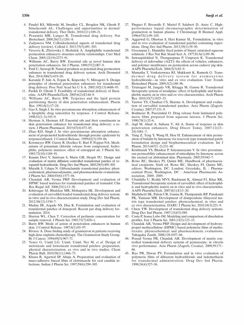
As shown in the Table I, ten matrix-type transdermalfilm formulations containing GRN, Methocel® E15, andEudragit® RL 100 or Eudragit® RS 100 were prepared.The preparation method as described previously byKusum Devi et al. was followed (17). Briefly, the drugand the polymers, propylene glycol (plasticizer), and D-limonene (penetration enhancer) were dispersed inethanol (casting solvent), and the polymeric dispersionwas degassed by sonication. The polymeric dispersion ofthe drug was then poured into a circular well formed by aglass mold (5.68 cm2, fabricated in the laboratory for thepurpose), placed in a standard Petri dish containingmercury (Fig. 1). The rate of solvent evaporation was
qualitatively controlled by covering the mold with aninverted funnel. The dry films were obtained after theevaporation of the casting solvent overnight in a solventhood. The dried formulation films were carefully cut intouniform sizes to contain a theoretical equivalent of ∼4 mgof the drug per film. This dose of GRN (4 mg per film)was targeted based on the recommended therapeuticdoses of GRN for chemotherapy-induced nausea andvomiting (24). Preliminary studies based on film dimen-sions showed that GRN (9.1% w/w) in the formulationsachieved this target dose per film. A backing membranewas attached to the prepared films, and the films werestored in a desiccator, between the sheets of glassinepaper.
Evaluation of Physicochemical Properties of Films
Weight Variation. The weight variation of the preparedfilms was evaluated by individually weighing three randomlyselected films of each prepared formulation. The mean weightand the standard deviation for each formulation film werecalculated and recorded.
Thickness. The thickness of prepared films was measuredusing a digital micrometer screw gauge with a least count of0.001 mm (Mitutoyo, Japan) at three different locations on thefilm. Three randomly selected films of each formulation wereevaluated for thickness. The mean thickness (mm) and thestandard deviation for each formulation film were calculatedand recorded.
Tensile Strength. The mechanical properties of the pre-pared polymer films were determined by measuring theirtensile strength as described previously by Yener et al.(25). According to the American Society for Testing andMaterials (ASTM), the tensile strength of polymeric filmsis a measure of the minimum stretching force applied tothe sample that results in the breaking of the film. Thetensile strength measurements were carried out ondumbbell-shaped films (2.5×1 cm). The formulation filmswere subjected to a pulling force by hanging standardweights from the films and measuring the force (kg/mm2)using a load cell.
Percentage Elongation. The determination of longitudi-nal strain (percentage elongation at break) was carried out asdescribed by Khanna et al. (26). The percentage elongation atbreak for the film samples was calculated using the followingformula (Eq. (5)):
%elongation at break ¼ increase in lengthinitial length
� 100 ð5Þ
Folding Endurance. The folding endurance was evalu-ated by repeatedly folding a small strip of film (2.5 cm2)at the same location to the point of breaking (17). Thefolding endurance value is typically described as thenumber of times a film may be folded at the same
1346 Saoji et al.

location before it breaks. Three randomly selectedsamples of each prepared formulation film wereevaluated for the folding endurance. The mean and thestandard deviation for each formulation were calculatedand recorded.
Drug Content Uniformity. The GRN content wasassayed on ten randomly selected films from each formu-lation. The prepared films were dissolved in ethanol(2 ml), and the volume was adjusted to 100 ml with PBS(pH 7.4). The solution was filtered and suitably diluted,and the GRN content of each film was determined using amodified reverse-phase HPLC (RP-HPLC) method previ-ously described by Pinguet et al. (27). Briefly, the HPLCsystem (Model: Prominence, Shimadzu Corporation,Kyoto, Japan) with LCsolution software, equipped with aLC-20AD HPLC pump, a manual rheodyne sample injec-tor, and a SPD-M20A detector, was used. The mobilephase was composed of methanol/0.02 M phosphate buffer(pH 4.0) (40:60, v/v), at a flow rate of 1 ml/min. AHypersil-Keystone C-18 column (250×4.6 mm, 5 μm) wasused as a stationary phase, and the GRN samples weredetected by ultraviolet (UV) detection at 305 nm. Thecalibration of the peak area against concentration ofGRN was found to be y=10,498x−73.42 with R2=0.9997for the GRN concentration range of 2–10 μg/ml (wherey=peak area and x=GRN concentration), and the limit ofdetection was found to be 0.02 μg/ml.
Flatness . A method prev ious ly descr ibed byKshirsagar et al. was used to calculate the flatness of the
prepared films (20). Briefly, the prepared films were cutinto uniform longitudinal strips. The length of each stripwas then measured accurately, and the variations inlength due to the non-uniformity of flatness were mea-sured. The flatness of the strips was calculated by mea-suring the percent constrict ion using Eq. (6). Aconstriction value of 0% was assumed to indicate 100%flatness. Three randomly selected films of each formula-tion were evaluated for flatness. The mean and the stan-dard deviation values for each formulation were calculated andrecorded:
%Constriction ¼ l1−l2l2
� 100 ð6Þ
Percent Moisture Content. Three randomly selected filmsof each prepared formulation were marked, then weighedindividually, and placed in a desiccator at room temperature(25±0.5°C). The films were then removed and weighed atpredetermined intervals until a constant weight was achieved.The moisture content (%) was calculated as a differencebetween the initial and the final weight with respect to thefinal weight (Eq. (7)) (28). The mean and the standard devi-ation values for each formulation were calculated and record-ed.
% moisture content ¼ final wt:−initial wt:initial wt:
� 100 ð7Þ
Percentage Moisture Uptake. The physical stability andthe integrity of the polymeric films in humid environments
Table I. Composition of the Transdermal Films
Formulation
Composition (% w/w)
F1 F2 F3 F4 F5 F6 F7 F8 F9 F10
Granisetron 9.1 9.1 9.1 9.1 9.1 9.1 9.1 9.1 9.1 9.1Methocel® E15 72.7 54.5 45.5 36.4 18.2 72.7 54.5 45.5 36.4 18.2Eudragit® RL 100 18.2 36.4 45.5 54.5 72.7 0.0 0.0 0.0 0.0 0.0Eudragit® RS 100 0.0 0.0 0.0 0.0 0.0 18.2 36.4 45.5 54.5 72.7Propylene glycola 25.0 25.0 25.0 25.0 25.0 25.0 25.0 25.0 25.0 25.0D-Limoneneb 5.0 5.0 5.0 5.0 5.0 5.0 5.0 5.0 5.0 5.0
aQuantity represents % w/w of the total polymer weightbQuantity represents % w/w of the total polymer weight
Fig. 1. Preparation of GRN films. a Circular glass mold in a Petri dish filled with mercury, b Mold coveredwith an inverted funnel to qualitatively control solvent evaporation, and c Dried GRN film
1347Influence of Component Excipients on Transdermal Films
are typically evaluated by calculating the percent moistureabsorption at elevated humidity conditions.
The extent of moisture uptake by the prepared filmsat elevated humidities and at room temperature (25±0.5°C) was determined using the method described byKusum Devi et al. (17). Briefly, saturated solutions ofammonium dihydrogen phosphate (60±5% RH), sodiumchloride (75±5% RH), and potassium nitrate (90±5%RH) were placed in three separate desiccators respectivelyand allowed to equilibrate for 24 h to attain the respectivehumidity values (29). Three randomly selected films fromeach formulation were weighed and placed in each desic-cator. The films were then removed at predetermined in-tervals and carefully weighed, until a constant weight wasachieved (17). The percentage moisture uptake by thefilms was calculated using Eq. (8):
% moisure uptake ¼ final wt:−initial wt:initial wt:
� 100 ð8Þ
Water Vapor Transmission Rate. The water vapor trans-mission rate (WVTR) is the amount of moisture transmittedthrough a unit area of film in a given duration at room tem-perature (25±0.5°C). The WVTR for the prepared films wascalculated using a method reported earlier (17). Briefly, glasscells were filled with 2 g of anhydrous calcium chloride, and aformulation film of specified area was affixed onto the cell rim.The cells were accurately weighed and placed in desiccatorscontaining saturated solutions of ammonium dihydrogenphosphate (60±5% RH), sodium chloride (75±5% RH), orpotassium nitrate (90±5% RH), respectively (29). The glasscells were removed from the desiccators at regular intervalsand weighed accurately. The process was continued until aconstant weight was achieved. The amount of water vaportransmitted was determined using the formula below(Eq. (9)):
WVTR ¼ final weight−initial weightarea� time
ð9Þ
In Vitro Release Studies
The in vitro release of GRN from the prepared formula-tion films was determined using a Franz diffusion cell. Threerandomly selected films of each formulation were used for theanalysis of drug release from the prepared films. The film,supported with a backing membrane, was placed in the donorcompartment of the cell. The donor compartment was sepa-rated from the receptor compartment by a dialysis membrane(Himedia®), with a molecular weight cutoff between 12,000and 14,000. The dialysis membrane was previously stored inPBS (pH 7.4) for 24 h. The receptor compartment containedmagnetically stirred PBS (18 ml, pH 7.4) maintained at 37±0.5°C. The amount of GRN released was determined byanalyzing the drug concentration in the filtered samples(1 ml), withdrawn at predetermined time intervals. The
samples were analyzed using a modified RP-HPLC at a wave-length of 305 nm. The cumulative drug release (%) was cal-culated and plotted against time.
Ex Vivo Permeation Studies
The ex vivo permeation studies were carried outusing a Franz diffusion cell mounted with excised rat skin,with the stratum corneum facing the donor compartment.The drug-releasing surface of the transdermal film underevaluation was placed in contact with the stratumcorneum side of the skin. The receiver compartmentcontained magnetically stirred PBS (18 ml, pH 7.4), main-tained at 37±0.5°C. The amount of GRN permeated wasdetermined by analyzing the drug concentration in thefiltered samples (1 ml), withdrawn at predetermined timeintervals. The samples were analyzed using a modifiedRP-HPLC at a wavelength of 305 nm. The cumulativeGRN permeated (%) was calculated and plotted againsttime.
The target flux was calculated using the equation de-scribed previously by Suwanpidokkul et al. (Eq. (10)) (30):
Jtarget ¼ CSSCltBWA
ð10Þ
whereA represents the surface area of the transdermal film (i.e.,2.5 cm2); BW, the standard human body weight of 60 kg;CSS, theGRN concentration at the therapeutic level (3.07 μg/l); and Clt,the total clearance (0.52 l/h). The calculated target flux value forGRN was 42.57 μg/h/cm2.
Statistical Analysis
The statistical analysis was carried out by a two-wayanalysis of variance (ANOVA), followed by Bonferronipost-test using GraphPad® Prism® software version 5.03(San Diego, CA). The results were expressed as mean±standard deviation (SD). The differences between themeans were considered to be significant if the P valuewas <0.05.
RESULTS
Effect of Penetration Enhancers on the Permeationof Granisetron
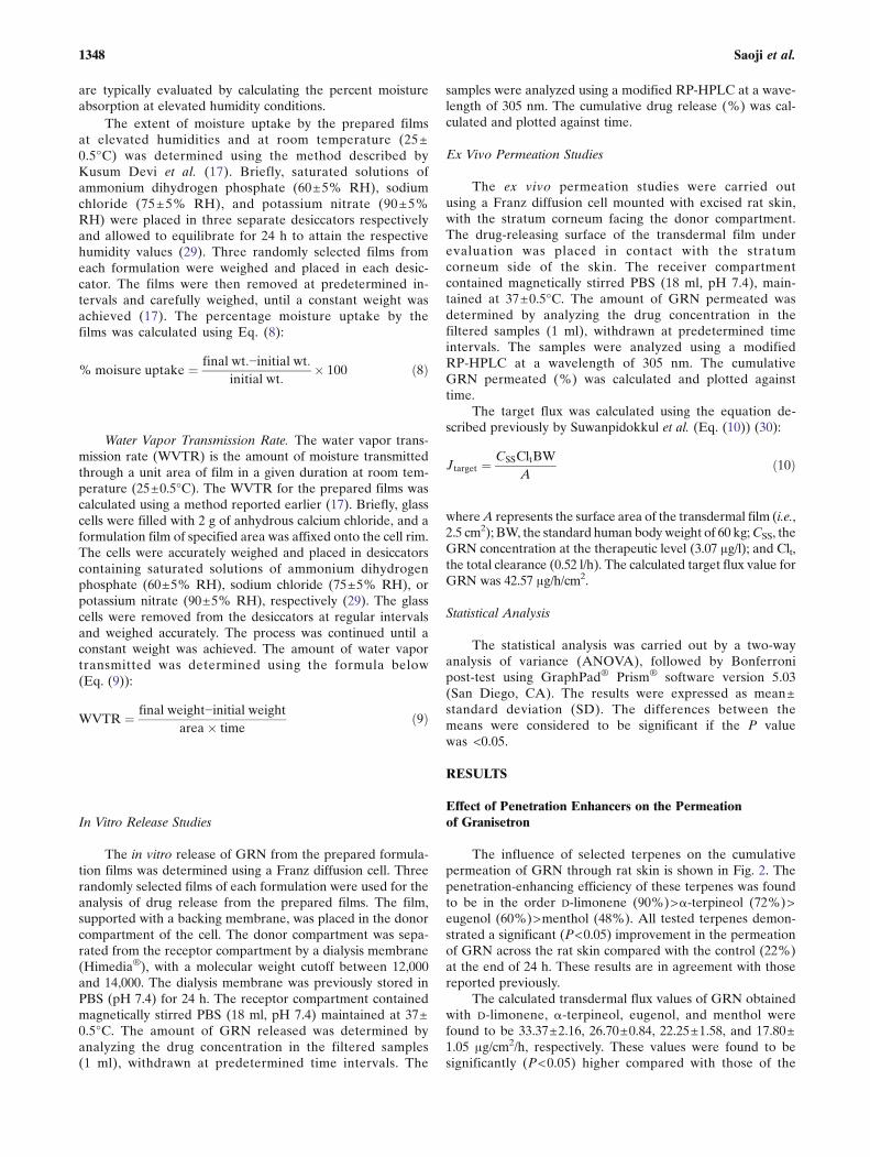
The influence of selected terpenes on the cumulativepermeation of GRN through rat skin is shown in Fig. 2. Thepenetration-enhancing efficiency of these terpenes was foundto be in the order D-limonene (90%)>α-terpineol (72%)>eugenol (60%)>menthol (48%). All tested terpenes demon-strated a significant (P<0.05) improvement in the permeationof GRN across the rat skin compared with the control (22%)at the end of 24 h. These results are in agreement with thosereported previously.
The calculated transdermal flux values of GRN obtainedwith D-limonene, α-terpineol, eugenol, and menthol werefound to be 33.37±2.16, 26.70±0.84, 22.25±1.58, and 17.80±1.05 μg/cm2/h, respectively. These values were found to besignificantly (P<0.05) higher compared with those of the
1348 Saoji et al.
control (8.16±0.62 μg/cm2/h). In addition, the calculatedpermeability coefficient values for D-limonene, α-terpineol,eugenol, and menthol were found to be 4.08±0.27, 3.27±0.21, 2.73±0.16, and 2.18±0.11 cm/h×10−2, respectively.
Drug-Polymer Compatibility Studies
The DSC thermogram of pure GRN showed a sharpendothermic peak at ∼300°C, corresponding to the GRNmelting point (thermograms not shown). The appearance ofa sharp endothermic peak is typically attributable to the crys-talline nature of the materials. The DSC thermogram ofMethocel® E15, Eudragit® RL 100, and Eudragit® RS 100revealed broad, undefined peaks over a temperature rangeof 60–240°C. Such broad endothermic peaks, mainly due tothe dehydration process, are typically observed withpredominantly amorphous polymers.
The FTIR spectral analysis of pure GRN showed themain peaks at wave numbers 3246, 1647, 1557, 1478, and1248 cm−1, confirming the purity of the drug (Fig. 3). TheFTIR spectra of the ternary mixtures, GRN:Methocel®
E15:Eudragit® RL 100 (1:1:1) and GRN:Methocel®
E15:Eudragit® RS 100 (1:1:1), showed that the major peaksof GRN were retained and observed at wave numbers 3245,1646, 1561, 1471, and 1248 cm−1. The presence of the polymersdid not appear to significantly affect the integrity of the GRNpeaks.
Physicochemical Properties of the Films
The results of the physicochemical properties of the pre-pared transdermal matrix films are shown in Table II. Themean weights of the prepared films from all ten formulationsranged between 55 and 58 mg, and no significant differenceswere observed among the formulations evaluated. The chang-es in the ratio of the incorporated polymers in the formulationdid not appear to have a statistically significant influence onthe weight of the films. The thickness of the prepared filmsranged between 92 and 107 μm. The film thickness appeared
to decrease with decreasing ratio of the Methocel®
E15:Eudragit® polymer in the formulations. These resultsare in agreement with those reported previously (31,32). Theresults of the flatness study showed that the formulation filmshad a negligible change in the length along the longitudinallycut edges, indicating a near 100% flatness. The films from alltested formulations appeared to have a smooth, flat surface,without any visible signs of constriction.
The results of the content uniformity assay of the pre-pared films are shown in Table II. The GRN content of thefilms from different formulations, as obtained from the HPLCassay, ranged from 98 to 100% w/w of the theoretical concen-tration. The drug content was found to be statistically similaracross different formulations. The results indicated that theprocess employed to prepare films in this study was robust andcapable of producing films without a significant variation. Thefolding endurance test results (Table II) showed that the filmsprepared from all formulations endured at least 250 strokes offolding/unfolding at the same location before revealing anysigns of cracking/breaking. These results were found to behigher than those reported in the literature for similar formu-lations (17). These results demonstrated the sturdiness of thefilms in maintaining their integrity with general skin foldingwhen applied.
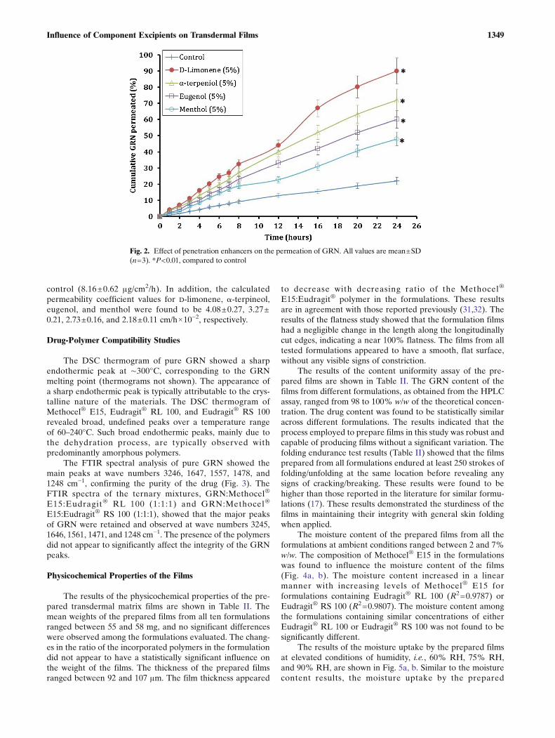
The moisture content of the prepared films from all theformulations at ambient conditions ranged between 2 and 7%w/w. The composition of Methocel® E15 in the formulationswas found to influence the moisture content of the films(Fig. 4a, b). The moisture content increased in a linearmanner with increasing levels of Methocel® E15 forformulations containing Eudragit® RL 100 (R2=0.9787) orEudragit® RS 100 (R2=0.9807). The moisture content amongthe formulations containing similar concentrations of eitherEudragit® RL 100 or Eudragit® RS 100 was not found to besignificantly different.
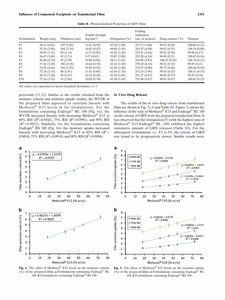
The results of the moisture uptake by the prepared filmsat elevated conditions of humidity, i.e., 60% RH, 75% RH,and 90% RH, are shown in Fig. 5a, b. Similar to the moisturecontent results, the moisture uptake by the prepared
Fig. 2. Effect of penetration enhancers on the permeation of GRN. All values are mean±SD(n=3). *P<0.01, compared to control
1349Influence of Component Excipients on Transdermal Films

formulation films appeared to depend on the amount ofMethocel® E15 in the formulations. For the formulationscontaining Eudragit® RL 100 (Fig. 5a), the moisture uptakeincreased linearly with increasing Methocel® E15 at 60% RH(R2=0.9568), 75% RH (R2=0.9989), and 90% RH (R2=0.9982). Similarly, for the formulations containing Eudragit®
RS 100 (Fig. 5b), the moisture uptake increased linearly withincreasing Methocel® E15 at 60% RH (R2=0.9485), 75% RH
(R2=0.992), and 90% RH (R2=0.9992). As observed withmoisture content results, the moisture uptake among theformulations containing similar concentrations of eitherEudragit® RL 100 or Eudragit® RS 100 was not found to besignificantly different.
The water vapor transmission rates (WVTR) in the pre-pared films were found to range between 0.09 and 0.56 mg/cm2/h. These results were in agreement with those reported
Fig. 3. FTIR analysis of the drug and the polymers. a Granisetron, b granisetron:Methocel® E15:Eudragit® RL 100 (1:1:1), and cgranisetron:Methocel® E15:Eudragit® RS 100 (1:1:1)
1350 Saoji et al.
previously (21,33). Similar to the results obtained from themoisture content and moisture uptake studies, the WVTR ofthe prepared films appeared to correlate linearly withMethocel® E15 levels in the formulations. For theformulations containing Eudragit® RL 100 (Fig. 6a), theWVTR increased linearly with increasing Methocel® E15 at60% RH (R2=0.8542), 75% RH (R2=0.9991), and 90% RH(R2=0.9822). Similarly, for the formulations containingEudragit® RS 100 (Fig. 6b), the moisture uptake increasedlinearly with increasing Methocel® E15 at 60% RH (R2=0.9454), 75% RH (R2=0.9914), and 90% RH (R2=0.9904).
In Vitro Drug Release
The results of the in vitro drug release from transdermalfilms are shown in Fig. 7a, b and Table III. Figure 7a shows theinfluence of the ratio of Methocel® E15 and Eudragit® RL 100on the release of GRN from the prepared transdermal films. Itwas observed that the formulation F1 (with the highest ratio ofMethocel® E15:Eudragit® RL 100) exhibited the highestcumulative amount of GRN released (Table III). For thesubsequent formulations, i.e., F2 to F5, the release of GRNwas found to be progressively slower. Similar results were
Table II. Physicochemical Properties of GRN Films
Formulation Weight (mg) Thickness (μm)Tensile strength(kg/mm2) Elongation (%)
Foldingendurance(no. of strokes) Drug content (%) Flatness
F1 56.21 (4.92) 107 (3.02) 11.01 (0.59) 62.50 (1.09) 257.12 (3.68) 99.47 (0.26) 100.08 (0.12)F2 55.18 (5.09) 104 (2.54) 10.43 (0.47) 60.09 (1.95) 263.47 (4.98) 99.87 (0.35) 100.14 (0.09)F3 58.03 (3.12) 99 (2.19) 11.73 (0.82) 61.82 (1.30) 252.41 (3.64) 99.42 (0.50) 99.98 (0.17)F4 56.47 (3.69) 95 (2.71) 9.57 (0.62) 59.07 (2.00) 255.28 (4.10) 98.89 (0.31) 100.02 (0.16)F5 56.08 (4.79) 92 (2.59) 10.68 (0.66) 64.13 (1.83) 254.09 (3.83) 100.23 (0.54) 100.16 (0.13)F6 57.41 (3.28) 106 (3.18) 10.64 (0.78) 62.24 (1.30) 259.28 (4.10) 99.32 (0.52) 99.95 (0.11)F7 55.20 (4.64) 104 (2.55) 10.85 (0.92) 61.85 (1.88) 253.27 (4.80) 99.73 (0.46) 100.04 (0.16)F8 57.18 (3.19) 98 (2.41) 11.81 (0.68) 63.04 (1.70) 253.14 (3.99) 99.05 (0.53) 100.11 (0.15)F9 58.14 (3.20) 96 (2.67) 10.22 (0.49) 63.54 (1.05) 251.27 (4.53) 98.94 (0.57) 99.93 (0.10)F10 57.16 (3.33) 93 (2.04) 10.69 (0.78) 63.26 (1.83) 251.40 (3.87) 98.91 (0.47) 100.02 (0.15)
All values are expressed as mean (standard deviation), n=3
Fig. 4. The effect of Methocel® E15 levels on the moisture content(%) of the prepared films. a Formulations containing Eudragit® RL
100. b Formulations containing Eudragit® RS 100
Fig. 5. The effect of Methocel® E15 levels on the moisture uptake(%) of the prepared films. a Formulations containing Eudragit® RL
100. b Formulations containing Eudragit® RS 100
1351Influence of Component Excipients on Transdermal Films
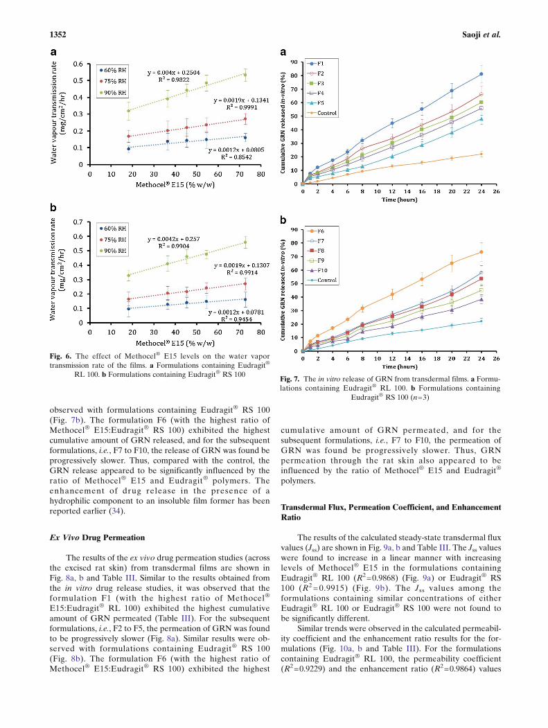
observed with formulations containing Eudragit® RS 100(Fig. 7b). The formulation F6 (with the highest ratio ofMethocel® E15:Eudragit® RS 100) exhibited the highestcumulative amount of GRN released, and for the subsequentformulations, i.e., F7 to F10, the release of GRN was found beprogressively slower. Thus, compared with the control, theGRN release appeared to be significantly influenced by theratio of Methocel® E15 and Eudragit® polymers. Theenhancement of drug release in the presence of ahydrophilic component to an insoluble film former has beenreported earlier (34).
Ex Vivo Drug Permeation
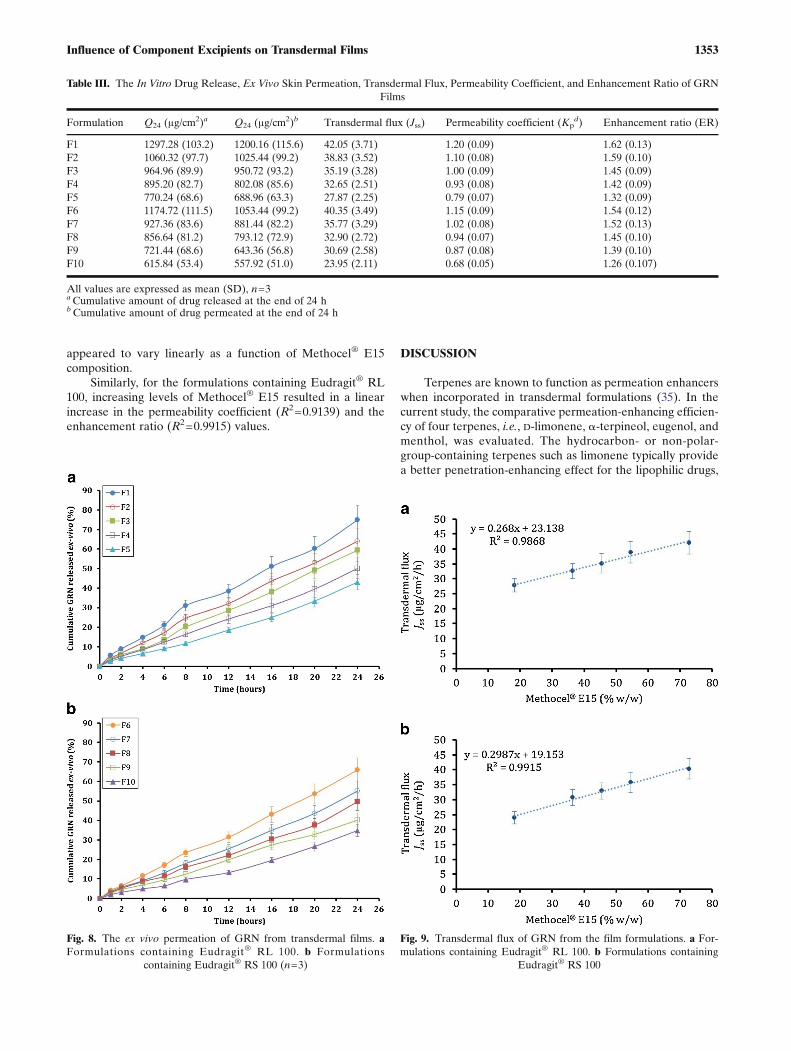
The results of the ex vivo drug permeation studies (acrossthe excised rat skin) from transdermal films are shown inFig. 8a, b and Table III. Similar to the results obtained fromthe in vitro drug release studies, it was observed that theformulation F1 (with the highest ratio of Methocel®
E15:Eudragit® RL 100) exhibited the highest cumulativeamount of GRN permeated (Table III). For the subsequentformulations, i.e., F2 to F5, the permeation of GRN was foundto be progressively slower (Fig. 8a). Similar results were ob-served with formulations containing Eudragit® RS 100(Fig. 8b). The formulation F6 (with the highest ratio ofMethocel® E15:Eudragit® RS 100) exhibited the highest
cumulative amount of GRN permeated, and for thesubsequent formulations, i.e., F7 to F10, the permeation ofGRN was found be progressively slower. Thus, GRNpermeation through the rat skin also appeared to beinfluenced by the ratio of Methocel® E15 and Eudragit®
polymers.
Transdermal Flux, Permeation Coefficient, and EnhancementRatio
The results of the calculated steady-state transdermal fluxvalues (Jss) are shown in Fig. 9a, b and Table III. The Jss valueswere found to increase in a linear manner with increasinglevels of Methocel® E15 in the formulations containingEudragit® RL 100 (R2=0.9868) (Fig. 9a) or Eudragit® RS100 (R2=0.9915) (Fig. 9b). The Jss values among theformulations containing similar concentrations of eitherEudragit® RL 100 or Eudragit® RS 100 were not found tobe significantly different.
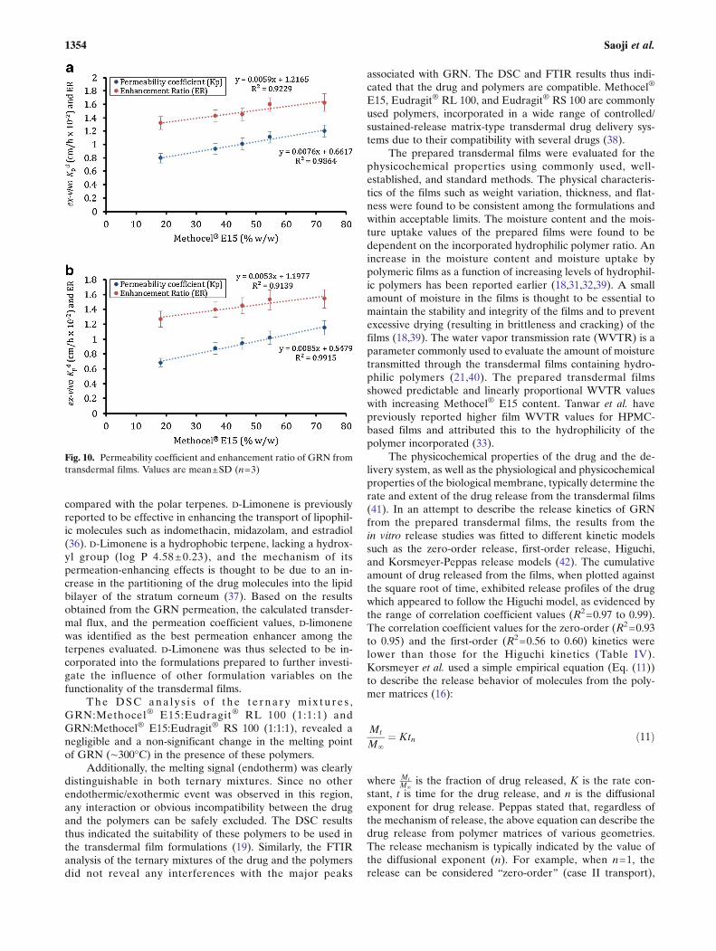
Similar trends were observed in the calculated permeabil-ity coefficient and the enhancement ratio results for the for-mulations (Fig. 10a, b and Table III). For the formulationscontaining Eudragit® RL 100, the permeability coefficient(R2=0.9229) and the enhancement ratio (R2=0.9864) values
Fig. 6. The effect of Methocel® E15 levels on the water vaportransmission rate of the films. a Formulations containing Eudragit®
RL 100. b Formulations containing Eudragit® RS 100Fig. 7. The in vitro release of GRN from transdermal films. a Formu-lations containing Eudragit® RL 100. b Formulations containing
Eudragit® RS 100 (n=3)
1352 Saoji et al.
appeared to vary linearly as a function of Methocel® E15composition.
Similarly, for the formulations containing Eudragit® RL100, increasing levels of Methocel® E15 resulted in a linearincrease in the permeability coefficient (R2=0.9139) and theenhancement ratio (R2=0.9915) values.
DISCUSSION
Terpenes are known to function as permeation enhancerswhen incorporated in transdermal formulations (35). In thecurrent study, the comparative permeation-enhancing efficien-cy of four terpenes, i.e., D-limonene, α-terpineol, eugenol, andmenthol, was evaluated. The hydrocarbon- or non-polar-group-containing terpenes such as limonene typically providea better penetration-enhancing effect for the lipophilic drugs,
Table III. The In Vitro Drug Release, Ex Vivo Skin Permeation, Transdermal Flux, Permeability Coefficient, and Enhancement Ratio of GRNFilms
Formulation Q24 (μg/cm2)a Q24 (μg/cm
2)b Transdermal flux (Jss) Permeability coefficient (Kpd) Enhancement ratio (ER)
F1 1297.28 (103.2) 1200.16 (115.6) 42.05 (3.71) 1.20 (0.09) 1.62 (0.13)F2 1060.32 (97.7) 1025.44 (99.2) 38.83 (3.52) 1.10 (0.08) 1.59 (0.10)F3 964.96 (89.9) 950.72 (93.2) 35.19 (3.28) 1.00 (0.09) 1.45 (0.09)F4 895.20 (82.7) 802.08 (85.6) 32.65 (2.51) 0.93 (0.08) 1.42 (0.09)F5 770.24 (68.6) 688.96 (63.3) 27.87 (2.25) 0.79 (0.07) 1.32 (0.09)F6 1174.72 (111.5) 1053.44 (99.2) 40.35 (3.49) 1.15 (0.09) 1.54 (0.12)F7 927.36 (83.6) 881.44 (82.2) 35.77 (3.29) 1.02 (0.08) 1.52 (0.13)F8 856.64 (81.2) 793.12 (72.9) 32.90 (2.72) 0.94 (0.07) 1.45 (0.10)F9 721.44 (68.6) 643.36 (56.8) 30.69 (2.58) 0.87 (0.08) 1.39 (0.10)F10 615.84 (53.4) 557.92 (51.0) 23.95 (2.11) 0.68 (0.05) 1.26 (0.107)
All values are expressed as mean (SD), n=3aCumulative amount of drug released at the end of 24 hbCumulative amount of drug permeated at the end of 24 h
Fig. 8. The ex vivo permeation of GRN from transdermal films. aFormulations containing Eudragit® RL 100. b Formulations
containing Eudragit® RS 100 (n=3)
Fig. 9. Transdermal flux of GRN from the film formulations. a For-mulations containing Eudragit® RL 100. b Formulations containing
Eudragit® RS 100
1353Influence of Component Excipients on Transdermal Films
compared with the polar terpenes. D-Limonene is previouslyreported to be effective in enhancing the transport of lipophil-ic molecules such as indomethacin, midazolam, and estradiol(36). D-Limonene is a hydrophobic terpene, lacking a hydrox-yl group (log P 4.58±0.23), and the mechanism of itspermeation-enhancing effects is thought to be due to an in-crease in the partitioning of the drug molecules into the lipidbilayer of the stratum corneum (37). Based on the resultsobtained from the GRN permeation, the calculated transder-mal flux, and the permeation coefficient values, D-limonenewas identified as the best permeation enhancer among theterpenes evaluated. D-Limonene was thus selected to be in-corporated into the formulations prepared to further investi-gate the influence of other formulation variables on thefunctionality of the transdermal films.
The DSC ana ly s i s o f the te rna ry mix tu re s ,GRN:Methocel® E15:Eudragit® RL 100 (1:1:1) andGRN:Methocel® E15:Eudragit® RS 100 (1:1:1), revealed anegligible and a non-significant change in the melting pointof GRN (∼300°C) in the presence of these polymers.
Additionally, the melting signal (endotherm) was clearlydistinguishable in both ternary mixtures. Since no otherendothermic/exothermic event was observed in this region,any interaction or obvious incompatibility between the drugand the polymers can be safely excluded. The DSC resultsthus indicated the suitability of these polymers to be used inthe transdermal film formulations (19). Similarly, the FTIRanalysis of the ternary mixtures of the drug and the polymersdid not reveal any interferences with the major peaks
associated with GRN. The DSC and FTIR results thus indi-cated that the drug and polymers are compatible. Methocel®
E15, Eudragit® RL 100, and Eudragit® RS 100 are commonlyused polymers, incorporated in a wide range of controlled/sustained-release matrix-type transdermal drug delivery sys-tems due to their compatibility with several drugs (38).
The prepared transdermal films were evaluated for thephysicochemical properties using commonly used, well-established, and standard methods. The physical characteris-tics of the films such as weight variation, thickness, and flat-ness were found to be consistent among the formulations andwithin acceptable limits. The moisture content and the mois-ture uptake values of the prepared films were found to bedependent on the incorporated hydrophilic polymer ratio. Anincrease in the moisture content and moisture uptake bypolymeric films as a function of increasing levels of hydrophil-ic polymers has been reported earlier (18,31,32,39). A smallamount of moisture in the films is thought to be essential tomaintain the stability and integrity of the films and to preventexcessive drying (resulting in brittleness and cracking) of thefilms (18,39). The water vapor transmission rate (WVTR) is aparameter commonly used to evaluate the amount of moisturetransmitted through the transdermal films containing hydro-philic polymers (21,40). The prepared transdermal filmsshowed predictable and linearly proportional WVTR valueswith increasing Methocel® E15 content. Tanwar et al. havepreviously reported higher film WVTR values for HPMC-based films and attributed this to the hydrophilicity of thepolymer incorporated (33).
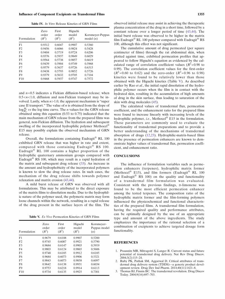
The physicochemical properties of the drug and the de-livery system, as well as the physiological and physicochemicalproperties of the biological membrane, typically determine therate and extent of the drug release from the transdermal films(41). In an attempt to describe the release kinetics of GRNfrom the prepared transdermal films, the results from thein vitro release studies was fitted to different kinetic modelssuch as the zero-order release, first-order release, Higuchi,and Korsmeyer-Peppas release models (42). The cumulativeamount of drug released from the films, when plotted againstthe square root of time, exhibited release profiles of the drugwhich appeared to follow the Higuchi model, as evidenced bythe range of correlation coefficient values (R2=0.97 to 0.99).The correlation coefficient values for the zero-order (R2=0.93to 0.95) and the first-order (R2=0.56 to 0.60) kinetics werelower than those for the Higuchi kinetics (Table IV).Korsmeyer et al. used a simple empirical equation (Eq. (11))to describe the release behavior of molecules from the poly-mer matrices (16):
Mt
M∞¼ Ktn ð11Þ
where MtM∞
is the fraction of drug released, K is the rate con-stant, t is time for the drug release, and n is the diffusionalexponent for drug release. Peppas stated that, regardless ofthe mechanism of release, the above equation can describe thedrug release from polymer matrices of various geometries.The release mechanism is typically indicated by the value ofthe diffusional exponent (n). For example, when n=1, therelease can be considered Bzero-order^ (case II transport),
Fig. 10. Permeability coefficient and enhancement ratio of GRN fromtransdermal films. Values are mean±SD (n=3)
1354 Saoji et al.
and n=0.5 indicates a Fickian diffusion-based release; when0.5<n<1.0, diffusion and non-Fickian transport may be in-volved. Lastly, when n>1.0, the apparent mechanism is Bsupercase II transport.^ The value of n is obtained from the slope oflogMt
M∞vs the log time curve. The n values for the GRN release
obtained using this equation (0.53 to 0.75) indicated that themain mechanism of GRN release from the prepared films wasgeneral, non-Fickian diffusion. The hydration and subsequentswelling of the incorporated hydrophilic polymer Methocel®
E15 may possibly explain the observed mechanism of GRNrelease.
Overall, the formulations containing Eudragit® RL 100exhibited GRN release that was higher in rate and extent,compared with those containing Eudragit® RS 100.Eudragit® RL 100 contains a higher proportion of thehydrophilic quaternary ammonium groups compared withEudragit® RS 100, which may result in a rapid hydration ofthe matrix and subsequent drug release (33). An increase inthe amount and hydrophobicity of the incorporated polymersis known to slow the drug release rates. In such cases, themechanism of the drug release shifts towards polymerrelaxation and matrix erosion (43,44).
A mild burst release of GRN was observed with allformulations. This may be attributed to the direct exposureof the matrix films to dissolution media. Due to the hydrophil-ic nature of the polymer used, the polymeric matrix may formloose channels within the network, resulting in a rapid releaseof the drug present in the surface layers of the film. The
observed initial release may assist in achieving the therapeuticplasma concentration of the drug in a short time, followed by aconstant release over a longer period of time (43,44). Theinitial burst release was observed to be higher in the matrixfilm Eudragit® RL 100 polymer compared with Eudragit® RS100, although this effect was not significant.
The cumulative amount of drug permeated (per squarecentimeter of films) through the rat abdominal skin, whenplotted against time, exhibited permeation profiles that ap-peared to follow Higuchi’s equation as evidenced by the cal-culated range of correlation coefficient values (R2=0.98 to0.99). The correlation coefficient values for the first-order(R2=0.60 to 0.62) and the zero-order (R2=0.96 to 0.98)kinetics were found to be relatively lower than thoseobtained with the Higuchi kinetics (Table V). As describedearlier by Rao et al., the initial rapid dissolution of the hydro-philic polymer occurs when the film is in contact with thehydrated skin, resulting in the accumulation of high amountsof drug in the skin surface, thus leading to saturation of theskin with drug molecules (45).
The calculated values of transdermal flux, permeationcoefficient, and the enhancement ratio for the prepared filmswere found to increase linearly with increasing levels of thehydrophilic polymer, i.e., Methocel® E15 in the formulation.These parameters are commonly used to evaluate thefunctionality of transdermal preparations and to provide abetter understanding of the mechanisms of transdermalabsorption of drugs (12,23). Hydrophilic-matrix-based filmsin the presence of permeation enhancers are known to dem-onstrate higher values of transdermal flux, permeation coeffi-cient, and enhancement ratio.
CONCLUSIONS
The influence of formulation variables such as perme-ation enhancers (terpenes), hydrophilic matrix former(Methocel® E15), and film formers (Eudragit® RL 100and Eudragit® RS 100) on the quality and functionalityof a transdermal film formulation was evaluated.Consistent with the previous findings, D-limonene wasfound to be the most efficient permeation enhanceramong the tested terpenes. The composition ratio of thehydrophilic matrix former and the film-forming polymersinfluenced the physicochemical and functional characteris-tics of the prepared films. A transdermal film formulation,having the required quality and performance attributes,can be optimally designed by the use of an appropriatetype and amount of the above ingredients. The studyemphasizes the importance of the rational selection of acombination of excipients to achieve targeted dosage formfunctionality.
REFERENCES
1. Prausnitz MR, Mitragotri S, Langer R. Current status and futurepotential of transdermal drug delivery. Nat Rev Drug Discov.2004;3(2):115–24.
2. Ruby PK, Pathak SM, Aggarwal D. Critical attributes of trans-dermal drug delivery system (TDDS)—a generic product devel-opment review. Drug Dev Ind Pharm. 2014;40(11):1421–8.
3. Thomas BJ, Finnin BC. The transdermal revolution. Drug DiscovToday. 2004;9(16):697–703.
Table IV. In Vitro Release Kinetics of GRN Films
Formulation
Zeroorder(R2)
Firstorder(R2)
Higuchimodel(R2)
Korsmeyer-Peppasmodel (n)
F1 0.9312 0.6047 0.9907 0.5360F2 0.9456 0.6066 0.9824 0.5428F3 0.9404 0.5719 0.9724 0.6206F4 0.9376 0.5937 0.9849 0.6029F5 0.9564 0.5738 0.9857 0.6619F6 0.9439 0.5904 0.9749 0.5968F7 0.9579 0.5637 0.9726 0.6315F8 0.9438 0.5793 0.9852 0.5726F9 0.9379 0.5632 0.9705 0.7104F10 0.9468 0.5937 0.9747 0.7572
Table V. Ex Vivo Permeation Kinetics of GRN Films
Formulation
Zeroorder(R2)
Firstorder(R2)
Higuchimodel(R2)
Korsmeyer-Peppas model(n)
F1 0.9679 0.6108 0.9907 0.5200F2 0.9745 0.6087 0.9921 0.5790F3 0.9684 0.6147 0.9903 0.5919F4 0.9803 0.6124 0.9865 0.5606F5 0.9748 0.6185 0.9912 0.6185F6 0.9684 0.6073 0.9906 0.5321F7 0.9843 0.6073 0.9858 0.6097F8 0.9642 0.6138 0.9931 0.6034F9 0.9753 0.6218 0.9914 0.6113F10 0.9734 0.6135 0.9925 0.7301
1355Influence of Component Excipients on Transdermal Films
4. Paudel KS, Milewski M, Swadley CL, Brogden NK, Ghosh P,Stinchcomb AL. Challenges and opportunities in dermal/transdermal delivery. Ther Deliv. 2010;1(1):109–31.
5. Prausnitz MR, Langer R. Transdermal drug delivery. NatBiotechnol. 2008;26(11):1261–8.
6. Zadymova NM. Colloidochemical aspects of transdermal drugdelivery (review). Colloid J. 2013;75(5):491–503.
7. Vavrova K, Zbytovska J, Hrabalek A. Amphiphilic transdermalpermeation enhancers: structure-activity relationships. Curr MedChem. 2005;12(19):2273–91.
8. Williams AC, Barry BW. Essential oils as novel human skinpenetration enhancers. Int J Pharm. 1989;57(2):R7–9.
9. Patil U, Saraogi R. Natural products as potential drug permeationenhancer in transdermal drug delivery system. Arch DermatolRes. 2014;306(5):419–26.
10. Karande P, Jain A, Ergun K, Kispersky V, Mitragotri S. Designprinciples of chemical penetration enhancers for transdermaldrug delivery. Proc Natl Acad Sci U S A. 2005;102(13):4688–93.
11. Parikh D, Ghosh T. Feasibility of transdermal delivery of fluox-etine. AAPS PharmSciTech. 2005;6(2):E144–9.
12. Williams AC, Barry BW. Terpenes and the lipid proteinpartitioning theory of skin penetration enhancement. PharmRes. 1991;8(1):17–24.
13. Gao S, Singh J. In vitro percutaneous absorption enhancement ofa lipophilic drug tamoxifen by terpenes. J Control Release.1998;51(2–3):193–9.
14. Herman A, Herman AP. Essential oils and their constituents asskin penetration enhancer for transdermal drug delivery: a re-view. J Pharm Pharmacol. 2015;67(4):473–85.
15. Zhao KD, Singh J. In vitro percutaneous absorption enhance-ment of propranolol hydrochloride through porcine epidermis byterpenes/ethanol. J Control Release. 1999;62(3):359–66.
16. Korsmeyer RW, Gurny R, Doelker E, Buri P, Peppas NA. Mech-anisms of potassium chloride release from compressed, hydro-philic, polymeric matrices: effect of entrapped air. J Pharm Sci.1983;72(10):1189–91.
17. Kusum Devi V, Saisivam S, Maria GR, Deepti PU. Design andevaluation of matrix diffusion controlled transdermal patches of ve-rapamil hydrochloride. Drug Dev Ind Pharm. 2003;29(5):495–503.
18. Mutalik S, Udupa N. Glibenclamide transdermal patches: physi-cochemical, pharmacodynamic, and pharmacokinetic evaluations.J Pharm Sci. 2004;93(6):1577–94.
19. Chandak AR, Verma PRP. Development and evaluation ofHPMC based matrices for transdermal patches of tramadol. ClinRes Regul Aff. 2008;25(1):13–30.
20. Kshirsagar SJ, Bhalekar MR, Mohapatra SK. Development andevaluation of carvedilol-loaded transdermal drug delivery system:in-vitro and in-vivo characterization study. Drug Dev Ind Pharm.2012;38(12):1530–7.
21. Madan JR, Argade NS, Dua K. Formulation and evaluation oftransdermal patches of donepezil. Recent pat drug delivery for-mulation. 2014.
22. Hayton WL, Chen T. Correction of perfusate concentration forsample removal. J Pharm Sci. 1982;71(7):820–1.
23. Barry BW. Mode of action of penetration enhancers in humanskin. J Control Release. 1987;6(1):85–97.
24. Riviere A. Dose finding study of granisetron in patients receivinghigh-dose cisplatin chemotherapy. The Granisetron Study Group.Br J Cancer. 1994;69(5):967–71.
25. Yener G, Uuml LG, Uuml, Uuml, Ner M, et al. Design ofmeloxicam and lornoxicam transdermal patches: preparation,physical characterization, ex vivo and in vivo studies. ChemPharm Bull. 2010;58(11):1466–73.
26. Khana R, Agarwal SP, Ahuja A. Preparation and evaluation ofmuco-adhesive buccal films of clotrimazole for oral candida in-fections. Indian J Pharm Sci. 1997;59(6):299–305.
27. Pinguet F, Bressolle F, Martel P, Salabert D, Astre C. High-performance liquid chromatographic determination ofgranisetron in human plasma. J Chromatogr B Biomed Appl.1996;675(1):99–105.
28. Aggarwal G, Dhawan S, Hari Kumar SL. Formulation, in vitroand in vivo evaluation of transdermal patches containing risper-idone. Drug Dev Ind Pharm. 2013;39(1):39–50.
29. Greenspan L. Humidity fixed points of binary saturated aqueoussolutions. J Res Nat Bur Stand Sect A. 1977;81A(1):89–96.
30. Suwanpidokkul N, Thongnopnua P, Umprayn K. Transdermaldelivery of zidovudine (AZT): the effects of vehicles, enhancers,and polymer membranes on permeation across cadaver pig skin.AAPS PharmSciTech. 2004;5(3):82–9.
31. Mamatha T, Venkateswara RJ, Mukkanti K, Ramesh G. Trans-d e rm a l d r u g d e l i v e r y s y s t em f o r a t om o x e t i n ehydrochloride—in vitro and ex vivo evaluation. Curr TrendsBiotechnol Pharm. 2009;3(2):188–96.
32. Tirunagari M, Jangala VR, Khagga M, Gannu R. Transdermaltherapeutic system of isradipine: effect of hydrophilic and hydro-phobic matrix on in vitro and ex vivo characteristics. Arch PharmRes. 2010;33(7):1025–33.
33. Tanwar YS, Chauhan CS, Sharma A. Development and evalua-tion of carvedilol transdermal patches. Acta Pharm (Zagreb,Croatia). 2007;57:151–9.
34. Bodmeier R, Paeratakul O. Drug release from laminated poly-meric films prepared from aqueous latexes. J Pharm Sci.1990;79(1):32–6.
35. Aqil M, Ahad A, Sultana V, Ali A. Status of terpenes as skinpenetration enhancers. Drug Discov Today. 2007;12(23–24):1061–7.
36. Yang Z, Teng Y, Wang H, Hou H. Enhancement of skin perme-ation of bufalin by limonene via reservoir type transdermal patch:formulation design and biopharmaceutical evaluation. Int JPharm. 2013;447(1–2):231–40.
37. Krishnaiah YS, Bhaskar P, Satyanarayana V. In vitro percutane-ous permeability enhancement of nimodipine by limonene acrossthe excised rat abdominal skin. Pharmazie. 2005;59:942–7.
38. Rowe RC, Sheskey PJ, Quinn ME. Handbook of pharmaceu-tical excipients. Sixth ed. Rowe RC, Sheskey PJ, Quinn ME,editors. Washington, DC: London; Greyslake, IL : Pharma-ceutical Press; Washington, DC : American Pharmacists As-sociation, 2009; 2009.
39. Ubaidulla U, Reddy MVS, Ruckmani K, Ahmad FJ, Khar RK.Transdermal therapeutic system of carvedilol: effect of hydrophil-ic and hydrophobic matrix on in vitro and in vivo characteristics.AAPS PharmSciTech. 2007;8(1):E13–20.
40. Madishetti SK, Palem CR, Gannu R, Thatipamula RP, PanakantiPK, Yamsani MR. Development of domperidone bilayered ma-trix type transdermal patches: physicochemical, in vitro andex vivo characterization. DARU J Pharm Sci. 2010;18(3):221–9.
41. Chien YW. Development of transdermal drug delivery systems.Drug Dev Ind Pharm. 1987;13(4/5):589.
42. Costa P, Sousa Lobo JM. Modeling and comparison of dissolutionprofiles. Eur J Pharm Sci. 2001;13(2):123–33.
43. Chandak AR, Verma PRP. Design and development of hydroxy-propyl methycellulose (HPMC) based polymeric films of metho-trexate: physicochemical and pharmacokinetic evaluations.Yakugaku Zasshi. 2008;128:1057–66.
44. Prasad Verma PR, Chandak AR. Development of matrix con-trolled transdermal delivery systems of pentazocine: in vitro/invivo performance. Acta Pharm (Zagreb, Croatia). 2009;59:171–86.
45. Rao PR, Diwan PV. Formulation and in vitro evaluation ofpolymeric films of diltiazem hydrochloride and indomethacinfor transdermal administration. Drug Dev Ind Pharm.1998;24(4):327–36.
1356 Saoji et al.