112 drug discoveries therapeutics. 2021; 15(2):112-117
TRANSCRIPT

www.ddtjournal.com
Drug Discoveries & Therapeutics. 2021; 15(2):112-117.112
DOI: 10.5582/ddt.2021.01033
SUMMARY
Keywords
Focal nodular hyperplasia mimicking hepatocellular adenoma and carcinoma in two cases
Menghua Zhu1,2,§, Hongyu Li1,§, Chunhui Wang3,§, Benqiang Yang4,§, Xuehan Wang5, Feifei Hou1, Shengye Yang1, Yuye Wang1,2, Xiaozhong Guo1,*, Xingshun Qi1,*
1 Department of Gastroenterology, General Hospital of Northern Theater Command, Shenyang, China; 2 Postgraduate College, Jinzhou Medical University, Jinzhou, China;3 Department of Hepatobiliary Surgery, General Hospital of Northern Theater Command, Shenyang, China;4 Department of Radiology, General Hospital of Northern Theater Command, Shenyang, China;5 Department of Pathology, General Hospital of Northern Theater Command, Shenyang, China.
Focal nodular hyperplasia, hepatobiliary contrast agents, contrast-enhanced ultrasound, hepatocellular adenoma, hepatocellular carcinoma
Focal nodular hyperplasia (FNH) is a solid benign tumor of the liver, predominantly in young women. A correct diagnosis of FNH is essential for making appropriate clinical decisions and avoiding unnecessary liver resection. Herein, we reported that two male cases with FNH, who initially presented with persistent abdominal discomfort, were misdiagnosed with hepatocellular adenoma (HCA) and hepatocellular carcinoma (HCC) on contrast-enhanced magnetic resonance imaging and computed tomography scans, respectively. After surgery, a histological diagnosis of FNH was finally established. In this paper, we also reviewed the knowledge regarding diagnosis and differential diagnosis of FNH on imaging examinations, which are helpful for avoiding misdiagnoses and guiding clinical interventions.
1. Introduction
Focal nodular hyperplasia (FNH) is a benign tumor of the liver with a prevalence of 0.3-3% in the general population (1,2). It is predominant in women aged 35-50 years old (3). In pathophysiology, arterial malformation leads to abnormal blood perfusion and secondary hyperplasia in the liver parenchyma (4). In histology, FNH is composed of hyperplastic hepatocytes separated by fibrous septum which contains hyperplastic bile ducts, tiny arterial branches, and infiltrating inflammatory cells (4). Most FNH patients are asymptomatic (4). Imaging examinations can usually achieve a definite diagnosis; if obscure, liver biopsy is recommended (3,4) with a great diagnostic accuracy of 95% (5). FNH patients mostly need conservative treatment alone (3,4,6), and undergo interventions when the symptoms are persistent and/or the diagnosis is unclear (3,7-9). Hepatocel lular adenoma (HCA) is another benign liver tumor with a prevalence estimated to be 0.001-0.004% (4). Unlike FNH, HCA has a risk of haemorrhage and malignant transformation. Lifestyle change, close imaging follow-up, and surgical resection are major treatment options for HCA (10).
Hepatocellular carcinoma (HCC), the most common primary liver malignancy, mainly develops in patients with liver cirrhosis secondary to viral hepatitis and alcohol abuse (3). Current treatments of HCC include liver transplantation, liver resection, transarterial chemoembolization, radiofrequency ablation, and molecular targeted therapy (11,12). The use of modern imaging techniques, including contrast-enhanced magnetic resonance imaging (CE-MRI), computed tomography (CECT), and ultrasound (CEUS), is valuable for the diagnosis of liver tumors, further guiding the treatment selection. However, atypical FNHs can mimic HCA or HCC on imaging, because all of them are hypervascular. Therefore, a differential diagnosis of FNH with HCA and HCC is of particular significance.
2. Case presentations
2.1. Case 1
On June 1, 2020, a 55-year-old male complained of persistent abdominal pain for half a year at our department. He had been treated with continuous oral
Case Report

www.ddtjournal.com
Drug Discoveries & Therapeutics. 2021; 15(2):112-117.
clopidogrel bisulfate and hydroxyurea for essential thrombocythemia for 5 years. He also had histories of tuberculosis, appendectomy, and smoking and alcohol cessation as well as a family history of liver cirrhosis. No positive abdominal signs were found on physical examinations. On laboratory tests, the platelet count was 549,000/mm3 (reference range: 125,000-350,000/mm3); fecal occult blood was negative; serum lipase and amylase, liver function parameters, and serum albumin were within the reference range; tumor markers, including alpha-fetoprotein (AFP), carcinoembryonic antigen (CEA), β2-microglobulin (β2-MG), carbohydrate antigen-50 (CA-50), CA19-9, and CA24-2, were negative; hepatitis B virus antigen and hepatitis C virus antibody were negative. No positive lesions were found on esophagogastroduodenoscopy and X-ray gastrointestinal fluoroscopy. Neither superior mesenteric artery occlusion/stenosis nor left renal vein compression was found on abdominal color Doppler ultrasonography. Abdominal CECT showed intrahepatic bile duct stones or calcification, splenomegaly, and splenic infarction (Figure 1). Abdominal CE-MRI further found a mass, which was not well-circumscribed, in the 7th segment of the liver, with isointensity on T1-weighted images, slight hyperintensity on T2-weighted images, homogeneous and strong hyperintensity on arterial phase, and slight hyperintensity on portal phase (Figure 1). A possible diagnosis of HCA was considered. On June 17, 2020, this patient underwent surgery at the Department of Hepatobiliary Surgery after obtaining his and his relatives' written informed consents. A mass with a diameter of about 3 cm was detected in the 7th segment of the liver under ultrasonic guidance, and then the 7th segment of the liver was completely resected. Histology confirmed a diagnosis of FNH (Figure 2). After surgery, abdominal pain disappeared, but liver dysfunction developed with increased serum alanine aminotransferase (ALT) level of 310.32 U/L (reference
range: 9-50 U/L), serum aspartate aminotransferase (AST) level of 514.61 U/L (reference range: 15-40 U/L), serum total bilirubin (TBIL) level of 152.0 μmol/L (reference range: 5.1-22.2 μmol/L), direct bilirubin (DBIL) level of 97.5 μmol/L (reference range: 0-8.6 μmol/L), alkaline phosphatase (AKP) level of 169.39 U/L (reference range: 45-125 U/L), and gamma-glutamytransferase (GGT) level of 60.93 U/L (reference range: 10-60 U/L), and a decreased serum albumin (ALB) level of 29.4 g/L (reference range: 40-55 g/L). Conservative treatment was given for liver dysfunction.
113
Figure 1. Imaging features of FNH in the case 1. (a-c) On multiphase CECT, no abnormal liver lesions were found, except for intrahepatic bile duct stones or calcification. (d-g) On conventional CE-MRI, a mass was not well-circumscribed in the 7th segment of the liver, which showed isointensity on T1-weighted images (d), slight hyperintensity on T2-weighted images (e), homogeneous and strong hyperintensity on arterial phase (f), and slight hyperintensity on portal phase (g).
Figure 2. Surgical specimen and microscopic image of FNH in the case 1. (a) Surgical specimen had a diameter of about 3 cm. (b) Histology confirmed the diagnosis of FNH combined with focal steatosis (hematoxylin and eosin, ×100).
Figure 3. Postoperative abdominal CT scan showing that the 7th segment of the liver was absent in the case 1.

www.ddtjournal.com
Drug Discoveries & Therapeutics. 2021; 15(2):112-117.114
and colonoscopy revealed verrucous gastritis and proliferative changes of terminal ileum lymphatic follicles, respectively. Abdominal CECT revealed an irregularly exogenous mass with a size of about 2.6 × 2.3 cm in the left lateral segment of the liver, with hyperintensity on arterial phase and isointensity on portal and delayed phases (Figure 4). Notably, there was a thickened vessel and its branches into the lesion on arterial phase. Thus, a possible diagnosis of HCC was considered. On May 18, 2020, this patient underwent surgery at the Department of Hepatobiliary Surgery after obtaining his and his relatives' written informed consents. Intraoperatively, an exogenous mass with a diameter of about 3 cm was seen in the left lateral segment of the liver, and then the left lateral segment of the liver was completely resected. Histology confirmed a diagnosis of FNH with atypical hyperplasia (Figure 5) and the immunohistochemical staining revealed the absence of focal malignancy. He was complicated with mild liver dysfunction after surgery, with increased ALT level of 76.14 U/L (reference range: 9-50 U/L) and AST level of 48.98 U/L (reference range: 15-40 U/L). He was discharged on May 28. Abdominal CT at one month and six months after surgery showed that the left lateral segment of the liver was absent (Figure 6).
3. Discussion
A minority of FNH cases may present with abdominal pain or discomfort, which is secondary to the compression of large FNHs on adjacent organs (1). Retraction of FNHs after therapy can relieve abdominal pain, which may explain a potential correlation of abdominal pain with FNHs (9). In the case 1, abdominal pain alleviated after resection of this lesion, indicating that his abdominal symptoms might originate from FNH. Certainly, abdominal pain could also be attributed to other comorbidities (13), such as dyspepsia (14). In the case 2, abdominal pain remained after surgery. Based on the CECT findings, the diagnosis was inaccurate in the case 2, and even hepatic lesion was not visualized in the case 1. CE-MRI has a higher diagnostic performance of focal liver lesions than
Abdominal CT was re-examined and showed that the 7th segment of the liver was absent (Figure 3). He was discharged on June 28. On a recent telephone follow-up visit, he was diagnosed with a hepatic vein stenosis, and at a liver transplantation waiting list.
2.2. Case 2
On May 10, 2020, a 26-year-old male complained of intermittent diarrhea for more than one year and abdominal pain for 20 days at our department. He had histories of upper limb fracture and smoking. He denied the history of alcohol or drug abuse and family history of liver diseases. No positive signs were found on physical examinations. On laboratory tests, fecal occult blood and fecal bacterium cultures were negative; complete blood cell count, serum lipase and amylase, liver function parameters, and serum albumin were within the reference range; tumor markers, including AFP, CEA, β2-MG, CA-50, CA19-9, and CA24-2, were negative; hepatitis B virus antigen and hepatitis C virus antibody were negative. Esophagogastroduodenoscopy
Figure 4. Abdominal multiphase CECT imaging features of FNH in the case 2. There was an irregularly exogenous mass in the left lateral segment of the liver on unenhanced phase (a), hyperintensity on arterial phase (b), and isointensity on portal (c) and delayed (d) phases. Notably, there were a thickened vessel (arrow) and its tiny branches into the lesion on arterial phase (b).
Figure 5. Surgical specimen and microscopic image of FNH in the case 2. (a) The lesion resected was lobulated in appearance with about 2.6 × 2.3 cm in size. (b) Histology showed nodular hyperplasia of hepatocytes with atypical hyperplasia and lymphocytes infiltration in the portal regions (hematoxylin and eosin, ×100).
Figure 6. Postoperative abdominal CT scans showing that the left lateral segment of the liver was absent in the case 2. (a) Abdominal CECT in the first postoperative month. (b) Abdominal unenhanced CT scan in the 6th postoperative month.
www.ddtjournal.com
Drug Discoveries & Therapeutics. 2021; 15(2):112-117. 115
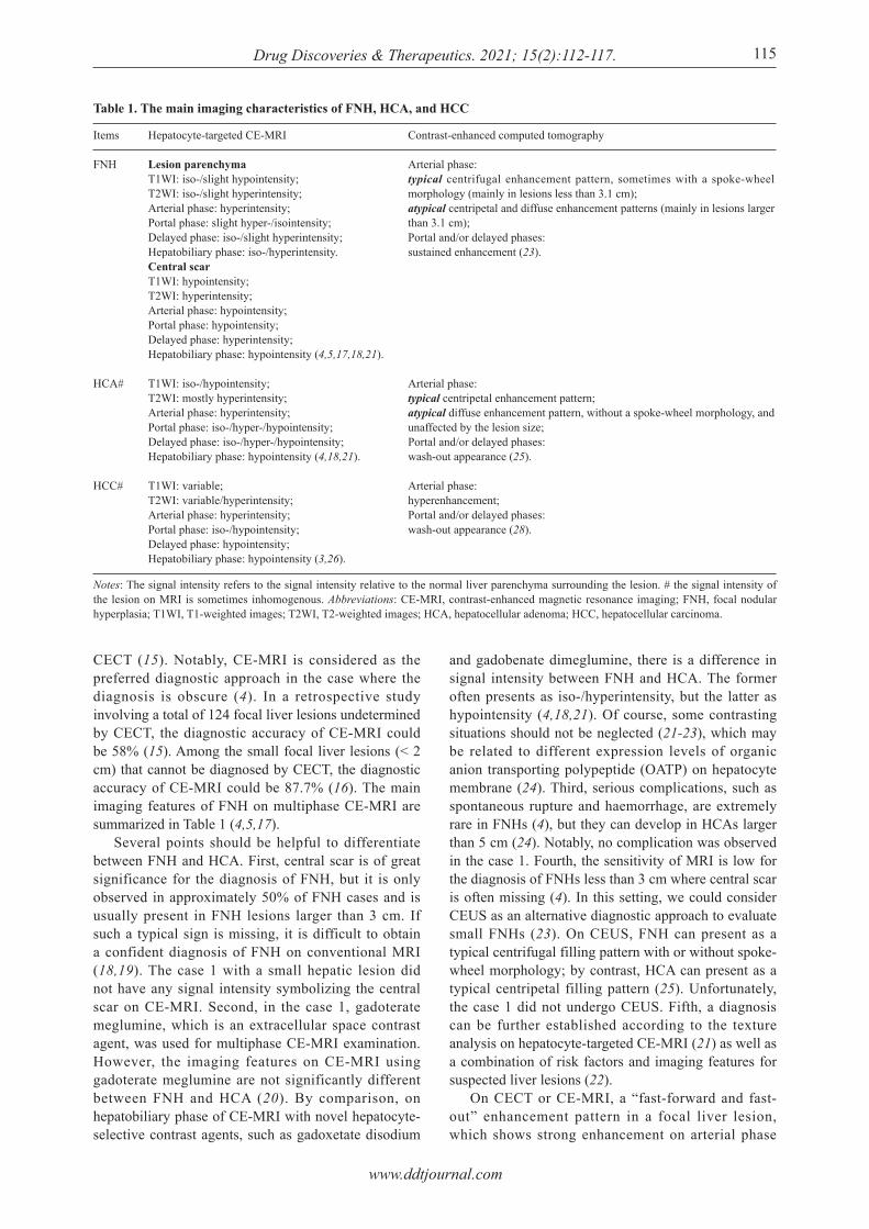
CECT (15). Notably, CE-MRI is considered as the preferred diagnostic approach in the case where the diagnosis is obscure (4). In a retrospective study involving a total of 124 focal liver lesions undetermined by CECT, the diagnostic accuracy of CE-MRI could be 58% (15). Among the small focal liver lesions (< 2 cm) that cannot be diagnosed by CECT, the diagnostic accuracy of CE-MRI could be 87.7% (16). The main imaging features of FNH on multiphase CE-MRI are summarized in Table 1 (4,5,17). Several points should be helpful to differentiate between FNH and HCA. First, central scar is of great significance for the diagnosis of FNH, but it is only observed in approximately 50% of FNH cases and is usually present in FNH lesions larger than 3 cm. If such a typical sign is missing, it is difficult to obtain a confident diagnosis of FNH on conventional MRI (18,19). The case 1 with a small hepatic lesion did not have any signal intensity symbolizing the central scar on CE-MRI. Second, in the case 1, gadoterate meglumine, which is an extracellular space contrast agent, was used for multiphase CE-MRI examination. However, the imaging features on CE-MRI using gadoterate meglumine are not significantly different between FNH and HCA (20). By comparison, on hepatobiliary phase of CE-MRI with novel hepatocyte-selective contrast agents, such as gadoxetate disodium
and gadobenate dimeglumine, there is a difference in signal intensity between FNH and HCA. The former often presents as iso-/hyperintensity, but the latter as hypointensity (4,18,21). Of course, some contrasting situations should not be neglected (21-23), which may be related to different expression levels of organic anion transporting polypeptide (OATP) on hepatocyte membrane (24). Third, serious complications, such as spontaneous rupture and haemorrhage, are extremely rare in FNHs (4), but they can develop in HCAs larger than 5 cm (24). Notably, no complication was observed in the case 1. Fourth, the sensitivity of MRI is low for the diagnosis of FNHs less than 3 cm where central scar is often missing (4). In this setting, we could consider CEUS as an alternative diagnostic approach to evaluate small FNHs (23). On CEUS, FNH can present as a typical centrifugal filling pattern with or without spoke-wheel morphology; by contrast, HCA can present as a typical centripetal filling pattern (25). Unfortunately, the case 1 did not undergo CEUS. Fifth, a diagnosis can be further established according to the texture analysis on hepatocyte-targeted CE-MRI (21) as well as a combination of risk factors and imaging features for suspected liver lesions (22). On CECT or CE-MRI, a “fast-forward and fast-out” enhancement pattern in a focal liver lesion, which shows strong enhancement on arterial phase
Table 1. The main imaging characteristics of FNH, HCA, and HCC
Items
FNH
HCA#
HCC#
Notes: The signal intensity refers to the signal intensity relative to the normal liver parenchyma surrounding the lesion. # the signal intensity of the lesion on MRI is sometimes inhomogenous. Abbreviations: CE-MRI, contrast-enhanced magnetic resonance imaging; FNH, focal nodular hyperplasia; T1WI, T1-weighted images; T2WI, T2-weighted images; HCA, hepatocellular adenoma; HCC, hepatocellular carcinoma.
Hepatocyte-targeted CE-MRI
Lesion parenchymaT1WI: iso-/slight hypointensity;T2WI: iso-/slight hyperintensity;Arterial phase: hyperintensity;Portal phase: slight hyper-/isointensity;Delayed phase: iso-/slight hyperintensity;Hepatobiliary phase: iso-/hyperintensity.Central scarT1WI: hypointensity;T2WI: hyperintensity;Arterial phase: hypointensity;Portal phase: hypointensity;Delayed phase: hyperintensity;Hepatobiliary phase: hypointensity (4,5,17,18,21).
T1WI: iso-/hypointensity;T2WI: mostly hyperintensity;Arterial phase: hyperintensity;Portal phase: iso-/hyper-/hypointensity;Delayed phase: iso-/hyper-/hypointensity;Hepatobiliary phase: hypointensity (4,18,21).
T1WI: variable;T2WI: variable/hyperintensity;Arterial phase: hyperintensity;Portal phase: iso-/hypointensity;Delayed phase: hypointensity;Hepatobiliary phase: hypointensity (3,26).
Contrast-enhanced computed tomography
Arterial phase:typical centrifugal enhancement pattern, sometimes with a spoke-wheel morphology (mainly in lesions less than 3.1 cm);atypical centripetal and diffuse enhancement patterns (mainly in lesions larger than 3.1 cm);Portal and/or delayed phases:sustained enhancement (23).
Arterial phase:typical centripetal enhancement pattern;atypical diffuse enhancement pattern, without a spoke-wheel morphology, and unaffected by the lesion size;Portal and/or delayed phases:wash-out appearance (25).
Arterial phase:hyperenhancement;Portal and/or delayed phases:wash-out appearance (28).

www.ddtjournal.com
Drug Discoveries & Therapeutics. 2021; 15(2):112-117.116
and fast wash-out on portal or delayed phases, is one of the most important diagnostic criteria of HCC (3). Such an imaging feature is observed in the case 2. However, he was finally diagnosed with FNH by histology. This may be related to abundant backflow veins inside his hepatic lesion. There are several points to be concerned for a differential diagnosis of FNH with HCC. First, hepatocyte-targeted CE-MRI can be considered for differentiating FNH with HCC. FNHs show iso-/hyperintensity on hepatobiliary phase, but HCCs show hypointensity (26). But it's important to note that a genetic subtype of HCC can also show iso-/hyperintensity on hepatobiliary phase due to its overexpression of OATP 1B3 (27). Second, central scar on imaging usually favors the diagnosis of FNH, rather than HCC. But the scar-like feature can also be observed in fibrolamellar HCC or scalloped HCC (27). The case 2 was lacking of central scar. Third, on CEUS, HCC shows hyperenhancement on arterial phase and wash-out appearance on portal and delayed phases, but FNH often shows continuous enhancement (28). No CEUS examination was further performed in the case 2. Fourth, the natural disease course is often different between FNH and HCC. FNH shows a varied change in size of lesions (slowly increased, stable, or decreased), while HCC often shows a progressively increased size of lesions (27). Fifth, the risk factors and laboratory tests of suspicious liver diseases are often valuable (3,11). The case 2 was a young male without any underlying liver disease, and his AFP level was within the reference range, which were not consistent with the diagnosis of HCC. In conclusion, FNH larger than 3 cm, rather than HCA and HCC, usually shows the presence of central scar. It is often difficult to distinguish FNH from HCA and HCC on CECT and conventional CE-MRI. By comparison, CE-MRI with hepatocyte-targeted contrast agents can provide more diagnostic clues, where FNH usually shows iso-/hyperintensity on hepatobiliary phase, but HCA and HCC often show hypointensity. Additionally, various CEUS findings and risk factors should be helpful for a differential diagnosis.
Funding: None.
Conflict of Interest: The authors have no conflicts of interest to disclose.
References
1. Oldhafer KJ, Habbel V, Horling K, Makridis G, Wagner KC. Benign liver tumors. Visc Med. 2020; 36:292-303.
2. Perrakis A, Vassos N, Grützmann R, Croner RS. What is changing in indications and treatment of focal nodular hyperplasia of the liver. Is there any place for surgery? Ann Hepatol. 2017; 16:333-341.
3. Marrero JA, Ahn J, Rajender Reddy K. ACG clinical guideline: the diagnosis and management of focal liver
lesions. Am J Gastroenterol. 2014; 109:1328-1347; quiz 1348.
4. European Association for the Study of the Liver (EASL). EASL Clinical Practice Guidelines on the management of benign liver tumours. J Hepatol. 2016; 65:386-398.
5. Sannier A, Cazejust J, Lequoy M, Cervera P, Scatton O, Rosmorduc O, Wendum D. Liver biopsy for diagnosis of presumed benign hepatocellular lesions lacking magnetic resonance imaging diagnostic features of focal nodular hyperplasia. Liver Int. 2016; 36:1668-1676.
6. Bröker MEE, Klompenhouwer AJ, Gaspersz MP, Alleleyn AME, Dwarkasing RS, Pieters IC, de Man RA, JNM IJ. Growth of focal nodular hyperplasia is not a reason for surgical intervention, but patients should be referred to a tertiary referral centre. World J Surg. 2018; 42:1506-1513.
7. Jung JM, Hwang S, Kim KH, Ahn CS, Moon DB, Ha TY, Song GW, Jung DH. Surgical indications for focal nodular hyperplasia of the liver: Single-center experience of 48 adult cases. Ann Hepatobiliary Pancreat Surg. 2019; 23:8-12.
8. Cherqui D, Husson E, Hammoud R, Malassagne B, Stéphan F, Bensaid S, Rotman N, Fagniez PL. Laparoscopic liver resections: a feasibility study in 30 patients. Ann Surg. 2000; 232:753-762.
9. Birn J, Williams TR, Croteau D, Schwartz S, Sturza S, Getzen T. Transarterial embolization of symptomatic focal nodular hyperplasia. J Vasc Interv Radiol. 2013; 24:1647-1655.
10. Vijay A, Elaffandi A, Khalaf H. Hepatocellular adenoma: An update. World J Hepatol. 2015; 7:2603-2609.
11. Villanueva A. Hepatocellular carcinoma. N Engl J Med. 2019; 380:1450-1462.
12. Qi X, Zhao Y, Li H, Guo X, Han G. Management of hepatocellular carcinoma: an overview of major findings from meta-analyses. Oncotarget. 2016; 7:34703-34751.
13. Campos Amico E, de Souza IK, Grigório Trigueiro JR, Cunha Miranda F, Lacerda Sousa R, de Araújo Lima Liguori A. Should focal nodular hyperplasia still be operated upon? Analysis of a case series. Dig Dis. 2019; 37:309-314.
14. Herman P, Pugliese V, Machado MA, Montagnini AL, Salem MZ, Bacchella T, D'Albuquerque LA, Saad WA, Machado MC, Pinotti HW. Hepatic adenoma and focal nodular hyperplasia: differential diagnosis and treatment. World J Surg. 2000; 24:372-376.
15. Elsayes KM, Leyendecker JR, Menias CO, Oliveira EP, Narra VR, Chapman WC, Hassanien MH, Elsharkawy MS, Brown JJ. MRI characterization of 124 CT-indeterminate focal hepatic lesions: evaluation of clinical utility. HPB (Oxford). 2007; 9:208-215.
16. Phongkitkarun S, Srianujata T, Jatchavala J. Supplement value of magnetic resonance imaging in small hepatic lesion (< or = 20 mm) detected on routine computed tomography. J Med Assoc Thai. 2009; 92:677-686.
17. Khosa F, Khan AN, Eisenberg RL. Hypervascular liver lesions on MRI. AJR Am J Roentgenol. 2011; 197:W204-220.
18. Suh CH, Kim KW, Park SH, Shin S, Ahn J, Pyo J, Shinagare AB, Krajewski KM, Ramaiya NH. A cost-effectiveness analysis of the diagnostic strategies for differentiating focal nodular hyperplasia from hepatocellular adenoma. Eur Radiol. 2018; 28:214-225.
19. Merkle EM, Zech CJ, Bartolozzi C, Bashir MR, Ba-Ssalamah A, Huppertz A, Lee JM, Ricke J, Sakamoto M, Sirlin CB, Ye SL, Zeng M. Consensus report from the

www.ddtjournal.com
Drug Discoveries & Therapeutics. 2021; 15(2):112-117. 117
7th International Forum for Liver Magnetic Resonance Imaging. Eur Radiol. 2016; 26:674-682.
20. Soussan M, Aubé C, Bahrami S, Boursier J, Valla DC, Vilgrain V. Incidental focal solid liver lesions: diagnostic performance of contrast-enhanced ultrasound and MR imaging. Eur Radiol. 2010; 20:1715-1725.
21. Cannella R, Rangaswamy B, Minervini MI, Borhani AA, Tsung A, Furlan A. Value of texture analysis on gadoxetic acid-enhanced MRI for differentiating hepatocellular adenoma from focal nodular hyperplasia. AJR Am J Roentgenol. 2019; 212:538-546.
22. Guo Y, Li W, Cai W, Zhang Y, Fang Y, Hong G. Diagnostic value of gadoxetic acid-enhanced MR imaging to distinguish HCA and its subtype from FNH: A systematic review. Int J Med Sci. 2017; 14:668-674.
23. Bröker MEE, Taimr P, de Vries M, Braun LMM, de Man RA, Ijzermans JNM, Dwarkasing RS. Performance of contrast-enhanced sonography versus MRI with a liver-specific contrast agent for diagnosis of hepatocellular adenoma and focal nodular hyperplasia. AJR Am J Roentgenol. 2020; 214:81-89.
24. Guo Y, Li W, Xie Z, Zhang Y, Fang Y, Cai W, Hong G. Diagnostic value of Gd-EOB-DTPA-MRI for hepatocellular adenoma: A meta-analysis. J Cancer. 2017; 8:1301-1310.
25. Roche V, Pigneur F, Tselikas L, Roux M, Baranes L, Djabbari M, Costentin C, Calderaro J, Laurent A, Rahmouni A, Luciani A. Differentiation of focal nodular hyperplasia from hepatocellular adenomas with low-mechanical-index contrast-enhanced sonography (CEUS):
effect of size on diagnostic confidence. Eur Radiol. 2015; 25:186-195.
26. Zech CJ, Ba-Ssalamah A, Berg T, et al. Consensus report from the 8th International Forum for Liver Magnetic Resonance Imaging. Eur Radiol. 2020; 30:370-382.
27. Kitao A, Matsui O, Yoneda N, Kita R, Kozaka K, Kobayashi S, Gabata T. Differentiation between hepatocellular carcinoma showing hyperintensity on the hepatobiliary phase of gadoxetic acid-enhanced MRI and focal nodular hyperplasia by CT and MRI. AJR Am J Roentgenol. 2018; 211:347-357.
28. Jiang HY, Chen J, Xia CC, Cao LK, Duan T, Song B. Noninvasive imaging of hepatocellular carcinoma: From diagnosis to prognosis. World J Gastroenterol. 2018; 24:2348-2362.
Received April 8, 2021; Revised April 17, 2021; Accepted April 25, 2021.
§These authors contributed equally to this work. *Address correspondence to:Xingshun Qi and Xiaozhong Guo , Depar tmen t o f Gastroenterology, General Hospital of Northern Theater Command (formerly General Hospital of Shenyang Military Area), No. 83 Wenhua Road, Shenyang, 110840 Liaoning Province, China. E-mail: [email protected], [email protected]
Released online in J-STAGE as advance publication April 29, 2021.