110 - edvotek
TRANSCRIPT

110.190812
110Edvo-Kit #110
Molecular Weight Determination of ProteinsExperiment Objective:
The objective of this experiment is to determine the molecular weight of a protein using SDS horizontal gel electrophoresis. Students will develop a basic understanding of protein structure and denaturation.
See page 3 for storage instructions.
SAMPLE LITERATURE
Please
refer
to in
cluded
weblin
k for c
orrect
versi
on.
PageExperiment Components 3Experiment Requirements 3Background Information 4
Experiment Procedures Experiment Overview 7 Module I: Preparing Agarose Gels for Protein Electrophoresis 8 Module II: Performing Electrophoresis 10 Module III: Supplemental Gel Staining (Optional) 11 Module IV: Size Determination of Unknown Proteins 12 Study Questions 14 Instructor's Guidelines 15 Pre-Lab Preparations 16 Experiment Results and Analysis 17 Study Questions and Answers 18
Appendices 19 A: Practice Gel Loading 20 B: Bulk Electrophoresis Preparation 21
Safety Data Sheets can be found on our website: www.edvotek.com/safety-data-sheets
EDVOTEK, The Biotechnology Education Company, and InstaStain are registered trademarks of EDVOTEK, Inc. LyphoProtein and Protein Agarose are trademarks of EDVOTEK, Inc.
Table of Contents
Molecular Weight Determination of Proteins EDVO-Kit #110
1.800.EDVOTEK • Fax 202.370.1501 • [email protected] • www.edvotek.com
2
Duplication of any part of this document is permitted for non-profit educational purposes only. Copyright © 1989-2019 EDVOTEK, Inc., all rights reserved. 110.190812
EDVO-Kit #110Molecular Weight Determination of Proteins

Experiment Components
Quick Reference:
There is enough sample for 6 gels if you are using an automatic micropipet for sample delivery. Use of transfer pipets will yield fewer gels.
Although the proteins in this experiment are pre-stained and can easily be visualized directly during and after electrophoresis, staining with EDVOTEK Protein InstaStain® may enhance the visibility of the bands.
• Horizontal gel electrophoresis apparatus (EDVOTEK® Cat. #502-504 highly recommended)• D.C. power supply (EDVOTEK® Cat. #507, 509, or 5010-Q highly recommended)• Micropipets with tips• Microwave• 250 mL or 1 L flask• Large beaker• Lab tape or markers• Hot gloves• Safety goggles and disposable laboratory gloves• Distilled or deionized water
For Staining with Protein InstaStain® (optional):• Methanol• Glacial acetic acid • Small plastic tray or large weigh boat • Plastic wrap• White light box (recommended)• Rocking platform• Pipet pump or bulb
All experiment compo-nents are intended for educational research only. They are not to be used for diagnostic or drug pur-poses, nor administered to or consumed by humans or animals.
Requirements
Components Storage Check (√)
A Pre-stained LyphoProtein™ Gel Marker -20° C q (Molecular Weight Standard Protein Markers)B Unknown Pre-stained LyphoProtein™ -20° C qC Unknown Pre-stained LyphoProtein™ -20° C qD Unknown Pre-stained LyphoProtein™ -20° C q
• Practice Gel Loading Solution Room Temp. q• Protein Agarose™ Powder Room Temp. q• Tris-Glycine-SDS Powdered Buffer Room Temp. q• Protein InstaStain® Room Temp. q
Supplies
• Transfer Pipets q• 1 mL Pipet q
Molecular Weight Determination of ProteinsEDVO-Kit #110
3
1.800.EDVOTEK • Fax 202.370.1501 • [email protected] • www.edvotek.com
Duplication of any part of this document is permitted for non-profit educational purposes only. Copyright © 1989-2019 EDVOTEK, Inc., all rights reserved. 110.190812
EDVO-Kit #110 Molecular Weight Determination of Proteins
Background Information
Proteins are a diverse group of large molecules, or macromolecules, that perform many of the essential functions in our cells. Scientists first observed proteins in the 18th century and quickly determined that these molecules were critical for maintaining body structure. Since then, it has be-come clear that proteins also have a role in many cellular processes, including motility, transport, and communication. It is estimated that about 2-4 million proteins are found in every cubic micron of most mammalian cells.
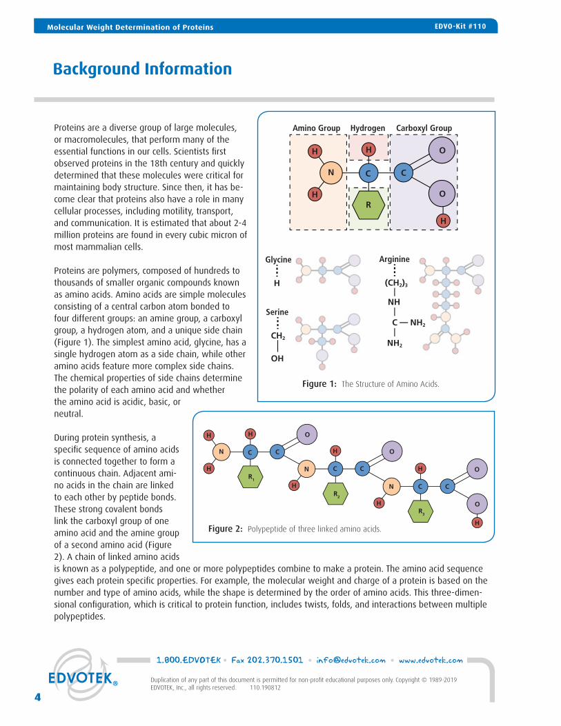
Proteins are polymers, composed of hundreds to thousands of smaller organic compounds known as amino acids. Amino acids are simple molecules consisting of a central carbon atom bonded to four different groups: an amine group, a carboxyl group, a hydrogen atom, and a unique side chain (Figure 1). The simplest amino acid, glycine, has a single hydrogen atom as a side chain, while other amino acids feature more complex side chains. The chemical properties of side chains determine the polarity of each amino acid and whether the amino acid is acidic, basic, or neutral.
During protein synthesis, a specific sequence of amino acids is connected together to form a continuous chain. Adjacent ami-no acids in the chain are linked to each other by peptide bonds. These strong covalent bonds link the carboxyl group of one amino acid and the amine group of a second amino acid (Figure 2). A chain of linked amino acids is known as a polypeptide, and one or more polypeptides combine to make a protein. The amino acid sequence gives each protein specific properties. For example, the molecular weight and charge of a protein is based on the number and type of amino acids, while the shape is determined by the order of amino acids. This three-dimen-sional configuration, which is critical to protein function, includes twists, folds, and interactions between multiple polypeptides.
Figure 1: The Structure of Amino Acids.
H H
H
CCN
O
OR
H
Amino Group Hydrogen Carboxyl Group
Glycine
H
Serine
CH2
OH
Arginine
(CH2)3
NH2
NH
C NH2
H H
H
H
H
H
H
H
CC
CC
CC
N
N
N
O
O
O
O
R1
R2
R3
Figure 2: Polypeptide of three linked amino acids.
Molecular Weight Determination of Proteins EDVO-Kit #110
1.800.EDVOTEK • Fax 202.370.1501 • [email protected] • www.edvotek.com
4
Duplication of any part of this document is permitted for non-profit educational purposes only. Copyright © 1989-2019 EDVOTEK, Inc., all rights reserved. 110.190812
EDVO-Kit #110Molecular Weight Determination of Proteins

To analyze proteins, researchers will first determine a protein’s molecular weight. Protein weight is most often de-scribed in unified atomic mass units or Daltons (Da). A mass of 1 DA is approximately the mass of one nucleon. If the protein’s amino acid composition or DNA sequence is known then the molecular weight can be calculated. (In some cases this calculation must also factor in non-amino molecules like heme, zinc, and covalently bonded carbohydrates.) However, when the molecular composition of the protein is unknown, the molecular weight must be determined experimentally. This can be done using ultracentrifugation and light scattering or by running an SDS gel electrophoresis experiment.
SDS gel electrophoresis is a simple but powerful method that provides information about the expression and purity of a molecule, along with its molecular weight. Often proteins are run on Polyacrylamide (PAGE) gels. However, acrylamide can be difficult and dangerous to work with when in liquid form. In addition, PAGE gels require specialized equipment. Alternatively, a high-density agarose gel can be used. Both PAGE gels and high-density agarose gels contain a tight network of microscopic pores and channels. During electro-phoresis, the proteins moved through the gel using these pores and channels.
To perform protein electrophoresis, a gel is prepared, placed in an electrophoresis chamber and flooded with buffer. Next, the protein samples are loaded into small indenta-tions, or wells, in the gel. Finally, an electrical current is applied to the gel box, pushing the charged proteins through the gel towards the positive electrode (Figure 3). As the proteins migrate they move through the gel’s pores and channels at different speeds. Smaller proteins have an easier time fitting than larger proteins and will migrate further in the same amount of time. By comparing the travel distance of unknown proteins to the travel distances of several known proteins the molecular weight of most protein can be determined.
PROTEIN DENATURATION FOR ELECTROPHORESIS
Proteins produce a unique challenge for electrophoresis because they have complex shapes and different charges, which affect how they migrate through the gel. Structural differences can cause two proteins with similar molecu-lar weights to migrate at different rates - a complicated, spread-out protein will move slower through the gel than one with a compact shape. Similarly, positively and negatively charged proteins will migrate in different directions through the electric field in a gel. Scientists solve both these problems by using chemicals that denature the pro-teins. This eliminates the complex structure and neutralize the charge of the native protein.
Figure 3: An Overview of SDS-PAGE
Stain to visualize.Protein Bands are separated by size.
Protein
Add SDS
Then, load samples onto SDS-PAGE gel.
negatively charged SDS molecules
5
1.800.EDVOTEK • Fax 202.370.1501 • [email protected] • www.edvotek.com
Duplication of any part of this document is permitted for non-profit educational purposes only. Copyright © 1989-2019 EDVOTEK, Inc., all rights reserved. 110.190812
Molecular Weight Determination of ProteinsEDVO-Kit #110

Sodium Dodecyl Sulfate (SDS) is a common detergent used to disrupt interactions between amino acids. The SDS molecule consists of a hydrocarbon chain bonded to a negatively charged sulfate group. When incubated with pro-teins and heated, SDS will unfold the protein’s three-dimensional structure. To break the stronger disulfide bonds in proteins researchers also use reducing agents such as β-mercaptoethanol (β-ME) or Dithiothreitol (DTT). Although the amino acid composition and sequence stay the same, a protein treated with SDS, β-ME, and DTT will no longer have biological activity because the specific three-dimensional shape has changed. Such proteins are said to be denatured. Proteins can also be denatured by briefly heating them.
In this experiment, you will be testing three unknown proteins to confirm their presence and purity and to esti-mate their molecular weight. For convenience, the proteins in this experiment have already been denatured and are pre-stained. You will run these proteins in a specialized protein agarose SDS gel along with several standard proteins of known molecular weight. Following electrophoresis you will compare the migration distances of the unknown proteins to the migration distances of the standard proteins in order to estimate their approximate size.
1.800.EDVOTEK • Fax 202.370.1501 • [email protected] • www.edvotek.com
6
Duplication of any part of this document is permitted for non-profit educational purposes only. Copyright © 1989-2019 EDVOTEK, Inc., all rights reserved. 110.190812
Molecular Weight Determination of Proteins EDVO-Kit #110

EXPERIMENT OBJECTIVE:
The objective of this experiment is to determine the molecular weight of a protein using SDS horizontal gel electrophoresis. Students will develop a basic understanding of protein structure and denaturation.
LABORATORY SAFETY
• Wear gloves and goggles while working in the laboratory.• Exercise caution when working in the laboratory – you will be using equipment that
can be dangerous if used incorrectly.• Wear protective gloves when working with hot reagents like boiling water and melted
agarose.• Always wash hands thoroughly with soap and water after working in the laboratory.
LABORATORY NOTEBOOKS:
Scientists document everything that happens during an experiment, includ-ing experimental conditions, thoughts and observations while conducting the experiment, and, of course, any data collected. Today, you’ll be documenting your experiment in a laboratory notebook or on a separate worksheet.
Before starting the Experiment:
• Carefully read the introduction and the protocol. Use this information to form a hypothesis for this experiment.
• Predict the results of your experiment.
During the Experiment:
• Record your observations.
After the Experiment:
• Interpret the results – does your data support or contradict your hy-pothesis?
• If you repeated this experiment, what would you change? Revise your hypothesis to reflect this change.
Experiment Overview
Wear gloves and safety goggles
30-45 minutes
15 min. plus electrophoresis time
15 min. plus stain/destain time
30 minutes
Molecular Weight Determination of ProteinsEDVO-Kit #110
7
1.800.EDVOTEK • Fax 202.370.1501 • [email protected] • www.edvotek.com
Duplication of any part of this document is permitted for non-profit educational purposes only. Copyright © 1989-2019 EDVOTEK, Inc., all rights reserved. 110.190812
EDVO-Kit #110 Molecular Weight Determination of Proteins

Module I: Preparing Agarose Gels For Protein Electrophoresis
1. COMBINE distilled water and agarose in a large flask (at least 250 mL). Use Table A to determine the correct volume and weight for your gel size. MIX by swirling the flask. DO NOT add the SDS buffer until step 4 (after the solution has been heated).
NOTE: High percentage agarose can easily bubble over during heating. To prevent this prepare each gel in a large flask, carefully monitor the solution as it is being heated, and add the Tris-Glycine-SDS buffer only after the solution has been heated. If you do observe a head of fine bubbles beginning to form in step 3 quickly remove the flask from the microwave and the let the bubbles subside. Swirl briefly before heating again.
2. MARK the height of the solution in the flask using lab tape or a nonpermanent marker. 3. DISSOLVE the agarose powder by boiling the solution. a. MICROWAVE the solution on high for 1 minute. b. Carefully REMOVE the flask from the microwave and MIX by swirling the flask. c. Continue to HEAT the solution in 15-second bursts until the agarose is completely dissolved. (The solution
should be clear like water.) d. REMOVE your solution from the microwave. e. OBSERVE if any evaporation has occurred. If
it has, ADD distilled water until the solution is back to its original volume as marked in step 2.
f. MIX by swirling.
4. ADD the concentrated Tris-Glycine-SDS buffer. Use Table A to determine the correct volume for your gel size. MIX by gently swirling the flask.
NOTE: Tris-Glycine-SDS contains a detergent that exacerbates bubbling. Gently handle the agarose solution after this buffer has been added. Even with careful pouring some small bubbles may form in the gel. These will not interfere with the experiment and may disappear as the gel solidifies. Bubbles can also be nudged to the side of the gel using a toothpick or plastic pipette tip during step 7.
Amt. ofProtein Agarose
Size of GelCasting tray +
DistilledWater+
TOTALVolume=
Tris-GlycineSDS Buffer
(STEP 4)
*Do not add the SDS buffer until step 4 (after the solution has been heated).
Molecular Weight Determination of Proteins EDVO-Kit #110
1.800.EDVOTEK • Fax 202.370.1501 • [email protected] • www.edvotek.com
8
Duplication of any part of this document is permitted for non-profit educational purposes only. Copyright © 1989-2019 EDVOTEK, Inc., all rights reserved. 110.190812
EDVO-Kit #110Molecular Weight Determination of Proteins
1:00
Caution! Flask will be HOT!
Concentratedbuffer
Distilled waterAgarose
Flask
Pour
Tris-GlycineSDS buffer
70°C
70°CDistilledwater
POURTris-Glycine-SDS
Buffer
OPTIONAL
1
2
3
P
2 0.
0
2 0.0
P

Module I: Preparing Agarose Gels For Protein Electrophoresis, continued
5. COOL the agarose solution to 70°C. Periodically MIX to promote even dissipation of heat. 6. While the agarose is cooling, SEAL the ends of a gel casting tray with the rubber end caps. PLACE the
well template (comb) in the first set of notches. 7. Slowly, POUR the cooled agarose solution into the prepared gel-casting tray. The gel should thoroughly
solidify within 20 minutes. 8. While the agarose solidifies, PREPARE the electrophoresis buffer by diluting the remaining Tris-Glycine-
SDS buffer in distilled water according to Table B.
9. REMOVE both end caps and then remove the comb by pulling straight up. Work slowly and carefully to prevent damage to the wells.
10. PLACE the gel (still on the tray) into the elec-trophoresis chamber. COVER the gel with 1X Electrophoresis Buffer using the recommended volume in Table B. The gel should be com-pletely submerged.
OPTIONAL STOPPING POINT: Gels can be stored overnight submerged in the electrophoresis chamber. Gels can also be stored for several days in the refrigerator. Keep refrigerated gels hydrated by storing each gel in a watertight plastic bag with a small amount of electrophoresis buffer.
9
1.800.EDVOTEK • Fax 202.370.1501 • [email protected] • www.edvotek.com
Duplication of any part of this document is permitted for non-profit educational purposes only. Copyright © 1989-2019 EDVOTEK, Inc., all rights reserved. 110.190812
Molecular Weight Determination of ProteinsEDVO-Kit #110
1:00
Caution! Flask will be HOT!
Concentratedbuffer
Distilled waterAgarose
Flask
Pour
Tris-GlycineSDS buffer
70°C
70°CDistilledwater
POURTris-Glycine-SDS
Buffer
OPTIONAL
1
2
3
P
2 0.
0
2 0.0
P

Module II: Performing Electrophoresis
1. COLLECT the three unknown protein samples and standard protein marker from your instructor.
2. Using a fresh pipet tip, MEASURE 20 µL of the standard protein marker.
3. PLACE the pipet tip under the buffer and directly above the sample well. Slowly DISPENSE the sample by depressing the plunger.
4. REPEAT steps 2-4 with the three unknown protein samples.
5. PLACE safety cover on the chamber. CHECK that the gel is properly oriented. Remember that the samples will migrate toward the positive (red) electrode.
6. CONNECT leads to the power supply and PERFORM electrophoresis (See Table C for time and voltage guide-lines.)
7. After electrophoresis is complete, REMOVE the gel and casting tray from the electrophoresis chamber.
1
2
3
4
Standard Protein Marker
Unknown Protein Sample 1
Unknown Protein Sample 2
Unknown Protein Sample 3
OPTIONAL STOPPING POINT: Gels can be stored overnight. For long term storage the gel should be submerged in a fixative solution.
Molecular Weight Determination of Proteins EDVO-Kit #110
1.800.EDVOTEK • Fax 202.370.1501 • [email protected] • www.edvotek.com
10
Duplication of any part of this document is permitted for non-profit educational purposes only. Copyright © 1989-2019 EDVOTEK, Inc., all rights reserved. 110.190812
EDVO-Kit #110Molecular Weight Determination of Proteins
1:00
Caution! Flask will be HOT!
Concentratedbuffer
Distilled waterAgarose
Flask
Pour
Tris-GlycineSDS buffer
70°C
70°CDistilledwater
POURTris-Glycine-SDS
Buffer
OPTIONAL
1
2
3
P
2 0.
0
2 0.0
P

Module III: Supplemental Gel Staining (Optional)
1. SLIDE the gel off of the casting tray into a small, clean gel-staining tray.
2. SUBMERGE the gel with approx. 100 mL of fixative solution. (Use enough solution to cover the gel entirely.)
3. Gently FLOAT a sheet of Protein InstaStain® with the stain side (blue) in the liquid.
4. SEAL the tray with plastic wrap to prevent evaporation.
5. Gently AGITATE on a rocking platform for 1-3 hours or overnight.
6. After staining, protein bands will appear as dark blue bands against a light background and will be ready for photography.
NOTE: If the gel is too dark, destain in several changes of fresh fixative solution until the contrast between the protein bands and background improves.
Molecular Weight Determination of ProteinsEDVO-Kit #110
11
1.800.EDVOTEK • Fax 202.370.1501 • [email protected] • www.edvotek.com
Duplication of any part of this document is permitted for non-profit educational purposes only. Copyright © 1989-2019 EDVOTEK, Inc., all rights reserved. 110.190812
EDVO-Kit #110 Molecular Weight Determination of Proteins

1. MEASURE the migration distance, in centimeters (to the nearest millimeter) of every major band in the gel. All measurements should be from the bottom of the sample well to the bottom of the protein band.
2. Using semilog graph paper, PLOT the migration distance or relative mobility (Rf) of each stan-dard protein on the non-logarithmic x-axis versus its molecular weight on the logarithmic y-axis. CHOOSE your scales so that the data points are well spread out.
3. DRAW the best average straight line through all the points. This line should roughly have an equal number of points scattered on each side of the line. As an ex-ample, refer to the figure at left. This method is a linear approxi-mation.
4. Using your standard graph, DE-TERMINE the molecular weight of the three unknown proteins. This can be done by finding the Rf (or migration distance) of the unknown band on the x-axis and drawing a straight vertical until the standard line is intersected.
5. A straight line is then made from the intersection across to the y-axis where the approximate molecular weight can be deter-mined.
Marker Protein 1 (Phosphorylase)
Marker Protein 2 (Bovine Serum Albumin)
Marker Protein 3 (Ovalbumin)
Marker Protein 4 (Carbonic anhydrase)
Marker Protein 5 (Soybean Trypsin Inhibitor)
Marker Protein 6 (Lysozyme)
Unknown Protein Sample 1
Unknown Protein Sample 2
Unknown Protein Sample 3
94,000
67,000
38,000
30,000
20,000
14,000
MigrationDistance
(cm)
MolecularWeight
(daltons)
8
10
76
5
4
3
2
1
9
876
5
4
3
2
1
9
876
5
4
3
2
1
9
100,000
10,000
6 7 8 9 10
Centimeters
Mol
ecul
ar W
eigh
t
Phosphorylase
Bovine Serum Albumin
Ovalbumin
Carbonic anhydrase
5
Soybean Trypsin Inhibitor
Lysozyme
Centimeters
Mol
ecul
ar W
eigh
t
Module IV: Size Determination of Unknown Proteins
Molecular Weight Determination of Proteins EDVO-Kit #110
1.800.EDVOTEK • Fax 202.370.1501 • [email protected] • www.edvotek.com
12
Duplication of any part of this document is permitted for non-profit educational purposes only. Copyright © 1989-2019 EDVOTEK, Inc., all rights reserved. 110.190812
EDVO-Kit #110Molecular Weight Determination of Proteins

80,000
100,000
70,00060,000
50,000
40,000
30,000
20,000
90,000
800 700 600
500
400
300
200
100
9001,000
10,000
8,000 7,000 6,000
5,000
4,000
3,000
2,000
9,000
X-axis: Migration distance (cm)
1 cm 2 cm 3 cm 4 cm 5 cm 6 cm
Y-a
xis:
Mo
lec
ula
r We
ight
13
1.800.EDVOTEK • Fax 202.370.1501 • [email protected] • www.edvotek.com
Duplication of any part of this document is permitted for non-profit educational purposes only. Copyright © 1989-2019 EDVOTEK, Inc., all rights reserved. 110.190812
Molecular Weight Determination of ProteinsEDVO-Kit #110

Study Questions
1. What is an amino acid? Draw or describe the four subparts of an amino acid.
2. What is Sodium Dodecyl Sulfate (SDS) and why is it so important to protein electrophoresis?
3. What would happen if a non-denatured (native) protein was run in a gel next to a denatured sample of the same protein?
Molecular Weight Determination of Proteins EDVO-Kit #110
1.800.EDVOTEK • Fax 202.370.1501 • [email protected] • www.edvotek.com
14
Duplication of any part of this document is permitted for non-profit educational purposes only. Copyright © 1989-2019 EDVOTEK, Inc., all rights reserved. 110.190812
EDVO-Kit #110Molecular Weight Determination of Proteins

Instructor's Guide
15
1.800.EDVOTEK • Fax 202.370.1501 • [email protected] • www.edvotek.com
Duplication of any part of this document is permitted for non-profit educational purposes only. Copyright © 1989-2019 EDVOTEK, Inc., all rights reserved. 110.190812
INSTRUCTOR'S GUIDEEDVO-Kit #110 Molecular Weight Determination of Proteins
Preparation for: What to do: Time Required:When?
Distribute student supplies OR
Anytime before Module I 10 minutes
10 minutes
10 minutes
30 minutesUp to one week beforeModule I.
Module I
Module II
Module III
Up to one week beforeModule II.
Rehydrate protein samples
Up to one week beforeModule III.
Prepare fixative solution (optional)
Batch prepare gels and electrophoresis buffer
ORGANIZING AND IMPLEMENTING THE EXPERIMENT
Prior to starting this experiment, carefully check the list of components and requirements on page 3 to ensure you have all the necessary components and equipment.
This experiment is supplied with reagents for six groups to each run four samples. Each group can be provided with protein agarose gel or can create their own.

Pre-Lab Preparations:
MODULE I - PREPARING AGAROSE GELS FOR PROTEIN ELECTROPHORESIS
This experiment requires a total of six 3.2% protein agarose gels for the class. These gels can be prepared by each student group as described in Module I. Alternatively, to save class time, all six gels and the electrophoresis buffer can be prepared beforehand by the teacher (see Appendix B).
Prepare Tris-Glycine-SDS Buffer
1. Measure 200 mL of distilled or deionized water and place in a large vessel. 2. Add the entire amount of Tris-Glycine-SDS Powdered Buffer powder to the vessel and mix well. 3. Label the vessel as "10X Tris-Glycine-SDS Buffer". 4. Use within 60 days of preparation.
MODULE II - PERFORMING ELECTROPHORESIS Reconstitute Proteins and Standard Protein Marker
1. Add 160 µL of distilled or deionized water to each tube (A-D) and allow the samples to hydrate for several minutes.
2. Vortex or flick tube vigorously to mix. 3. Either place re-hydrated proteins in their original tubes in a central location to
be shared by the class or aliquot 25 µL of each protein into six appropriately labeled new tubes. The latter will require 24 additional small tubes.
NOTE: Rehydrated proteins may be kept at room temperature for immediate use or frozen until needed.
MODULE III - SUPPLEMENTAL GEL STAINING Prepare Fixative Solution (Optional - This solution is only needed for long term gel storage or for supplemental staining.)
1. Mix 500 mL methanol, 400 mL distilled water, and 100 mL glacial acetic acid. 2. Cover to prevent evaporation.
FOR MODULE IEach group will need:
• Protein Agarose • Distilled Water• 10X Tris-Glycine-SDS Buffer• Gel casting tray, seals, and comb• Two large flasks (for gel prepa-
ration and electrophoresis buffer preparation)
FOR MODULE IIEach group will need:
• Protein agarose Gel• Electrophoresis apparatus • Power supply• Micropipette and tips• Standard Protein Marker• Three Protein Samples
FOR MODULE IIIEach group will need:
• Approximately 100 μL of Fixative Solution• Protein InstaStain® Card• Staining tray• Plastic wrap• Rocking platform
1.800.EDVOTEK • Fax 202.370.1501 • [email protected] • www.edvotek.com
16
Duplication of any part of this document is permitted for non-profit educational purposes only. Copyright © 1989-2019 EDVOTEK, Inc., all rights reserved. 110.190812
INSTRUCTOR'S GUIDE Molecular Weight Determination of Proteins EDVO-Kit #110
Experiment Results and Analysis
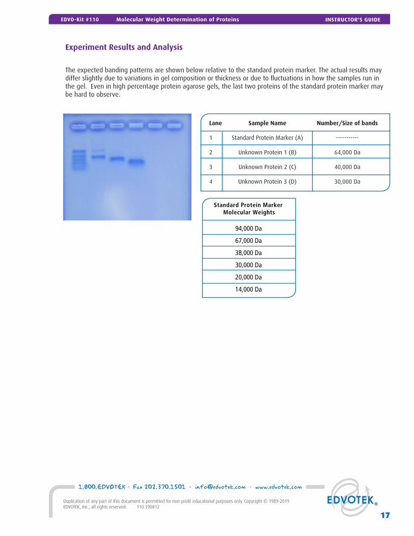
The expected banding patterns are shown below relative to the standard protein marker. The actual results may differ slightly due to variations in gel composition or thickness or due to fluctuations in how the samples run in the gel. Even in high percentage protein agarose gels, the last two proteins of the standard protein marker may be hard to observe.
Lane Sample Name Number/Size of bands
1 Standard Protein Marker (A) -----------
2 Unknown Protein 1 (B) 64,000 Da
3 Unknown Protein 2 (C) 40,000 Da
4 Unknown Protein 3 (D) 30,000 Da
17
1.800.EDVOTEK • Fax 202.370.1501 • [email protected] • www.edvotek.com
Duplication of any part of this document is permitted for non-profit educational purposes only. Copyright © 1989-2019 EDVOTEK, Inc., all rights reserved. 110.190812
INSTRUCTOR'S GUIDEEDVO-Kit #110 Molecular Weight Determination of Proteins
Standard Protein Marker Molecular Weights
94,000 Da
67,000 Da
38,000 Da
30,000 Da
20,000 Da
14,000 Da

Please refer to the kit insert for the Answers to
Study Questions
A Practice Gel Loading
B Bulk Electrophoresis Preparation
Safety Data Sheets can be found on our website: www.edvotek.com/safety-data-sheets
Appendices
19
1.800.EDVOTEK • Fax 202.370.1501 • [email protected] • www.edvotek.com
Duplication of any part of this document is permitted for non-profit educational purposes only. Copyright © 1989-2019 EDVOTEK, Inc., all rights reserved. 110.190812
APPENDICESEDVO-Kit #110 Molecular Weight Determination of Proteins
Appendix APractice Gel Loading
If your students are unfamiliar with loading samples in agarose we suggest they practices the delivery techniques be-fore performing this experiment. Below is one suggested activity for practice gel loading. Although the same gel can be used for both this activity and the actual experiment we suggest using a separate gel in case of damage. 1. Cast a separate practice gel with the maximum number of combs in it. For practice gel loading you can use any gel
grade agar at any concentration or you can use Edvotek’s reusable DuraGels™ (Cat. #S-43).
2. Place the gel under water or buffer either in the chamber or in a similarly deep tray.
3. Let students practice delivering the practice gel solution to the sample wells.
4. If the students need more practice, remove the practice gel loading solution by squirting buffer into the wells with a transfer pipet.
1.800.EDVOTEK • Fax 202.370.1501 • [email protected] • www.edvotek.com
20
Duplication of any part of this document is permitted for non-profit educational purposes only. Copyright © 1989-2019 EDVOTEK, Inc., all rights reserved. 110.190812
APPENDICES Molecular Weight Determination of Proteins EDVO-Kit #110
Appendix B
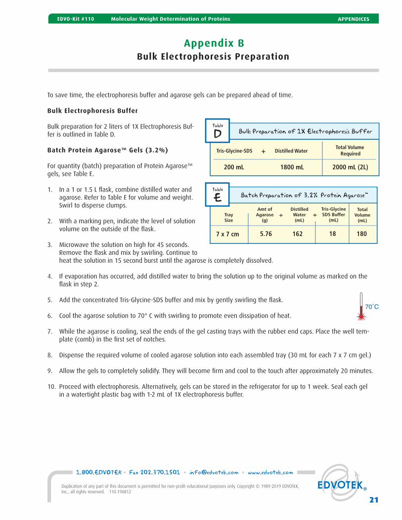
To save time, the electrophoresis buffer and agarose gels can be prepared ahead of time.
Bulk Electrophoresis Buffer
Bulk preparation for 2 liters of 1X Electrophoresis Buf-fer is outlined in Table D.
Batch Protein Agarose™ Gels (3.2%)
For quantity (batch) preparation of Protein Agarose™ gels, see Table E.
1. In a 1 or 1.5 L flask, combine distilled water and agarose. Refer to Table E for volume and weight. Swirl to disperse clumps.
2. With a marking pen, indicate the level of solution volume on the outside of the flask.
3. Microwave the solution on high for 45 seconds. Remove the flask and mix by swirling. Continue to heat the solution in 15 second burst until the agarose is completely dissolved.
4. If evaporation has occurred, add distilled water to bring the solution up to the original volume as marked on the flask in step 2.
5. Add the concentrated Tris-Glycine-SDS buffer and mix by gently swirling the flask.
6. Cool the agarose solution to 70° C with swirling to promote even dissipation of heat.
7. While the agarose is cooling, seal the ends of the gel casting trays with the rubber end caps. Place the well tem-plate (comb) in the first set of notches.
8. Dispense the required volume of cooled agarose solution into each assembled tray (30 mL for each 7 x 7 cm gel.)
9. Allow the gels to completely solidify. They will become firm and cool to the touch after approximately 20 minutes.
10. Proceed with electrophoresis. Alternatively, gels can be stored in the refrigerator for up to 1 week. Seal each gel in a watertight plastic bag with 1-2 mL of 1X electrophoresis buffer.
Bulk Electrophoresis Preparation
200 mL 1800 mL 2000 mL (2L)
70˚C
21
1.800.EDVOTEK • Fax 202.370.1501 • [email protected] • www.edvotek.com
Duplication of any part of this document is permitted for non-profit educational purposes only. Copyright © 1989-2019 EDVOTEK, Inc., all rights reserved. 110.190812
APPENDICESEDVO-Kit #110 Molecular Weight Determination of Proteins