11-12-development of the cns
TRANSCRIPT

Development of the CNS
Normal and Abnormal

Lectures Objectives• Describe the formation of neural tube and neural crest.• Describe the development of brain and spinal cord.• Describe the positional changes of spinal cord.• Describe the development of the spinal nerves and their
spinal ganglia.• Describe the development of meninges.• Describe the development of brain vesicles from the neural
tube.• Describe the development of the different parts of brain.• Describe the development of brain ventricles and choroid
plexuses.• Describe the development of the cranial nerves and their
ganglia.• Describe the congenital anomalies of brain and spinal cord.

Neurulation
• The notochord induces the ectodermal cells over it to form the neural plate
• Neural plate the neural folds and neural groove that will fuse to form the neural tube (the primordium of the CNS)

Neurulation…
• Neural tube forms and separate from the above ectoderm (the primordium of epidermis)in the third week
• During the neural tube formation cells on the crest of the neural folds (neural crest) migrate laterally
• Neural crest cells form the ganglia in the PNS and other tissues

Development of NS
• Neural plate forms the nervous system
• Neural tube forms the CNS
• Neural crest forms the PNS and ANS
• Neural canal becomes the ventricles and spinal canal

Contribution of the Neural Crest• Derived from neuroectoderm• Give rise to or participate in the
formation of many cells types and organs:– Sensory neurons of the spinal
nerves, cranial nerves (V, VII, IX, and X)
– Autonomic ganglia– Sheathing cells of the peripheral
nervous system– Pigment cells of the dermis– Muscles, connective tissues, and
bones of pharyngeal arch origin – Suprarenal medulla– Meninges (coverings) of the brain
and spinal cord


Development of NS
• By the end of the 4thweek – Rostral neuropore closed and forebrain develop
– Caudal neuropore closed and caudal eminencedevelop

Development of the Spinal Cord
• Neural tube caudal to the 4th somites forms SC
• Thickening of the neural tube forming three layers – Ependymal (ventricular) layer
– Mantle layer – Marginal Layer

Histogenesis of the Neural Tube• Ependymal (ventricular) layer →
blast cells (migrate to mantle layer)– Remaining cells → ependymal cells
Microglia – from bone marrow• Mantle layer → cells of CNS
– Neuroblasts• Motor neurons (somatic &
Autonomic)• Interneurons
– Gliablasts (invade both mantle and marginal layers)
• Astrocytes• Oligodendrocytes
• Marginal Layer – white mater

Regions of the Spinal Cord• Ependyma• Marginal layer• Roof and floor plates• Sulcus limitans• Mantle layer
– Alar plates – dorsal gray columns – sensory input
• Internuncials, Commissurals, Tract fibers
Thickening forms dorsal septum– Basal plates – ventral gray
columns• Somatic & visceral motor neurons Thickening forms ventral median
fissure

Neural Tube Defects
• Spina bifida: results from failure of the neural tube to close in the vertebral column

Spina Bifida
• Spina bifida occulta– Defect in vertebral arch– L5‐S1– No clinical symptoms
• Spina bifida cystica– With meningocele
• 1/1000• Sac contains meninges & CSF
– With meningomyelocele • 2‐4/1000• Sac contains spinal cord and/or nerves• Neurological deficits

Development of the Brain: Vesicles

Brain Flexures
• Happens due to the rapid growth of the brain
• Midbrain flexure– Ventrally
• Cervical flexure– Between hindbrain & SC– Ventrally
• Pontine flexure– Between metencephalon &
myelencephalon– Dorsally

Myelencephalon • Caudal part
– Closed part (spinal canal)– Neuroblasts from alar plates
migrate into the marginal zone• Gracile & cuneate nuclei
– Pyramids – ventrally• Rostral part
– Open part• Lateral plates diverts away & roof plate widens
– Alar plates lateral» Afferent neurons» Olivary nuclei
– Basal plates medial» Efferent nuclei

MyelencephalonFunctional components of cranial nerves
• Basal plates– GSE – IIX nerve– SVE – IX nerve– GVE – X, XI nerves
Sulcus limitans• Alar plates
– GVA– SVA– GSA– SSA
This order is valid for entire brainstem

Metencephalon • Walls → pons & cerebellum• Cavity → rostral part of 4th
ventricle• Alar plates gives rise to
cerebellar swellings• Cerebellar swelling fuse in the
midline• Neuroblasts from alar plates
forms – Cerebellar cortex– Central nuclei of cerebellum– Pontine nuclei (cochlear &
vestibular nuclei)– Sensory nuclei of trigeminal
nerve

Midbrain
• Neural canal → cerebral aqueduct
• Alar plates – Tectum nuclei
• Superior & inferior colliculi
• Basal plates– Tegmentum nuclei
• III, IV nerves• Red nucleus• Substantia nigra

Diencephalon
• Three swelling from the lateral wall of the 3rdventricle– EpithalamusEpithalamic sulcus– ThalamusHypothalamic sulcus– Hypothalamus

Telencephalon
• Cerebral vesicles → cerebral hemispheres – Enlarge and becomes C‐shape
– Caudal end enlarge and fold – temporal lobe
• Cavity → anterior part of 3rd ventricle & lateral ventricles (c‐shape)– Temporal horn follow temporal lobe

Telencephalon
• Corpus striatum develops from swelling in the floor of each cerebral hemisphere– Fibers (C‐shape internal capsule) divide it into C‐shape caudate nucleus & lentiform nucleus
• Neuroblast cells migrate to marginal zone to form cerebral cortex

Telencephalon• Cerebral commissures
– Lamina terminalis• Anterior commissure• Hippocampal commissure (fornix)
• Corpus callosum– Begins at lamina terminalis
and grows beyond it
• Septum pellucidum– Between fornix & corpus
callosum
• Optic chiasm

Telencephalon
• Growth of the cerebral hemispheres results in – The development of sulci and gyri – The hidden insula

Congenital Anomalies of the Brain

Defective Closure of Rostral Neuropore
• Anencephaly • Meroanencephaly
– 1/1000
• Cranium bifidium1/2000Mostly in mid‐occipital– Cranial meningocele– Meningoencephalocele– meningohydroencephalocele

Hydrocephalus • Blockage of drainage of CSF
(tumor, inflammation, developmental malformation, meningitis, hemorrhage or injury)
• Continued production cause an increase in pressure ‐‐‐hydrocephalus
• In newborn or fetus, the fontanels allow this internal pressure to cause expansion of the skull and damage to the brain tissue
• Neurosurgeon implants a drain shunting the CSF to the veins of the neck or the abdomen

Development of the Spinal Nerves• Motor nerve fibers
– Begin to appear in the fourth week,
– Arising from nerve cell in basal plates
– Form ventral nerve roots • Sensory fibers form dorsal
root– Originate from nerve cells in
spinal ganglia – Derived from neural crest
cells– Proximal grow into the dorsal
horns– Distal processes join the
ventral nerve roots to form a spinal nerve

Development of the Spinal Nerves• Spinal nerves divide into
dorsal and ventral primary rami– Contain both motor and
sensory fibers• Dorsal rami innervate the
back (muscles, vertebral joints, and skin)
• Ventral rami innervate the limbs and ventral body wall– Form nerve plexuses
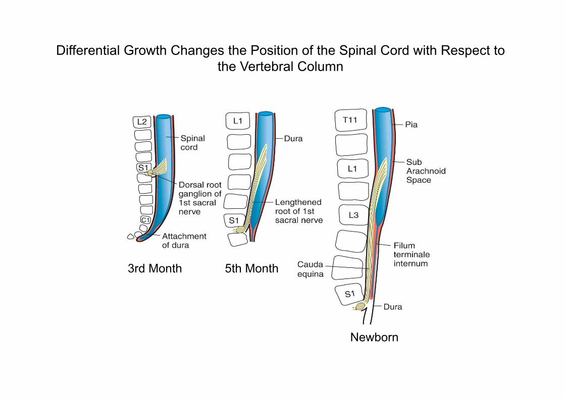
Differential Growth Changes the Position of the Spinal Cord with Respect to the Vertebral Column
3rd Month 5th Month
Newborn