11-1 introduction to muscle movement is a fundamental characteristic of all living things muscle...
TRANSCRIPT

11-1
Introduction to Muscle
• movement is a fundamental characteristic of all living things
• muscle cells are capable of converting the chemical energy of ATP into mechanical energy
• types of muscle– skeletal, cardiac and smooth
• physiology of skeletal muscle– basis of warm-up, quickness, strength,
endurance and fatigue

Functions of Muscle
• Producing Movement- just about all movements of the human body and its parts are a result of muscle contraction.
• Maintaining Posture- muscles function almost continuously to maintain posture.
• Stabilizing Joints- muscles help stabilize and strengthen joints.
• Generating Heat- muscles generate heat as they contract.
11-2

11-3
Characteristics of Muscle• responsiveness (excitability)
– to chemical signals, stretch and electrical changes across the plasma membrane
• conductivity– local electrical change triggers a wave of excitation that travels
along the muscle fiber
• contractility – shortens when stimulated
• extensibility – capable of being stretched between contractions
• elasticity – returns to its original resting length after being stretched
11-4
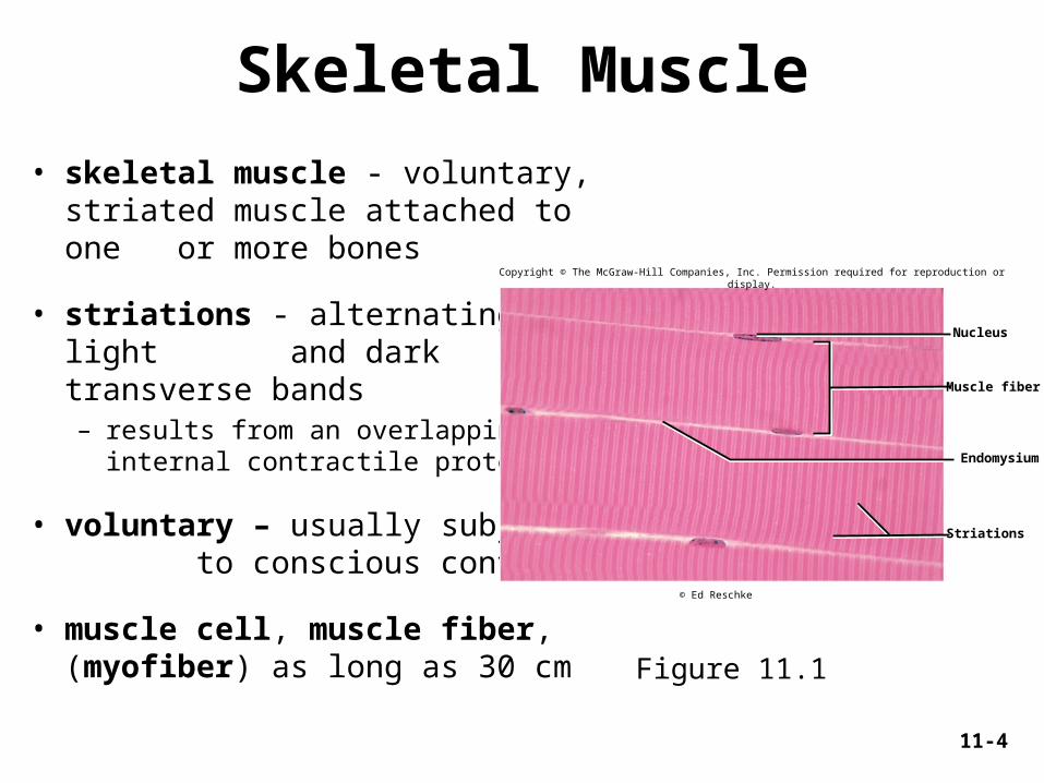
Skeletal Muscle
• skeletal muscle - voluntary, striated muscle attached to one or more bones
• striations - alternating light and dark transverse bands– results from an overlapping of
internal contractile proteins
• voluntary – usually subject to conscious control
• muscle cell, muscle fiber, (myofiber) as long as 30 cm Figure 11.1
Nucleus
Muscle fiber
Endomysium
Striations
Copyright © The McGraw-Hill Companies, Inc. Permission required for reproduction or display.
© Ed Reschke
11-5
Connective Tissue Elements
• tendons are attachments between muscle and bone matrix
– endomysium – connective tissue around muscle cells
– perimysium – connective tissue around muscle fascicles
– epimysium – connective tissue surrounding entire muscle
– continuous with collagen fibers of tendons
– in turn, with connective tissue of bone matrix

Sarcoplasm
Sarcolemma
Openings intotransverse tubules
Sarcoplasmicreticulum
Mitochondria
Myofibrils
Myofilaments
A band
I band
Z disc
Nucleus
Triad:Terminal cisternaeTransverse tubule
Musclefiber
11-6
Structure of a Skeletal Muscle Fiber
Figure 11.2
Copyright © The McGraw-Hill Companies, Inc. Permission required for reproduction or display.
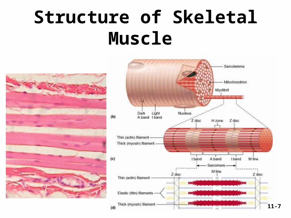
Structure of Skeletal Muscle
11-7
11-8
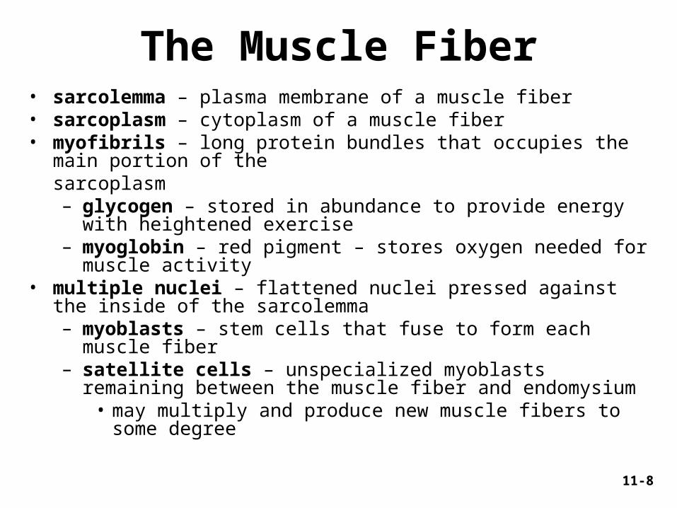
The Muscle Fiber• sarcolemma – plasma membrane of a muscle fiber• sarcoplasm – cytoplasm of a muscle fiber• myofibrils – long protein bundles that occupies the main portion of the sarcoplasm
– glycogen – stored in abundance to provide energy with heightened exercise
– myoglobin – red pigment – stores oxygen needed for muscle activity
• multiple nuclei – flattened nuclei pressed against the inside of the sarcolemma– myoblasts – stem cells that fuse to form each muscle fiber– satellite cells – unspecialized myoblasts remaining between the
muscle fiber and endomysium• may multiply and produce new muscle fibers to some degree

11-9
Thick Myofilaments
• made of several hundred myosin molecules– shaped like a golf club
• two chains intertwined to form a shaft-like tail• double globular head
– heads directed outward in a helical array around the bundle• heads on one half of the thick filament angle to the left• heads on the other half angle to the right• bare zone with no heads in the middle
Figure 11.3 a-b
(a) Myosin molecule
HeadTail
(b) Thick filament
Myosin head
Copyright © The McGraw-Hill Companies, Inc. Permission required for reproduction or display.
11-10
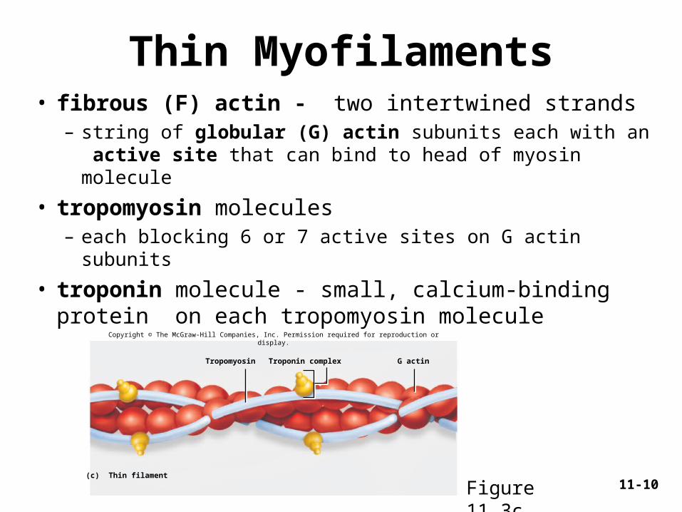
Thin Myofilaments• fibrous (F) actin - two intertwined strands
– string of globular (G) actin subunits each with an active site that can bind to head of myosin molecule
• tropomyosin molecules– each blocking 6 or 7 active sites on G actin subunits
• troponin molecule - small, calcium-binding protein on each tropomyosin molecule
(c) Thin filament
Troponin complex G actinTropomyosin
Copyright © The McGraw-Hill Companies, Inc. Permission required for reproduction or display.
Figure 11.3c

11-11
Elastic Myofilaments
• titin (connectin) – huge springy protein– flank each thick filament and anchor it to the
Z disc– helps stabilize the thick filament– center it between the thin filaments– prevents over stretching
11-12
Regulatory and Contractile Proteins
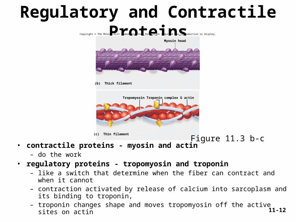
• contractile proteins - myosin and actin – do the work
• regulatory proteins - tropomyosin and troponin – like a switch that determine when the fiber can contract and when it cannot– contraction activated by release of calcium into sarcoplasm and its binding
to troponin, – troponin changes shape and moves tropomyosin off the active
sites on actin
(b) Thick filament
Myosin head
(c) Thin filament
Troponin complex G actinTropomyosin
Copyright © The McGraw-Hill Companies, Inc. Permission required for reproduction or display.
Figure 11.3 b-c
11-13
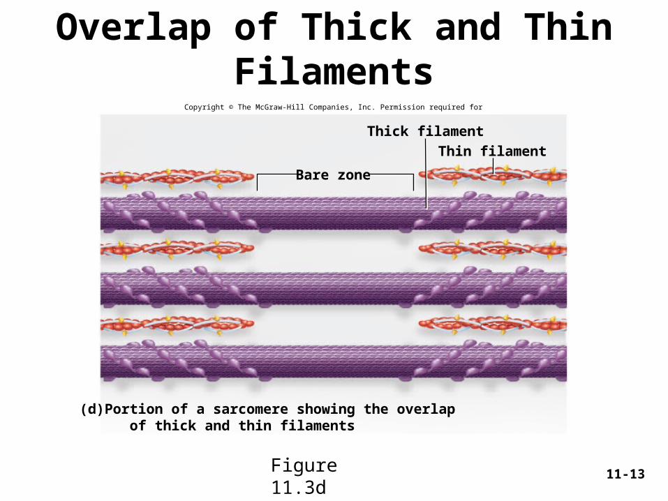
Overlap of Thick and Thin FilamentsCopyright © The McGraw-Hill Companies, Inc. Permission required for reproduction or display.
(d) Portion of a sarcomere showing the overlap of thick and thin filaments
Bare zone
Thin filament
Thick filament
Figure 11.3d

Sliding of the actin filaments during contraction
11-14
11-15
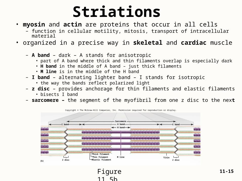
Striations • myosin and actin are proteins that occur in all cells
– function in cellular motility, mitosis, transport of intracellular material• organized in a precise way in skeletal and cardiac muscle
– A band – dark – A stands for anisotropic• part of A band where thick and thin filaments overlap is especially dark• H band in the middle of A band – just thick filaments• M line is in the middle of the H band
– I band – alternating lighter band – I stands for isotropic• the way the bands reflect polarized light
– z disc – provides anchorage for thin filaments and elastic filaments• bisects I band
– sarcomere – the segment of the myofibril from one z disc to the next Copyright © The McGraw-Hill Companies, Inc. Permission required for reproduction or display.
Sarcomere
I band I band A band
H band
Thick filament
TitinThin filamentElastic filament
(b) Z discZ disc
M line
Figure 11.5b

Striations and Sarcomeres
• sarcomere – functional contractile unit of the muscle fiber– muscle shortens because individual sarcomeres shorten
– pulls z discs closer to each other 11-16
Figure 11.5a
Copyright © The McGraw-Hill Companies, Inc. Permission required for reproduction or display.
Ind
ivid
ual
myo
fib
rils
12
34
5
Sarcomere
I band I bandA band
H band
M line
(a)
Z disc
Nucleus
Visuals Unlimited

11-17
Sarcomeres• sarcomere - segment from Z disc to Z disc
– functional contractile unit of muscle fiber
• muscle cells shorten because their individual sarcomeres shorten – Z disc (Z lines) are pulled closer together as
thick and thin filaments slide past each other
• neither thick nor thin filaments change length during shortening– only the amount of overlap changes
11-18
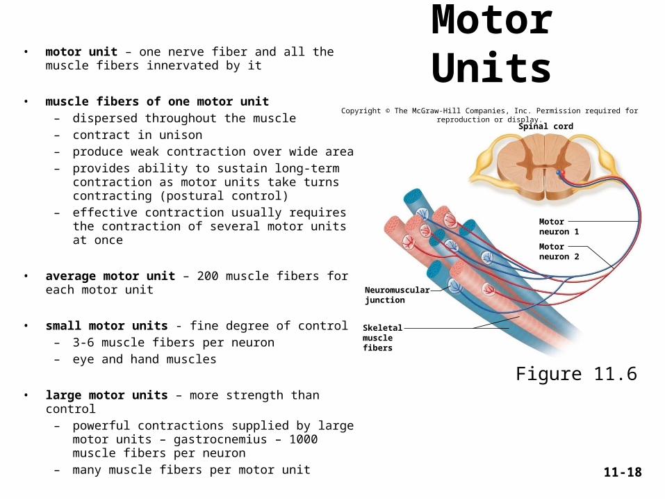
Motor Units• motor unit – one nerve fiber and all the muscle fibers innervated by it
• muscle fibers of one motor unit– dispersed throughout the muscle– contract in unison– produce weak contraction over wide area– provides ability to sustain long-term contraction
as motor units take turns contracting (postural control)
– effective contraction usually requires the contraction of several motor units at once
• average motor unit – 200 muscle fibers for each motor unit
• small motor units - fine degree of control – 3-6 muscle fibers per neuron– eye and hand muscles
• large motor units – more strength than control– powerful contractions supplied by large motor
units – gastrocnemius – 1000 muscle fibers per neuron
– many muscle fibers per motor unit
Figure 11.6
Spinal cord
Neuromuscularjunction
Skeletalmusclefibers
Motorneuron 1
Motorneuron 2
Copyright © The McGraw-Hill Companies, Inc. Permission required for reproduction or display.
11-19
The Neuromuscular Junction • synapse – point where a nerve fiber meets its
target cell
• neuromuscular junction (NMJ) - when target cell is a muscle fiber
• each terminal branch of the nerve fiber within the NMJ forms separate synapse with the muscle fiber
• one nerve fiber stimulates the muscle fiber at several points within the NMJ
11-20
Components of Neuromuscular Junction• synaptic knob - swollen end of nerve fiber
– contains synaptic vesicles filled with acetylcholine (ACh)
• synaptic cleft - tiny gap between synaptic knob and muscle sarcolemma
• Schwann cell envelops & isolates all of the NMJ from surrounding tissue fluid
• synaptic vesicles undergo exocytosis releasing ACh into synaptic cleft
• 50 million ACh receptors – proteins incorporated into muscle cell plasma membrane

11-21
Neuromuscular Junction - LM
Figure 11.7a
Copyright © The McGraw-Hill Companies, Inc. Permission required for reproduction or display.
Neuromuscularjunction
Motor nervefibers
Muscle fibers
(a) 100 µmVictor B. Eichler
11-22
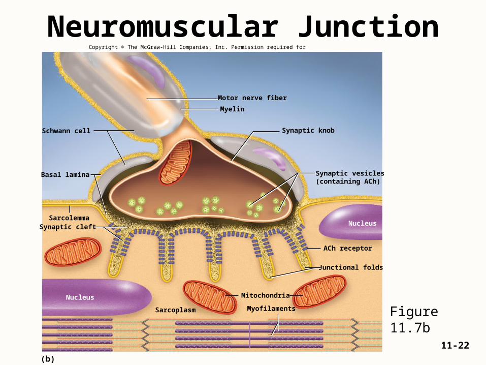
Neuromuscular JunctionCopyright © The McGraw-Hill Companies, Inc. Permission required for reproduction or display.
(b)
Myelin
Motor nerve fiber
Schwann cell
Basal lamina
Synaptic knob
Synaptic vesicles(containing ACh)
Sarcolemma
Junctional folds
ACh receptor
Myofilaments
Nucleus
Synaptic cleft Nucleus
Sarcoplasm
Mitochondria
Figure 11.7b

11-23
Neuromuscular Toxins• toxins that interfere with synaptic function can paralyze the muscles
• tetanus (lockjaw) is a form of spastic paralysis caused by toxin of Clostridium tetani– glycine in the spinal cord normally stops motor neurons from
producing unwanted muscle contractions– tetanus toxin blocks glycine release in the spinal cord and causes
overstimulation and spastic paralysis of the muscles
• flaccid paralysis – a state in which the muscles are limp and cannot contract– curare – compete with ACh for receptor sites, but do not stimulate
the muscles – plant poison used by South American natives to poison blowgun
darts
• botulism – type of food poisoning caused by a neuromuscular toxin secreted by the bacterium Clostridium botulinum– blocks release of ACh causing flaccid paralysis– Botox Cosmetic injections for wrinkle removal

11-24
Electrically Excitable Cells• muscle fibers and neurons are electrically excitable cells
– their plasma membrane exhibits voltage changes in response to stimulation
• electrophysiology - the study of the electrical activity of cells• in an unstimulated (resting) cell
– there are more anions (negative ions) on the inside of the plasma membrane than on the outside
– the plasma membrane is electrically polarized (charged)
– there are excess sodium ions (Na+) in the extracellular fluid (ECF)
– there are excess potassium ions (K+) in the intracellular fluid (ICF)
– also in the ICF, there are anions such as proteins, nucleic acids, and phosphates that cannot penetrate the plasma membrane
– these anions make the inside of the plasma membrane negatively charged by comparison to its outer surface
• voltage (electrical potential) – a difference in electrical charge from one point to another
• resting membrane potential – about -90mV– maintained by sodium-potassium pump
11-25
Electrically Excitable Cells• stimulated (active) muscle fiber or nerve cell
– ion gates open in the plasma membrane– Na+ instantly diffuses down its concentration gradient into the cell– these cations override the negative charges in the ICF– depolarization - inside of the plasma membrane becomes briefly positive– immediately, Na+ gates close and K+ gates open– K+ rushes out of cell– repelled by the positive sodium charge and partly because of its concentration
gradient– loss of positive potassium ions turns the membrane negative again
(repolarization)– action potential – quick up-and-down voltage shift from the negative RMP to a
positive value, and back to the negative value again.– RMP is a stable voltage seen in a waiting muscle or nerve cell– action potential is a quickly fluctuating voltage seen in an active stimulated
cell– an action potential at one point on a plasma membrane causes another
one to happen immediately in front of it, which triggers another one alittle farther along and so forth

11-26
Muscle Contraction & Relaxation• four major phases of contraction and relaxation
– excitation• the process in which nerve action potentials lead to muscle
action potentials
– excitation-contraction coupling• events that link the action potentials on the sarcolemma to
activation of the myofilaments, thereby preparing them to contract
– contraction• step in which the muscle fiber develops tension and may shorten
– relaxation• when its work is done, a muscle fiber relaxes and returns to its
resting length
11-27
Rigor Mortis• rigor mortis - hardening of muscles and stiffening of body
beginning 3 to 4 hours after death – deteriorating sarcoplasmic reticulum releases Ca+2
– muscle contracts, but can not relax.
• muscle relaxation requires ATP, and ATP production is no longer produced after death– fibers remain contracted until myofilaments begins to decay
• rigor mortis peaks about 12 hours after death, then diminishes over the next 48 to 60 hours
11-28
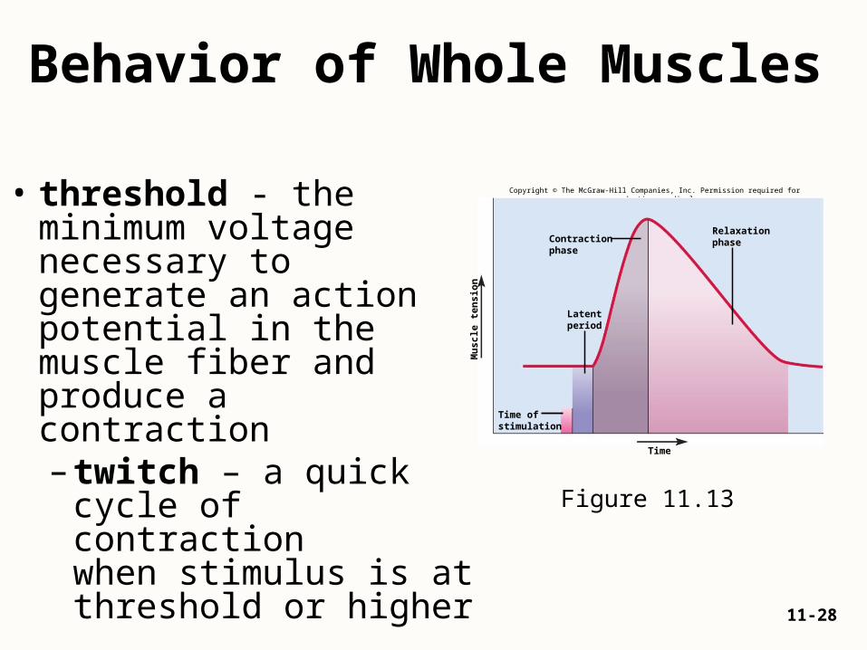
Behavior of Whole Muscles
• threshold - the minimum voltage necessary to generate an action potential in the muscle fiber and produce a contraction– twitch – a quick cycle
of contraction when stimulus is at threshold or higher
Figure 11.13
Copyright © The McGraw-Hill Companies, Inc. Permission required for reproduction or display.
Contractionphase
Relaxationphase
Time
Latentperiod
Time ofstimulation
Mu
scle
ten
sio
n

11-29
• isometric muscle contraction– muscle is producing internal tension while an external resistance
causes it to stay the same length or become longer– can be a prelude to movement when tension is absorbed by elastic
component of muscle– important in postural muscle function and antagonistic muscle joint
stabilization• isotonic muscle contraction
– muscle changes in length with no change in tension– concentric contraction – muscle shortens while maintains tension– eccentric contraction – muscle lengthens as it maintains tension
Copyright © The McGraw-Hill Companies, Inc. Permission required for reproduction or display.
Muscle shortens,tension remainsconstant
Movement
Movement
Muscle developstension but doesnot shorten
No movement
Muscle lengthenswhile maintainingtension
(a) Isometric contraction (b) Isotonic concentric contraction (c) Isotonic eccentric contraction
Isometric and Isotonic Contractions
Figure 11.16

11-30
Muscle Metabolism• all muscle contraction depends on ATP
• ATP supply depends on availability of:– oxygen– organic energy sources such as glucose and fatty acids
• two main pathways of ATP synthesis– anaerobic fermentation
• enables cells to produce ATP in the absence of oxygen• yields little ATP and toxic lactic acid, a major factor in muscle fatigue
– aerobic respiration• produces far more ATP• less toxic end products (CO2 and water)• requires a continual supply of oxygen
11-31
Short-Term Energy Needs• as the phosphagen system is exhausted
• muscles shift to anaerobic fermentation– muscles obtain glucose from blood and their own
stored glycogen– in the absence of oxygen, glycolysis can generate a
net gain of 2 ATP for every glucose molecule consumed
– converts glucose to lactic acid
• glycogen-lactic acid system – the pathway from glycogen to lactic acid
• produces enough ATP for 30 – 40 seconds of maximum activity

11-32
Long-Term Energy Needs
• after 40 seconds or so, the respiratory and cardiovascular systems “catch up” and deliver oxygen to the muscles fast enough for aerobic respiration to meet most of the ATP demands
• aerobic respiration produces 36 ATP per glucose– efficient means of meeting the ATP demands of prolonged exercise
– one’s rate of oxygen consumption rises for 3 to 4 minutes and levels off to a steady state in which aerobic ATP production keeps pace with demand
– little lactic acid accumulates under steady state conditions
– depletion of glycogen and blood glucose, together with the loss of fluid and electrolytes through sweating, set limits on endurance and performance even when lactic acid does not
11-33
Endurance & Fatigue
• endurance – the ability to maintain high-intensity exercise for more than 4 to 5 minutes
• muscle fatigue - progressive weakness and loss of contractility from prolonged use of the muscles– repeated squeezing of rubber ball– holding text book out level to the floor
• causes of muscle fatigue– ATP synthesis declines as glycogen is consumed– ATP shortage slows down the Na+ - K+ pumps– lactic acid lowers pH of sarcoplasm– motor nerve fibers use up their ACh– central nervous system, where all motor commands originate, fatigues by
unknown processes, so there is less signal output to the skeletal muscles
11-34
Cardiac Muscle
• limited to the heart where it functions to pump blood
• required properties of cardiac muscle
– contraction with regular rhythm
– muscle cells of each chamber must contract in unison
– contractions must last long enough to expel blood
– must work in sleep or wakefulness, with out fail, and without conscious attention
– must be highly resistant to fatigue

11-35
Cardiac Muscle • characteristics of cardiac muscle cells
– striated like skeletal muscle, but myocytes (cardiocytes) are shorter and thicker
– each myocyte is joined to several others at the uneven, notched linkages – intercalated discs
• appear as thick dark lines in stained tissue sections
• electrical gap junctions allow each myocyte to directly stimulate its neighbors
• mechanical junctions that keep the myocytes from pulling apart
– sarcoplasmic reticulum less developed, but T tubules are larger and admit supplemental Ca2+ from the extracellular fluid
– damaged cardiac muscle cells repair by fibrosis• a little mitosis observed following heart attacks
• not in significant amounts to regenerate functional muscle

11-36
2 Types of Smooth Muscle
• multiunit smooth muscle
– occurs in some of the largest arteries and pulmonary air passages, in piloerector muscles of hair follicle, and in the iris of the eye
– autonomic innervation similar to skeletal muscle
• terminal branches of a nerve fiber synapse with individual myocytes and form a motor unit
• each motor unit contracts independently of the others
Copyright © The McGraw-Hill Companies, Inc. Permission required for reproduction or display.
Synapses
Autonomicnerve fibers
(a) Multiunit smooth muscle
Figure 11.21a

11-37
2 Types of Smooth Muscle
• single-unit smooth muscle
– more widespread
– occurs in most blood vessels, in the digestive, respiratory, urinary, and reproductive tracts – also called visceral muscle
• often in two layers– inner circular– outer longitudinal
– myocytes of this cell type are electrically coupled to each other by gap junctions
– they directly stimulate each other and a large number of cells contract as a single unit
Figure 11.21b
Copyright © The McGraw-Hill Companies, Inc. Permission required for reproduction or display.
Varicosities
Gap junctions
Autonomicnerve fibers
(b) Single-unit smooth muscle
11-38
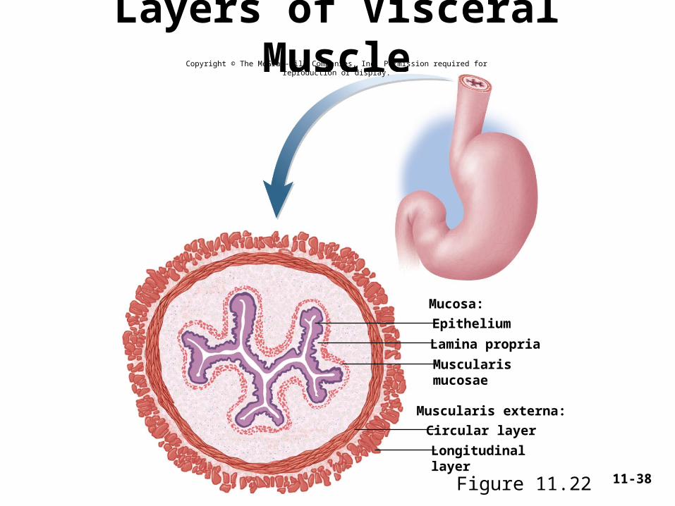
Layers of Visceral Muscle
Epithelium
Mucosa:
Muscularis externa:
Lamina propria
Muscularismucosae
Circular layer
Longitudinallayer
Figure 11.22
Copyright © The McGraw-Hill Companies, Inc. Permission required for reproduction or display.
11-39
Stimulation of Smooth Muscle• smooth muscle is involuntary and can contract without nervous
stimulation– can contract in response to chemical stimuli
• hormones, carbon dioxide, low pH, and oxygen deficiency• in response to stretch• single unit smooth muscle in stomach and intestines has
pacemaker cells that set off waves of contraction throughout the entire layer of muscle
• most smooth muscle is innervated by autonomic nerve fibers– can trigger and modify contractions– stimulate smooth muscle with either acetylcholine or norepinephrine– can have contrasting effects
• relax the smooth muscle of arteries• contract smooth muscles of the bronchioles
11-40
• skeletal muscle cannot contract forcefully if overstretched
• smooth muscle contracts forcefully even when greatly stretched– allows hollow organs such as the stomach and bladder to fill and then expel their
contents efficiently
• smooth muscle can be anywhere from half to twice its resting length and still contract powerfully
• three reasons:– there are no z discs, so thick filaments cannot butt against them and stop
contraction– since the thick and thin filaments are not arranged in orderly sarcomeres,
stretching does not cause a situation where there is too little overlap for cross-bridges to form
– the thick filaments of smooth muscle have myosin heads along their entire length, so cross-bridges can form anywhere
• plasticity – the ability to adjust its tension to the degree of stretch– a hollow organ such as the bladder can be greatly stretched yet not become
flabby when it is empty
Contraction and Stretching
11-41
Muscular Dystrophy• muscular dystrophy - group of hereditary diseases in which skeletal
muscles degenerate and weaken, and are replaced with fat and fibrous scar tissue
• Duchenne muscular dystrophy is caused by a sex-linked recessive trait (1 of 3500 live-born boys)– most common form– disease of males – diagnosed between 2 and 10 years of age– mutation in gene for muscle protein dystrophin
• actin not linked to sarcolemma and cell membranes damaged during contraction, necrosis and scar tissue results
– rarely live past 20 years of age due to affects on respiratory and cardiac muscle – incurable
• facioscapulohumeral MD - autosomal dominant trait affecting both sexes equally– facial and shoulder muscles more than pelvic muscles
• limb-girdle dystrophy– combination of several diseases of intermediate severity– affects shoulder, arm, and pelvic muscles