10 things every paramedic should know about capnography · american heart association guidelines...
TRANSCRIPT
10 Things Every Paramedic Should Know About CapnographyCapnography is the vital sign of ventilation.
By tracking the carbon dioxide in a patient’s exhaled breath, capnography enables
paramedics to objectively evaluate a patient’s ventilatory status (and indirectly
circulatory and metabolic status), as the medics utilize their clinical judgement to
assess and treat their patients.
Part One: The Science
Definitions:
Capnography – the measurement of carbon dioxide (CO2) in exhaled breath.
Capnometer – the numeric measurement of CO2.
Capnogram – the wave form.
End Tidal CO2 (ETCO2 or PetCO2) - the level of (partial pressure of) carbon dioxide
released at end of expiration.
Oxygenation Versus Ventilation
Oxygenation is how we get oxygen to the tissue. Oxygen is inhaled into the lungs
where gas exchange occurs at the capillary-alveolar membrane. Oxygen is
transported to the tissues through the blood stream. Pulse oximetry measures
oxygenation.
At the cellular level, oxygen and glucose combine to produce energy. Carbon
dioxide, a waste product of this process (The Krebs cycle), diffuses into the blood.
Ventilation (the movement of air) is how we get rid of carbon dioxide. Carbon
dioxide is carried back through the blood and exhaled by the lungs through the
alveoli. Capnography measures ventilation.
This document may be distributed for educational purposes only.
EMS SEO - emsseo.com
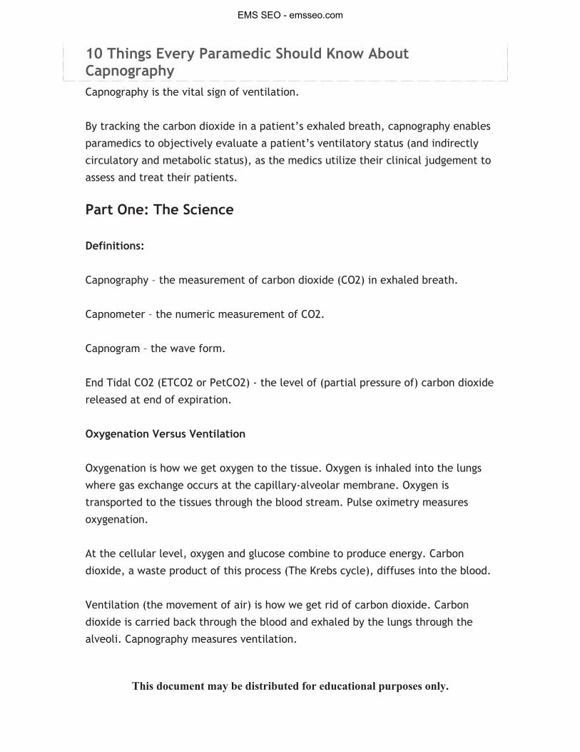
Capnography versus Pulse Oximetry
Capnography provides an immediate picture of patient condition. Pulse oximetry is
delayed. Hold your breath. Capnography will show immediate apnea, while pulse
oximetry will show a high saturation for several minutes.
Circulation and Metabolism
While capnography is a direct measurement of ventilation in the lungs, it also
indirectly measures metabolism and circulation. For example, an increased
metabolism will increase the production of carbon dioxide increasing the ETCO2. A
decrease in cardiac output will lower the delivery of carbon dioxide to the lungs
decreasing the ETCO2.
“CO2 is the smoke from the flames of metabolism.”– Ray Fowler, M.D. Dallas, Street Doc’s
Society
PaCO2 vs. PeTCO2
PaCO2= Partial Pressure of Carbon Dioxide in arterial blood gases. The PaCO2 is
measured by drawing the ABGs, which also measure the arterial PH.
If ventilation and perfusion are stable PaCO2 should correlate to PetCO2.
In a study comparing PaCO2 and PetCO2 in 39 patients with severe asthma, the mean
difference between PaCO2 and PetCO2 was 1.0 mm Hg, the median difference was 0 mm Hg.
Only 2 patients were outside the 5 mg HG agreement (1-6, 1-12). -Jill Corbo, MD, et al,
Concordance Between Capnography and Arterial Blood Gas Measurements of Carbon Dioxide in
This document may be distributed for educational purposes only.
EMS SEO - emsseo.com
Acute Asthma, Annals of Emergency Medicine, October 2005
“Research has (also) shown good concordance...in patients with normal lung function, upper
and lower airway disease, seizures, and diabetic ketoacidosis.” –ibid.
V/Q Mismatch
If ventilation or perfusion are unstable, a Ventilation/Perfusion (V/Q) mismatch
can occur.
This V/Q mismatch can be caused by blood shunting and dead space in the lungs.
(Ventilating unperfused lung area. Perfusing unventilated lung area.)
The V/Q mismatch will alter the correlation between PaC02 and PetCO2.
Normal Capnography Values
ETCO2 35-45 mm Hg is the normal value for capnography.
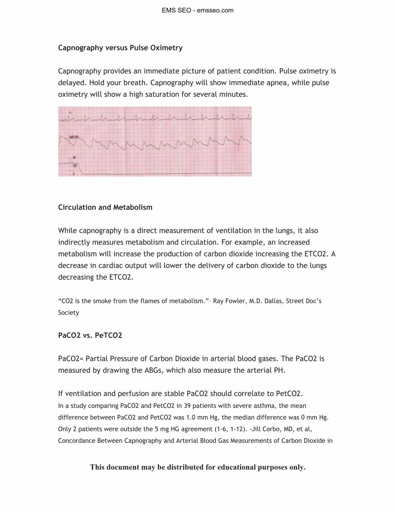
Capnography Wave Form
The normal wave form appears as straight boxes on the monitor screen:
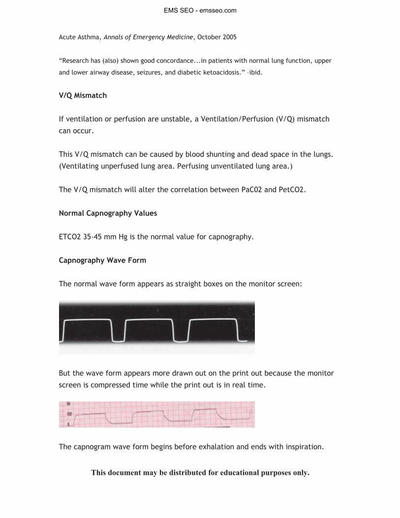
But the wave form appears more drawn out on the print out because the monitor
screen is compressed time while the print out is in real time.
The capnogram wave form begins before exhalation and ends with inspiration.
This document may be distributed for educational purposes only.
EMS SEO - emsseo.com
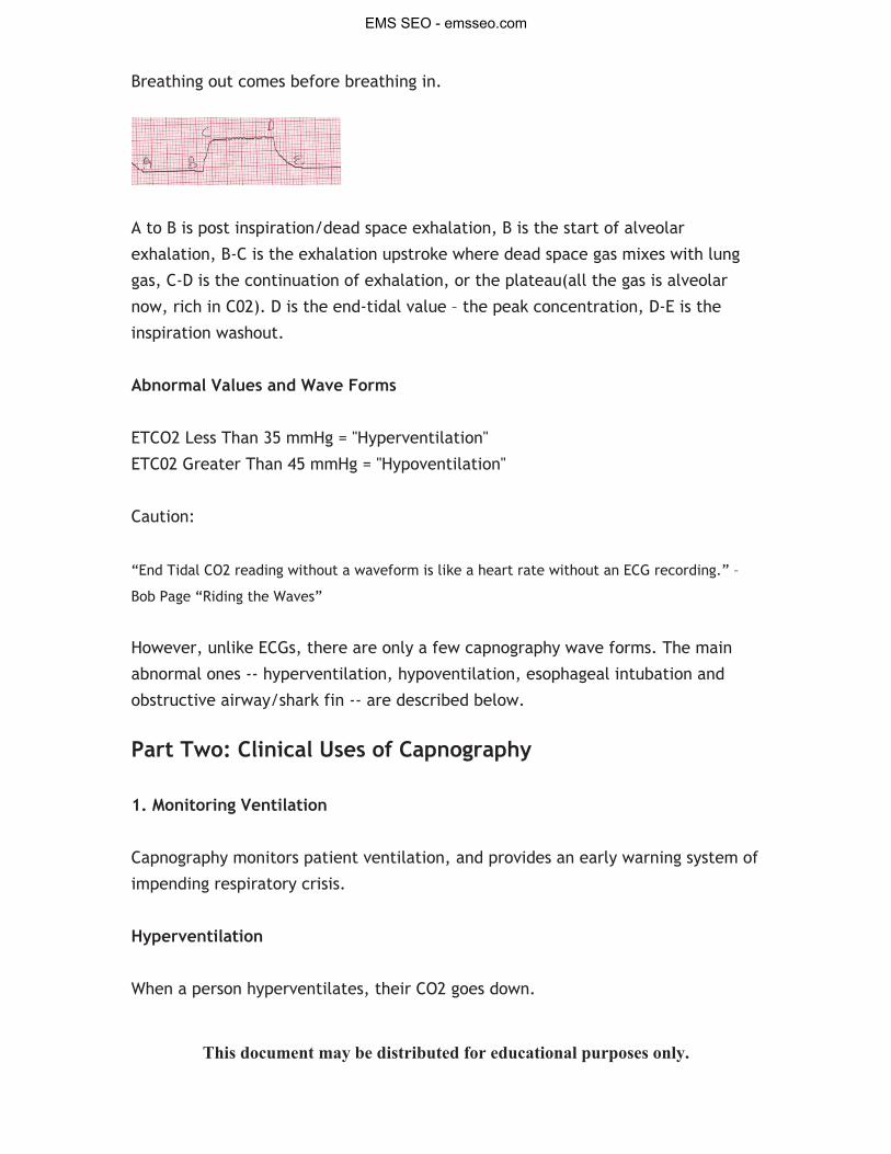
Breathing out comes before breathing in.
A to B is post inspiration/dead space exhalation, B is the start of alveolar
exhalation, B-C is the exhalation upstroke where dead space gas mixes with lung
gas, C-D is the continuation of exhalation, or the plateau(all the gas is alveolar
now, rich in C02). D is the end-tidal value – the peak concentration, D-E is the
inspiration washout.
Abnormal Values and Wave Forms
ETCO2 Less Than 35 mmHg = "Hyperventilation"
ETC02 Greater Than 45 mmHg = "Hypoventilation"
Caution:
“End Tidal CO2 reading without a waveform is like a heart rate without an ECG recording.” –
Bob Page “Riding the Waves”
However, unlike ECGs, there are only a few capnography wave forms. The main
abnormal ones -- hyperventilation, hypoventilation, esophageal intubation and
obstructive airway/shark fin -- are described below.
Part Two: Clinical Uses of Capnography
1. Monitoring Ventilation
Capnography monitors patient ventilation, and provides an early warning system of
impending respiratory crisis.
Hyperventilation
When a person hyperventilates, their CO2 goes down.
This document may be distributed for educational purposes only.
EMS SEO - emsseo.com

Hyperventilation can be caused by many factors from anxiety to bronchospasm to
pulmonary embolus. Other reasons C02 may be low: cardiac arrest, decreased
cardiac output, hypotension, cold, severe pulmonary edema.
Hypoventilation
When a person hypoventilates, their CO2 goes up.
Hypoventilation can be caused by altered mental status such as overdose,
sedation, intoxication, postictal states, head trauma, or stroke, or by a tiring CHF
patient. Other reasons CO2 may be high: Increased cardiac output with increased
breathing, fever, sepsis, pain, severe difficulty breathing, depressed respirations.
Some diseases may cause the CO2 to go down, then up, then down. (See asthma
below).
Capnography can help a paramedic anticipate when a patient may soon require
assisted ventilations or intubation.
2. Confirming, Maintaining , and Assisting Intubation
Continuous End Tidal CO2 monitoring can confirm a tracheal intubation. A good
wave form indicating the presence of CO2 ensures the ET tube is in the trachea.
This document may be distributed for educational purposes only.
EMS SEO - emsseo.com
A 2005 study comparing field intubations that used continuous capnography to confirm
intubations versus non-use showed zero unrecognized misplaced intubations in the monitoring
group versus 23% misplaced tubes in the unmonitored group. -Silverstir, Annals of Emergency
Medicine, May 2005
“When exhaled CO2 is detected (positive reading for CO2) in cardiac arrest, it is usually a
reliable indicator of tube position in the trachea.” - The American Heart Association 2005 CPR
and ECG Guidelines
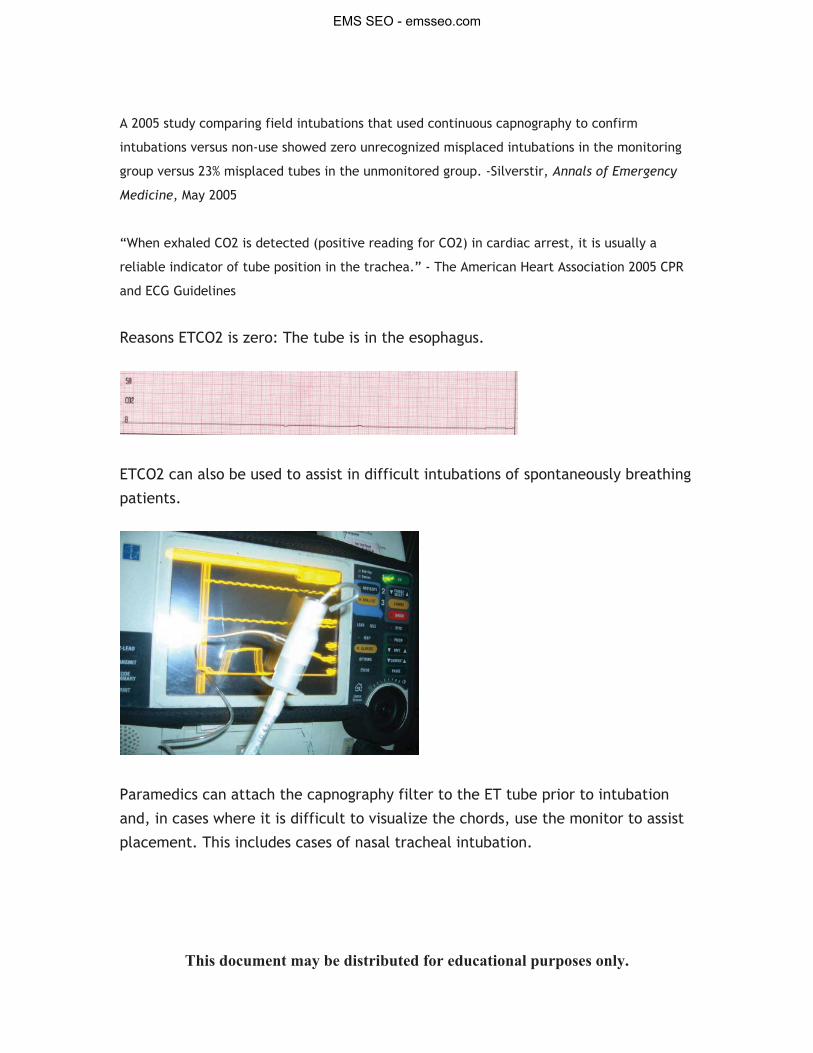
Reasons ETCO2 is zero: The tube is in the esophagus.
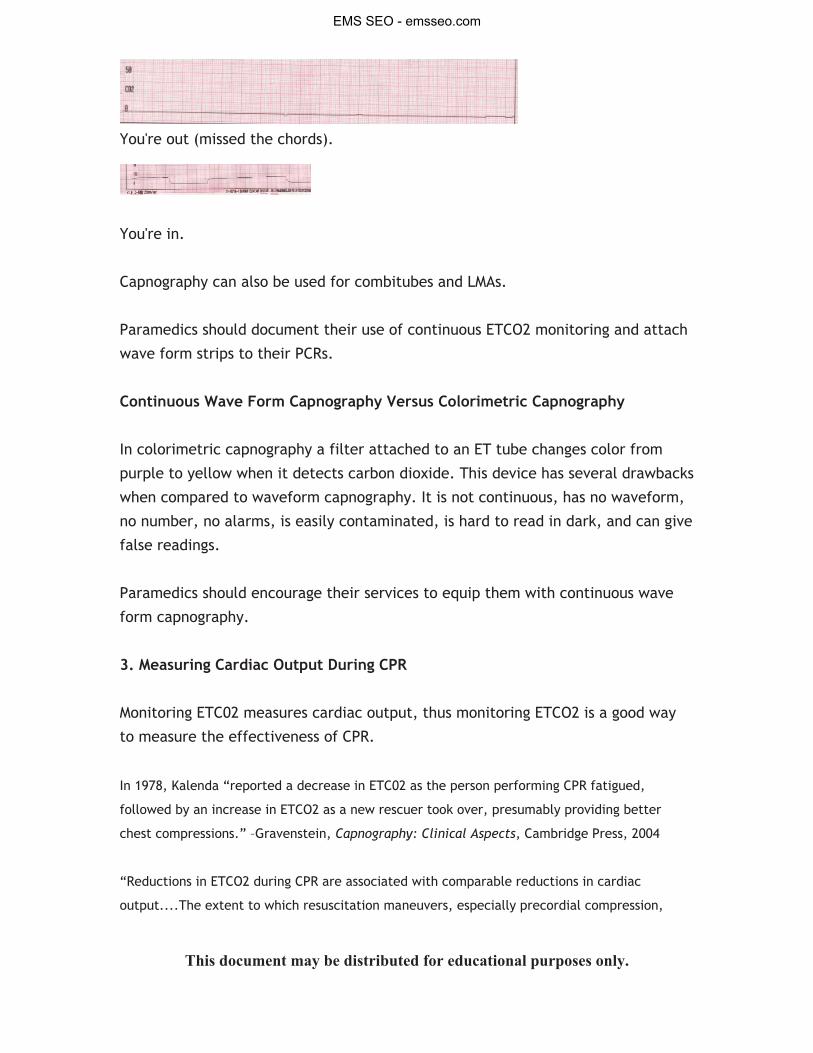
ETCO2 can also be used to assist in difficult intubations of spontaneously breathing
patients.
Paramedics can attach the capnography filter to the ET tube prior to intubation
and, in cases where it is difficult to visualize the chords, use the monitor to assist
placement. This includes cases of nasal tracheal intubation.
This document may be distributed for educational purposes only.
EMS SEO - emsseo.com
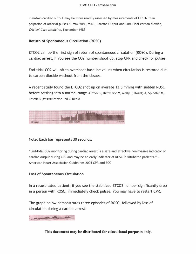
You're out (missed the chords).
You're in.
Capnography can also be used for combitubes and LMAs.
Paramedics should document their use of continuous ETCO2 monitoring and attach
wave form strips to their PCRs.
Continuous Wave Form Capnography Versus Colorimetric Capnography
In colorimetric capnography a filter attached to an ET tube changes color from
purple to yellow when it detects carbon dioxide. This device has several drawbacks
when compared to waveform capnography. It is not continuous, has no waveform,
no number, no alarms, is easily contaminated, is hard to read in dark, and can give
false readings.
Paramedics should encourage their services to equip them with continuous wave
form capnography.
3. Measuring Cardiac Output During CPR
Monitoring ETC02 measures cardiac output, thus monitoring ETCO2 is a good way
to measure the effectiveness of CPR.
In 1978, Kalenda “reported a decrease in ETC02 as the person performing CPR fatigued,
followed by an increase in ETCO2 as a new rescuer took over, presumably providing better
chest compressions.” –Gravenstein, Capnography: Clinical Aspects, Cambridge Press, 2004
“Reductions in ETCO2 during CPR are associated with comparable reductions in cardiac
output....The extent to which resuscitation maneuvers, especially precordial compression,
This document may be distributed for educational purposes only.
EMS SEO - emsseo.com
maintain cardiac output may be more readily assessed by measurements of ETCO2 than
palpation of arterial pulses.” -Max Weil, M.D., Cardiac Output and End-Tidal carbon dioxide,
Critical Care Medicine, November 1985
Return of Spontaneous Circulation (ROSC)
ETCO2 can be the first sign of return of spontaneous circulation (ROSC). During a
cardiac arrest, if you see the CO2 number shoot up, stop CPR and check for pulses.
End-tidal CO2 will often overshoot baseline values when circulation is restored due
to carbon dioxide washout from the tissues.
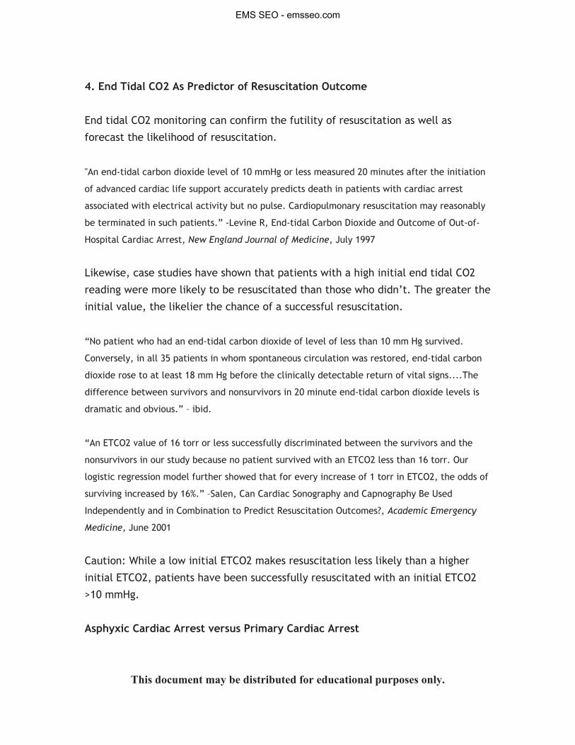
A recent study found the ETCO2 shot up on average 13.5 mmHg with sudden ROSC
before settling into a normal range.-Grmec S, Krizmaric M, Mally S, Kozelj A, Spindler M,
Lesnik B.,Resuscitation. 2006 Dec 8
Note: Each bar represents 30 seconds.
“End-tidal CO2 monitoring during cardiac arrest is a safe and effective noninvasive indicator of
cardiac output during CPR and may be an early indicator of ROSC in intubated patients.” -
American Heart Association Guidelines 2005 CPR and ECG
Loss of Spontaneous Circulation
In a resuscitated patient, if you see the stabilized ETCO2 number significantly drop
in a person with ROSC, immediately check pulses. You may have to restart CPR.
The graph below demonstrates three episodes of ROSC, followed by loss of
circulation during a cardiac arrest:
This document may be distributed for educational purposes only.
EMS SEO - emsseo.com
4. End Tidal CO2 As Predictor of Resuscitation Outcome
End tidal CO2 monitoring can confirm the futility of resuscitation as well as
forecast the likelihood of resuscitation.
"An end-tidal carbon dioxide level of 10 mmHg or less measured 20 minutes after the initiation
of advanced cardiac life support accurately predicts death in patients with cardiac arrest
associated with electrical activity but no pulse. Cardiopulmonary resuscitation may reasonably
be terminated in such patients.” -Levine R, End-tidal Carbon Dioxide and Outcome of Out-of-
Hospital Cardiac Arrest, New England Journal of Medicine, July 1997
Likewise, case studies have shown that patients with a high initial end tidal CO2
reading were more likely to be resuscitated than those who didn’t. The greater the
initial value, the likelier the chance of a successful resuscitation.
“No patient who had an end-tidal carbon dioxide of level of less than 10 mm Hg survived.
Conversely, in all 35 patients in whom spontaneous circulation was restored, end-tidal carbon
dioxide rose to at least 18 mm Hg before the clinically detectable return of vital signs....The
difference between survivors and nonsurvivors in 20 minute end-tidal carbon dioxide levels is
dramatic and obvious.” – ibid.
“An ETCO2 value of 16 torr or less successfully discriminated between the survivors and the
nonsurvivors in our study because no patient survived with an ETCO2 less than 16 torr. Our
logistic regression model further showed that for every increase of 1 torr in ETCO2, the odds of
surviving increased by 16%.” –Salen, Can Cardiac Sonography and Capnography Be Used
Independently and in Combination to Predict Resuscitation Outcomes?, Academic Emergency
Medicine, June 2001
Caution: While a low initial ETCO2 makes resuscitation less likely than a higher
initial ETCO2, patients have been successfully resuscitated with an initial ETCO2
>10 mmHg.
Asphyxic Cardiac Arrest versus Primary Cardiac Arrest
This document may be distributed for educational purposes only.
EMS SEO - emsseo.com
Capnography can also be utilized to differentiate the nature of the cardiac arrest.
A 2003 study found that patients suffering from asphyxic arrest as opposed to
primary cardiac arrest had significantly increased initial ETCO2 reading that came
down within a minute. These high initial readings, caused by the buildup of carbon
dioxide in the lungs while the nonbreathing/nonventilating patient's heart
continued pump carbon dioxide to the lungs before the heart bradyed down to
asystole, should come down within a minute. The ETCO2 values of asphyxic arrest
patients then become prognostic of ROSC.-Grmec S, Lah K, Tusek-Bunc K,Crit Care. 2003
Dec;7(6
5. Monitoring Sedated Patients
Capnography should be used to monitor all patients receiving pain management or
sedation for evidence of hypoventilation and/or apnea.
In a 2006 published study of 60 patients undergoing sedation, in 14 of 17 patients
who suffered acute respiratory events, ETCO2 monitoring flagged a problem before
changes in SPO2 or observed changes in respiratory rate.
“End-tidal carbon dioxide monitoring of patients undergoing PSA detected many clinically
significant acute respiratory events before standard ED monitoring practice did so. The
majority of acute respiratory events noted in this trial occurred before changes in SP02 or
observed hypoventilation and apnea.” - -Burton, Does End-Tidal Carbon Dioxide Monitoring
Detect Respiratory Events Prior to Current Sedation Monitoring Practices, Academic Emergency
Medicine, May 2006
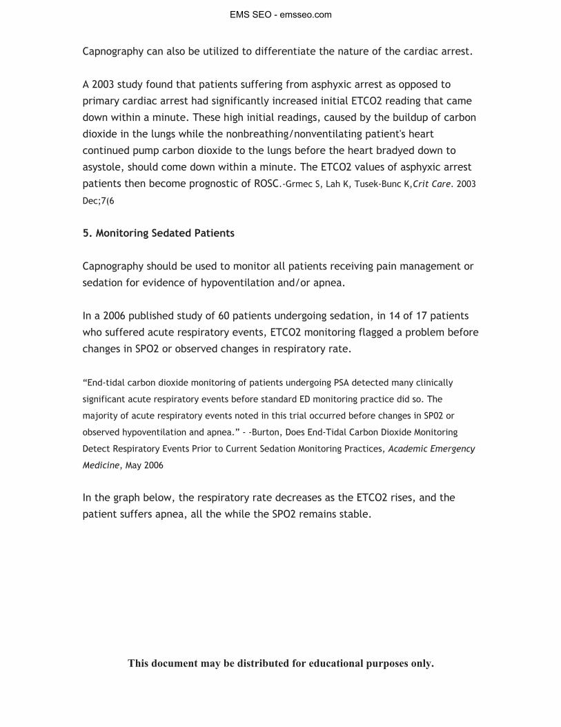
In the graph below, the respiratory rate decreases as the ETCO2 rises, and the
patient suffers apnea, all the while the SPO2 remains stable.
This document may be distributed for educational purposes only.
EMS SEO - emsseo.com
Note: Each bar represents thirty seconds.
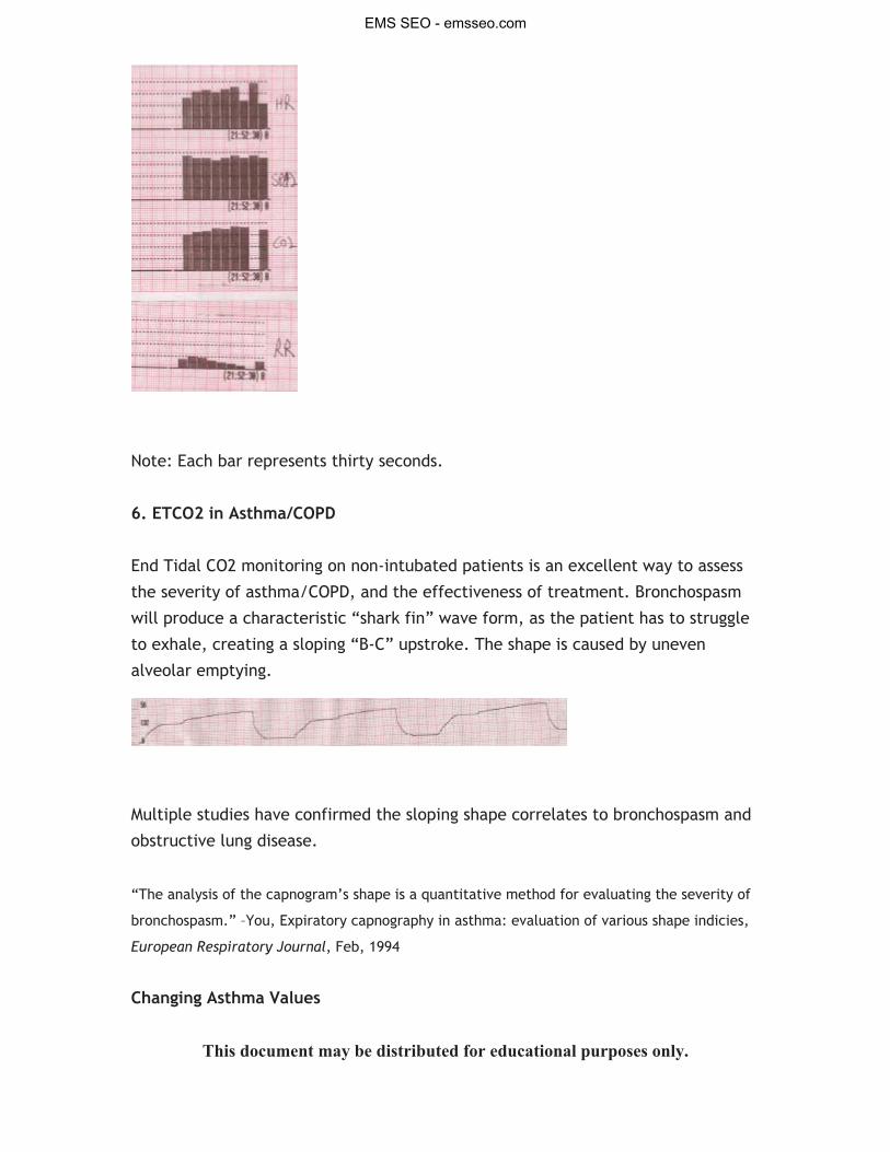
6. ETCO2 in Asthma/COPD
End Tidal CO2 monitoring on non-intubated patients is an excellent way to assess
the severity of asthma/COPD, and the effectiveness of treatment. Bronchospasm
will produce a characteristic “shark fin” wave form, as the patient has to struggle
to exhale, creating a sloping “B-C” upstroke. The shape is caused by uneven
alveolar emptying.
Multiple studies have confirmed the sloping shape correlates to bronchospasm and
obstructive lung disease.
“The analysis of the capnogram’s shape is a quantitative method for evaluating the severity of
bronchospasm.” –You, Expiratory capnography in asthma: evaluation of various shape indicies,
European Respiratory Journal, Feb, 1994
Changing Asthma Values
This document may be distributed for educational purposes only.
EMS SEO - emsseo.com
Asthma values change with severity. With a mild asthma, the CO2 will drop (below
35) as the patient hyperventilates to compensate. As the asthma worsens, the C02
levels will rise to normal. When the asthma becomes severe, and the patient is
tiring and has little air movement, the C02 numbers will rise to dangerous levels
(above 60).
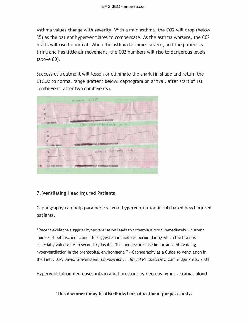
Successful treatment will lessen or eliminate the shark fin shape and return the
ETCO2 to normal range (Patient below: capnogram on arrival, after start of 1st
combi-vent, after two combivents).
7. Ventilating Head Injured Patients
Capnography can help paramedics avoid hyperventilation in intubated head injured
patients.
“Recent evidence suggests hyperventilation leads to ischemia almost immediately...current
models of both ischemic and TBI suggest an immediate period during which the brain is
especially vulnerable to secondary insults. This underscores the importance of avoiding
hyperventilation in the prehospital environment.” --Capnography as a Guide to Ventilation in
the Field, D.P. Davis, Gravenstein, Capnography: Clinical Perspectives, Cambridge Press, 2004
Hyperventilation decreases intracranial pressure by decreasing intracranial blood
This document may be distributed for educational purposes only.
EMS SEO - emsseo.com
flow. The decreased cerebral blood flow may result in cerebral ischemia.
In a study of 291 intubated head injured patients, 144 had ETCO2 monitoring. Patients with
ETCO2 monitoring had lower incidence of inadvertant severe hyperventilation (5.6%) than those
without ETCO2 monitoring (13.4%). Patients in both groups with severe hyperventilation had
significantly higher mortality (56%) than those without (30%). –Davis, The Use of Quantitative
End-Tidal Capnometry to Avoid Inadvertant Severe Hyperventilation in Patients with Head
Injury After Paramedic Rapid Sequence Intubation, Journal of Trauma, April 2004
“A target value of 35 mmHg is recommended...The propensity of prehospital personnel to use
excessively high respiratory rates suggests that the number of breaths per minute should be
decreased. On the other hand, the mounting evidence against tidal volumes in excessive of
10cc/kg especially in the absence of peep, would suggest the hypocapnia be addressed by
lower volume ventilation.” – --Capnography as a Guide to Ventilation in the Field, D.P. Davis,
Gravenstein, Capnography: Clinical Perspectives, Cambridge Press, 2004
8. Perfusion Warning Sign
End tidal CO2 monitoring can provide an early warning sign of shock. A patient
with a sudden drop in cardiac output will show a drop in ETCO2 numbers that may
be irregardless of any change in breathing. This has implications for trauma
patients, cardiac patients – any patient at risk for shock.
In the study cited below, 5 pigs had hemorrhagic shock induced by bleeding, 5 pigs
had septic shock induced by infusion of e-coli, and 6 pigs had cardiogenic shock
induced by repeated episodes of v-fib. The pigs cardiac output was continuously
measured as well as their PETCO2.
“Cardiac output and PetCO2 were highly related in diverse experimental models of circulatory
shock in which cardiac output was reduced by >40 % of baseline values… measurement of
PetC02 is a noninvasive alternative for continuous assessment of cardiac output during low flow
circulatory shock states of diverse causes.” -Xiahua, End-tidal carbon dioxide as a noninvasive
indicator of cardiac index during circulatory shock, Critical Care Medicine, 2000, Vol 28, No 7
“A patient with low cardiac output caused by cardiogenic shock or hypovolemia resulting from
hemorrhage won’t carry as much CO2 per minute back to the lungs to be exhaled. This
This document may be distributed for educational purposes only.
EMS SEO - emsseo.com
patient’s ETC02 will be reduced. It doesn’t necessarily mean the patient is hyperventilating or
that their arterial CO2 level will be reduced. Reduced perfusion to the lungs alone causes this
phenomenon. The patient’s lung function may be perfectly normal.” --Baruch Krauss, M.D,
JEMS, November 2003
9. Other Issues:
CHF – Typically non-wheezing CHF patients will show an upright wave form.
Capnography numbers may drop precipitously in flash pulmonary edema as fluid
prevents offloading of carbon dioxide from the lungs.
DKA - Patients with DKA hyperventilate to lessen their acidosis. The
hyperventilation causes their PAC02 to go down.
“End-tidal C02 is linearly related to HC03 and is significantly lower in children with DKA. If
confirmed by larger trials, cut-points of 29 torr and 36 torr, in conjunction with clinical
assessment, may help discriminate between patients with and without DKA, respectively.” –
Fearon, End-tidal carbon dioxide predicts the presence and severity of acidosis in children with
diabetes, Academic Emergency Medicine, December 2002
Pulmonary Embolus – Pulmonary embolus will cause an increase in the dead space
in the lungs decreasing the alveoli available to offload carbon dioxide. The ETCO2
will go down.
Hyperthermia – Metabolism is on overdrive in fever, which may cause ETCO2 to
rise.
Trauma - A 2004 study of blunt trauma patients requiring RSI showed that only 5
percent of patients with ETCO2 below 26.25 mm Hg after 20 minutes survived to
discharge. The median ETCO2 for survivors was 30.75. - Deakin CD, Sado DM, Coats TJ,
Davies G. “Prehospital end-tidal carbon dioxide concentration and outcome in major trauma.”
Journal of Trauma. 2004;57:65-68.
Field Disaster Triage - It has been suggested that capnography is an excellent
triage tool to assess respiratory status in patients in mass casualty chemical
incidents, such as those that might be caused by terrorism.
This document may be distributed for educational purposes only.
EMS SEO - emsseo.com
“Capnography…can serve as an effective, rapid assessment and triage tool for critically injured
patients and victims of chemical exposure. It provides the ABCs in less than 15 seconds and
identifies the common complications of chemical terrorism. EMS systems should consider
adding capnography to their triage and patient assessment toolbox and emphasize its use
during educational programs and MCI drills.”- Krauss, Heightman, 15 Second Triage Tool, JEMS,
September 2006
10. The Future
Capnography should be the prehospital standard of care for confirmation and
continuous monitoring of intubation, as well as for monitoring ventilation in
sedated patients. Additionally, it should see increasing use in the monitoring of
unstable patients of many etiologies. As more research is done, the role of
capnography in prehospital medicine will continue to grow and evolve.
***
10 Things Every Paramedic Should Know About Capnography
Peter Canning, EMT-P
January 2007 (Version 5)
This document may be distributed for educational purposes only.
EMS SEO - emsseo.com