10: dynamic camouflage in benthic and pelagic cephalopods: an
TRANSCRIPT
1
DISTRIBUTION STATEMENT A. Approved for public release; distribution is unlimited.
Dynamic Camouflage in Benthic and Pelagic Cephalopods: An Interdisciplinary Approach to Crypsis Based on Color,
Reflection, and Bioluminescence
Sönke Johnsen Biology Department
Duke University Durham, NC 27708
phone: (919) 660-7321 fax: (919) 660-7293 email: [email protected]
Alison Sweeney and Daniel Morse Marine Science Institute
U. of California at Santa Barbara Santa Barbara, CA 93106
phone: (805) 893-3416 fax: (805) 893-7998 email: [email protected] phone: (805) 893-7442 fax: (805) 893-7998 email: [email protected]
Dariusz Stramski and Jules Jaffe
Marine Physical Lab Scripps Inst. of Oceanography, UCSD
La Jolla, CA 92093 phone: (858) 534-3353 fax: (858) 534-7641 email: [email protected] phone (858) 534-6101 fax: (858) 534-7641 email: [email protected]
Award Number: N00014-09-1-1053
http://www.biology.duke.edu/johnsenlab
LONG-TERM GOALS Our overall goal is to understand the perceptual and mechanistic principles that underlay camouflage framed in the context of the animals’ environment. In particular, we plan to characterize and understand the perceptual abilities of several species of benthic and pelagic cephalopods, the aspects of their optical environment that affect their camouflage behavior, the characterization of that behavior, and the molecular mechanisms inside the skin by which those responses are accomplished. OBJECTIVES 1. To characterize the spatiotemporal characteristics of the near-surface and shallow benthic
underwater light field, including ultraviolet radiation and polarization. 2. To determine the visual abilities of several species of cephalopod and model both the shallow and
deep-water world from the animals’ points of view.

2
3. To incorporate the knowledge gained from objectives 1 and 2 in order to study the camouflage behavior of these species under simulated ocean conditions.
4. To understand the underlying molecular and biophysical mechanisms governing changes in the
skin that produce the observed optical effects, to provide a platform for future translational efforts. APPROACH Objective 1 Light measurements: Our approach to characterize the underwater light field during the cruise in Baja California involved the deployment of an underwater optical package consisting of several hyperspectral optical sensors from the RAMSES radiometer family (TriOS, Germany). The optical sensors provide measurements of plane and scalar irradiances as well as radiance from a single direction with an averaging time typically from about 0.002 to > 1 s (depending on light intensity) and with high spectral resolution (~3 nm from 350 to 850 nm) made at several discrete depths throughout the water column. Two modes of deployment were used: (i) the sensors were oriented vertically to make typical measurements of the downwelling and upwelling light fields including downwelling plane and scalar irradiances, upwelling plane and scalar irradiances, and upwelling nadir radiance, and (ii) the sensors were oriented horizontally to measure the irradiances and radiance produced by photons traveling predominantly at horizontal directions toward and away from the sun. Also, the Underwater Porcupine Radiometer System was deployed on one occasion to acquire time-series data of wave-induced fluctuations in underwater light at near-surface depths. Because the spectral and spatial characteristics of underwater light field depend largely on the inherent optical properties (IOPs) of water, our approach on the cruise also included the collection of discrete water samples for the determinations of the spectral particulate absorption coefficient, ap(λ), as well as parameters characterizing the bulk concentration of suspended particulate matter. Finally, radiometrically calibrated movies of natural environments come from another novel device that we call the omnicamera: a collection of six underwater video cameras that image the environment in all six axes. Objective 2 Visual physiology: our approach involves using microscopy, microspectrophotometry and the optomotor response to measure six primary visual parameters of the study species: field of view, spectral sensitivity, acuity, temporal resolution, and contrast and polarization sensitivity. Field of view is determined from the placement and orientation of the eyes and the geometry of the retina and pupil. Spectral sensitivity will be investigated using microspectrophotometry (MSP), which measures the absorption spectra of individual photoreceptors. Spatial and temporal resolution will be estimated from the spacing of photoreceptors in the retina and via the optomotor response. Contrast sensitivity is estimated by determining photon catch and also via optomotor assays using stripes of decreasing contrast. Polarization sensitivity will also be assayed via retinal morphology and optomotor response. Objective 3 Camouflage behavior: Camouflage behavior is studied both in situ and within various controlled environments, including a “holodeck”, a tank surrounded by monitors that project natural environments or controlled visual stimuli. The top of the tank will have a plexiglass “floatee” that will make the surface optically flat and permit undistorted observation of the animals from the outside as well as permitting images to be projected into the tank by two DLP projection systems. The tank will

3
be built and tested at SIO in the experimental aquarium facility and then transported to UCSB where it will be set up in a lab equipped with a flow-through filtered and thermostated sea water system. Objective 4 Biophotonics: The fourth objective is to quantify the correspondence between the optical properties of cephalopod skin and their optical environments and to uncover the biophysical principles driving the neurotransmitter-induced self-assembly of the photonically active reflectin proteins. We will characterize the optics of the skin using fiber-optic spectroscopy coupled with goniometry, measuring the polarization-specific bidirectional reflectance of the skin of the target species and correlate these with the statistical analyses of the light measurements from objective 1 to determine which aspects of this complex reflectance have specifically evolved for camouflage. We will also determine the ultrastructure of the reflectin-based structures using transmission electron microscopy and model their optical effects to determine what aspects of the biological structures are important for the observed environmental optical match. We will also investigate the biophysical mechanisms governing tunable reflectance using assays already developed by the Morse lab. We will first sequence and express reflectin proteins from our two uncharacterized core species, P. microlampas and O. bimaculoides, as well as species of particular interest from our field trip to Palau and opportunistically sampled through our collaboration with MBARI. We will take advantage of next-generation “454” high-throughput DNA sequencing and transcriptional profiling technology to generate these sequences, because reflectin genes often have many copies in the genome, long stretches of unalignable sequence, and repetitive elements, which makes traditional degenerate PCR-based sequencing methods difficult. We have also developed dynamic light scattering, electron microscopic, and refractive index assays for the neurotransmitter- and phosphorylation-induced hierarchical self-assembly of the recombinant reflectin protein, and propose to use these assays with the novel reflectins we will to chart the extent to which a unified molecular and biophysical mechanism of action underlies the diversity of camouflage in cephalopods. WORK COMPLETED Objective 1 Light measurements: During 11 days on the cruise in Baja California, the Stramski team measured 21 vertical profiles of underwater light field with the RAMSES hyperspectral system. These measurements were typically made in a vertical profiling mode from the surface to depths between 15 and 70 m at different times of the day to cover a broad range of solar zenith angles including noon and sunset conditions. Five of these deployments were made with the optical sensors oriented horizontally. Ten time-series with a surface radiometric float (each of 2-3 minutes duration) and 6 time-series with the Porcupine (each of 10-min duration) were also made. The Stramski team also operated the CTD-rosette system equipped with an oxygen sensor, a fluorometer providing a proxy for chlorophyll-a concentration, and a beam transmissometer for determining the beam attenuation coefficient at 660 nm. Discrete samples of seawater were collected at two depths (surface and subsurface phytoplankton maximum) for ap(λ), phytoplankton pigments, POC, and SPM. In total, 25 samples for ap(λ), 25 samples for pigments, 39 samples for POC, and 47 samples for SPM, were collected. The analysis of SPM and ap(λ) was completed after cruise in the Stramski lab at SIO. Particulate absorption was partitioned into phytoplankton and non-phytoplankton contributions using methanol extraction method. POC data will be soon available as the analysis is currently underway at the Marine Science Institute Analytical Laboratory in Santa Barbara. The Stramski team made progress in the development of the SQUID (SeQuence of Underwater Irradiance Detectors) instrument, which will be used during this

4
project to measure the spatial statistics of wave-induced irradiance fluctuations. We completed the construction of new optical sensors which are significantly smaller than the Porcupine sensors developed recently by us for the ONR RaDyO program. The SQUID sensors include custom-designed small photodiodes (photosensitive area is 11 mm in diameter) fabricated specially to meet our specifications at the Institute of Electron Technology in Warsaw. Although the SQUID sensors were reduced in size compared with the Porcupine sensors, their sensitivity is higher owing primarily to lower dark current (max. 10 nA). The OmniCam System has been built and consists of an 18” glass sphere inside of which 6 HDTV quality low-light-level CCD cameras are positioned, each pointing at the face of a cube (Figure 1). The cameras use wide-angle lenses, in order to accommodate the field of view necessary to have almost omni-directional coverage underwater. Each camera is equipped with its own computer for control and image storage. All 6 computers, one for each camera, are synchronized with a 7th computer that also records depth, temperature, and orientation. All of the computers share a wireless network inside the sphere that is rated to 6500 meters depth. The camera was tested on the New Horizon cruise by a team of 2 engineers and a graduate student from Dr. Jaffe’s group. Over the course of the 11 days, this group performed 20 vertical profiles from 5 – 70m at various times of the day. These cameras will be quantified in order to compute the angular dependence of the radiant flux and the data will be used as imput to the Holodeck, as described above. In addition, Dr. Jaffe’s student, Justin Haag, currently supported on this MURI, performed a number of vertical profiles using a set of low-light-level CCD cameras that were fitted with linar polarization filters. The filters were oriented at several angles in order to quantify the angular dependence of polarization at depths from 5 – 50 meters. Data analysis is underway.
Figure 1: Images from Dr. Jaffe’s group’s OmniCam sytem. Left) images from deployment in the SIO kelp tank, as preparation for the images taken in the Sea of Cortez on our recent cruise (right)
Objective 2 Visual physiology: A large-scale recirculating seawater aquarium facility has been designed and constructed at Duke University. This six hundred gallon system is specifically designed for housing cephalopods, which are extremely sensitive to water condition. The system includes large-scale biological filtration, protein skimmers, UV sterilizers, activated carbon, and a chiller system. This allows us to house cephalopods for long-term behavioral experiments. In addition, both the microspectrophotometry and optomotor apparatuses have been completed and used to collect initial data. During the June 2010 New Horizon cruise, eyes were collected from Pterygioteuthis and three

5
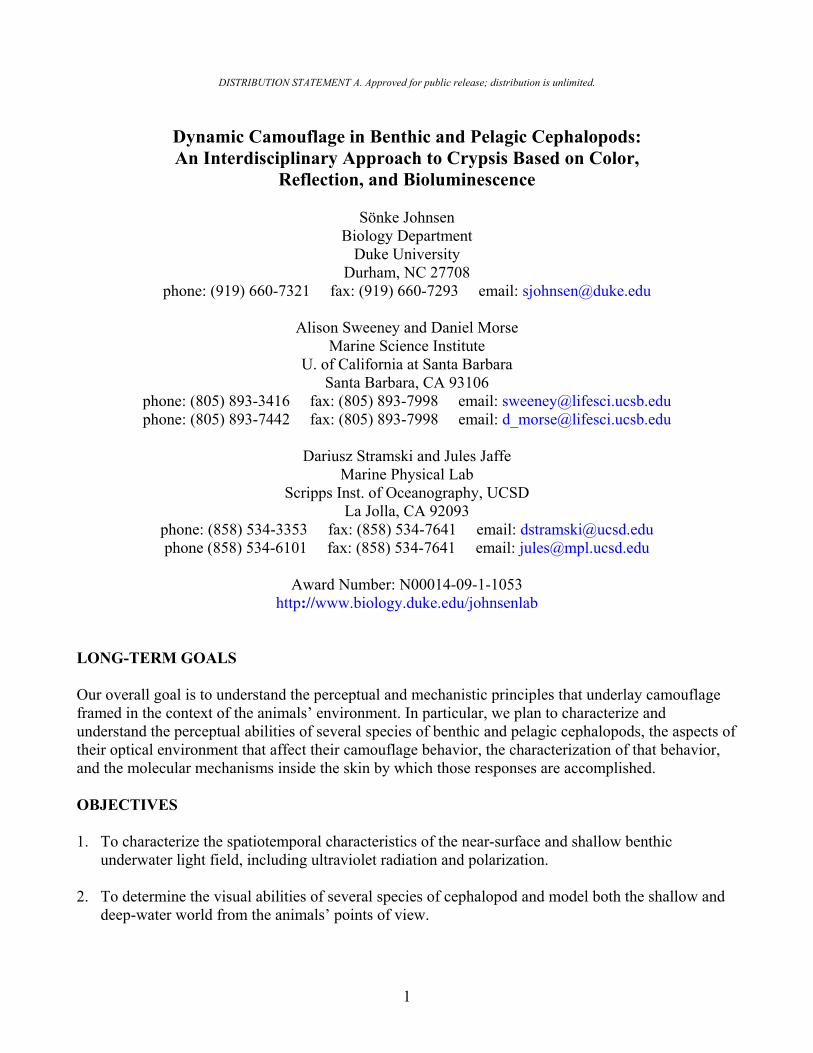
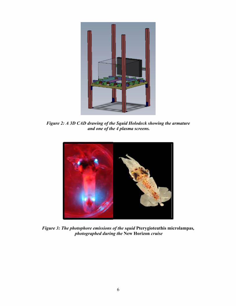
further species –Dosidicus gigas, Abraliopsis sp., and Japetella sp.-- which will be sectioned using the cryostat and investigated with MSP (microspectrophotometry) at Duke University. This will allow us to determine the spectral sensitivities of these species and allow for interspecific comparisons (for example we might expect habitat-restricted species to be 'tuned' to the prevailing light environment). It will also enable us to ascertain whether or not any of these species have multiple visual pigments to allow for color vision. Additional eye samples were preserved in RNAlater for work on gene expression should MSP reveal interesting results, and fixed for light microscopy for further anatomical work. On the second cruise in the South Pacific a further 11 species of cephalopod were caught during trawls of waters between 100 and 1600 meters. Eye samples were preserved where possible for MSP, future molecular work, and light microscopy. MSP analysis should begin before the end of the year. A common behavioral test of visual acuity is to determine the optomotor response of an animal, where it will move either its body or head in response to a revolving stimulus in order to stabilize the image on the retina. An undergraduate student took on the task of testing the optomotor response of O. bimaculoides and developed a robust protocol that is now being used to generate data on visual acuity. Objective 3 Camouflage behavior: The seawater facility at UCSB is being redesigned to contain the holodeck, which is still under development. The system will consist of 4 plasma displays (wall illumination) and 3 projectors (top and bottom) in order to encase the salt water aquarium in omni-directional images. Figure 2 is a 3D computer aided design rendering of the system’s armature that is currently under construction. Software development, for controlling all 7 images is already running on a Mac Pro computer system. The system will use the Mac Quartz Developer environment. The holodeck will display the images collected from cruises in addition to various synthetic images. A prototype system will be finished in late fall of 2010. The pelagic study animal, Pterygioteuthis microlampas, was caught frequently during trawls on this cruise, and initial experiments on chromatophore configuration and photophore use in these squid were carried out (Figure 3). During a second research cruise, sampling deep waters of the South Pacific on the German vessel Sonne, behavioral experiments were conducted on the dynamic camouflage and chromatophore expression in pelagic cephalopods. Data was obtained on chromatophore use in the pelagic octopus Japetella, and the squid Leachia dislocata and Chiroteuthis. All of these species have the ability to be virtually transparent when their chromatophores are retracted, an effective camouflage utilized by many pelagic animals. Yet all also have the ability to 'switch on' a range of densities of chromatophores. The texture-matching abilities of the octopus Octopus bimaculoides was also examined (Figure 4).
6
Figure 2: A 3D CAD drawing of the Squid Holodeck showing the armature and one of the 4 plasma screens.
Figure 3: The photophore emissions of the squid Pterygioteuthis microlampas, photographed during the New Horizon cruise

7
Figure 4: Octopus bimaculoides adjusting its skin texture to match a textured but not a patterned environment.
Objective 4 Biophotonics: During the New Horizon cruise we achieved the following: 1) TEM tissue sampling from all camouflaging reflective tissues from all the species of squids we collected (primarily Dosidicus and Pterygioteuthis, but also Japetella, Abraliopsis and a few others), 2) RNA preservation of iridescent tissues for deep sequencing, 3) reflectance measurements, reflectance microscopy and darkfield photomicrography of these tissues, and 4) biophotonic cells fixed for 3D SEM of reflective tissues. We completed several rounds of sample preparation and initial sequence analysis of biophotonic transcriptomes generated with 454 deep sequencing technology from several different iridescent camouflaging tissues from several species of cephalopod. This sequencing technology allows us to generate approximately one million transcript reads with an average length of 450 base pairs, leading to a near-complete sequence characterization of all genes transcribed (and thus, proteins produced) in a tissue of interest. Our sequence results were of high quality, boding well for our ability to obtain insights about photonic assembly from the data. Bioinformatic sequence analysis has commenced. RESULTS Objective 1 Light measurements: The radiometric data from the hyperspectral RAMSES system deployed in Baja California have not yet been processed to a point suitable for presentation in this report. We present here the selected results of the absorption coefficient of phytoplankton and the dry mass concentration of suspended particulate matter. Figure 5 shows the location of stations in Baja California superimposed over a map of the surface concentration of chlorophyll-a, [Chl], obtained from satellite observations of ocean color. There is a general decreasing trend in [Chl] from north to south. Within the study region, the [Chl] values range generally from ~0.1 to 0.5 mg m-3. SPM ranges from 60 to 200 mg m-3 for surface samples and from 100 to 700 mg m-3 for samples collected at the [Chl] max (Figure 6). These values are in the upper range of the typical open ocean values. The [Chl] max was situated mostly between ~25 and 40 m with one exception of 54 m (station 10, 23.85o N, 109.12o W). Subsurface SPM can reach values up to 5 times higher than the surface values, which suggests enhanced optical turbidity and phytoplankton contributions at a depth compared with the near-surface layer. The difference in bio-optical properties between the surface and subsurface samples is also

8
evident when the spectral shapes of phytoplankton absorption are compared (Figure 7). The subsurface samples show relatively higher absorption within the 450 - 700 nm spectral range compared with the surface samples. These results will be useful to interpret the light field measurements and to model the radiative transfer and visual perception by animals.
Figure 5. 10-day average surface concentration of chlorophyll-a [Chl] showing the location of the stations visited during the MURI cruise on R/V New Horizon in Baja California. [Chl] is computed
from the SeaWiFS, MODIS and MERIS ocean color sensors (GlobColor archive data).
Figure 6. SPM as a function of latitude during the cruise in Baja California. Data for the surface and [Chl] maximum samples are shown. The three basins visited during the cruise are indicated.

9
Figure 7. Normalized spectra of phytoplankton absorption coefficient for the surface and
subsurface samples in black and green, respectively. The normalization is made at 440 nm. Objective 3 Camouflage behavior: Initial results suggest that, in the case of Chiroteuthis, light of different wavelengths result in different levels of chromatophore expression. We tested this by acclimating individual animals in ambient light, then videoing body pattern responses as light with different filters was passed over them. We found that violet, blue, and green filtered light resulted in a strong expression of dark chromatophores on the mantle of the animals. The strongest chromatophore reaction seems to be driven by exposure to blue light, and this was often accompanied by further behavioral changes such as rapid jetting, typical of an escape response. Orange light resulted in a very weak reaction. Animals did not change their body patterns when exposed to red light (negative control). Although we expect pelagic animals to have maximum sensitivity to blue light, given the dominance with depth, it is not obvious why exposure should result in a change of body pattern. One plausible hypothesis (and the motivation for undertaking the experiment) is that many predators in the deep sea use bioluminescence to detect or lure prey so being sensitive to flashes or sudden changes in light will be useful for avoiding predation. Whether or not the response of chromatophore expression acts in some way to increase the cephalopod's camouflage, or whether it is purely a threat response remains to be seen. In addition, an analysis of polarization imagery from previous cruises found that tissue birefringence, while striking, was not apparent in situ. However because scattering structures depolarized light, there was nevertheless significant polarization contrast (Figure 8).

10
Figure 8. Example of a polarization and channel analysis of an in situ image. Animal shown is the scyphomedusa Aurelia aurita. The columns show each of the R, G, and B colour channels
from the camera. The rows show the unpolarized image (U/2), the image when the polarizer was oriented so that the background light was minimized (M), and an image of the degree of polarization
(P) (The horizontal bar is the grey code of the degree of polarization). The gray values in the unpolarized images were halved to increase legibility given the limited dynamic range of
printed paper (i.e. to prevent the unpolarized image from appearing too bright or the minimum image from appearing too dim).
Objective 4 Biohotonics: In a manuscript recently submitted to the Journal of the Royal Society: Interface, we showed that the silvery eye covering of Loliginid squid has a novel nested-spindle geometry comprising a randomly distributed Bragg reflector (DBR). This reflector has a range of spatial frequencies resulting in broadband visible reflectance spectra and making a nearly ideal passive camouflage material for the particular region of the water column where these animals live. We developed a novel application of the transfer-matrix method of optical modeling to investigate specular reflection from this novel photonic structure (Figure 9), demonstrating that a DBR with widely distributed thickness variations of high refractive index elements is sufficient to yield broadband reflectance over visible wavelengths, and that unlike DBRs with one or a few spatial frequencies, this broadband reflectance occurs from all viewing angles. Analysis of the protein content of these spindle-shaped cells reveals that they are largely comprised of members of the reflectin family – the proteins we also have characterized in the dynamically tunable reflectors on the squid body. The spindle shape of the cells facilitates self-assembly of a random DBR to achieve smooth spatial distributions in refractive indices. This design lends itself to technological imitation to achieve a DBR with smoothly distributed spatial frequencies in a facile, inexpensive manner. Continuing research we reported last year, we have further characterized the neurotransmitter-induced changes in phosphorylation of the reflectin proteins that appear to drive the changes in reflectance intensity and color in cells located on the squid body. These results are reported in two recent publications. Preliminary analysis of our

11
transcriptome data has revealed dozens of novel reflectins with biophysical properties distinct from those previously characterized in the squid Loligo opalescens and Euprymna scolopes. Our continuing bioinformatic and biochemical analyses will provide the necessary informatic foundation to further elucidate mechanisms of photonic self-assembly via reflectins over the remaining course of this project. Our complementary work with optical modeling of reflectin-based photonic structures responsible for camouflage of the squid eye has begun to reveal the ways in which reflectin evolution directs evolution of camouflage in cephalopods.
Figure 9. Schematic outlining our approach to using the transfer matrix method of optical modeling to probe the camouflaging silvery covering of squid eyes. a) Transmission electron
micrographs were converted to binary images, and then converted to one-dimensional spatial components based on vertical transects through the image, simulating a normal angle of
incident light. b) The spatial components obtained from these vertical transects were then pooled (represented by the red and blue box), and their resulting spatial distributions fit to two different gamma distributions, one for high index regions, and the other for low index regions, as shown
by the red and blue bars. The fit components of the gamma distributions are also shown in this panel. c) Thicknesses were chosen from a gamma distribution as generated in panel b, and
input into a transfer matrix model of reflectance. High and low index layers alternated and were chosen with frequencies dictated by the respective gamma-fit distributions. This panel
shows a schematic of mathematics involved in the transfer matrix method, with k representing the layer being modeled, representing the components of the transfer matrices, and E represents
the electric field at each interface. IMPACT/APPLICATIONS The systems evolved by marine animals in order to hunt, hide, and mate over hundreds of million years surpass our contemporary engineering designs for underwater vehicles. Hiding and hunting are natural tasks for our military and we believe that valuable clues will be provided by the results of our studies. The impact will hopefully affect all branches of the armed forces that have aquatic missions. This

12
includes Special Forces, mine hunting vehicles, the submarine community, and a newest generation of underwater vehicles that could all benefit from the option of “stealth”. Since visual methods play an important role in the mission profiles of all of these groups, the ability to enhance and hide from detection should be an important payoff. RELATED PROJECTS "Bioinspired Dynamically Tunable Polymer-Based Filters for Multi-Spectral Infrared Imaging"; DARPA; W911NF-08-1-0494; $150,000; 10-01/08-09/30/09. This work represents a "translation" of what we learned from the biomolecular mechanisms governing dynamically tunable reflectance in cephalopods to novel routes for synthetic optical materials. Performed in collaboration with Raytheon, Inc. This funding has ended; proposal for continuation is pending. “Bio-inspired Visual Information Processing and Dynamically Tunable Multispectral IR Detection: Learning from the Octopus.” ARL/ARO; W911NF-09-D-0001; $200,000; 1/1/09-12/31/09. To D.E. Morse and R. N. Hanlon. This work represents a "translation" of what we learned from the biomolecular mechanisms governing dynamically tunable reflectance in cephalopods to novel routes for synthetic optical materials. This funding has ended. ”Bio-Inspired Photonics: Polymer-Based, Dynamically Tunable Multi-Spectral Filters for IR Detection”; DARPA; proposal pending for continuation of effort described immediately above. This work represents a "translation" of what we learned from the biomolecular mechanisms governing dynamically tunable reflectance in cephalopods to novel routes for synthetic optical materials. PUBLICATIONS Izumi, M., A. M. Sweeney, D. G. DeMartini, J. C. Weaver, M. L. Powers, A.R. Tao, T. V. Silvas, R.
M. Kramer, W. J. Crookes-Goodson, L. M. Mäthger, R. R. Naik, R. T. Hanlon and Morse, D. E. (2010). Changes in reflectin protein phosphorylation are associated with dynamic iridescence in squid. Journal of the Royal Society: Interface 7: 549-560.
Johnsen, S., Marshall, N. J., and Widder, E. A. (in press). Polarization sensitivity as a contrast
enhancer in pelagic predators: Lessons from in situ polarization imaging of transparent zooplankton. Philosophical Transactions of the Royal Society of London, Series B.
Marshall, N. J., and Johnsen, S. (in press). Camouflage in Marine fish. In Animal Camouflage: Current
issues and new perspectives. Cambridge University Press: Cambridge UK. Tao, A.R., D. G. DeMartini, M. Izumi, A. M. Sweeney, and Morse, D. E. (2010). The role of protein
assembly in dynamically tunable bio-optical tissues. Biomaterials 31:793-801 Sweeney, A.M., Holt, A.L., Johnsen, S., and Morse, D.E. (in review). A highly-distributed Bragg stack
with unique geometry provides effective camouflage for Loligo squid eyes. Journal of the Royal Society: Interface
Zylinski, S., and Osorio, D. (in press). What can camouflage tell us about non-human visual
perception? A case study of the cuttlefish. In Animal Camouflage: Current issues and new perspectives. Cambridge University Press: Cambridge UK.

13
Zylinski, S. How, M., Osorio, D., Hanlon, R. T., and Marshall, N. J. (in review). To be seen or to hide:
visual characteristics of body patterns for camouflage and communication in the Australian giant cuttlefish, Sepia apama. American Naturalist.