1 the nano-particles (nps) for cancer diagnosis and photo-thermal therapy (ptt) int. j. cancer,...
TRANSCRIPT
1
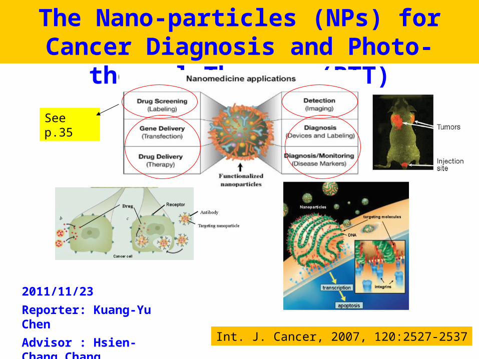
The Nano-particles (NPs) for Cancer Diagnosis and Photo-thermal Therapy (PTT)
Int. J. Cancer, 2007, 120:2527-2537
2011/11/23
Reporter: Kuang-Yu Chen
Advisor : Hsien-Chang Chang
See p.35

2
The different between normal tissue and cancer The nanotechnology applications in cancer The nanoparticles used for photothermal therapy (PTT) The biological window in near infrared ray (NIR) region Some In vitro and In vivo examples of PTT Photodynamic therapy (PDT) Conclusion
Outline

3
Nanotechnology Applications in CancerSchematic diagrams showing
enhanced permeability and retention of nanoparticles in tumors.
Normal tissue vasculatures are lined by tight endothelial cells, thereby preventing nanoparticle drugs from escaping or extravasation, whereas tumor tissue vasculatures are leaking and hyperpermeable allowing preferential accumulation of nanoparticles in the tumor interstitial space.
Annu. Rev. Biomed. Eng., 9(2007)257–288
4
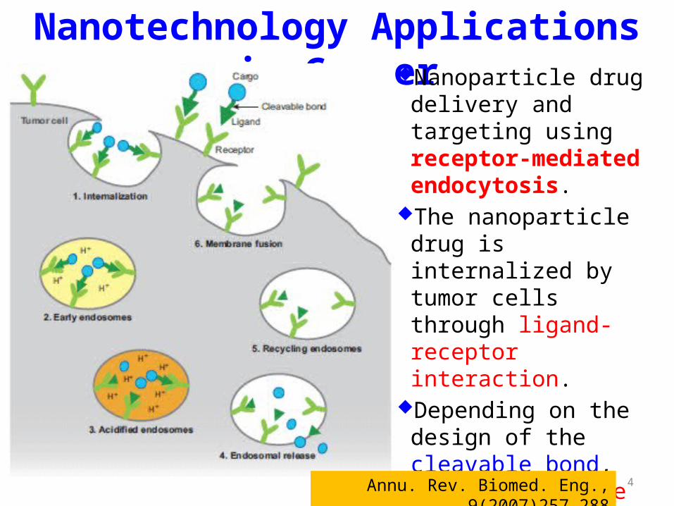
Nanotechnology Applications in CancerNanoparticle drug delivery
and targeting using receptor-mediated endocytosis.
The nanoparticle drug is internalized by tumor cells through ligand-receptor interaction.
Depending on the design of the cleavable bond, the drug will be released intracellularly on exposure to lysosomal enzymes or lower pH.
Annu. Rev. Biomed. Eng., 9(2007)257–288

5
Nanotechnology Applications in Cancer
Self-assembled polymeric nanoparticles with dual tumor-targeting and therapeutic functions.
Annu. Rev. Biomed. Eng., 9(2007)257–288
Delivery of the nanoparticle drugs by receptor-mediated endocytosis and controlled drug release inside the cytoplasm.
6
Nanotechnology Applications in Cancer
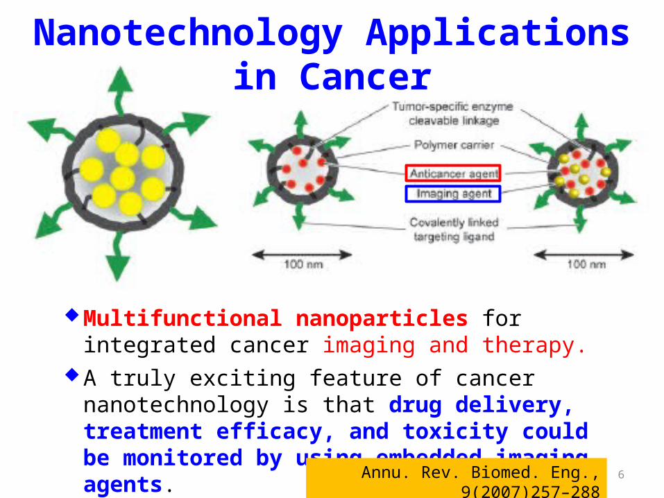
Multifunctional nanoparticles for integrated cancer imaging and therapy.
A truly exciting feature of cancer nanotechnology is that drug delivery, treatment efficacy, and toxicity could be monitored by using embedded imaging agents.
Annu. Rev. Biomed. Eng., 9(2007)257–288

7
Summary: Nanotechnology Applications in CancerNanometer-sized particles have novel optical, electronic,
magnetic, or structural properties and are currently under intense development for applications in cancer, cardiovascular diseases, and degenerative neurological disorders such as Alzheimer’s disease.
Targeted nanoparticle drugs offer significant advantages in improving cancer therapeutic efficacy and simultaneously reducing drug toxicity.
Future work needs to address the potential long-term toxicity, degradation, and metabolism of nanoparticle agents, to identify and develop new biomarker-probe systems, and to develop multifunctional nanoscale platforms for integrated imaging, detection, and therapy.
Annu. Rev. Biomed. Eng., 9(2007)257–288
8Nat. Biotechnol., 19(2001)316-317
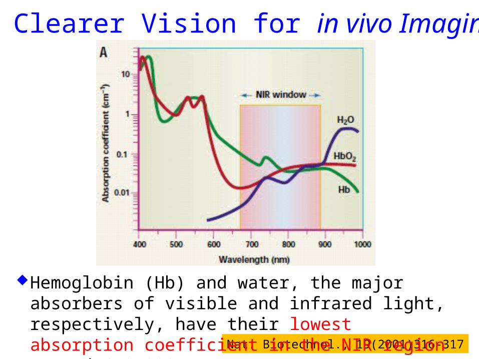
A Clearer Vision for in vivo Imaging
Hemoglobin (Hb) and water, the major absorbers of visible and infrared light, respectively, have their lowest absorption coefficient in the NIR region around 670-900 nm.
9
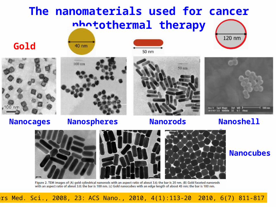
The nanomaterials used for cancer photothermal therapy
Small, 2010, 6(7) 811-817Lasers Med. Sci., 2008, 23:217-228
Nanocubes
Nanospheres Nanorods Nanoshells
Gold
Nanocages
ACS Nano., 2010, 4(1):113-20
10
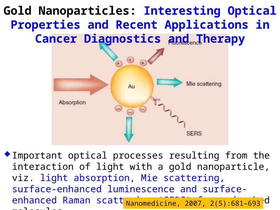
Gold Nanoparticles: Interesting Optical Properties and Recent Applications in Cancer Diagnostics and Therapy
Important optical processes resulting from the interaction of light with a gold nanoparticle, viz. light absorption, Mie scattering, surface-enhanced luminescence and surface-enhanced Raman scattering (SERS) from adsorbed molecules.
Nanomedicine, 2007, 2(5):681–693
11
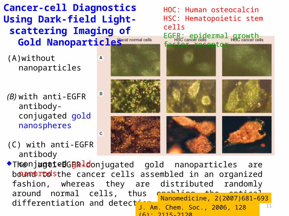
Cancer-cell Diagnostics Using Dark-field Light-scattering
Imaging of Gold Nanoparticles
The anti-EGFR-conjugated gold nanoparticles are bound to the cancer cells assembled in an organized fashion, whereas they are distributed randomly around normal cells, thus enabling the optical differentiation and detection of the cancer cells.
HOC: Human osteocalcinHSC: Hematopoietic stem cellsEGFR: epidermal growth factor receptor
(A) without nanoparticles
(B) with anti-EGFR antibody-conjugated gold nanospheres
(C) with anti-EGFR antibody conjugated gold nanorods
Nanomedicine, 2(2007)681–693J. Am. Chem. Soc., 2006, 128 (6): 2115-2120
12
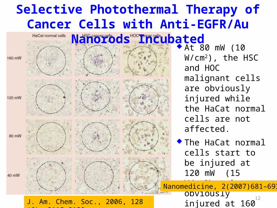
At 80 mW (10 W/cm2), the HSC and HOC malignant cells are obviously injured while the HaCat normal cells are not affected.
The HaCat normal cells start to be injured at 120 mW (15 W/cm2) and are obviously injured at 160 mW (20 W/cm2).
Selective Photothermal Therapy of Cancer Cells with Anti-EGFR/Au Nanorods Incubated
J. Am. Chem. Soc., 2006, 128 (6): 2115-2120
Nanomedicine, 2(2007)681–693
13Nano Letters, 2007, 7(7):1929-1934
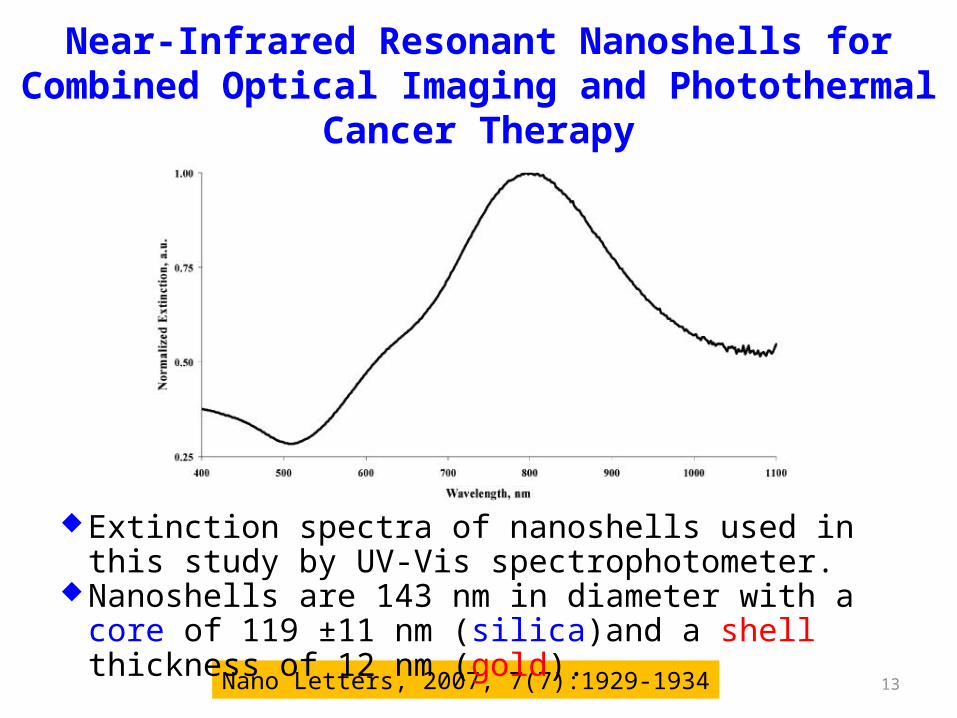
Extinction spectra of nanoshells used in this study by UV-Vis spectrophotometer.
Nanoshells are 143 nm in diameter with a core of 119 ±11 nm (silica)and a shell thickness of 12 nm (gold).
Near-Infrared Resonant Nanoshells for Combined Optical Imaging and Photothermal Cancer Therapy

14
Histological Examination of Tumors Using Silver Staining Confirmed The Presence of Nanoshells throughout The Tumors
The silver staining of representative areas of tumors from mice treated with nanoshells (A) or with PBS (B).
The neutron activation analysis (NAA) verified nanoshells present in the tumor shown in figure A at 12.5 ppm (equivalent to approximately 3x10⁶ nanoshells/gram of tumor tissue).
12.5 ppm 0 ppm
Nano Letters, 2007, 7(7):1929-1934

15
The long-term survival rate of nanoshell + NIR group: 83%
Tumor Size Before Irradiation and 12 Days Post-rradiation of Mice
nanoshell + NIR
sham + NIR
untreated control
nanoshell + NIR
sham + NIR
untreated control
Nano Letters, 2007, 7(7):1929-1934
The Survival Data for The Treatment Groups Post
Irradiation Nanoshell + NIR Laser Irradiation
16
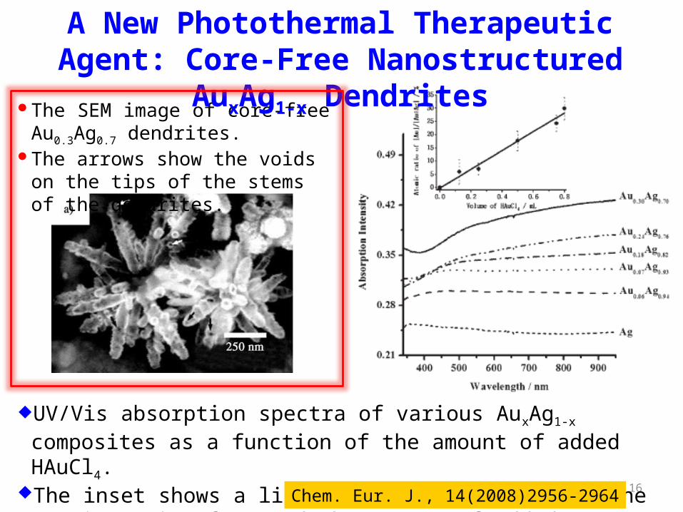
UV/Vis absorption spectra of various AuxAg1-x composites as a function of the amount of added HAuCl4.
The inset shows a linear relationship between the atomic ratio of Au and the amount of added HAuCl4. Chem. Eur. J., 14(2008)2956-2964
The SEM image of core-free Au0.3Ag0.7 dendrites.
The arrows show the voids on the tips of the stems of the dendrites.
A New Photothermal Therapeutic Agent: Core-Free Nanostructured AuxAg1-x Dendrites
17
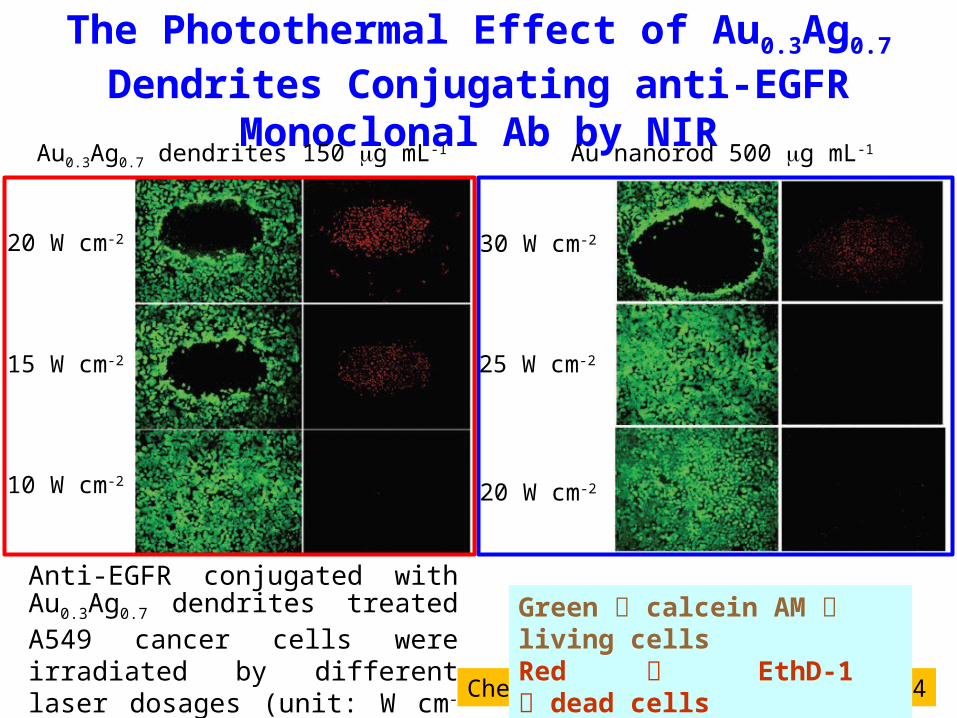
Anti-EGFR conjugated with Au0.3Ag0.7
dendrites treated A549 cancer cells were irradiated by different laser dosages (unit: W cm-2) for 4 min.
Chem. Eur. J., 14(2008)2956-2964
The Photothermal Effect of Au0.3Ag0.7 Dendrites Conjugating anti-EGFR Monoclonal Ab by NIR
Au nanorod 500 mg mL-1
30 W cm-2
25 W cm-2
20 W cm-2
Au0.3Ag0.7 dendrites 150 mg mL-1
20 W cm-2
15 W cm-2
10 W cm-2
Green calcein AM living cellsRed EthD-1 dead cells
18Chem. Eur. J., 13(2007)3878-3885
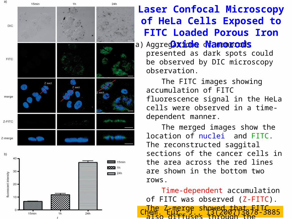
a) Aggregations of nanorods presented as dark spots could be observed by DIC microscopy observation.
The FITC images showing accumulation of FITC fluorescence signal in the HeLa cells were observed in a time-dependent manner.
The merged images show the location of nuclei and FITC. The reconstructed saggital sections of the cancer cells in the area across the red lines are shown in the bottom two rows.
Time-dependent accumulation of FITC was observed (Z-FITC). The Z-merge showed that FITC also diffuses through the nucleus (scale bar: 10 mm).
b) FITC image of quantified accumulation of the FITC fluorescent signal.
Laser Confocal Microscopy of HeLa Cells Exposed to FITC Loaded Porous Iron Oxide Nanorods

19
Gold Nanocages as Photothermal Transducers for Cancer Treatment
UV–vis–NIR spectra showing the local surface plasma resonance (LSPR) peaks of Au nanocages in different media.
The inset shows a typical TEM image of the Au nanocages with an edge length of 48 ± 3.5 nm. Small, 2010, 6(7) 811-817
PVP-coated nanocages in PBS at pH 7.4 (solid line)
PEGylated nanocages in PBS at pH 7.4 (dashed line)
PEGylated nanocages in fetal bovine serum (dotted line)
20
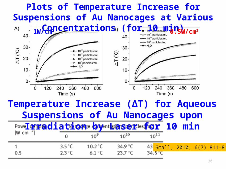
Plots of Temperature Increase for Suspensions of Au Nanocages at Various Concentrations (for 10 min)
1W/cm2 0.5W/cm2
Temperature Increase (ΔT) for Aqueous Suspensions of Au Nanocages upon Irradiation by Laser for 10 min
Small, 2010, 6(7) 811-817
21
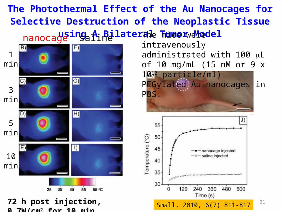
The Photothermal Effect of the Au Nanocages for Selective Destruction of the Neoplastic Tissue using A Bilateral Tumor Model
The mice were intravenously administrated with 100 mL of 10 mg/mL (15 nM or 9 x 10¹² particle/ml) PEGylated Au nanocages in PBS.
72 h post injection, 0.7W/cm2 for 10 min
salinenanocage
3 min
5 min
10 min
1 min
Small, 2010, 6(7) 811-817
22
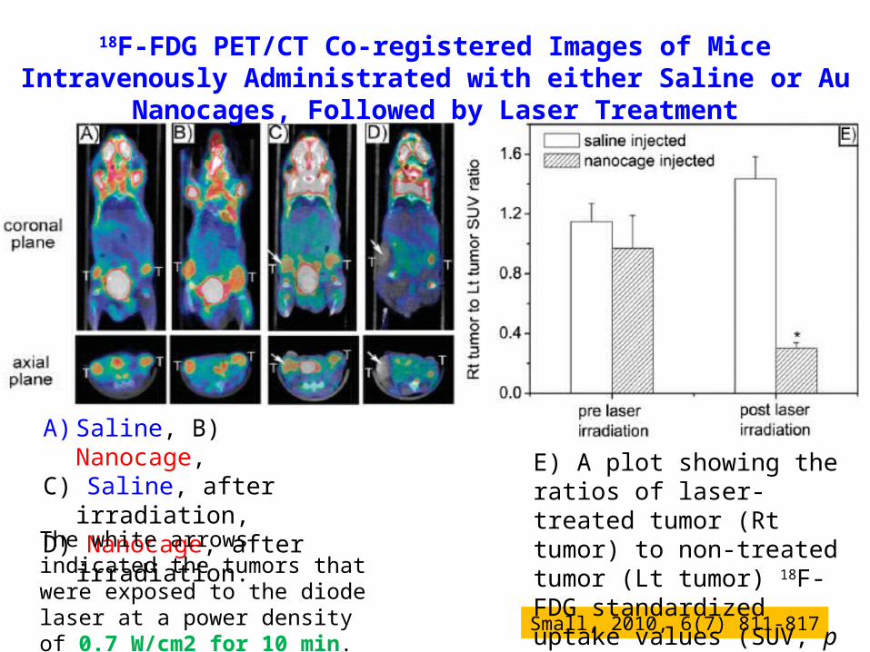
18F-FDG PET/CT Co-registered Images of Mice Intravenously Administrated with either Saline or Au Nanocages, Followed by Laser Treatment
A) Saline, B) Nanocage, C) Saline, after irradiation,D) Nanocage, after irradiation.
Small, 2010, 6(7) 811-817
E) A plot showing the ratios of laser-treated tumor (Rt tumor) to non-treated tumor (Lt tumor) 18F-FDG standardized uptake values (SUV, p < 0.001).
The white arrows indicated the tumors that were exposed to the diode laser at a power density of 0.7 W/cm2 for 10 min.

23
Representative Histology Images of Tumor Tissues from the Two Mice Intravenously Administrated with Saline and Au Nanocages, Respectively
A) saline, w/o irradiation C) nanocage w/o irradiationB) saline, w/ irradiation D) nanocage, w/ irradiation(0.7W/cm2 for 10 min)
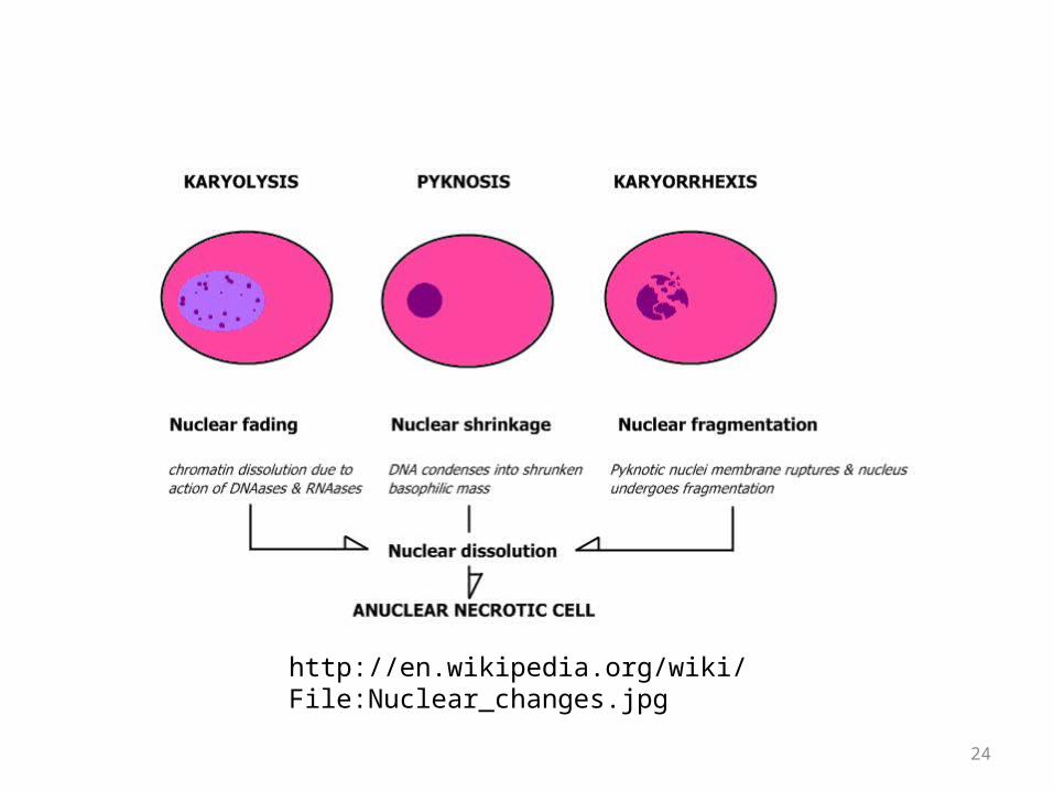
Tumors from mice treated with nanocages and laser irradiation showed distinctive characteristics of cellular damage, such as abundant pyknosis (arrow), karyorhexis (open arrow), karyolysis (arrowhead), and interstitial edema (asterisk).
Small, 2010, 6(7) 811-817
24
http://en.wikipedia.org/wiki/File:Nuclear_changes.jpg
25
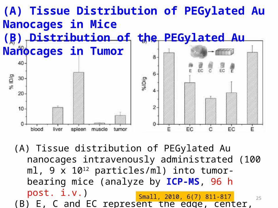
(A) Tissue Distribution of PEGylated Au Nanocages in Mice (B) Distribution of the PEGylated Au Nanocages in Tumor
(A) Tissue distribution of PEGylated Au nanocages intravenously administrated (100 ml, 9 x 10¹² particles/ml) into tumor-bearing mice (analyze by ICP-MS, 96 h post. i.v.)
(B) E, C and EC represent the edge, center, and region between edge and center. Small, 2010, 6(7) 811-817
26
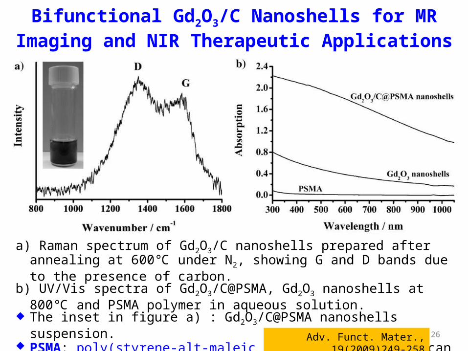
a) Raman spectrum of Gd2O3/C nanoshells prepared after annealing at 600℃ under N2, showing G and D bands due to the presence of carbon.
b) UV/Vis spectra of Gd2O3/C@PSMA, Gd2O3 nanoshells at 800℃ and PSMA polymer in aqueous solution.
The inset in figure a) : Gd2O3/C@PSMA nanoshells suspension. PSMA: poly(styrene-alt-maleic acid) (PSMA) polymer can improve water
dispersion for further antibody conjugation Adv. Funct. Mater., 19(2009)249-258
Bifunctional Gd2O3/C Nanoshells for MR Imaging and NIR Therapeutic Applications
27
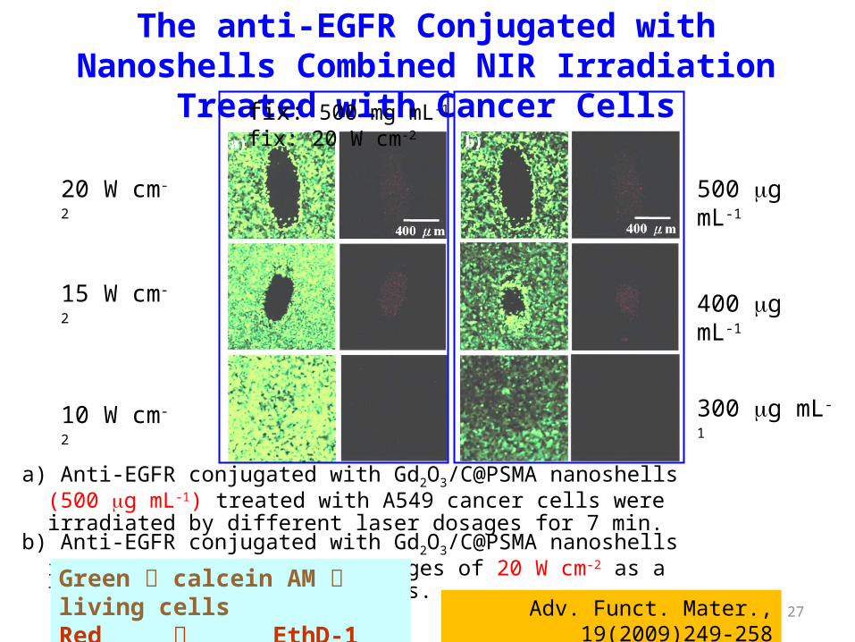
a) Anti-EGFR conjugated with Gd2O3/C@PSMA nanoshells (500 mg mL-1) treated with A549 cancer cells were irradiated by different laser dosages for 7 min.
b) Anti-EGFR conjugated with Gd2O3/C@PSMA nanoshells irradiated by the laser dosages of 20 W cm-2 as a function of nanoshell dosages.
The anti-EGFR Conjugated with Nanoshells Combined NIR Irradiation Treated with Cancer Cells
Adv. Funct. Mater., 19(2009)249-258Green calcein AM living cellsRed EthD-1 dead cells
20 W cm-2
15 W cm-2
10 W cm-2
500 mg mL-1
400 mg mL-1
300 mg mL-1
fix: 500 mg mL-1 fix: 20 W cm-2
28
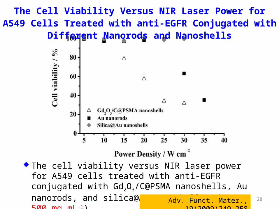
The cell viability versus NIR laser power for A549 cells treated with anti-EGFR conjugated with Gd2O3/C@PSMA nanoshells, Au nanorods, and silica@Au nanoshells (dosage: 500 mg mL-1).
The Cell Viability Versus NIR Laser Power for A549 Cells Treated with anti-EGFR Conjugated with Different Nanorods and Nanoshells
Adv. Funct. Mater., 19(2009)249-258
29
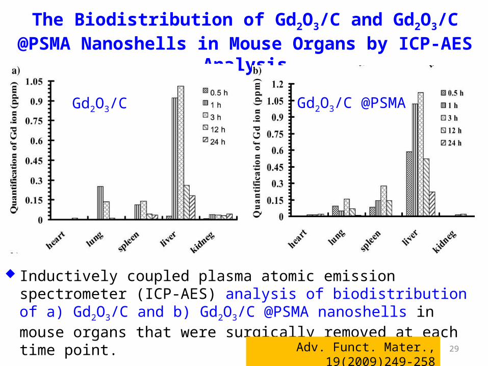
The Biodistribution of Gd2O3/C and Gd2O3/C @PSMA Nanoshells in Mouse Organs by ICP-AES Analysis
Inductively coupled plasma atomic emission spectrometer (ICP-AES) analysis of biodistribution of a) Gd2O3/C and b) Gd2O3/C @PSMA nanoshells in mouse organs that were surgically removed at each time point.
Adv. Funct. Mater., 19(2009)249-258
Gd2O3/C Gd2O3/C @PSMA
30
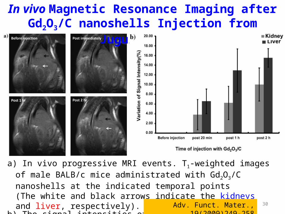
a) In vivo progressive MRI events. T1-weighted images of male BALB/c mice administrated with Gd2O3/C nanoshells at the indicated temporal points (The white and black arrows indicate the kidneys and liver, respectively).
b) The signal intensities of liver and kidney in T1-weighted imaging at the indicated temporal points.
In vivo Magnetic Resonance Imaging after Gd2O3/C nanoshells Injection from Jugular Vein
Adv. Funct. Mater., 19(2009)249-258
31
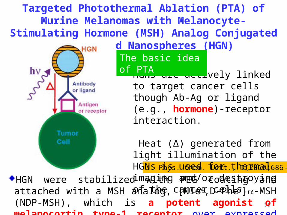
Targeted Photothermal Ablation (PTA) of Murine Melanomas with Melanocyte-Stimulating Hormone (MSH) Analog
Conjugated Hollow Gold Nanospheres (HGN)
J. Phys. Chem. Lett., 1(2010)686-695
HGNs are actively linked to target cancer cells though Ab-Ag or ligand (e.g., hormone)-receptor interaction.
Heat (Δ) generated from light illumination of the HGNs is used for thermal imaging and/or destroying of the cancer cells.
The basic idea of PTA
HGN were stabilized with PEG coating and attached with a MSH analog, [Nle4,D-Phe7]a-MSH (NDP-MSH), which is a potent agonist of melanocortin type-1 receptor over expressed in melanoma.

32
Synthesis, Characterization, and Tunable Optical Properties of Hollow Gold Nanospheres
J. Phys. Chem. B, 110(2006)19935-19944
UV-visible absorption spectra of nine HGN samples with varying diameters and wall thicknesses
Image showing the color range of HGN solutions. The vial on the far left contains solid gold nanoparticles, the rest are HNGs with varying diameters and wall thicknesses.

33Clin. Cancer Res., 2009, 15(3):876-886
D: TEM images of NDP-MSH-PEG-HAuNS and PEG-HAuNS subjected to immunogold staining. Only NDP-MSH-PEG-HAuNS were stained with 5-nm gold nanoparticles (arrow). Bar, 20 nm.
Conjugation of NDP-MSH peptide to HAuNS through PEG linker
A: schematic of nanoshell synthesis and bioconjugation.
B: TEM image of NDP-MSH-PEG-HAuNS. Bar, 50 nm.
C:absorbance of HAuNS in water before and after bioconjugation.
outer diameter: 43.5 ± 2.3 nm, shell thickness: 3-4 nm
34
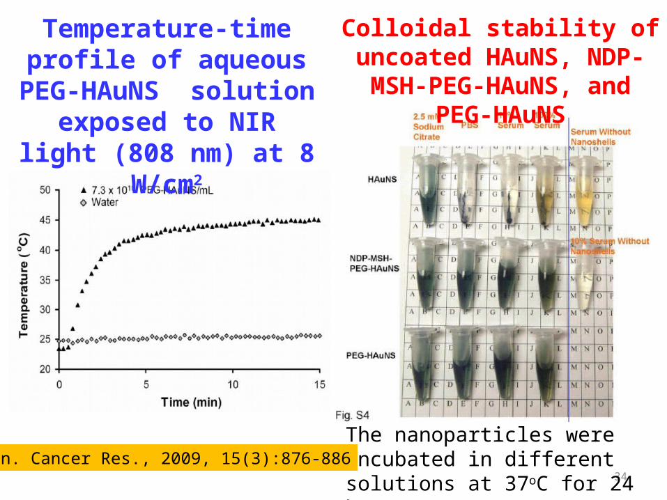
Temperature-time profile of aqueous PEG-HAuNS
solution exposed to NIR light (808 nm) at 8 W/cm2
Colloidal stability of uncoated HAuNS, NDP-MSH-PEG-HAuNS,
and PEG-HAuNS
The nanoparticles were incubated in different solutions at 37oC for 24 h.Clin. Cancer Res., 2009, 15(3):876-886
35
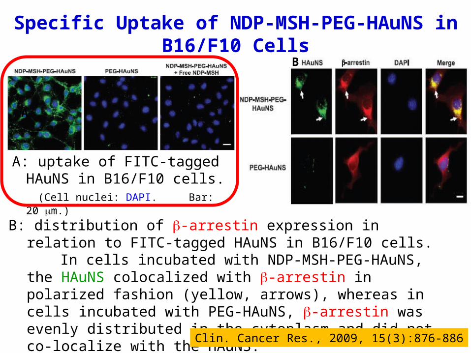
B: distribution of b-arrestin expression in relation to FITC-tagged HAuNS in B16/F10 cells.
In cells incubated with NDP-MSH-PEG-HAuNS, the HAuNS colocalized with b-arrestin in polarized fashion (yellow, arrows), whereas in cells incubated with PEG-HAuNS, b-arrestin was evenly distributed in the cytoplasm and did not co-localize with the HAuNS.
(Cell nuclei: DAPI. Bar: 20 mm.)
B
Clin. Cancer Res., 2009, 15(3):876-886
Specific Uptake of NDP-MSH-PEG-HAuNS in B16/F10 Cells
A
A: uptake of FITC-tagged HAuNS in B16/F10 cells.
(Cell nuclei: DAPI. Bar: 20 mm.)
36
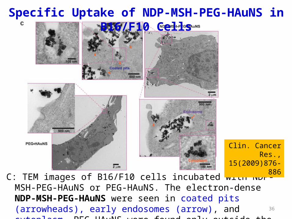
Specific Uptake of NDP-MSH-PEG-HAuNS in B16/F10 Cells
C: TEM images of B16/F10 cells incubated with NDP-MSH-PEG-HAuNS or PEG-HAuNS. The electron-dense NDP-MSH-PEG-HAuNS were seen in coated pits (arrowheads), early endosomes (arrow), and cytoplasm. PEG-HAuNS were found only outside the cell membrane.
Clin. Cancer Res., 15(2009)876-886
37
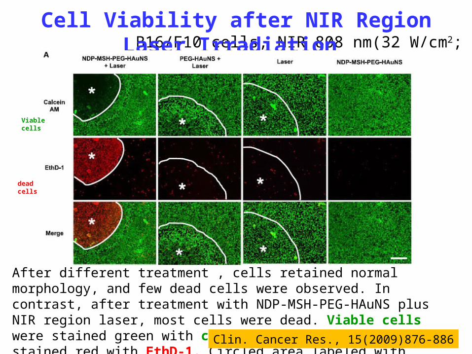
B16/F10 cells, NIR 808 nm(32 W/cm2; 3 mins)Cell Viability after NIR Region Laser Irradiation
After different treatment , cells retained normal morphology, and few dead cells were observed. In contrast, after treatment with NDP-MSH-PEG-HAuNS plus NIR region laser, most cells were dead. Viable cells were stained green with calcein AM; dead cells were stained red with EthD-1. Circled area labeled with asterisk, laser-irradiated area; bar: 100 mm Clin. Cancer Res., 15(2009)876-886
Viable cells
dead cells

38
Biodistribution and Intratumoral Distribution of FITC-tagged HAuNS
Tissue and tumors were removed 4 h after i.v. injection of HAuNS.A: representative fluorescence micrographs of cryosectioned B16/F10 melanoma.
Microvessels were stained with rat anti-mouse CD31 monoclonal antibody. Cell nuclei: DAPI . Significantly more HAuNS were found in the tumors of mice injected with NDP-MSH-PEG-HAuNS than in the tumors of mice injected with PEG-HAuNS. NDP-MSH-PEG-HAuNS were distributed throughout the tumor matrix in the interstitial space, whereas PEG-HAuNS were distributed around tumor vessels (arrows). Bar, 100 mm.
B: biodistribution of FITC-tagged HAuNS in different tissues. Data were calculated as the number of particle aggregates per mm2 visual area at X200, and values are presented as mean FSD (n = 5). Clin. Cancer Res., 2009, 15(3):876-886

39
C: biodistributionof NDP-MSH-PEG-HAuNS and PEG-HAuNS. Data were plottedas %ID/g).Mean FSD(n =5).*,P < 0.01.
D: Z-stack images of tumor sections at higher magnification. Melanocortin type-1 receptor was stained with rabbit anti-mouse melanocortin type-1 receptor polyclonal antibody (pseudo colored red). Blood vessels were stained with rat anti-mouse CD31 monoclonal antibody (pseudo colored blue). FITC-tagged HAuNS were green. NDP-MSH-PEG-HAuNS but not PEG-HAuNS co-localized with melanocortin type-1receptor (yellow and orange, arrowheads), indicating melanocortin type-1 receptor mediated endocytosis of NDP-MSH-PEG-HAuNS in vivo. Asterisks, the lumens of tumor vasculature with discontinuous CD31 staining; bar,10 mm
Biodistribution and Intratumoral Distribution of FITC-tagged HAuNS
Clin. Cancer Res., 15(2009)876-886
40
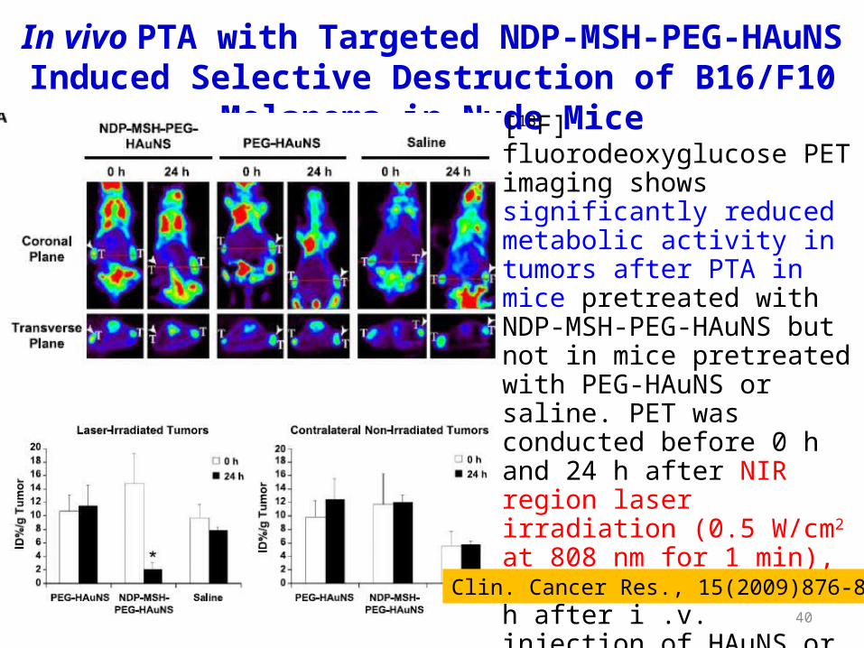
In vivo PTA with Targeted NDP-MSH-PEG-HAuNS Induced Selective Destruction of B16/F10 Melanoma in Nude Mice
[18F] fluorodeoxyglucose PET imaging shows significantly reduced metabolic activity in tumors after PTA in mice pretreated with NDP-MSH-PEG-HAuNS but not in mice pretreated with PEG-HAuNS or saline. PET was conducted before 0 h and 24 h after NIR region laser irradiation (0.5 W/cm2 at 808 nm for 1 min), which was commenced 4 h after i .v. injection of HAuNS or saline.T, tumor. Arrowheads, tumors irradiated with NIR region light.
Clin. Cancer Res., 15(2009)876-886
41
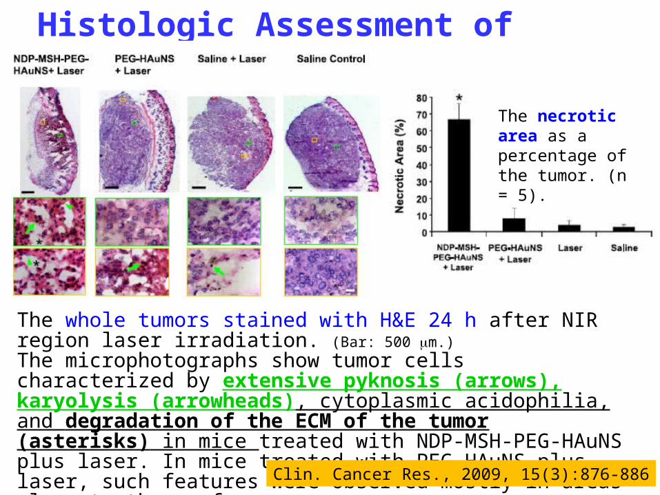
The whole tumors stained with H&E 24 h after NIR region laser irradiation. (Bar: 500 mm.)The microphotographs show tumor cells characterized by extensive pyknosis (arrows), karyolysis (arrowheads), cytoplasmic acidophilia, and degradation of the ECM of the tumor (asterisks) in mice treated with NDP-MSH-PEG-HAuNS plus laser. In mice treated with PEG-HAuNS plus laser, such features were observed mostly in areas close to the surface. (Bar: 50 mm. )
Histologic Assessment of Tumor Necrosis
Clin. Cancer Res., 2009, 15(3):876-886
The necrotic area as a percentage of the tumor. (n = 5).
42
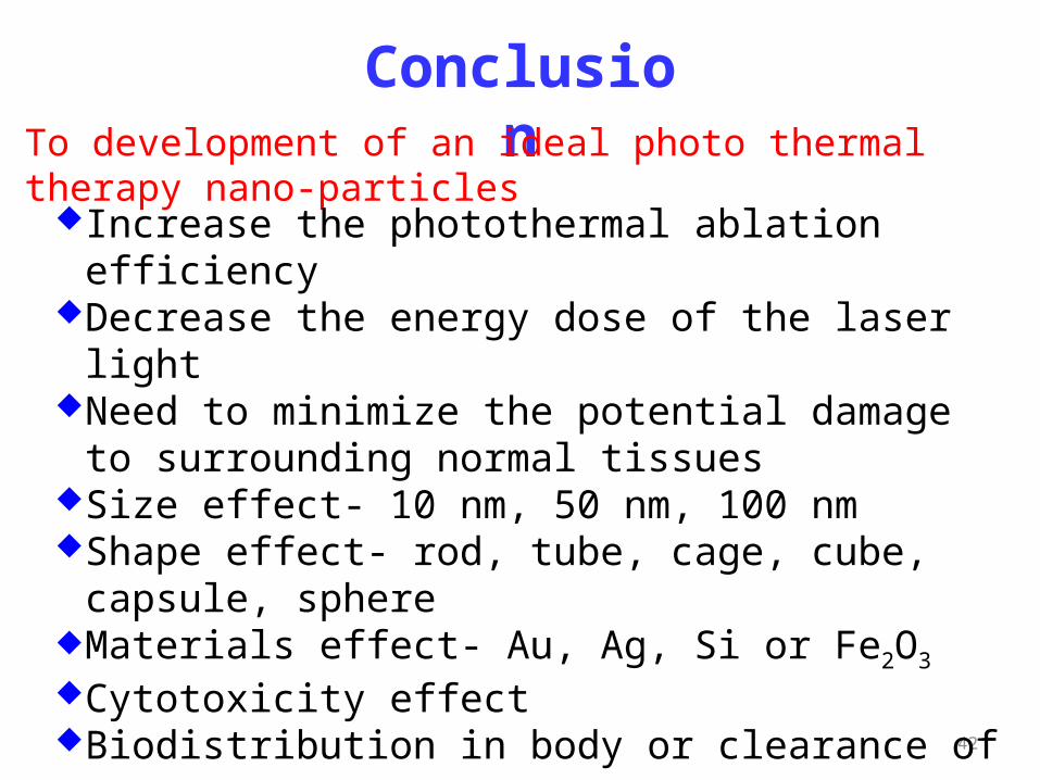
ConclusionTo development of an ideal photo thermal therapy nano-particles
Increase the photothermal ablation efficiencyDecrease the energy dose of the laser lightNeed to minimize the potential damage to surrounding
normal tissuesSize effect- 10 nm, 50 nm, 100 nmShape effect- rod, tube, cage, cube, capsule, sphereMaterials effect- Au, Ag, Si or Fe2O3
Cytotoxicity effectBiodistribution in body or clearance of organSpecificity (targeting)Cost
43
PDT is based on photosensitizers (PSs) generating singlet oxygen (1O2), and subsequently reactive oxygen species (ROS) upon localized exposure to light in the presence of ground state oxygen (O2).
After excitation of the PS from its ground singlet state to an excited singlet state, the PS undergoes intersystem crossing to a longer-lived excited triplet state.
Energy transfer from the PS’s excited triplet state to a nearby oxygen molecule (which posses a ground triplet state) results in the relaxation of the PS to its ground singlet state and the formation of an excited singlet state oxygen molecule.
The intrinsic cytotoxity of singlet oxygen leads to selective and irreversible destruction of diseased tissues in the vicinity of 1O2.
What is the PDT?
44
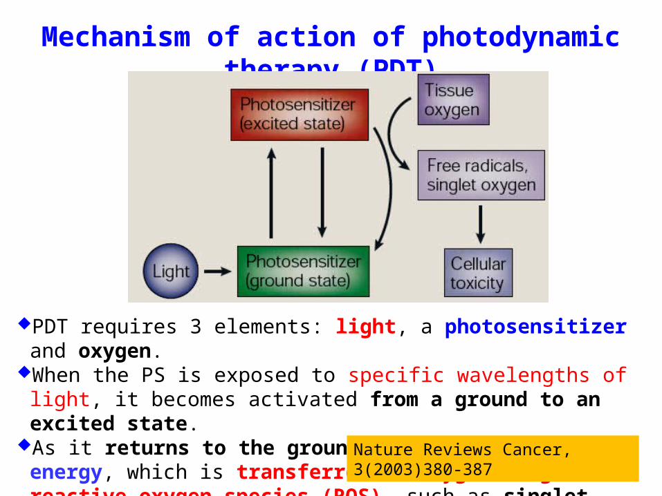
Mechanism of action of photodynamic therapy (PDT)
PDT requires 3 elements: light, a photosensitizer and oxygen.When the PS is exposed to specific wavelengths of light, it becomes
activated from a ground to an excited state.As it returns to the ground state, it releases energy, which is transferred
to oxygen to generate reactive oxygen species (ROS), such as singlet oxygen and free radicals.
These ROS mediate cellular toxicity.Nature Reviews Cancer, 3(2003)380-387
45
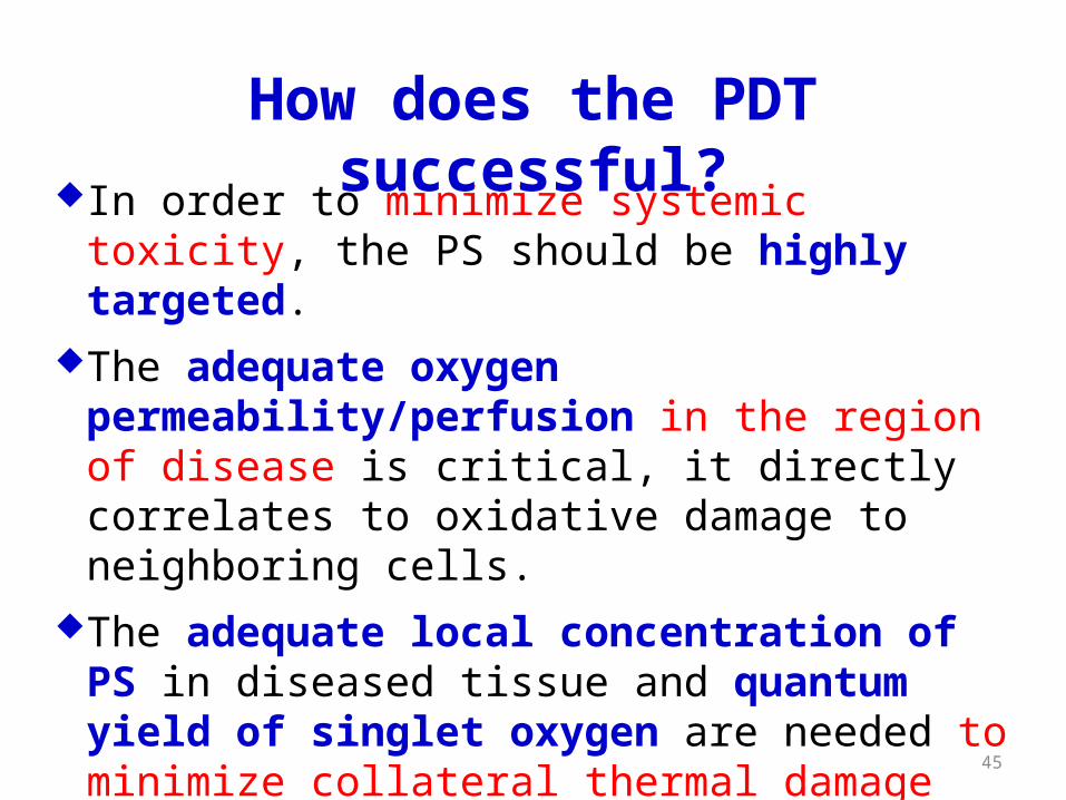
In order to minimize systemic toxicity, the PS should be highly targeted.
The adequate oxygen permeability/perfusion in the region of disease is critical, it directly correlates to oxidative damage to neighboring cells.
The adequate local concentration of PS in diseased tissue and quantum yield of singlet oxygen are needed to minimize collateral thermal damage from photoirradiation.
How does the PDT successful?

46
J. Mater. Chem., 19(2009)1252–1257
Mesoporous Silica Nanoparticles (MSNs) Functionalized with an Oxygen-sensing Probe for Cell Photodynamic
Therapy (PDT): Potential Cancer Theranostics
Theranostics = Therapeutics + Diagnostics
MSNs serve as a matrix in which high densities of transportable molecules reside, shielded from their local environment.
47
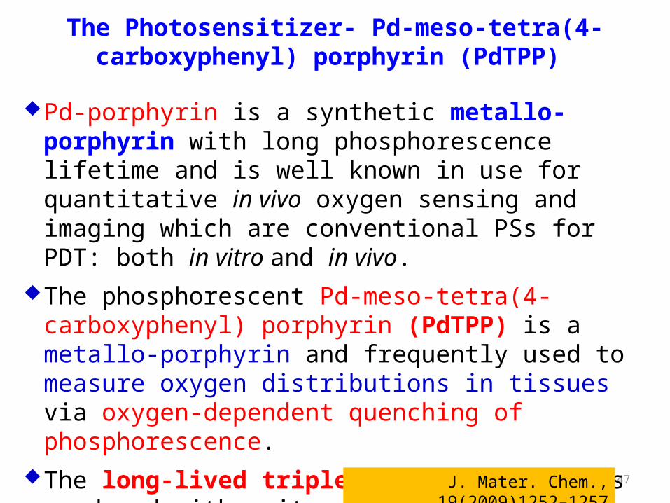
Pd-porphyrin is a synthetic metallo-porphyrin with long phosphorescence lifetime and is well known in use for quantitative in vivo oxygen sensing and imaging which are conventional PSs for PDT: both in vitro and in vivo.
The phosphorescent Pd-meso-tetra(4-carboxyphenyl) porphyrin (PdTPP) is a metallo-porphyrin and frequently used to measure oxygen distributions in tissues via oxygen-dependent quenching of phosphorescence.
The long-lived triplet state of PdTPP is produced with unity quantum yield, while the concentration of singlet oxygen generated in PDT depends on the accessibility of the excited PdTPP to oxygen.
The Photosensitizer- Pd-meso-tetra(4-carboxyphenyl) porphyrin (PdTPP)
J. Mater. Chem., 19(2009)1252–1257
48
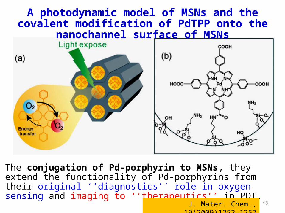
A photodynamic model of MSNs and the covalent modification of PdTPP onto the nanochannel surface of MSNs
The conjugation of Pd-porphyrin to MSNs, they extend the functionality of Pd-porphyrins from their original ‘‘diagnostics’’ role in oxygen sensing and imaging to ‘‘therapeutics’’ in PDT.
J. Mater. Chem., 19(2009)1252–1257

49
The spectra shows the characteristic peak of amide bond which was self-assembled with PdTPP and MSNs.
The spectra provide strong support for the assertion that PdTPP attachment to MSNs was by means of amide bonds.
FTIR spectra was used to evaluate the covalent bonding of MSN and PdTPP
Two strong absorption bands: amide I (1650 cm-1, carbonyl stretch), amide II (1540 cm-1, CN stretch and NH bend)
J. Mater. Chem., 19(2009)1252–1257

50
After PdTPP modification, the surface area and mean pore diameter of MSNs decreased from 1026 m2/g and 3 nm to 540 m2/g and 2.3 nm, respectively (by N2 adsorption–desorption isotherm measurement, BET), which implied the majority of PdTPP conjugation took place within the MSN’s nanochannels.
A TEM image of MSN–PdTPP
uniform tertiary structure of MSN–PdTPP (average particle size ~100 nm).
J. Mater. Chem., 19(2009)1252–1257

52
The absorption and phosphorescence spectra of PdTPP bound to 2% albumin in PBS (solid lane) and MSNs–
PdTPP dispersed in ethanol (dashed lane)
PdTPP and MSNs–PdTPP in PBS with 2% bovine serum albumin show similar absorption and phosphorescence spectra.
J. Mater. Chem., 19(2009)1252–1257
53
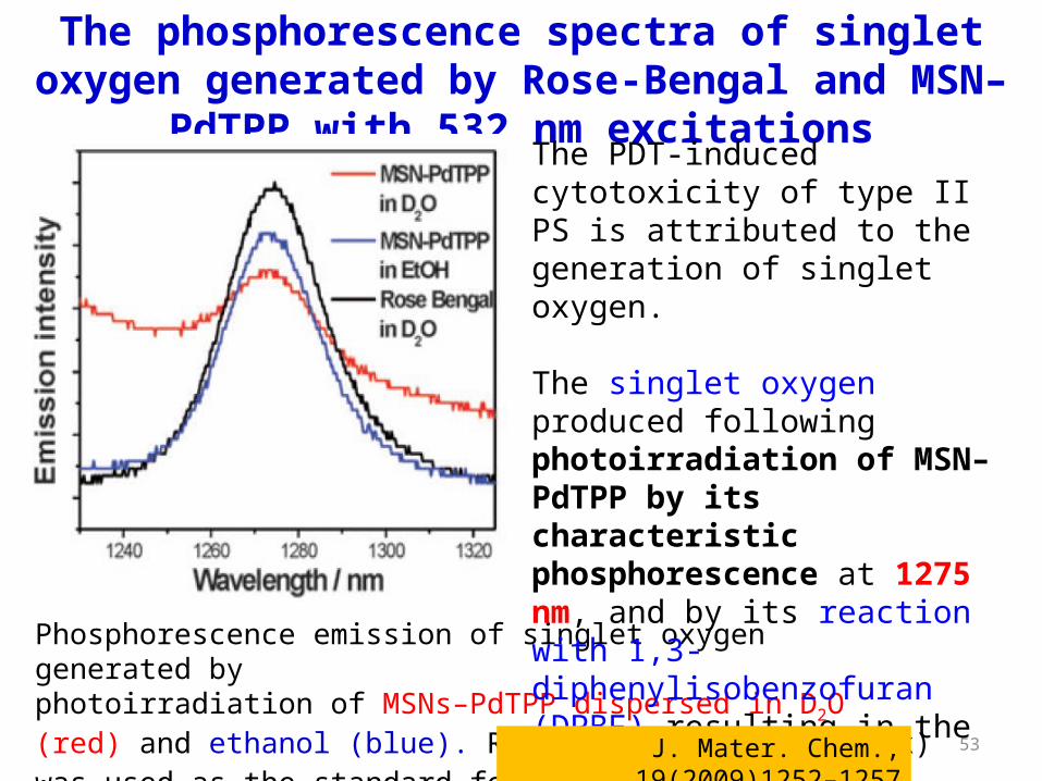
Phosphorescence emission of singlet oxygen generated byphotoirradiation of MSNs–PdTPP dispersed in D2O (red) and ethanol (blue). Rose Bengal in D2O (black) was used as the standard for singlet oxygen phosphorescence measurement.
The phosphorescence spectra of singlet oxygen generated by Rose-Bengal and MSN–PdTPP with 532 nm excitations
The PDT-induced cytotoxicity of type II PS is attributed to the generation of singlet oxygen.
The singlet oxygen produced following photoirradiation of MSN–PdTPP by its characteristic phosphorescence at 1275 nm, and by its reaction with 1,3-diphenylisobenzofuran (DPBF) resulting in the decrease of DPBF’s absorption at 400 nm.
J. Mater. Chem., 19(2009)1252–1257

54
Photobleaching of DPBF by singlet oxygen generated from MSN–PdTPP upon photoirradiation at 532 nm.
The steep decrease of DPBF absorption with time in the MSN–PdTPP solution indicates the efficient generation of 1O2.
The photobleaching of DPBF in D2O in the presence of MSN–PdTPP
J. Mater. Chem., 19(2009)1252–1257
Inset:decay curves of DPBF absorption as function of irradiative timein D2O (□),in MSNs solution (○) andin MSNs–PdTPP solution ( ).△
55
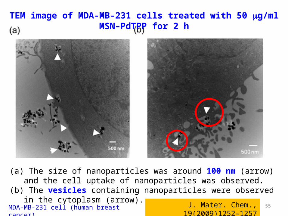
(a) The size of nanoparticles was around 100 nm (arrow) and the cell uptake of nanoparticles was observed.
(b) The vesicles containing nanoparticles were observed in the cytoplasm (arrow).
TEM image of MDA-MB-231 cells treated with 50 mg/ml MSN–PdTPP for 2 h
J. Mater. Chem., 19(2009)1252–1257MDA-MB-231 cell (human breast cancer)
56
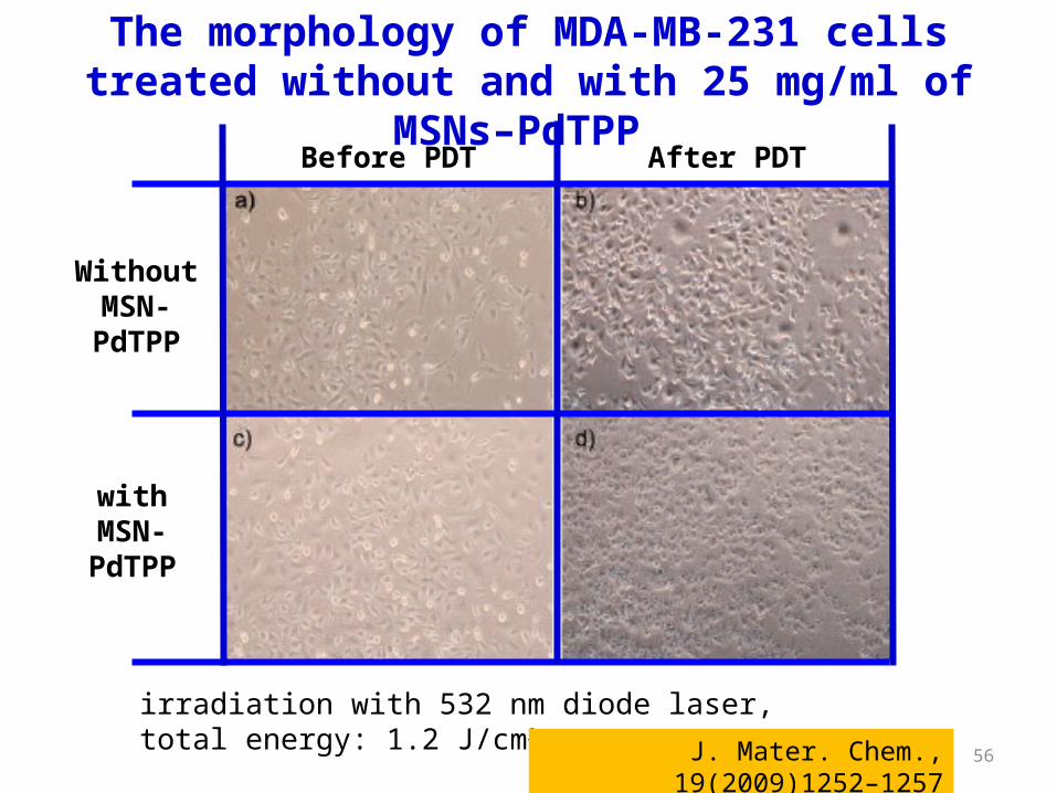
irradiation with 532 nm diode laser, total energy: 1.2 J/cm2
The morphology of MDA-MB-231 cells treated without and with 25 mg/ml of MSNs–PdTPP
with MSN-PdTPP
After PDTBefore PDT
Without MSN-PdTPP
J. Mater. Chem., 19(2009)1252–1257
57
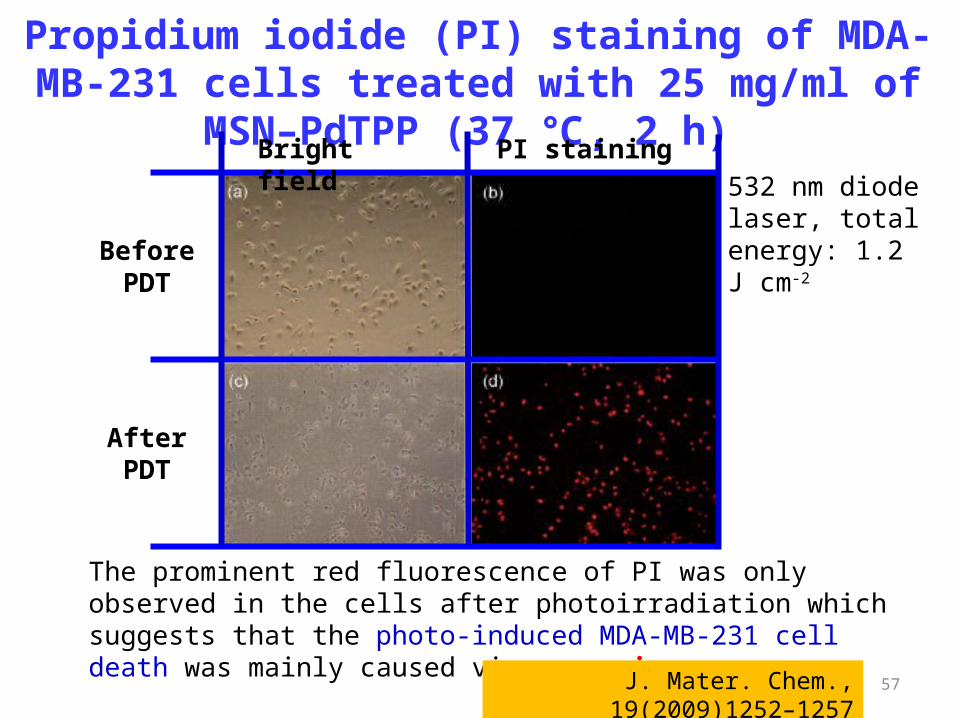
532 nm diode laser, total energy: 1.2 J cm-2
The prominent red fluorescence of PI was only observed in the cells after photoirradiation which suggests that the photo-induced MDA-MB-231 cell death was mainly caused via necrosis.
Propidium iodide (PI) staining of MDA-MB-231 cells treated with 25 mg/ml of MSN–PdTPP (37 , 2 h) ℃
Bright field PI staining
After PDT
Before PDT
J. Mater. Chem., 19(2009)1252–1257
58
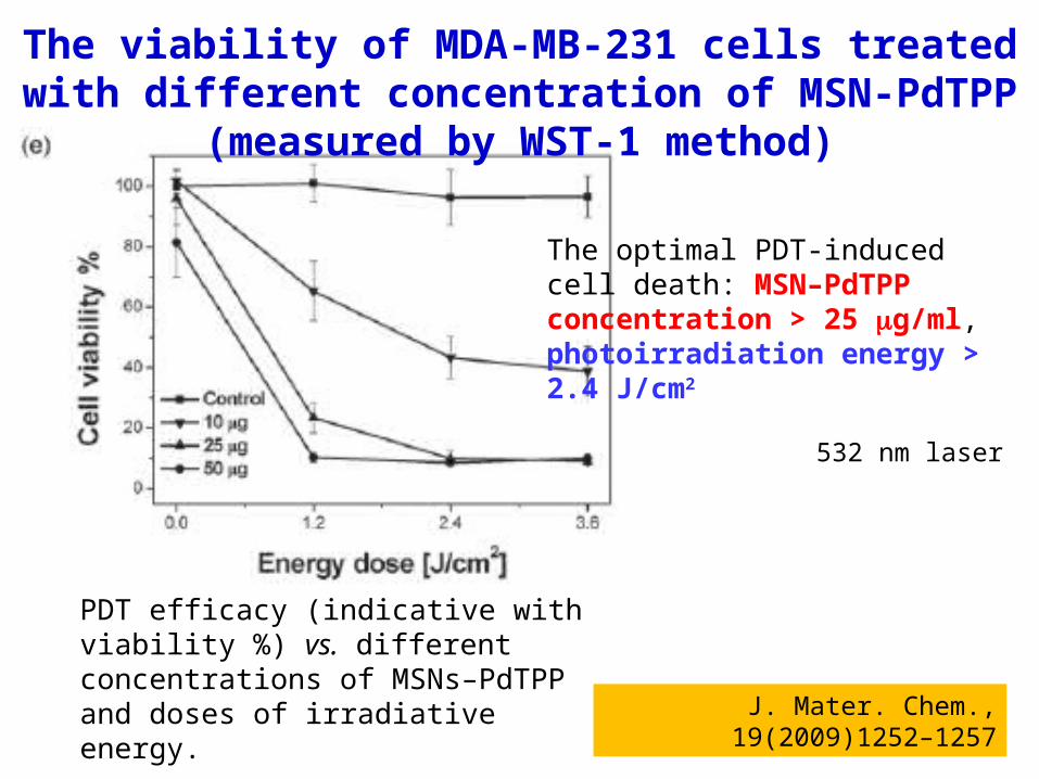
PDT efficacy (indicative with viability %) vs. different concentrations of MSNs–PdTPP and doses of irradiative energy.
The viability of MDA-MB-231 cells treated with different concentration of MSN-PdTPP (measured by WST-1 method)
532 nm laser
J. Mater. Chem., 19(2009)1252–1257
The optimal PDT-induced cell death: MSN–PdTPP concentration > 25 mg/ml, photoirradiation energy > 2.4 J/cm2
59
The significant photo-induced cytotoxicity in MDA-MB-231 breast cancer studies is most likely due to a number of factors, principal of which are the high concentration of PdTPP conjugated within MSNs and the role that MSNs play as a facilitator for endocytotic cell uptake that dramatically increases the intracellular density of singlet oxygen upon photoirradiation.
MSN–PdTPP is a promising PS for PDT.And with the ability of PdTPP to serve as an in vivo contrast
agent for oxygen sensing and imaging, MSN–PdTPP holds great promise as a useful nano-platform for cancer theranostics.
Conclusions
J. Mater. Chem., 19(2009)1252–1257

60
Size-dependent processes of particle transport in the human body
Nature Materials, 8(2009)15-23

61
Thanks for your attention !