1 - skin: basic structure and function...1 chapter 1 skin: basic structure and function 3 from...
TRANSCRIPT

1
1
1 Skin: Basic Structure and Function
The epidermis can be divided into the innermost basal layer (stratum germinativum), the malpighian or prickle layer (stratum spinosum), the granular layer (stratum granulosum), and the horny layer (stratum corneum). On the palms and soles, a pale clear to pink layer, the stratum lucidum, is noted just above the granular layer (Fig. 1.3). When the skin in other sites is scratched or rubbed, the malpighian and granular layers thicken, a stratum lucidum forms, and the stratum corneum becomes thick and compact. Histones appear to regulate epidermal differentiation, and histone deacetylation suppresses expression of profilaggrin. Slow-cycling stem cells provide a reservoir for regeneration of the epidermis. Sites rich in stem cells include the deepest portions of the rete, especially on palmoplantar skin, as well as the hair bulge. Stem cells divide infrequently in normal skin, but in cell culture they form active, growing colonies. They can be identified by their high expression of β1-integrins and lack of terminal differentiation markers. Stem cells can also be identified by their low levels of desmosomal proteins, such as desmoglein 3. The basal cells divide, and as their progeny move upward, they flatten and their nucleus disappears. Abnormal keratinization can manifest as parakeratosis (retained nuclei), as corps ronds (round, clear to pink, abnormally keratinized cells), or as grains (elongated, basophilic, abnormally keratinized cells).
During keratinization, the keratinocyte first passes through a synthetic and then a degradative phase on its way to becoming a horn cell. In the synthetic phase, within its cytoplasm the keratinocyte accumulates intermediate filaments composed of a fibrous protein, keratin, arranged in an α-helical coiled pattern. These tonofilaments are fashioned into bundles, which converge on and terminate at the plasma membrane, where they end in specialized attachment plates called desmosomes. The degradative phase of keratinization is characterized by the disappearance of cell organelles and the consolidation of all contents into a mixture of filaments and amorphous cell envelopes. This programmed process of maturation resulting in death of the cell is called terminal differentiation. Terminal differentiation is also seen in the involuting stage of keratoacanthomas, where the initial phase of proliferation gives way to terminal keratinization and involution. Degradation of the mitochondrial network within keratinocytes occurs with aging. Oxidation injury to keratinocytes occurs with environmental exposure and thermal burns, and can be partially prevented by vitamin C in the form of L-ascorbic acid.
Premature programmed cell death, or apoptosis, appears in hematoxylin and eosin (H&E)–stained sections as scattered bright-red cells, some of which may contain small, black pyknotic nuclei. These cells are present at various levels of the epidermis, because this form of cell death does not represent part of the normal process of maturation. Widespread apoptosis is noted in the verrucous phase of incontinentia pigmenti. It is also a prominent finding in catagen hairs, where apoptosis results in the involution of the inferior segment of the hair follicle.
In normal skin, the plasma membranes of adjacent cells are separated by an intercellular space. Electron microscopic histo-chemical studies have shown that this interspace contains glyco-proteins and lipids. Lamellar granules (Odland bodies or membrane-coating granules) appear in this space, primarily at the interface between the granular and cornified cell layers. Lamellar granules contribute to skin cohesion and impermeability. Conditions such as lamellar ichthyosis and Flegel hyperkeratosis demonstrate abnormal lamellar granules. Glycolipids such as ceramides
Bonus images for this chapter can be found online at expertconsult.inkling.com
Skin is composed of three layers: the epidermis, dermis, and subcutaneous fat (panniculus) (Fig. 1.1). The outermost layer, the epidermis, is composed of viable keratinocytes covered by a layer of keratin, the stratum corneum. The principal component of the dermis is the fibrillar structural protein collagen. The dermis lies on the panniculus, which is composed of lobules of lipocytes separated by collagenous septa that contain the neurovascular bundles.
There is considerable regional variation in the relative thickness of these layers. The epidermis is thickest on the palms and soles, measuring approximately 1.5 mm. It is very thin on the eyelid, where it measures less than 0.1 mm. The dermis is thickest on the back, where it is 30–40 times as thick as the overlying epidermis. The amount of subcutaneous fat is generous on the abdomen and buttocks compared with the nose and sternum, where it is meager.
EPIDERMIS AND ADNEXADuring the first weeks of life, the fetus is covered by a layer of nonkeratinizing cuboidal cells called the periderm (Fig. 1.2). Later, the periderm is replaced by a multilayered epidermis. Adnexal structures, particularly follicles and eccrine sweat units, originate during the third month of fetal life as downgrowths from the developing epidermis. Later, apocrine sweat units develop from the upper portion of the follicular epithelium and sebaceous glands from the midregion of the follicle. Adnexal structures appear first in the cephalic portion of the fetus and later in the caudal portions.
The adult epidermis is composed of three basic cell types: keratinocytes, melanocytes, and Langerhans cells. An additional cell, the Merkel cell, can be found in the basal layer of the palms and soles, oral and genital mucosa, nail bed, and follicular infun-dibula. Located directly above the basement membrane zone, Merkel cells contain intracytoplasmic dense-core neurosecretory-like granules and, through their association with neurites, act as slow-adapting touch receptors. They have direct connections with adjacent keratinocytes by desmosomes and contain a paranuclear whorl of intermediate keratin filaments. Both polyclonal keratin immunostains and monoclonal immunostaining for keratin 20 stain this whorl of keratin filaments in a characteristic paranuclear dot pattern. Merkel cells also label for neuroendocrine markers such as chromogranin and synaptophysin.
KeratinocytesKeratinocytes are of ectodermal origin and have the specialized function of producing keratin, a complex filamentous protein that not only forms the surface coat (stratum corneum) of the epidermis but also is the structural protein of hair and nails. Multiple distinct keratin genes have been identified and consist of two subfamilies, acidic and basic. The product of one basic and one acidic keratin gene combines to form the multiple keratins that occur in many tissues. Mutations in the genes for keratins 5 and 14 are associated with epidermolysis bullosa simplex. Keratin 1 and 10 mutations are associated with epidermolytic hyperkeratosis. Mild forms of this disorder may represent localized or widespread expressions of mosaicism for these gene mutations.

2 Andrews’ Diseases of the Skin
because it is thought to be responsible for keratin filament aggrega-tion. Conversion to filaggrin takes place in the granular layer, and this forms the electron-dense interfilamentous protein matrix of mature epidermal keratin. Kallikrein-related peptidase 5, a serine protease secreted from lamellar granules, appears to function in profilaggrin cleavage.
Keratohyalin is hygroscopic, and repeated cycles of hydration and dehydration contribute to normal desquamation of the stratum corneum. Ichthyosis vulgaris is characterized by a diminished or absent granular layer, contributing to the retention hyperkeratosis
contribute a water-barrier function to skin and are typically found in topical products meant to restore the epidermal barrier. Lamellar bodies form abnormally in the absence of critical ceramides such as glucosylceramide, or there is disproportion of critical lipids. Desmosomal adhesion depends on cadherins, including the calcium-dependent desmogleins and desmocollins. Antibodies to these molecules result in immunobullous diseases, but desmogleins function not only in adhesion but also in differentiation. The binding of the desmoglein 1 cytoplasmic tail to the scaffolding-protein Erbin downregulates the Ras-Raf pathway to promote stratification and differentiation of keratinocytes in the epidermis.
Keratinocytes of the granular zone contain, in addition to the keratin filament system, keratohyaline granules, composed of amorphous particulate material of high sulfur-protein content. This material, profilaggrin, is a precursor to filaggrin, so named
Apocrineunit
Straight duct
Coiled gland
Spiraled duct
Straight duct
Coiled duct
Eccrine gland
Superficial plexus
Deep plexus
Epidermis
Subcutaneous tissue
Meissner nerveending
papillary
reticular
Sebaceous glandArrector pili muscle
Hair shaftPacini nerve ending
DermisEccrine sweat unit
Dermalvasculature
Fig. 1.1 Diagrammatic cross section of the skin and panniculus.
Fig. 1.2 In early fetal life, a cuboidal periderm is present, rather than an epidermis. Fetal skin, H&E × 40.
Fig. 1.3 Volar skin demonstrating a thick corneum and dermis, H&E × 100.

1
CHAPTER 1 Skin: Basic Structure and Function 3
from eumelanin to pheomelanin production, whereas activating gene mutations can enhance eumelanin synthesis. Most redheads are compound heterozygotes or homozygotes for a variety of loss-of-function mutations in this gene.
Antimicrobial peptides, including cathelicidin and β-defensins, are key components of the innate immune system. They protect against infection, are implicated in the pathogenesis of atopic dermatitis, and play a role in control of pigmentation. The β-defensins encompass a class of small, cationic proteins important to both the innate and the adaptive immune system. β-Defensin 3 also functions as a melanocortin receptor ligand.
Eumelanin production is optimal at pH 6.8, and changes in cellular pH also result in alterations of melanin production and the eumelanin/pheomelanin ratio. Within keratinocytes, melanin typically forms a cap over the nucleus, where it presumably functions principally in a photoprotective role. Evidence of keratinocyte photodamage in the form of pyrimidine dimer formation can be assessed using gas chromatography–mass spectrometry or enzyme-linked immunosorbent assays. Pigment within melanocytes also serves to protect the melanocytes themselves against photodamage, such as ultraviolet A (UVA)–induced membrane damage.
Areas of leukoderma, or whitening of skin, can be caused by very different phenomena. In vitiligo, the affected skin becomes white because of destruction of melanocytes. In albinism, the number of melanocytes is normal, but they are unable to synthesize fully pigmented melanosomes because of defects in the enzymatic formation of melanin. Local areas of increased pigmentation can result from a variety of causes. The typical freckle results from a localized increase in production of pigment by a near-normal number of melanocytes. Black “sunburn” or “ink spot” lentigines demonstrate basilar hyperpigmentation and prominent melanin within the stratum corneum. Nevi are benign proliferations of melanocytes. Melanomas are their malignant counterpart. Mela-nocytes and keratinocytes express neurotrophins (ectodermal nerve growth factors). Melanocytes release neurotrophin 4, but the release is downregulated by ultraviolet B (UVB) irradiation, suggesting neurotrophins as possible targets for therapy of disorders of pigmentation. Melanocytes express toll-like receptors (TLRs) and stimulation by bacterial lipopolysaccharides increases pigmentation. Melatonin and its metabolites protect melanocytes from UVB damage.
Langerhans CellsLangerhans cells are normally found scattered among keratinocytes of the stratum spinosum. They constitute 3%–5% of the cells in this layer. As with melanocytes, Langerhans cells are not connected to adjacent keratinocytes by the desmosomes. The highest density of Langerhans cells in the oral mucosa occurs in the vestibular region, and the lowest density is in the sublingual region, suggesting the latter is a relatively immunologically “privileged” site.
At the light microscopic level, Langerhans cells are difficult to detect in routinely stained sections. However, they appear as dendritic cells in sections impregnated with gold chloride, a stain specific for Langerhans cells. They can also be stained with CD1α or S-100 immunostains. Ultrastructurally, they are characterized by a folded nucleus and distinct intracytoplasmic organelles called Birbeck granules. In their fully developed form, the organelles are rod shaped with a vacuole at one end, resembling a tennis racquet. The vacuole is an artifact of processing.
Functionally, Langerhans cells are of the monocyte-macrophage lineage and originate in bone marrow. They function primarily in the afferent limb of the immune response by providing for the recognition, uptake, processing, and presentation of antigens to sensitized T lymphocytes and are important in the induction of delayed-type sensitivity as well as humoral immunity. Once an antigen is presented, Langerhans cells migrate to the lymph nodes. Hyaluronan (hyaluronic acid) plays a critical role in Langerhans
noted in this disorder. Keratohyalin results in the formation of soft, flexible keratin. Keratin that forms in the absence of kera-tohyaline granules is typically hard and rigid. Hair fibers and nails are composed of hard keratin.
Keratinocytes play an active role in the immune function of the skin. In conditions such as allergic contact dermatitis, these cells participate in the induction of the immune response, rather than acting as passive casualties. Keratinocytes secrete a wide array of cytokines and inflammatory mediators, including tumor necrosis factor–alpha (TNF-α). They also can express molecules on their surface, such as intercellular adhesion molecule 1 (ICAM-1) and major histocompatibility complex (MHC) class II molecules, suggesting that keratinocytes actively respond to immune effector signals.
During wound healing, epithelial cell migration occurs before dermal remodeling. Tight junction proteins claudin-1 and occludin are critical for effective migration. Downregulation of claudin-1 expression results in delayed migration and reduced epithelial proliferation. For occludin, downregulation impairs wound healing when cells are also subjected to mechanical stress. Wound healing occurs best in a moist environment but can be impaired by excessive maceration.
MelanocytesMelanocytes are derived from the neural crest and by the eighth week of development can be found within the fetal epidermis. In normal, sun-protected trunk epidermis, melanocytes reside in the basal layer at a frequency of about 1 in every 10 basal keratinocytes. Areas such as the face, shins, and genitalia have a greater density of melanocytes, and in heavily sun-damaged facial skin, Mart-1 immunostaining can demonstrate ratios of melanocytes to basal keratinocytes that approach 1 : 1. Recognition of the variation in melanocyte/keratinocyte ratio is critical in the interpretation of biopsies of suspected lentigo maligna (malignant melanoma in situ) on sun-damaged skin.
Racial differences in skin color are not caused by differences in the number of melanocytes. It is the number, size, and distribu-tion of the melanosomes or pigment granules within keratinocytes that determine differences in skin color. Pale skin has fewer melanosomes, and these are smaller and packaged within membrane-bound complexes. Dark skin has more melanosomes, and these tend to be larger and singly dispersed. Chronic sun exposure can stimulate melanocytes to produce larger melanosomes, thereby making the distribution of melanosomes within keratinocytes resemble the pattern seen in dark-skinned individuals.
In histologic sections of skin routinely stained by H&E, the melanocyte appears as a cell with ample amphophilic cytoplasm or as a clear cell in the basal layer of the epidermis. The apparent halo is an artifact formed during fixation of the specimen. This occurs because the melanocyte, lacking tonofilaments, cannot form desmosomal attachments with keratinocytes. Keratinocytes also frequently demonstrate clear spaces but can be differentiated from melanocytes because they demonstrate cell-cell junctions and a layer of cytoplasm peripheral to the clear space.
The melanocyte is a dendritic cell. Its dendrites extend for long distances within the epidermis, and any one melanocyte is therefore in contact with a great number of keratinocytes; together they form the so-called epidermal melanin unit. Keratinocytes actively ingest the tips of the melanocytic dendrites, thus imbibing the melanosomes.
Melanosomes are synthesized in the Golgi zone of the cell and pass through a series of stages in which the enzyme tyrosinase acts on melanin precursors to produce the densely pigmented granules. Melanocytes in red-haired individuals tend to be rounder and to produce more pheomelanin. The melanocortin 1 receptor (MC1R) is important in the regulation of melanin production. Loss-of-function mutations in the MC1R gene bring about a change

4 Andrews’ Diseases of the Skin
lamina lucida/lamina densa interface, and collagen IV is the major component of the lamina densa. Staining in the lower, transient portion of the hair follicle, however, is different. All BMZ com-ponents diminish and may become discontinuous in the inferior segment of the follicle. Hemidesmosomes are also not apparent in the BMZ of the hair bulb. The lack of hemidesmosomes in the deep portions of the follicle may relate to the transient nature of the inferior segment, whereas abundant hemidesmosomes stabilize the upper portion of the follicle.
The BMZ is considered to be a porous semipermeable filter, which permits exchange of cells and fluid between the epidermis and dermis. It further serves as a structural support for the epidermis and holds the epidermis and dermis together. The BMZ also helps regulate growth, adhesion, and movement of keratinocytes and fibroblasts, as well as apoptosis. Much of this regulation takes place through activation of integrins and syndecans. Extracel-lular matrix protein 1 demonstrates loss-of-function mutations in lipoid proteinosis, resulting in reduplication of the basement membrane.
Breitkreutz D, et al: Skin basement membrane. Biomed Res Int 2013; 2013: 179784.
El Domyati M, et al: Immunohistochemical localization of basement membrane laminin 5 and collagen IV in adult linear IgA disease. Int J Dermatol 2015; 54: 922.
Hashmi S, et al: Molecular organization of the basement mem-brane zone. Clin Dermatol 2011; 29: 398.
EPIDERMAL APPENDAGES (ADNEXA)Eccrine and apocrine glands, ducts, and pilosebaceous units constitute the skin adnexa. Embryologically, they originate as downgrowths from the epidermis and are therefore ectodermal in origin. Hedgehog signaling by the transducer known as smooth-ened appears critical for hair development. Abnormalities in this pathway contribute to the formation of pilar tumors and basal cell carcinoma. In the absence of hedgehog signaling, embryonic hair germs may develop instead into modified sweat gland or mammary epithelium.
Although the various adnexal structures serve specific functions, all can function as reserve epidermis, in that reepithelialization occurs after injury to the surface epidermis, principally because of the migration of keratinocytes from the adnexal epithelium to the skin surface. It is not surprising, therefore, that skin sites such as the face or scalp, which contain pilosebaceous units in abundance, reepithelialize more rapidly than skin sites such as the back, where adnexa of all types are comparatively scarce. Once a wound has reepithelialized, granulation tissue is no longer produced. Deep, saucerized biopsies in an area with few adnexa will slowly fill with granulation tissue until they are flush with the surrounding skin. In contrast, areas rich in adnexa will quickly be covered with epithelium. No more granulation tissue will form, and the contour defect created by the saucerization will persist.
The pseudoepitheliomatous hyperplasia noted in infections and inflammatory conditions consists almost exclusively of adnexal epithelium. Areas of thin intervening epidermis are generally evident between areas of massively hypertrophic adnexal epithelium.
Eccrine Sweat UnitsThe intraepidermal spiral duct, which opens directly onto the skin surface, is called the acrosyringium. It is derived from dermal duct cells through mitosis and upward migration. The acrosyrin-gium is composed of small polygonal cells with a central round nucleus surrounded by ample pink cytoplasm. In the stratum corneum overlying an actinic keratosis, the lamellar spiral acro-syringeal keratin often stands out prominently against the compact red parakeratotic keratin produced by the actinic keratosis.
cell maturation and migration. Langerhans cells express langerin, membrane adenosine triphosphatase (ATPase, CD39), and CCR6, whereas CD1α+ dermal dendritic cells express macrophage mannose receptor, CD36, factor XIIIa, and chemokine receptor 5, suggesting different functions for these two CD1α+ populations. If skin is depleted of Langerhans cells by exposure to UV radiation, it loses the ability to be sensitized until its population of Langerhans cell is replenished. Macrophages that present antigen in Langerhans cell–depleted skin can induce immune tolerance. In contrast to Langerhans cells, which make interleukin-12 (IL-12), the macro-phages found in the epidermis 72 hours after UVB irradiation produce IL-10, resulting in downregulation of the immune response. At least in mice, viral immunity appears to require priming by CD8α+ dendritic cells, rather than Langerhans cells, suggesting a complex pattern of antigen presentation in cutaneous immunity.
Vaccine studies suggest the importance of various cutaneous dendritic cells. Microneedle delivery of vaccine into skin can provoke CD8+ T-cell expansion mediated by CD11c(+) CD11b(+) langerin-negative dendritic cells. Intradermal immunization is dependent on Langerhans cells to stimulate follicular T helper cells and germinal center formation.
Chen Y, et al: Biomaterials as novel penetration enhancers for transdermal and dermal drug delivery systems. Drug Deliv 2013; 20: 199.
Homberg M, et al: Beyond expectations: novel insights into epidermal keratin function and regulation. Int Rev Cell Mol Biol 2014; 311: 265.
Janjetovic Z, et al: Melatonin and its metabolites protect human melanocytes against UVB-induced damage. Sci Rep 2017; 7: 1274.
Levin C, et al: Critical role for skin-derived migratory DCs and Langerhans cells in T(FH) and GC responses after intradermal immunization. J Invest Dermatol 2017; 137: 1905.
Mellem D, et al: Fragmentation of the mitochondrial network in skin in vivo. PLoS One 2017; 12: e0174469.
Pielesz A, et al: The role of topically applied l-ascorbic acid in ex-vivo examination of burn-injured human skin. Spectrochim Acta A Mol Biomol Spectrosc 2017; 185: 279.
Roberts N, et al: Developing stratified epithelia. Wiley Interdiscip Rev Dev Biol 2014; 3: 389.
Volksdorf T, et al: Tight junction proteins claudin-1 and occludin are important for cutaneous wound healing. Am J Pathol 2017; 187: 1301.
Whitehead F, et al: Identifying, managing and preventing skin maceration. J Wound Care 2017; 26: 159.
DERMOEPIDERMAL JUNCTIONThe junction of the epidermis and dermis is formed by the basement membrane zone (BMZ). Ultrastructurally, this zone is composed of four components: the plasma membranes of the basal cells with the specialized attachment plates (hemidesmosomes); an electron-lucent zone called the lamina lucida; the lamina densa (basal lamina); and the fibrous components associated with the basal lamina, including anchoring fibrils, dermal microfibrils, and collagen fibers. At the light microscopic level, the periodic acid–Schiff (PAS)–posi-tive basement membrane is composed of the fibrous components. The basal lamina is synthesized by the basal cells of the epidermis. Type IV collagen is the major component of the basal lamina. Type VII collagen is the major component of anchoring fibrils. The two major hemidesmosomal proteins are BP230 (bullous pemphigoid antigen 1) and BP180 (bullous pemphigoid antigen 2, type XVII collagen).
In the upper permanent portion of the anagen follicle, plectin, BP230, BP180, α6β4-integrin, laminin 5, and type VII collagen show essentially the same expression as that found in the interfol-licular epidermis. Laminin 5 (laminin-332) is a component of the

1
CHAPTER 1 Skin: Basic Structure and Function 5
Apocrine excretion is episodic, although the actual secretion of the gland is continuous. Apocrine gland secretion in humans serves no known function. In other species, it has a protective as well as a sexual function, and in some species, it is important in thermo-regulation as well.
Although occasionally found in an ectopic location, apocrine units of the human body are generally confined to the following sites: axillae, areolae, anogenital region, external auditory canal (ceruminous glands), and eyelids (glands of Moll). They are also generally prominent in stroma of the sebaceous nevus of Jadassohn. Apocrine glands do not begin to function until puberty.
Hair FolliclesDuring embryogenesis, mesenchymal cells in the fetal dermis collect immediately below the basal layer of the epidermis. Epidermal buds grow down into the dermis at these sites. The developing follicle forms at an angle to the skin surface and continues its downward growth. At this base, the column of cells widens, forming the bulb, and surrounds small collections of mesenchymal cells. These papillary mesenchymal bodies contain mesenchymal stem cells with broad functionality. At least in mice, they demonstrate extramedullary hematopoietic stem cell activ-ity, representing a potential therapeutic source of hematopoietic stem cells and a possible source of extramedullary hematopoiesis in vivo.
Along one side of the fetal follicle, two buds are formed; an upper bud develops into the sebaceous gland, and a lower bud becomes the attachment for the arrector pili muscle. A third epithelial bud develops from the opposite side of the follicle above the level of the sebaceous gland anlage and gives rise to the apocrine gland. The uppermost portion of the follicle, which extends from its surface opening to the entrance of the sebaceous duct, is called the infundibular segment. It resembles the surface epidermis, and its keratinocytes may be of epidermal origin. The portion of the follicle between the sebaceous duct and the insertion of the arrector pili muscle is the isthmus. The inner root sheath fully keratinizes and sheds within this isthmic portion. The inferior portion includes the lowermost part of the follicle and the hair bulb. Throughout life, the inferior portion undergoes cycles of involution and regeneration.
Hair follicles develop sequentially in rows of three. Primary follicles are surrounded by the appearance of two secondary follicles; other secondary follicles subsequently develop around the principal
The straight dermal portion of the duct is composed of a double layer of cuboidal epithelial cells and is lined by an eosino-philic cuticle on its luminal side. The coiled secretory acinar portion of the eccrine sweat gland may be found within the superficial panniculus. In areas of skin such as the back that possess a thick dermis, the eccrine coil is found in the deep dermis, surrounded by an extension of fat from the underlying panniculus. An inner layer of epithelial cells, the secretory portion of the gland, is surrounded by a layer of flattened myoepithelial cells. The secretory cells are of two types: large, pale, glycogen-rich cells and smaller, darker-staining cells. The pale glycogen-rich cells are thought to initiate the formation of sweat. The darker cells may function similar to cells of the dermal duct, which actively reabsorb sodium, thereby modifying sweat from a basically isotonic to a hypotonic solution by the time it reaches the skin surface. Sweat is similar in composition to plasma, containing the same electrolytes, but in a more dilute concentration. Physical conditioning in a hot environment results in production of larger amounts of extremely hypotonic sweat in response to a thermal stimulus. This adaptive response allows greater cooling with conservation of sodium.
In humans, eccrine sweat units are found at virtually all skin sites. In most other mammals, the apocrine gland is the major sweat gland.
Physiologic secretion of sweat occurs as a result of many factors and is mediated by cholinergic innervation. Heat is a prime stimulus to increased sweating, but other physiologic stimuli, including emotional stress, are important as well. During early development, there is a switch between adrenergic and cholinergic innervation of sweat glands. Some responsiveness to both cholinergic and adrenergic stimuli persists. Cholinergic sweating involves a biphasic response, with initial hyperpolarization and secondary depolariza-tion mediated by the activation of calcium and chloride ion conductance. Adrenergic secretion involves monophasic depolariza-tion and is dependent on cystic fibrosis transmembrane conductance regulator GCl. Cells from patients with cystic fibrosis demonstrate no adrenergic secretion. Vasoactive intestinal polypeptide may also play a role in stimulating eccrine secretion.
Apocrine UnitsApocrine units develop as outgrowths not of the surface epidermis, but of the infundibular or upper portion of the hair follicle. Although immature apocrine units are found covering the entire skin surface of the human fetus, these regress and are absent by the time the fetus reaches term. The straight excretory portion of the duct, which opens into the infundibular portion of the hair follicle, is composed of a double layer of cuboidal epithelial cells.
The coiled secretory gland is located at the junction of the dermis and subcutaneous fat (Fig. 1.4). It is lined by a single layer of cells, which vary in appearance from columnar to cuboidal. This layer of cells is surrounded by a layer of myoepithelial cells. Apocrine coils appear more widely dilated than eccrine coils, and apocrine sweat stains more deeply red in H&E sections, contrasting with the pale pink of eccrine sweat.
The apices of the columnar cells project into the lumen of the gland and in histologic cross section appear as if they are being extruded (decapitation secretion). Controversy surrounds the mode of secretion in apocrine secretory cells, whether merocrine, apocrine, holocrine, or all three. The composition of the product of secretion is only partially understood. Protein, carbohydrate, ammonia, lipid, and iron are all found in apocrine secretion. It appears milky white, although lipofuscin pigment may rarely produce dark shades of brown and gray-blue (apocrine chromhi-drosis). Apocrine sweat is odorless until it reaches the skin surface, where it is altered by bacteria, which makes it odoriferous. Apocrine secretion is mediated by adrenergic innervation and by circulating catecholamines of adrenomedullary origin. Vasoactive intestinal polypeptide may also play a role in stimulating apocrine secretion.
Fig. 1.4 Axillary skin is rugose and demonstrates large apocrine glands. Axillary skin, H&E × 40.

6 Andrews’ Diseases of the Skin
Human hair growth is cyclic, but each follicle functions as an independent unit and regulatory T cells play a role in control of follicular stem cells and hair regeneration (Fig. 1.6). Humans do not shed hair synchronously, as most animals do. Each hair follicle undergoes intermittent stages of activity and quiescence. Syn-chronous termination of anagen or telogen results in telogen effluvium. Most commonly, telogen effluvium is the result of early release from anagen, such as that induced by a febrile illness, surgery, or weight loss.
Pregnancy is typically accompanied by retention of an increased number of scalp hairs in anagen, as well as a prolongation of telogen. Soon after delivery, telogen loss can be detected as abnormally prolonged telogen hairs are released. At the same time, abnormally prolonged anagen hairs are converted synchro-nously to telogen. Between 3 and 5 months later, a more profound effluvium is noted. Patients receiving chemotherapy often have hair loss because the drugs interfere with the mitotic activity of the hair matrix, leading to the formation of a tapered fracture. Only anagen hairs are affected, leaving a sparse coat of telogen hairs on the scalp. As the matrix recovers, anagen hairs resume growth without having to cycle through catagen and telogen.
The growing anagen hair is characterized by a pigmented bulb and an inner root sheath. Histologically, catagen hairs are best identified by the presence of many apoptotic cells in the outer root sheath. Telogen club hairs have a nonpigmented bulb with a shaggy lower border. The presence of bright-red trichilemmal keratin bordering the club hair results in a flamethrower-like
units. The density of pilosebaceous units decreases throughout life, possibly because of dropout of the secondary follicles. In mouse models, signaling by molecules designated as ectodysplasin A and noggin is essential for the development of primary hair follicles and induction of secondary follicles. Arrector pili muscles contained within the follicular unit interconnect at the level of the isthmus.
The actual hair shaft, as well as an inner and an outer root sheath, is produced by the matrix portion of the hair bulb (Fig. 1.5). The sheaths and contained hair form concentric cylindrical layers. The hair shaft and inner root sheath move together as the hair grows upward until the fully keratinized, inner root sheath sheds at the level of the isthmus. The epidermis of the upper part of the follicular canal is contiguous with the outer root sheath. The upper two portions of the follicle (infundibulum and isthmus) are permanent; the inferior segment is completely replaced with each new cycle of hair growth. On the scalp, anagen, the active growth phase, lasts about 3–5 years. Normally, about 85%–90% of all scalp hairs are in the anagen phase, a figure that decreases with age and decreases faster in individuals with male-pattern baldness (as length of anagen decreases dramatically). Scalp anagen hairs grow at a rate of about 0.37 mm/day. Catagen, or involution, lasts about 2 weeks. Telogen, the resting phase, lasts about 3–5 months. Most sites on the body have a much shorter anagen and much longer telogen, resulting in short hairs that stay in place for long periods without growing longer. Prolongation of the anagen phase results in long eyelashes in patients with acquired immunodeficiency syndrome (AIDS).
Outer root sheath
Inner root sheath
Hair cuticle
Cortex
Medulla
Bulb with matrix cells
Dermal papilla
Hair shaftCross-section
Fig. 1.5 Anatomy of the hair follicle. Additional eFigures are available.

1
CHAPTER 1 Skin: Basic Structure and Function 7
in a pattern resembling a shark’s teeth. This same undulating cuticle is seen in steatocystoma and some dermoid cysts.
Sebaceous glands are found in greatest abundance on the face and scalp, although they are distributed throughout all skin sites except the palms and soles. They are always associated with hair follicles, except at the following sites: tarsal plate of the eyelids (meibomian glands), buccal mucosa and vermilion border of the lip (Fordyce spots), prepuce and mucosa lateral to the penile frenulum (Tyson glands), labia minora, and female areola (Mont-gomery tubercles).
Most lipids produced by the sebaceous gland are also produced elsewhere in the body. Wax esters and squalene are unique secretory products of sebaceous glands. Sebocytes express histamine receptors, and antihistamines can reduce squalene levels, suggesting that antihistamines could play a role in modulating sebum production. Skin lipids contribute to the barrier function, and some have antimicrobial properties. Antimicrobial lipids include free sphingoid bases derived from epidermal ceramides and fatty acids (e.g., sapienic acid) derived from sebaceous triglycerides.
Ali N, et al: Regulatory T cells in skin facilitate epithelial stem cell differentiation. Cell 2017; 169: 1119.
Horsley V, et al: T(regs) expand the skin stem cell niche. Dev Cell 2017; 41: 455.
Maryanovich M, et al: T-regulating hair follicle stem cells. Immunity 2017; 46: 979.
Patzelt A, et al: Drug delivery to hair follicles. Expert Opin Drug Deliv 2013; 10: 787.
NailsNails act to assist in grasping small objects and in protecting the fingertip from trauma and serve a sensory function. Pacinian corpuscle-like structures are present in the nail bed of human fetuses, but are difficult to identify in adults. Fingernails grow an average of 0.1 mm/day, requiring about 4–6 months to replace a complete nail plate. The growth rate is much slower for toenails, with 12–18 months required to replace the great toenail.
appearance in vertical H&E sections. As the new anagen hair grows, the old telogen hair is shed.
The scalp hair of white people is round; pubic hair, beard hair, and eyelashes are oval. The scalp hair of black people is also oval, and this, along with curvature of the follicle just above the bulb, causes black hair to be curly. Uncombable hair is triangular with a central canal. Hair shape is at least partially controlled by the trichohyalin gene.
Hair color depends on the degree of melanization and distribu-tion of melanosomes within the hair shaft. Melanocytes of the hair bulb synthesize melanosomes and transfer them to the keratinocytes of the bulb matrix. Larger melanosomes are found in the hair of black persons; smaller melanosomes, which are aggregated within membrane-bound complexes, are found in the hair of white persons. Red hair is characterized by spherical melanosomes. Graying of hair results from a decreased number of melanocytes, which produces fewer melanosomes. Repetitive oxidative stress causes apoptosis of hair follicle melanocytes, resulting in normal hair graying. Premature graying is related to exhaustion of the melanocyte stem cell pool.
Although the genetics of balding is complex, it is known that polymorphisms in the androgen receptor gene are carried on the X chromosome, inherited from the mother. The genetics of female pattern hair loss is less clear, because polymorphisms in the androgen receptor do not appear to be associated with female-pattern hair loss, and adrenal androgens may play a larger role.
Sebaceous GlandsSebaceous glands are formed embryologically as an outgrowth from the upper portion of the hair follicle. They are composed of lobules of pale-staining cells with abundant lipid droplets in their cytoplasm. At the periphery of the lobules, basaloid germina-tive cells are noted. These cells give rise to the lipid-filled pale cells, which are continuously being extruded through the short sebaceous duct into the infundibular portion of the hair follicle. The sebaceous duct is lined by a red cuticle that undulates sharply
Growinghair
Growinghair
Sebaceousgland
Dermalpapilla
Club hair
Anagen Catagen Telogen Anagen
Fig. 1.6 Phases of the growth cycle of a hair.
8 Andrews’ Diseases of the Skin
reticular dermis (dermis below level of postcapillary venule). Collagen I messenger ribonucleic acid (mRNA) and collagen III mRNA are both expressed in the reticular and papillary dermis and are downregulated by UV light, as is the collagen regulatory proteoglycan decorin. This downregulation may play a role in photoaging.
Type IV collagen is found in the BMZ. Type VII collagen is the major structural component of anchoring fibrils and is produced predominantly by keratinocytes. Abnormalities in type VII collagen are seen in dystrophic epidermolysis bullosa, and autoantibodies to this collagen type characterize acquired epidermolysis bullosa. Collagen fibers are continuously being degraded by proteolytic enzymes called “spare collagenases” and replaced by newly syn-thesized fibers. Additional information on collagen types and diseases can be found in Chapter 25.
The fibroblast also synthesizes elastic fibers and the ground substance of the dermis, which is composed of acid mucopolysac-charides and fibronectin. They can be stimulated to produce fibronectin by agents such as phytosphingosine-1-phosphate and epidermal growth factor.
Elastic fibers differ both structurally and chemically from collagen. They consist of aggregates of two components: protein filaments and elastin, an amorphous protein. The amino acids desmosine and isodesmosine are unique to elastic fibers. Elastic fibers in the papillary dermis are fine, whereas those in the reticular dermis are coarse. The extracellular matrix or ground substance of the dermis is composed of sulfated acid mucopoly-saccharide, principally chondroitin sulfate and dermatan sulfate, neutral mucopolysaccharides, and electrolytes. Sulfated acid mucopolysaccharides stain with colloidal iron and with alcian blue at both pH 2.5 and pH 0.5. They stain metachromatically with toluidine blue at both pH 3.0 and pH 1.5. Hyaluronan (hyaluronic acid) is a minor component of normal dermis but is the major mucopolysaccharide that accumulates in pathologic states. It stains with colloidal iron, and with both alcian blue and toluidine blue (metachromatically), but only at the higher pH for each stain.
Collagen is the major stress-resistant material of the skin. Elastic fibers contribute little to resisting deformation and tearing of skin but have a role in maintaining elasticity. Connective tissue disease is a term generally used to refer to a clinically heterogeneous group of autoimmune diseases, including lupus erythematosus, sclero-derma, and dermatomyositis. Scleroderma involves the most visible collagen abnormalities, as collagen bundles become hyalinized and the space between collagen bundles diminishes. Both lupus and dermatomyositis produce increased dermal mucin, mostly hyaluronic acid. Bullous lupus has autoantibodies directed against type VII collagen.
Defects in collagen synthesis have been described in a number of inheritable diseases, including Ehlers-Danlos syndrome, X-linked cutis laxa, and osteogenesis imperfecta. Defects in elastic tissue are seen in Marfan syndrome and pseudoxanthoma elasticum.
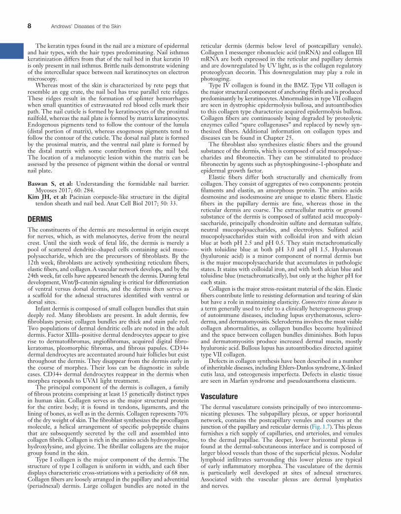
VasculatureThe dermal vasculature consists principally of two intercommu-nicating plexuses. The subpapillary plexus, or upper horizontal network, contains the postcapillary venules and courses at the junction of the papillary and reticular dermis (Fig. 1.7). This plexus furnishes a rich supply of capillaries, end arterioles, and venules to the dermal papillae. The deeper, lower horizontal plexus is found at the dermal-subcutaneous interface and is composed of larger blood vessels than those of the superficial plexus. Nodular lymphoid infiltrates surrounding this lower plexus are typical of early inflammatory morphea. The vasculature of the dermis is particularly well developed at sites of adnexal structures. Associated with the vascular plexus are dermal lymphatics and nerves.
The keratin types found in the nail are a mixture of epidermal and hair types, with the hair types predominating. Nail isthmus keratinization differs from that of the nail bed in that keratin 10 is only present in nail isthmus. Brittle nails demonstrate widening of the intercellular space between nail keratinocytes on electron microscopy.
Whereas most of the skin is characterized by rete pegs that resemble an egg crate, the nail bed has true parallel rete ridges. These ridges result in the formation of splinter hemorrhages when small quantities of extravasated red blood cells mark their path. The nail cuticle is formed by keratinocytes of the proximal nailfold, whereas the nail plate is formed by matrix keratinocytes. Endogenous pigments tend to follow the contour of the lunula (distal portion of matrix), whereas exogenous pigments tend to follow the contour of the cuticle. The dorsal nail plate is formed by the proximal matrix, and the ventral nail plate is formed by the distal matrix with some contribution from the nail bed. The location of a melanocytic lesion within the matrix can be assessed by the presence of pigment within the dorsal or ventral nail plate.
Baswan S, et al: Understanding the formidable nail barrier. Mycoses 2017; 60: 284.
Kim JH, et al: Pacinian corpuscle-like structure in the digital tendon sheath and nail bed. Anat Cell Biol 2017; 50: 33.
DERMISThe constituents of the dermis are mesodermal in origin except for nerves, which, as with melanocytes, derive from the neural crest. Until the sixth week of fetal life, the dermis is merely a pool of scattered dendritic-shaped cells containing acid muco-polysaccharide, which are the precursors of fibroblasts. By the 12th week, fibroblasts are actively synthesizing reticulum fibers, elastic fibers, and collagen. A vascular network develops, and by the 24th week, fat cells have appeared beneath the dermis. During fetal development, Wnt/β-catenin signaling is critical for differentiation of ventral versus dorsal dermis, and the dermis then serves as a scaffold for the adnexal structures identified with ventral or dorsal sites.
Infant dermis is composed of small collagen bundles that stain deeply red. Many fibroblasts are present. In adult dermis, few fibroblasts persist; collagen bundles are thick and stain pale red. Two populations of dermal dendritic cells are noted in the adult dermis. Factor XIIIa–positive dermal dendrocytes appear to give rise to dermatofibromas, angiofibromas, acquired digital fibro-keratomas, pleomorphic fibromas, and fibrous papules. CD34+ dermal dendrocytes are accentuated around hair follicles but exist throughout the dermis. They disappear from the dermis early in the course of morphea. Their loss can be diagnostic in subtle cases. CD34+ dermal dendrocytes reappear in the dermis when morphea responds to UVA1 light treatment.
The principal component of the dermis is collagen, a family of fibrous proteins comprising at least 15 genetically distinct types in human skin. Collagen serves as the major structural protein for the entire body; it is found in tendons, ligaments, and the lining of bones, as well as in the dermis. Collagen represents 70% of the dry weight of skin. The fibroblast synthesizes the procollagen molecule, a helical arrangement of specific polypeptide chains that are subsequently secreted by the cell and assembled into collagen fibrils. Collagen is rich in the amino acids hydroxyproline, hydroxylysine, and glycine. The fibrillar collagens are the major group found in the skin.
Type I collagen is the major component of the dermis. The structure of type I collagen is uniform in width, and each fiber displays characteristic cross-striations with a periodicity of 68 nm. Collagen fibers are loosely arranged in the papillary and adventitial (periadnexal) dermis. Large collagen bundles are noted in the
1
CHAPTER 1 Skin: Basic Structure and Function 9
unmyelinated nerve fibers that terminate in the papillary dermis and around hair follicles. Impulses pass to the central nervous system by way of the dorsal root ganglia. Histamine-evoked itch is transmitted by slow-conducting unmyelinated C-polymodal neurons. Signal transduction differs for sensations of heat and cold and in peripheral nerve axons.
Postganglionic adrenergic fibers of the autonomic nervous system regulate vasoconstriction, apocrine gland secretions, and contraction of arrector pili muscles of hair follicles. Cholinergic fibers mediate eccrine sweat secretion.
Mast CellsMast cells play an important role in the normal immune response, as well as immediate-type sensitivity, contact allergy, and fibrosis. Measuring 6–12 microns in diameter, with ample amphophilic cytoplasm and a small round central nucleus, normal mast cells resemble fried eggs in histologic sections. In telangiectasia macularis eruptiva perstans (TMEP mastocytosis), they are spindle shaped and hyperchromatic, resembling large, dark fibroblasts. Mast cells are distinguished by containing up to 1000 granules, each measuring 0.6–0.7 micron in diameter. Coarse particulate granules, crystalline granules, and granules containing scrolls may be seen. On the cell’s surface are 100,000–500,000 glycoprotein receptor sites for immunoglobulin E (IgE). There is heterogeneity to mast cells with type I, or connective tissue mast cells found in the dermis and submucosa, and type II, or mucosal mast cells found in the bowel and respiratory tract mucosa.
Mast cell granules stain metachromatically with toluidine blue and methylene blue (in Giemsa stain) because of their high content of heparin. They also contain histamine, neutrophil chemotactic factor, eosinophil chemotactic factor of anaphylaxis, tryptase, kininogenase, and β-glucosaminidase. Slow-reacting substance of anaphylaxis (leukotrienes C4 and D4), leukotriene B4, platelet-activating factor, and prostaglandin D2 are formed only after IgE-mediated release of granules. Mast cells stain reliably with the Leder ASD–chloracetase esterase stain. Because this stain does not rely on the presence of mast cell granules, it is particularly useful in situations when mast cells have degranulated. In forensic medicine, fluorescent labeling of mast cells with antibodies to the mast cell enzymes chymase and tryptase is useful in determining the timing of skin lesions in regard to death. Lesions sustained while living show an initial increase and then a decline in mast cells. Lesions sustained postmortem demonstrate few mast cells.
Cutaneous mast cells respond to environmental changes. Dry environments result in an increase in mast cell number and cutaneous histamine content. In mastocytosis, mast cells accumulate in skin because of abnormal proliferation, migration, and failure of apoptosis. The terminal deoxynucleotidyl transferase–mediated deoxyuridine triphosphate–biotin nick end labeling (TUNEL) method is used to assess apoptosis and demonstrates decreased staining in mastocytomas. Proliferation usually is only moderately enhanced.
SUBCUTANEOUS TISSUE (FAT)Beneath the dermis lies the panniculus, with lobules of fat cells or lipocytes separated by fibrous septa composed of collagen and large blood vessels (Fig. 1.8). The collagen in the septa is continuous with the collagen in the dermis. Just as the epidermis and dermis vary in thickness according to skin site, so does the subcutane-ous tissue. The panniculus provides buoyancy and functions as a repository of energy and an endocrine organ. It is an important site of hormone conversion, such as that of androstenedione into estrone by aromatase. Leptin, a hormone produced in lipocytes, regulates body weight through the hypothalamus and influences how we react to flavors in food. Various substances can affect lipid accumulation within lipocytes. Obestatin is a polypeptide that
MusclesSmooth muscle occurs in the skin as arrectores pilorum (erectors of the hairs), as the tunica dartos (or dartos) of the scrotum, and in the areolas around the nipples. The arrectores pilorum are attached to the hair follicles below the sebaceous glands and, in contracting, pull the hair follicle upward, producing gooseflesh. The presence of scattered smooth muscle throughout the dermis is typical of anogenital skin.
Smooth muscle also comprises the muscularis of dermal and subcutaneous blood vessels. The muscularis of veins is composed of small bundles of smooth muscle that crisscross at right angles. Arterial smooth muscle forms a concentric, wreathlike ring. Specialized aggregates of smooth muscle cells (glomus bodies) are found between arterioles and venules and are especially prominent on the digits and at the lateral margins of the palms and soles. Glomus bodies serve to shunt blood and regulate temperature. Most smooth muscle expresses desmin intermediate filaments, but vascular smooth muscle instead expresses vimentin. Smooth muscle actin is consistently expressed by all types of smooth muscle.
Striated (voluntary) muscle occurs in the skin of the neck as the platysma muscle and in the skin of the face as the muscles of expression. This complex network of striated muscle, fascia, and aponeuroses is known as the superficial muscular aponeurotic system (SMAS).
NervesIn the dermis, nerve bundles are found together with arterioles and venules as part of the neurovascular bundle. In the deep dermis, nerves travel parallel to the surface, and the presence of long, sausage-like granulomas following this path is an important clue to the diagnosis of Hansen disease.
Touch and pressure are mediated by Meissner corpuscles found in the dermal papillae, particularly on the digits, palms, and soles, and by Vater-Pacini corpuscles located in the deeper portion of the dermis of weight-bearing surfaces and genitalia. Mucocutane-ous end organs are found in the papillary dermis of modified hairless skin at the mucocutaneous junctions: the glans, prepuce, clitoris, labia minora, perianal region, and vermilion border of the lips. Temperature, pain, and itch sensation are transmitted by
Fig. 1.7 Below the epidermis, the papillary dermis is composed of fine, nonbundled collagen. Capillaries are present within the papillary dermis, and the postcapillary venule sits at the junction of the papillary and reticular dermis. H&E × 40.

10 Andrews’ Diseases of the Skin
Certain inflammatory dermatoses, known as the panniculitides, principally affect this level of the skin, producing subcutaneous nodules. The pattern of the inflammation, specifically whether it primarily affects the septa or the fat lobules, serves to distinguish various conditions that may be clinically similar.
Abraham SN, et al: Mast cell-orchestrated immunity to pathogens. Nat Rev Immunol 2010; 10: 440.
Kwon SB, et al: Phytosphingosine-1-phosphate and epidermal growth factor synergistically restore extracellular matrix in human dermal fibroblasts in vitro and in vivo. Int J Mol Med 2017; 39: 741.
Mikesh LM, et al: Proteomic anatomy of human skin. J Proteomics 2013; 84: 190.
Purohit T, et al: Smad3-dependent CCN2 mediates fibronectin expression in human skin dermal fibroblasts. PLoS One 2017; 12: e0173191.
Bonus images for this chapter can be found online at expertconsult.inkling.com
eFig. 1.1 Cross section of anagen bulb demonstrating pigment within matrix.
eFig. 1.2 Cross section of isthmus of anagen follicle demonstrating glycogenated outer root sheath and keratinized inner root sheath.
eFig. 1.3 Catagen hair with many apoptotic keratinocytes within the outer root sheath.
eFig. 1.4 Vertical section of telogen hair demonstrating “flame-thrower” appearance of club hair.
reduces feed intake and weight gain in rodents. (−)Ternatin, a highly N-methylated cyclic heptapeptide that inhibits fat accumulation, produced by the mushroom Coriolus versicolor, has similar effects in mice. Study of these molecules provides insight into the molecular basis of weight gain and obesity. Abnormal fat distribution and insulin resistance are seen in Cushing syndrome and as a result of antiretroviral therapy. In obese children and adolescents developing diabetes, severe peripheral insulin resistance is associated with intramyocellular and intraabdominal lipocyte lipid accumulation.
Fig. 1.8 The lobules of the subcutaneous fat are separated by fibrous septae. H&E × 40.

1
CHAPTER 1 Skin: Basic Structure and Function 10.e1
eFig. 1.1 Cross section of anagen bulb demonstrating pigment within matrix.
eFig. 1.2 Cross section of isthmus of anagen follicle demonstrating glycogenated outer root sheath and keratinized inner root sheath.
eFig. 1.3 Catagen hair with many apoptotic keratinocytes within the outer root sheath.
eFig. 1.4 Vertical section of telogen hair demonstrating “flamethrower” appearance of club hair. Additional eFigures are available in the electronic version.