1 seaweed thalli and cells - cambridge university...
TRANSCRIPT

1
Seaweed thalli and cells
1.1 Introduction: the algae and theirenvironments
1.1.1 Seaweeds
The term “seaweed” traditionally includes only macro-
scopic, multicellular marine red, green, and brown
algae. However, each of these groups has microscopic,
if not unicellular, representatives. All seaweeds at some
stage in their life cycles are unicellular, as spores or
gametes and zygotes, and may be temporarily plank-
tonic (Amsler and Searles 1980; Maximova and Sazhin
2010). Some remain small, forming sparse but product-
ive turfs on coral reefs (Hackney et al. 1989) while
others, such as the “kelps” of temperate reefs, can form
extensive underwater forests (Graham et al. 2007a).
Siphonous algae such as Codium, Caulerpa and
Bryopsis that form large thalli are, in fact, acellular.
The prokaryotic Cyanobacteria have occasionally been
acknowledged in “seaweed” floras (e.g. Setchell and
Gardner 1919; Littler and Littler 2011a). They are
widespread on temperate rocky and sandy shores
(Whitton and Potts 1982) and are particularly important
in the tropics, where large macroscopic tufts of Oscilla-
toriaceae and smaller but abundant nitrogen-fixing
Nostocaceae are major components of the reef flora
(Littler and Littler 2011a, b; Charpy et al. 2012). Benthic
diatoms also form large and sometimes abundant tube-
dwelling colonies that resemble seaweeds (Lobban
1989). An ancient lineage of (mostly) deep-water green
algae, the Palmophyllales, that includes Verdigellas and
Palmophyllum, have a palmelloid organization with
complex thalli built from an amorphous matrix
with a nearly uniform distribution of spherical cells
(Womersley 1971; Zechman et al. 2010). On a smaller
scale are the colonial filaments of some simple red
algae, such as Stylonema (previously Goniotrichum).
A “seaweed” is therefore problematic to precisely
define: here “seaweed” refers to algae from the red,
green, and brown lineages that, at some stage of their
life cycle, form multicellular or siphonous macrothalli.
In this book we shall consider macroscopic and micro-
scopic marine benthic environments and how sea-
weeds respond to those environments.
The algae are evolutionarily diverse, but are related
to one another through the endosymbiotic events that
gave rise to plastids. The traditional classification of
seaweeds as “red”, “green”, and “brown” is still fitting,
but our understanding of how these groupings arose
and their relatedness to each other and other eukary-
otes has been transformed over the past 20 years as
our understanding of endosymbiosis has grown (e.g.
Walker et al. 2011). The evolutionary origin of the algae
continues to be the subject of considerable research
effort and debate (e.g. Brodie and Lewis 2007; Archi-
bald 2009; Keeling 2010; Yoon et al. 2010; Burki et al.
2012; Collén et al. 2013). The taxonomic position of a
species can be viewed as a “working hypothesis”, and
as such is subject to change as new information arises
(Cocquyt et al. 2010). Unraveling algal evolution is
complex because, in addition to the multiple endo-
symbiotic events, there are other complicating events
such as horizontal (lateral) gene transfer (HGT; see
Brodie and Lewis 2007). Knowledge of the relatedness
of the different seaweed groups, and their relations to
1
www.cambridge.org© in this web service Cambridge University Press
Cambridge University Press978-0-521-14595-4 - Seaweed Ecology and Physiology: Second EditionCatriona L. Hurd, Paul J. Harrison, Kai Bischof and Christopher S. LobbanExcerptMore information
Primary Endosymbiosis
Primary Endosymbiosis
Secondary Endosymbiosis
Secondary Endosymbiosis
Secondary Endosymbiosis
Serial Secondary Endosymbiosis
(Green Alga)
Tertiary Endosymbiosis(Diatom)
Stramenopiles
Ciliates
Dinophysis
Lepididinium
Euglenids
Chlorarachniophytes
Paulinella
DinoflagellatesApicomplexa
Green Algae
Durinskia
Karlodinium
Red Algae
Glaucophytes
Tertiary Endosymbiosis(Cryptomonad)
Tertiary Endosymbiosis(Haptophyte)
Haptophytes
Cryptomonads
?
Land Plants
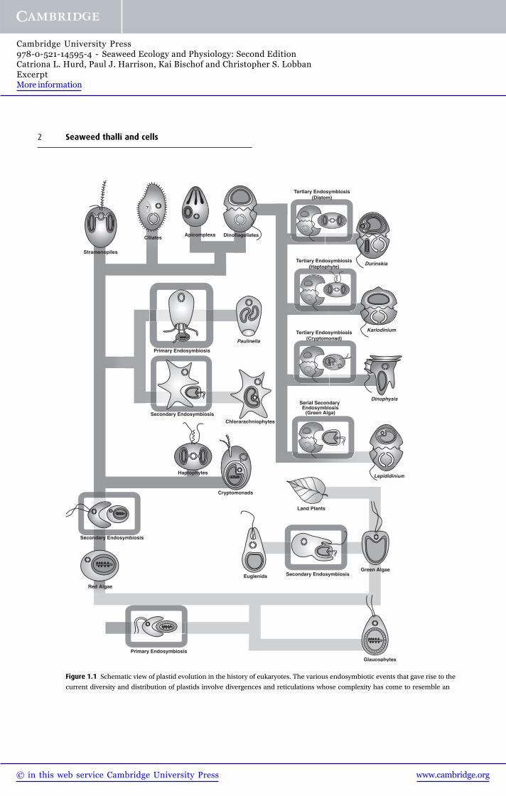
Figure 1.1 Schematic view of plastid evolution in the history of eukaryotes. The various endosymbiotic events that gave rise to the
current diversity and distribution of plastids involve divergences and reticulations whose complexity has come to resemble an
2 Seaweed thalli and cells
www.cambridge.org© in this web service Cambridge University Press
Cambridge University Press978-0-521-14595-4 - Seaweed Ecology and Physiology: Second EditionCatriona L. Hurd, Paul J. Harrison, Kai Bischof and Christopher S. LobbanExcerptMore information

other eukaryotes, is helpful in predicting aspects of
their physiology and ecology.
There are several hypotheses on how algal plastids
have arisen. A leading hypothesis is that a primary
endosymbiotic event (~1.5 billion years ago), in which
a free-living cyanobacterium was engulfed and incorp-
orated within a heterotrophic eukaryote, gave rise to
three major lineages: (1) the glaucophytes, (2) the
green lineage in which the green algae are ancestral
to the terrestrial plants, and (3) the red lineage which
includes the red seaweeds (Yoon et al. 2004; Keeling
2010; Fig. 1.1). But there may have been more than
one primary event, and the glaucophytes could have
arisen separately from the green and red lineages (see
Graham et al. 2009). At least three secondary endo-
symbiotic events (eukaryote þ eukaryote) have
occurred. It is fairly certain that two separate second-
ary events involving unicellular green algae gave rise to
the euglenoids and chlorarachniophytes (reviewed by
Keeling 2009, 2010; Fig. 1.1). Less clear, however, are
the secondary endosymbiotic event(s) involving uni-
cellular red algae (Burki et al. 2012). The chromalveo-
late hypothesis proposed by T. Cavalier-Smith (1999)
suggests that a single secondary endosymbiotic event
involving a red alga gave rise to six lineages (Fig. 1.1):
ciliates, dinoflagellates, apicomplexa, haptophytes,
cryptomonads, and the stramenopiles (heterokonts),
with the first three belonging to the Alveolata. The
chromalveolate hypothesis is “highly contentious”
but considered by Keeling (2009) and others as the
“hypothesis to beat”. At the time of writing (2013),
the consensus is that the stramenopiles and Alveolata
group with Rhizaria, forming the “SAR” clade; the
haptophytes form a closely related sister group to the
SAR clade, and the position of the cryptomonads is
equivocal (Walker et al. 2011; Burki et al. 2012). Within
the stramenopiles, the unicellular diatoms share a
common ancestor with the multicellular brown sea-
weeds (Phaeophyceae; Patterson 1989a; Andersen
2004). However, phylogenies based on carbon storage
and cell wall polysaccharides suggest that the strame-
nopiles arose separately from the Alveolates, and that a
related, but distinct, red algal plastid was incorporated
into an ancestral stramenopile in a second endosym-
biotic event (Michel et al. 2010a, b). The dinoflagellates
arose from tertiary or serial secondary endosymbioses
(Fig. 1.1). As new information arises, and new molecu-
lar and bioinformatic techniques are added to the
existing repertoire, hypotheses on eukaryotic evolution
and speciation will continue to develop.
Ocean vegetation is dominated by the algae. No
mosses, ferns, or gymnosperms are found in the
oceans, and only a few angiosperms (the seagrasses)
occur in marine habitats. That there are relatively few
marine angiosperms may reflect the problems of adap-
tion to the sea, including ion regulation and pollin-
ation (Ackerman 1998). The water column is chiefly
the domain of the phytoplankton, but populations of
floating seaweeds that have been detached from the
substratum are common and provide an important
mechanism of dispersal (sec. 3.3.7). Intertidal rocky
shores are abundantly covered with a macrovegetation
Caption for Figure 1.1 (cont.) electronic circuit diagram. Endosymbiosis events are boxed, and the lines are shaded to distinguish
lineages with no plastid (dark gray), plastids from the green algal lineage (light gray) or the red algal lineage (mid-gray). At the
bottom is the single primary endosymbiosis leading to three lineages (glaucophytes, red algae, and green algae). On the lower
right, a discrete secondary endosymbiotic event within the euglenids led to their plastid. On the lower left, a red alga was taken up
in the ancestor of chromalveolates. From this ancestor, haptophytes and cryptomonads (as well as their non-photosynthetic
relatives such as katablepharids and telonemids) first diverged. After the divergence of the rhizarian lineage, the plastid appears to
have been lost, but in two subgroups of Rhizaria, photosynthesis was regained: in the chlorarachniophytes by secondary
endosymbiosis with a green alga, and in Paulinella by taking up a cyanobacterium (many other rhizarian lineages remain non-
photosynthetic). At the top left, the stramenopiles diverged from alveolates, where plastids were lost in ciliates and predominantly
became non-photosynthetic in the apicomplexan lineage. At the top right, four different events of plastid replacement are
shown in dinoflagellates, involving a diatom, haptophyte, cryptomonad (three cases of tertiary endosymbiosis) and green alga
(a serial secondary endosymbiosis). Most of the lineages shown have many members or relatives that are non-photosynthetic, but
these have not all been shown for the sake of clarity. (From Keeling, 2010, reproduced with permission.)
1.1 Introduction: the algae and their environments 3
www.cambridge.org© in this web service Cambridge University Press
Cambridge University Press978-0-521-14595-4 - Seaweed Ecology and Physiology: Second EditionCatriona L. Hurd, Paul J. Harrison, Kai Bischof and Christopher S. LobbanExcerptMore information

that is almost exclusively seaweeds, although in west-
ern North America surf grass (Phyllospadix spp.) is an
exception. Seaweed surfaces themselves are colonized
by benthic microalgae and bacteria, with which they
may have intimate ecological relationships, and sea-
weed microstages grow on and within larger seaweeds.
Muddy and sandy areas have fewer seaweeds, because
most species cannot anchor there, though some sipho-
nous greens (e.g. some species of Halimeda, Caulerpa,
and Udotea) produce penetrating, root-like holdfasts
that also serve in nutrient uptake (Littler et al. 1988). In
such areas, seagrasses become the dominant vegeta-
tion, particularly in tropical and subtropical areas (Lar-
kum et al. 2006). There is also a paucity of freshwater
macroalgae. Freshwater red and brown algae are rep-
resented by relatively few genera and species, and
Ulvophyceae are also scarce with only a few genera
(e.g. Cladophora) having penetrated fresh waters
(Wehr and Sheath 2003).
Most seaweeds are multicellular most of the time.
What does this imply for physiological ecology?
Multicellularity confers the advantage of allowing
extensive development in the third dimension of the
water column. Such development can be achieved in
other ways, however. Siphonous green algae form
large multinucleate thalli that are at least technically
single cells (acellular rather than unicellular), sup-
ported by turgor pressure (Valonia), ingrowths of the
rhizome wall (trabeculae) in Caulerpa, or interweaving
of numerous narrow siphons (Codium, Avrainvillea)
(Fig. 1.2). Colonial diatoms, both tube-dwelling and
chain-forming, also build three-dimensional struc-
tures, as do zooxanthellae (dinoflagellates) in associ-
ation with corals. Multicellular algae often grow
vertically away from the substratum; this habit brings
them closer to the light, enables them to grow large
without extreme competition for space, and allows
them to harvest nutrients from a greater volume of
10
5
8
76
5
4
3
21
4321
15
cm
Figure 1.2 Thallus morphology and construction in siphonous green algae. Thalli drawn to scale; insets (not to scale) show
principles of construction: (1) Caulerpa cactoides: network of trabeculae. (2) Avrainvillea gardineri: tightly woven felt of filaments.
(3) Chlorodesmis sp.: bush of dichotomously branched siphons, constricted at the bases of the branches (inset). (4) Penicillus
capitus: calcified siphons form a multiaxial pseudotissue in the stem (inset), but separate to form bushy head. (5) Halimeda tuna:
segmented, calcified thallus of woven medulla and cortical utricles (inset). (6) Halicystis stage of Derbesia, a single ovoid cell
(shown at gametogenesis). (7) Bryopsis plumosa gametophyte: pinnately branched free siphons. (8) Codium fragile: interwoven
uncalcified siphons form multiaxial branches. (From Menzel 1988, with permission of Springer-Verlag, Berlin.)
4 Seaweed thalli and cells
www.cambridge.org© in this web service Cambridge University Press
Cambridge University Press978-0-521-14595-4 - Seaweed Ecology and Physiology: Second EditionCatriona L. Hurd, Paul J. Harrison, Kai Bischof and Christopher S. LobbanExcerptMore information

water. On the other hand, there are creeping filament-
ous algae, even endophytic and endolithic filaments
(e.g. Entocladia), as well as crustose algae such as
Ralfsia, and Porolithon, that do not grow up into the
water column. Support tissue usually is not necessary
for this upward growth, because most small seaweeds
are slightly buoyant, and the water provides support.
Support tissue is metabolically expensive, however
strength and resilience are required to withstand water
motion. Some of the larger seaweeds (e.g. Pterygo-
phora) have stiff, massive stipes, but others (e.g. Hor-
mosira) employ flotation to keep them upright. Many
of the kelps and fucoids have special gas-filled struc-
tures, pneumatocysts (Dromgoole 1990; Raven 1996),
whereas in other seaweeds (e.g. erect species of
Codium) gas trapped among the filaments achieves
the same effect (Dromgoole 1982).
A second important feature of multicellularity is that
it allows division of labor between tissues; such div-
ision is developed to various degrees in seaweeds.
Nutrient (and water) uptake and photosynthesis take
place over virtually the entire surface of the seaweed
thallus, in contrast to vascular land plants. Differenti-
ation and specialization among the vegetative cells of
algal thalli range from virtually nil (as in Ulothrix,
where all cells except the rhizoids serve both vegetative
and reproductive functions), through to Porphyra
[many species of this genus are now treated in other
genera, with most being in Pyropia; Sutherland et al.
2011; see sec. 10.2] and Ulva whose blades are mor-
phologically simple but are differentiated into regions
with distinct physiologies (e.g. Hong et al. 1995; Han
et al. 2003), to the highly differentiated photosynthetic,
storage, and translocation tissues in a variety of organs,
including stipe, blades, and pneumatocysts, that occur
in fucoids and kelps (Graham et al. 2009). Of course,
no seaweed shows the degrees of differentiation seen
in vascular plants. Even in vascular plants, the cells are
biochemically more general than animal cells: the
organs of vascular plants (stems, leaves, roots, flowers)
all contain much the same mix of cells, whereas
animal organs each contain only a few specialized cell
types. The low diversity of cells in a seaweed thallus
means that each cell is physiologically and biochemi-
cally even more general than vascular plant cells.
The evolution of multicellularity entails the co-
ordinated growth of cells, which, in turn, requires
cell-to-cell communication. The detection of genes
coding for receptor kinases (signaling molecules
that are found in all multicellular eukaryotes) in
Ectocarpus, and their absence in the related unicellular
diatoms, suggests that these molecules were a pre-
requisite for multicellularity. Another pre-requisite is
cell–cell adhesion via a sticky extracellular matrix.
Integrin-related proteins that have a key role in cell
adhesion in animals are also present in Ectocarpus, but
not in diatoms (Cock et al. 2010a). In the red seaweeds,
pit plugs are considered a vital step in the evolution
of multicellularity, by providing structural integrity
within the otherwise loosely packaged cells of pseudo-
parenchymatous construction (Graham et al. 2009,
p. 319; Gantt et al. 2010).
1.1.2 Environmental-factor interactions
Benthic algae interact with other marine organisms,
and all interact with their physico-chemical environ-
ment. As a rule, they live attached to the seabed
between the top of the intertidal zone and the max-
imum depth to which adequate light for growth can
penetrate. Among the major environmental (abiotic)
factors affecting seaweeds are light, temperature, sal-
inity, water motion, and nutrient availability. Among
the biological (biotic) interactions are relations
between seaweeds and their epiphytic bacteria, fungi,
algae, and sessile animals; interactions between herbi-
vores and seaweeds (both macroalgae and epiphytes);
and the impact of predators, including humans. Each
propagule contains the genetic information that will
allow the maturing seaweed to develop a phenotype
that is suited to its environment: in fact, there can be a
high degree of phenotypic plasticity even within a
genetically uniform population grown under the same
environmental conditions (Fig. 1.3). Individual pat-
terns of growth, morphology, and reproduction are
overall effects of all these factors combined.
An organism’s physico-chemical environment, con-
sisting of all the external abiotic factors that influence
the organism, is very complex and constantly varying.
In order for us to discuss or study it, we need to reduce it
1.1 Introduction: the algae and their environments 5
www.cambridge.org© in this web service Cambridge University Press
Cambridge University Press978-0-521-14595-4 - Seaweed Ecology and Physiology: Second EditionCatriona L. Hurd, Paul J. Harrison, Kai Bischof and Christopher S. LobbanExcerptMore information

to smaller parts, to think about one variable at a time.
And yet, each of the environmental “factors” that we
might consider – temperature, salinity, light, and so
forth – is really a composite of many variables, and they
tend to interact. Most importantly, the organization of
life is now best understood as constitutive hierarchies
(Mayr 1982, p. 65), in which at each new level or system
there are emergent properties that are not predictable
from study of the component parts. This is most evident
in comparing the properties of individuals (say,
humans) to the properties of the next level of compon-
ents (organ systems, e.g. nervous system), but it also
works upward from the individual through populations,
communities, ecosystems, and the biosphere. It has
major implications when we attempt to predict com-
munity or ecosystem properties from studies at the
species (population) level, as we usually must. The
following paragraphs are intended to paint the big
picture, before we go on to study it pixel by pixel.
Factor interactions can be grouped into four cat-
egories: (1) multifaceted factors, (2) interactions
between environmental variables, (3) interactions
between environmental variables and biological
factors, and (4) sequential effects.
1. Many environmental factors have several compon-
ents that do not necessarily change together (i.e.
multifaceted factors). Light quality and quantity,
which are important in photosynthetic responses
and metabolic patterns, both change with depth,
but the changes depend on turbidity and the nature
of the particles. In submarine caves, light quantity
diminishes with little change in quality. Natural
light has the additional important component of
day length, which influences reproductive states.
Salinity is another complex factor, of which the
two chief components are the osmotic potential of
the water and the ionic composition. Osmotic
potential affects water flow in and out of the cell,
turgor pressure, and growth, while the concentra-
tions of Ca2þ and HCO3- affect membrane integrity
and photosynthesis, respectively. Hydrodynamic
forces affect thallus survival and spore settlement
on wave-swept shores, and water motion also has
important effects on the boundary layers over sea-
weed surfaces and thus on nutrient uptake and gas
exchange. Nutrients must be considered not simply
in their absolute concentrations but also in the
amounts present in biologically available forms;
concentrations of trace metals may create toxicity
problems, particularly in polluted areas. Pollution,
as a factor, may include not only the toxic effects of
component chemicals but also an increase in
Figure 1.3 Variation in cap morphologies of Acetabuaria
acetabulum, the progeny of which were raised in the same
experimental conditions. “Concave” included a minor variant
(“concave-bell”) in which the rim of a concave cap was
flattened. “Convex” are mirror images of concave. “Flat” caps
are usually perpendicular to the stalk. “Saddles” have two
opposing quadrants of the cap curved up and the other two
down. In “beaked”, one or both halves of the cap are
adpressed and parallel to the stalk. “Split” caps have rays that
are not all fused so that the cap is divided into halves, quarters
or sixths. In “lapped”, the rays adjacent to the two that had not
fused overlap each other. “Rose” and “medusas” are the most
convoluted cap shapes. “Undefined” are caps which combine
two or more of the above morphologies. (From Nishimura
and Mandoli 1992, reproduced with permission.)
6 Seaweed thalli and cells
www.cambridge.org© in this web service Cambridge University Press
Cambridge University Press978-0-521-14595-4 - Seaweed Ecology and Physiology: Second EditionCatriona L. Hurd, Paul J. Harrison, Kai Bischof and Christopher S. LobbanExcerptMore information

turbidity, hence a reduction in irradiance. Emersion
often involves desiccation, heating, or chilling,
removal of most nutrients (except CO2), and, fre-
quently, changes in the salinity of the water in the
surface film on the seaweeds and in the free space
between cells.
2. Interactions among environmental variables are the
rule rather than the exception. Bright light is often
associated with increased heating, particularly of
seaweeds exposed at low tide. Light, especially blue
light, regulates the activities of many enzymes,
including some involved in carbon fixation and
nitrogen metabolism. Temperature and salinity
affect the density of seawater, hence the mixing of
nutrient-rich bottom water with nutrient-depleted
surface water. Thermoclines can affect plankton
movements, including migration of the larvae of
epiphytic animals. Temperature also affects cellular
pH and hence some enzyme activities. The sea-
water carbonate system and especially the concen-
tration of free CO2 are greatly affected by pH,
salinity, and temperature, while the availability of
ammonium is pH-dependent, because at high pH
the ion escapes as free ammonia. Water motion can
affect turbidity and siltation as well as nutrient
availability. These are examples of one environ-
mental variable affecting another. There are also
examples of two environmental variables acting
synergistically on seaweed; for instance, the com-
bination of low salinity and high temperature can
be harmful at levels where each alone would be
tolerable. In some seaweeds, the combined effects
of temperature and photoperiod regulate develop-
ment and reproduction.
3. Interactions between physico-chemical and bio-
logical factors are also the rule rather than the
exception. The environment of a given seaweed
includes other organisms, as we have seen, with
which the seaweed interacts through intraspecific
and interspecific competition, predator–prey rela-
tionships, associations with parasites and patho-
gens, and basiphyte–epiphyte relationships. These
other organisms are also affected by the environ-
ment, as are their effects on other organisms. More-
over, other organisms may greatly modify the
physico-chemical environment of a given individ-
ual. Protection from strong irradiance and desicca-
tion by canopy seaweeds is important to the
survival of understory algae, including germlings
of the larger species. Organisms shade each other
(and sometimes themselves) and have large effects
on nutrient concentrations and water flow. Other
interactions stem from the way the biological par-
ameters, such as age, phenotype, and genotype,
affect a seaweed’s response to the abiotic environ-
ment, as well as the effects that organisms have on
the environment. The chief biological parameters
that condition a given seaweed’s response to its
environment are age, reproductive condition, nutri-
ent status (including stores of N, P, and C), and past
history. By “past history” is meant the effects of past
environmental conditions on seaweed develop-
ment. Genetic differentiation within populations
leads to different responses in seaweeds from dif-
ferent parts of a population. The seasons can also
affect certain physiological responses, aside from
those involved in life-history changes; these
responses include acclimation of temperature
optima and tolerance limits.
4. Finally, there are factor interactions through
sequential effects. Nitrogen limitation may cause
red algae to catabolize some of their phycobilipro-
teins, which will in turn reduce their light-harvesting
ability. In general, any factor that alters the growth,
form, or reproductive or physiological condition is
apt to change the responses of the seaweed to other
factors both currently and in the future. A good
example of a sequential effect, and also biotic–
abiotic interaction, was seen by Littler and Littler
(1987) following an unusual flash flood in southern
California. Intertidal urchins (Strongylocentrotus
purpuratus) were almost completely wiped out, but
the persistent macroalgae suffered little damage
from the freshwater. Subsequently, however, there
was a great increase in ephemeral algae (Ulva, Ecto-
carpaceae) because of the reduction in grazing pres-
sure. The complexity of the interactions of variables
in nature often confounds interpretation of the
effects even of “major” events, such as El Niño
warm-water periods (Paine 1986; sec. 7.3.7).
1.1 Introduction: the algae and their environments 7
www.cambridge.org© in this web service Cambridge University Press
Cambridge University Press978-0-521-14595-4 - Seaweed Ecology and Physiology: Second EditionCatriona L. Hurd, Paul J. Harrison, Kai Bischof and Christopher S. LobbanExcerptMore information

Testing the effects of the various factor interactions
described above requires a multifaceted approach that
includes quantitative field observations, field manipu-
lations and targeted laboratory experiments; for each
approach a rigorous experimental design is essential
so that the appropriate statistical analyses can be
applied to detect differences among experimental
treatments. In laboratory experiments, usually one
variable is tested at a time, and all other factors are
held constant, or at least equal in all treatments.
Experiments in which two (or occasionally three)
factors are varied are possible but the number of cul-
ture vessels required for independent replication of
treatments can be technically difficult to achieve espe-
cially with large seaweeds: it is important to avoid
pseudoreplication in both laboratory and field experi-
ments (Hurlbert 1984). It is also important to under-
stand how field manipulations can confound results,
and to include appropriate controls. For example,
Underwood (1980) criticized some field experiments
designed to determine the effects of grazer exclusion
because the fences and cages used to keep out grazers
also affected the watermotion over the rock surface and
provided some shade. Furthermore, field studies that
use correlation analyses to elucidate whether an envir-
onmental factor causes a specific biological pattern
(e.g. growth, onset of reproduction) can be misleading
because the key environmental factors that regulate
seaweed biological processes are themselves tightly
correlated, for example light, temperature, and nitrate
concentration. As Schiel and Foster (1986, p. 273)
explain “The existence of patterns and abundance of
species constitutes evidence that these physical factors
and biological interactions may affect the structure of
these communities. They do not at the same time,
however, demonstrate the importance or unimport-
ance of these factors in producing observed patterns.”
1.1.3 Laboratory culture versus fieldexperiments
Several considerations confound the interpretation of
field reality via laboratory studies. First, while labora-
tory studies provide much more controlled conditions
than are found in nature, they are limited in some
important ways and contain some implicit assump-
tions, such as the following: (1) High nutrient levels
common in lab experiments do not alter the seaweeds’
responses to the factor under study. (2) The reactions
of seaweeds to uniform conditions (including the
factor under study) are not different from their
responses to the factor(s) under fluctuating conditions.
To a certain extent these assumptions are valid. Cul-
ture media can be very rich in nutrients, to compen-
sate for lack of water movement and exchange, but it is
unlikely that this substitution can give precisely the
same results. Other culture conditions are also gener-
ally optimal, except for the variable under study, and
the results may not elucidate the behavior of seaweeds
in the field, which are subject to competition and often
suboptimal conditions (Neushul 1981). Another
important difference between laboratory and field is
that in culture, species usually are tested in isolation,
away from interspecific competition and grazing. Fur-
thermore, culture conditions are uniform (at least on a
large scale), whereas in nature there often are large
and unpredictable fluctuations in the environment
(e.g. Gorospec and Karl 2011). Microscale heterogen-
eity in culture conditions should not be overlooked
(Allen 1977; Norton and Fetter 1981). In the culture
flask, one cell may shade another, and cells form
nutrient-depleted zones around them, creating a
mosaic of nutrient concentrations through which cells
pass. In the field, scale also needs to be considered at
the large end – for instance, the amount of space
needed for a patch of a given alga to establish itself
(Schiel and Foster 1986). In essence, for both field and
laboratory experiments, informed decisions must be
made on the experimental conditions that are pro-
vided, and it is important to be aware that these con-
ditions will affect the outcome and interpretation of
the results.
Second, the timescale over which an experiment
is conducted affects the interpretation of the data
(Raven and Geider 2003). In short-term physiological
experiments (seconds to minutes), a single factor
can be varied (e.g. different levels of UV-B radiation)
and a response (e.g. the production of reactive oxygen
species) is measured. This physiological response is at
the level of regulation i.e. the up- or down-regulation
8 Seaweed thalli and cells
www.cambridge.org© in this web service Cambridge University Press
Cambridge University Press978-0-521-14595-4 - Seaweed Ecology and Physiology: Second EditionCatriona L. Hurd, Paul J. Harrison, Kai Bischof and Christopher S. LobbanExcerptMore information

of pre-existing enzymes, and reveals the physiological
potential of that organism to respond to an immediate
environmental change. In medium-term experiments
(hours to days) acclimation to new environmental
conditions may occur. Acclimation involves gene
expression, and the synthesis of new proteins such
as enzymes. Adaptation to particular environmental
factors occurs over a longer timescale (up to millennia)
and is a mechanism for speciation (sec. 7.1).
Third, when a single species occurs in widely differ-
ent latitudes or longitudes, its physiology and ecology
may be quite different. For many topics, only one study
or a few studies have been done, and a phenomenon
demonstrated in a particular alga under certain condi-
tions will not necessarily turn out to be the same in
other algae or under other conditions. In Australia, for
example, the kelp Ecklonia radiata dominates across
3000 km of coastline, from the southeast to southwest.
However, the morphology and ecology of Ecklonia on
the east coast is very different to Ecklonia on the west
and south coasts, with the result that different coastal
management plans are required for these different
regions (Connell and Irving 2009). Equally, very few
natural populations or communities have been studied
often enough to assess how much variability is present
from place to place (ecotypic variation). The kelp beds
of southern California are exceptional in that they have
been repeatedly analyzed by different people along the
coast since the 1960s (Steneck et al. 2002; Graham et al.
2007a). For Macrocystis, there is no typical kelp bed;
environmental parameters differ from one kelp bed to
another, and parameters such as specific growth rate
versus nitrogen supply vary among populations (Kopc-
zak et al. 1991).
In this first chapter we shall review the foundations
of seaweed construction, cell biology, molecular biol-
ogy and genetics on which any understanding of sea-
weed physiological ecology must rest. In Chapter 2, we
continue this review by tracing the development of
seaweed thalli from gametes and spores to reproduct-
ive individuals. In both these chapters, we build upon
the fundamental information on seaweed anatomy
and development that is described in algal text books,
particularly van den Hoek et al. (1995) and Graham
et al. (2009).
1.2 Seaweed morphology and anatomy
1.2.1 Thallus construction
Diversity of thallus construction in algae contrasts
strongly with uniformity in vascular plants. In the
latter, parenchymatous meristems (e.g. at the shoot
and root apices) produce tissue that differentiates in
a wide variety of shapes. For seaweeds, parenchymat-
ous construction is prevalent only in the brown orders.
For example, in kelps, fucoids, and Dictyotales, this
mode of construction has given rise to internal and
morphological complexity (Fig. 1.4). The larger sea-
weeds, especially Laminariales and Fucales, have
several different tissue and cell types, including photo-
synthetic epidermis, cortex, medulla, sieve tubes, and
mucilage ducts (Graham et al. 2009). The ontogeny of
the parenchyma in the Dictyotales (Fig. 1.4d–m) has
been followed in detail by Gaillard and L’Hardy-Halos
(1990), who cite many sources, and by Katsaros and
Galatis (1988). However, the great majority of sea-
weeds either are filamentous or are built up of united
or corticated filaments. Large and complex structures
can be built up this way, for example Codium ampli-
vesiculatum (previously C. magnum) can reach several
meters long (Dawson 1950). Cell division may take
place throughout the alga, or the meristematic region
may be localized. If localized, it is most commonly at
the apex, but may be at the base or somewhere in
between (intercalary).
A simple filament consists of an unbranched chain
of cells attached by their end walls and results from
cell division only in the plane perpendicular to the
axis of the filament. Unbranched filaments are uncom-
mon among seaweeds; examples are Ulothrix and
Chaetomorpha. Usually, some cell division takes place
parallel to the filament axis to produce branches (Cla-
dophora, Ectocarpus, Antithamnion; see Fig. 1.17).
Filaments consisting of a single row of cells (branched
or not) are called uniseriate. Pluriseriate filaments, i.e.
two or more rows of cells, are seen in genera such as
Blidingia, Bangia, and Sphacelaria (Fig. 1.4a; Graham
et al. 2009). Branches need not grow out free, but may
creep down the main filament, forming cortication, as
seen in Ceramium (Fig. 1.5a) and Ballia. In some of the
1.2 Seaweed morphology and anatomy 9
www.cambridge.org© in this web service Cambridge University Press
Cambridge University Press978-0-521-14595-4 - Seaweed Ecology and Physiology: Second EditionCatriona L. Hurd, Paul J. Harrison, Kai Bischof and Christopher S. LobbanExcerptMore information

A
Ax
Cp
CpCm
Cmf
CpCm
Co
A
Sa
(g)
(h)
(i)
(j)
(m)
50 mm
(l)
(d)
(k)
(c)
(b)
2
11
2
3r
r
3
4
2
1
a
t
s
s pi
i
i
a
a
3
4
br
4
4 7
7769 9
1
22
33
6
47
(a)
(f)
(e)Sa
efghi
m
lk
j
Figure 1.4 Parenchymatous development in seaweeds. (a) Sphacelaria plumula apex showing first transverse division (t), followed
by pairs of cells (i, s), of which s forms branches, but i does not. (b, c) Fucus vesiculosus germination showing successive cell
divisions (numbered) (divisions 5 and 8 in the plane of the page). (d–m) Dictyota: development of parenchyma; (d) long section
through adventive branch, showing locations of cross sections at each level (diagrammatic); (e–m) serial cross sections
to show sequence of periclinal divisions. Arrows indicate junction between original two pericentral cells (first shown in h).
For the sake of clarity, the proportions of the cells were changed; the adventive branch is actually half as long and twice as wide as
shown. A, apical cell; Sa, subapical cell; Ax, axial cell; Cp, pericentral cell, Cm, medullary cell; Co, cortical cell. (Parts a–c from
Fritsch 1945, based on classical literature; d–m from Gaillard and L’Hardy-Halos 1990, with permission of Blackwell Scientific
Publications.)
10 Seaweed thalli and cells
www.cambridge.org© in this web service Cambridge University Press
Cambridge University Press978-0-521-14595-4 - Seaweed Ecology and Physiology: Second EditionCatriona L. Hurd, Paul J. Harrison, Kai Bischof and Christopher S. LobbanExcerptMore information