1 running title: plant cuticle structures and - plant physiology
TRANSCRIPT

1
Running Title: Plant cuticle structures and functions
Plant Physiology Preview. Published on July 26, 2013, as DOI:10.1104/pp.113.222737
Copyright 2013 by the American Society of Plant Biologists
www.plantphysiol.orgon March 28, 2019 - Published by Downloaded from Copyright © 2013 American Society of Plant Biologists. All rights reserved.
2
The Formation and Function of Plant Cuticles
Trevor H. Yeats† and Jocelyn K. C. Rose*
Department of Plant Biology, Cornell University, Ithaca, NY 14853
† Present address: Energy Biosciences Institute, University of California, Berkeley, CA
94720
* Corresponding author, email [email protected]
www.plantphysiol.orgon March 28, 2019 - Published by Downloaded from Copyright © 2013 American Society of Plant Biologists. All rights reserved.
3
Abstract
The plant cuticle is an extracellular hydrophobic layer that covers the aerial
epidermis of all land plants, providing protection against desiccation and external
environmental stresses. The past decade has seen considerable progress in assembling
models for the biosynthesis of its two major components, the polymer cutin and cuticular
waxes. Most recently, two breakthroughs in the long-sought molecular bases of alkane
formation and polyester synthesis have allowed construction of nearly complete
biosynthetic pathways for both waxes and cutin. Concurrently, a complex regulatory
network controlling the synthesis of the cuticle is also beginning to emerge. It has also
become clear that the physiological role of the cuticle extends well beyond its primary
function as a transpiration barrier, playing important roles in processes ranging from
development to interactions with microbes. Here, we review recent progress in the
biochemistry and molecular biology of cuticle synthesis and function and highlight some
of the major questions that will drive future research in this field.
www.plantphysiol.orgon March 28, 2019 - Published by Downloaded from Copyright © 2013 American Society of Plant Biologists. All rights reserved.

4
Introduction
The first plant colonizers of land, approximately 450 million years ago in the mid-
Paleozoic era, faced a daunting set of challenges associated with their new terrestrial
environment, including desiccation, temperature extremes, gravity and increased
exposure to UV radiation (Waters, 2003; Leliaert et al., 2011). The transition from an
exclusively aquatic to a terrestrial life style would therefore have necessitated the
evolution of a toolbox of morphological and physiological features, some of which are
apparent through studies of the fossil record, or by examining extant plant lineages. For
example, the development of architecturally complex cell walls for biomechanical
support and structural protection, which typify modern land plants, can be traced back to
divergence and radiation within the Charophycean green algae, their immediate ancestors
(Sørensen et al., 2011). However, the most critical adaptive trait for survival during
terrestrialization would have been the ability to retain water in increasingly dehydrating
habitats. Consequently, the capacity to synthesize, deposit and maintain a hydrophobic
surface layer, or cuticle, over the surfaces of aerial organs was arguably one of the most
important innovations in the history of plant evolution. This idea is borne out by both
fossil evidence (Edwards, 1993) and the ubiquity of cuticles among all extant
embryophytes, from bryophytes (Budke et al., 2012) to angiosperms.
Armed with a protective skin, together with a range of adaptive strategies for
acquiring and conserving water, as well as for avoiding or tolerating water stress,
embryophytes now thrive in a wide range of desiccating environments (Ogburn and
Edwards, 2010; Aroca et al., 2012; Delaux et al., 212; Jones and Dolan, 2012; Obata and
Fernie, 2012; Gaff and Oliver, 2013). Accordingly, cuticles from a broad range of
species, and in various ecological and agricultural contexts have been studied from the
perspective of their role as the primary barrier to transpirational water loss. However, it is
now clear that cuticles play numerous other roles in plant development, physiology and
interactions with the abiotic environment and other organisms. Indeed, in recent years
there have been many instances of unexpected associations between the cuticle and
diverse aspects of plant biology. In parallel, the past decade has seen considerable
progress in understanding the biosynthesis of the major cuticle components and the
complex regulatory networks that control cuticle synthesis and assembly.
www.plantphysiol.orgon March 28, 2019 - Published by Downloaded from Copyright © 2013 American Society of Plant Biologists. All rights reserved.
5
This review summarizes recent progress in elucidating the biochemistry and
molecular biology of cuticle synthesis and function and highlights some of the
connections to other aspects of plant biology, including signaling, pathogen defense and
development. Given the broad scope and space limitation, not every aspect of cuticle
biosynthesis is covered in depth, and recent specialized reviews focusing on cuticle
biomechanical properties (Dominguez et al., 2011), defensive functions (Reina-Pinto and
Yephremov, 2009) and transport barrier properties (Burghardt and Riederer, 2006) may
be of further interest. In addition, key ongoing questions in the field are discussed and
potential future approaches to resolving those questions are suggested.
Cuticle Structure, Biosynthesis and Assembly
Plant cuticles are composite structures, composed of a covalently-linked
macromolecular scaffold of cutin and a variety of organic solvent soluble lipids that are
collectively termed waxes. Although the cuticle is usually considered independently from
the underlying polysaccharide cell wall of the epidermis, the two structures are physically
associated and have some overlapping functions. Indeed the cuticle can be considered a
specialized lipidic modification of the cell wall, just as lignification is a common
modification of plant secondary cell walls. The microscopic structure of the cuticle is
often divided into two domains based on histochemical staining and their presumed
chemical composition: a cutin rich domain with embedded polysaccharides, which is
referred to as the ‘cuticular layer’, while an overlying layer that is less abundant in
polysaccharides, but enriched in waxes, is referred to as the ‘cuticle proper’ (Figure 1A).
The waxes are either deposited within the cutin matrix (intracuticular wax) or accumulate
on its surface as epicuticular wax crystals, or films. These epicuticular waxes can confer
distinct macroscopic surface properties: epicuticular films are responsible for the glossy
appearance common to many leaves and fruits, while epicuticular wax crystals account
for the dull, glaucous appearance of broccoli (Brassica oleracea) leaves and Arabidopsis
(Arabidopsis thaliana) stems. Cuticle architectural organization can be discerned using a
number of microscopic techniques. Scanning electron microscopy (SEM) can reveal the
elaborate and diverse morphologies of epicuticular wax crystals (Figure 1B; Jeffree,
2006), while transmission electron microscopy (TEM) shows the distinct patterning of
www.plantphysiol.orgon March 28, 2019 - Published by Downloaded from Copyright © 2013 American Society of Plant Biologists. All rights reserved.

6
interior layers of the cuticle, although this approach does not allow the visualization of
wax structures (Figure 1C). Cuticles vary considerably in their architecture and,
depending on species and ontogeny, differ dramatically in thickness, from the nanometer
to micrometer scale (Jeffree, 2006). In the latter case, light microscopy and can be used to
elucidate the fine structures of the cuticle and epidermal cell wall (Figure 1D), while
histochemical staining coupled with confocal microscopy can further resolve three-
dimensional cuticle architecture (Buda et al., 2009).
Wax Biosynthesis
Wax composition can vary substantially with species, ontogeny and
environmental growth conditions (Jenks and Ashworth, 1999). In most cases, the
majority of compounds comprising the cuticular wax are derived from very long chain
fatty acids (VLCFA, C20-C34), including alkanes, aldehydes, primary and secondary
alcohols, ketones and esters (Table 1). In some species, various lipophilic secondary
metabolites such as pentacyclic triterpenoids, flavonoids and tocopherols can also be
substantial components (Jetter et al., 2006). There has been impressive progress in
revealing the molecular biology underlying VLCFA-derived wax biosynthesis and to this
end Arabidopsis has provided an excellent experimental model (Bernard and Joubes,
2013). In addition to its well-known advantages as a genetic system, the presence of stem
epicuticular wax crystals, which impart a glaucous appearance in the wild type, has
enabled an easy screen for wax deficient mutants. Such mutants, termed eceriferum (cer;
Koornneef et al., 1989), typically exhibit a glossy stem phenotype and it has primarily
been through molecular analyses of these and other wax mutants that an increasingly
complete pathway for acyl wax biosynthesis has been established.
Wax biosynthesis begins with de novo C16 or C18 fatty acid biosynthesis in the
plastid of epidermal cells (Figure 2). These long chain fatty acid compounds are
converted to CoA thioesters by a long chain acyl-CoA synthase (LACS) isozyme and are
ultimately transferred to the endoplasmic reticulum (ER). The mechanism of intracellular
trafficking of fatty acid from the chloroplast to the ER remains unknown, although
heterologous expression of Arabidopsis LACS1, LACS2 and LACS3 facilitates fatty acid
uptake in yeast, suggesting that this class of enzymes may play dual roles in fatty acid
www.plantphysiol.orgon March 28, 2019 - Published by Downloaded from Copyright © 2013 American Society of Plant Biologists. All rights reserved.
7
trafficking and activation (Pulsifer et al., 2012). For reference, Table 2 provides a list of
the corresponding genes, as well as others discussed in this review. The C16 acyl-CoA is
then a substrate for the fatty acid elongase (FAE) complex. Through successive addition
of two carbons per cycle derived from malonyl-CoA, the ultimate products of this
complex are VLCFAs. The complex consists of four core subunits: β-ketoacyl-CoA
synthase, β-ketoacyl-CoA reductase, β-hydroxyacyl-CoA dehydratase and enoyl-CoA
reductase. In Arabidopsis, 21 genes are predicted to encode β-ketoacyl-CoA synthase,
and for wax biosynthesis the most important gene, based on the mutant phenotype, is
CER6 (Fiebig et al., 2000). Genes encoding the remaining subunits of the FAE complex,
represented by KCR1, PAS2 and CER10, respectively, are less redundant and their
pleiotropic mutant phenotypes underscore the shared importance of the FAE in
generating VLCFA precursors for sphingolipid biosynthesis (Zheng et al., 2005; Bach et
al., 2008; Beaudoin et al., 2009). An additional family of proteins, comprised of CER2,
CER26 and CER26-like, appears to be required for elongation of fatty acids to lengths
greater than 28C (Haslam et al., 2012; Pascal et al., 2013). Curiously, these enzymes have
sequence homology to BAHD acyltransferases, but conserved catalytic amino acid
residues of this family of enzymes are dispensable for the elongation-promoting activity
of CER2 (Haslam et al., 2012). The elongation cycles can be terminated by a thioesterase
(THS) to form free VLCFAs, or the VLCFA-CoA esters can undergo further
modifications.
Primary alcohols can be produced from VLCFA-CoA by fatty acyl-coenzyme A
reductase, an enzyme encoded by CER4 in Arabidopsis (Rowland et al., 2006). Free
primary alcohols can occur in the wax mixture or they can be esterified to a fatty acid in
order to form wax esters. In this case, the alcohol is coupled to an acyl group derived
from fatty acyl-CoA. The Arabidopsis enzyme responsible for this is WSD1, an enzyme
of the wax synthase/diacyl glycerol acyltransferase family (Li et al., 2008).
A second branch of acyl wax biosynthesis leads to formation of aldehydes and
ultimately alkanes. Interestingly, in Arabidopsis, LACS1, which is also required for C16
cutin monomer biosynthesis, appears to have an additional specificity for C30 VLCFA
and is required for normal accumulation of downstream wax compounds (Lu et al.,
2009). This suggests that conversion of an intracellular pool of free VLCFA back to
www.plantphysiol.orgon March 28, 2019 - Published by Downloaded from Copyright © 2013 American Society of Plant Biologists. All rights reserved.

8
VLCFA-CoA is an important route to aldehyde and alkane biosynthesis, rather than
VLCFA-CoA directly derived from FAE. A long unresolved question in wax
biosynthesis is the enzymatic basis of alkane synthesis. Classical biochemistry, using
crude extracts from pea (Pisum sativum) indicated that the reaction likely occurs via
reduction of VLCFA-CoA to an aldehyde intermediate followed by decarbonylation,
yielding an alkane that is 1C shorter (Cheesbrough and Kolattukudy, 1984; Schneider-
Belhaddad and Kolattukudy, 2000). Although this enzyme was not purified and
identified, compelling evidence was recently obtained, through studies of Arabidopsis,
that a complex of CER1 and CER3 act together to catalyze the formation of alkanes from
VLCFA-CoA. It was shown by a split ubiquitin yeast two-hybrid assay and an
Arabidopsis split luciferase assay that CER1 interacts with CER3 as well as several
isoforms of cytochrome b5. Furthermore, heterologous expression of the combination of
CER1, CER3, a cytochrome b5 and LACS1 in yeast resulted in the formation of very
long chain alkanes (Bernard et al., 2012). This strongly suggests that a complex including
CER1 and CER3 with cytochrome b5 as an electron donor catalyzes the reduction and
decarbonylation of VLCFA-CoA in order to form cuticular alkanes. Aside from being a
major component of the wax mixture, alkanes can undergo further modification to form
secondary alcohols and ketones. In Arabidopsis, both of these oxidations are performed
by the cytochrome P450 enzyme mid-chain alkane hydroxylase (MAH1; Greer et al.,
2007).
Synthesis of Cutin Precursors
Cutin is typically composed of inter-esterified hydroxy fatty acids, with lesser
amounts of glycerol, phenylpropanoids and dicarboxylic acids (Kolattukudy, 2001).
Chemical processes that cleave ester bonds, such as saponification, readily release these
monomeric constituents, although in some species an additional lipidic polymer, referred
to as cutan, remains recalcitrant to such treatments. Cutan is rich in ether and C-C bonds,
but its structure is otherwise unknown and it appears to be restricted to relatively few
extant species (Gupta et al., 2006). The hydroxy fatty acids of cutin are typically ω-
hydroxy fatty acids, usually with one or two additional midchain hydroxyl groups or an
epoxy group (Figure 3A). Despite extensive surveys of the chemical composition of plant
www.plantphysiol.orgon March 28, 2019 - Published by Downloaded from Copyright © 2013 American Society of Plant Biologists. All rights reserved.

9
cutins in the 1970s and 1980s (Kolattukudy, 2001), the composition of Arabidopsis cutin
was not determined until relatively recently (Bonaventure et al., 2004; Franke et al.,
2005). It is important to note that in this important model species, the cutin of stems and
leaves is atypical in that its major component is a dicarboxylic acid (Figure 3A), implying
that the predominant structural motif must be a copolymer with an unknown polyhydroxy
compound; presumably glycerol (Pollard et al., 2008). However, despite the atypical
composition of its cutin, Arabidopsis has proven to be an important model for
deciphering the pathway of cutin biosynthesis, and more recently it was discovered that
the cutin of its floral organs is more typical, in that it is composed primarily of 10,16-
dihydroxyhexadecanoic acid (Li-Beisson et al., 2009).
While there is considerable diversity in the structure of cutin monomers, the
pathway for the biosynthesis of 10,16-dihydroxyhexadecanoic acid-based cutin is the
most complete, and the major themes of cutin biosynthesis are likely shared for other
cutin monomers. Here, we summarize this pathway based on recent molecular genetic
and biochemical studies using Arabidopsis and tomato (Solanum lycopersicum).
Intracellular acyltransferases and hydroxylases
Biosynthesis of cutin begins with de novo fatty acid synthesis in the plastid of
epidermal cells (Figure 2). The next three steps occur in the ER and consist of ω- and
midchain hydroxylation and synthesis of an acyl-CoA intermediate. The relative order of
these steps is not known, although it has been shown that the ω-hydroxylation precedes
the midchain hydroxylation and that the final product of these steps is most likely a
dihydroxyhexadecanoic acid-CoA ester (Li-Beisson et al., 2009). The ω-hydroxylase is
encoded by members of the CYP86 subfamily of cytochrome P450s (CYP86A4 in
Arabidopsis flowers; Li-Beisson et al., 2009), while the midchain hydroxylase is encoded
by the CYP77 subfamily (CYP77A6 in Arabidopsis flowers; Li-Beisson et al., 2009). The
acyltransferases that synthesize acyl-CoA are encoded by the long chain acyl-CoA
synthase (LACS) family, which consists of nine members in Arabidopsis, and both
LACS1 and LACS2 appear to be responsible for C16 cutin monomer biosynthesis (Lu et
al., 2009).
www.plantphysiol.orgon March 28, 2019 - Published by Downloaded from Copyright © 2013 American Society of Plant Biologists. All rights reserved.

10
An additional intracellular acyltransferase required for synthesis of cutin polyester
is a glycerol 3-phosphate acyltransferase (GPAT). Recently it was shown that plants
possess a unique subfamily of bifunctional GPATs encoding enzymes with both sn-2
specific glycerol-3-phosphate:acyl-CoA acyltransferase activity as well as phosphatase
activity, yielding a 2-monoacylglyceryl ester (Yang et al., 2010). In the case of
Arabidopsis floral cutin, this activity is encoded by GPAT6 (Li-Beisson et al., 2009).
Although the specific sequence of all intracellular biosynthetic steps will require
additional characterization of substrate specificity of each enzyme, biochemical
characterization of Arabidopsis bifunctional GPATs indicates that they have a strong
preference for ω-hydroxylated acyl-CoA, suggesting that hydroxylation precedes transfer
to glycerol (Yang et al., 2012). In any case, the ultimate product of the intracellular steps
of cutin biosynthesis is likely to be 2-monoacylglyceryl esters of cutin monomers. In the
case of 10,16-dihydroxyhexadecanoic acid-based cutin, this is 2-mono-(10,16)-
dihydroxyhexadecanoyl glycerol (2-MHG).
Transport of Cuticle Precursors
After synthesis of wax and cutin precursors, they are exported from the ER,
across the plasma membrane, through the polysaccharide cell wall and to the nascent
cuticular membrane. Most of these transport processes are poorly understood, although
trafficking of both wax and cutin-precursors across the plasma membrane has been
shown to depend on ATP-binding cassette (ABC) transporters. In Arabidopsis,
CER5/ABCG12 (Pighin et al., 2004) and ABCG11 (Bird et al., 2007) are required for wax
export. Both of these encode half transporters and, based on double mutant analysis and
bimolecular fluorescent complementation (BiFC) analyses, it has been suggested that an
ABCG11/ABCG12 heterodimer is required for wax secretion (McFarlane et al., 2010).
ABCG11 is also required for cutin accumulation and since it is also able to dimerize with
itself, it has been proposed that this homodimer is the functional complex responsible for
cutin export (McFarlane et al., 2010). Additionally, a third Arabidopsis half transporter,
ABCG13, was shown to be required for cutin deposition in flowers (Panikashvili et al.,
2011).
www.plantphysiol.orgon March 28, 2019 - Published by Downloaded from Copyright © 2013 American Society of Plant Biologists. All rights reserved.

11
More recently, full transporters required for cutin deposition were identified in
Arabidopsis (ABCG32; Bessire et al., 2011), as well as wild barley (Hordeum
spontaneum) and rice (Oryza sativa; Chen et al., 2011). Despite the clear genetic
evidence supporting a role for ABC transporters in cuticular lipid export, the substrate
specificity of these transporters has not yet been demonstrated in vitro. However, all the
ABC transporters that have been implicated in cuticle biosynthesis to date are members
of the ABCG subfamily, which has been associated with the transport of lipids and
hydrophobic compounds in other systems (Moitra et al., 2011). Moreover, in several
cases intracellular lipidic inclusions were observed in ABC transporter mutants, further
supporting their direct involvement in cuticular lipid export (Pighin et al., 2004; Bird et
al., 2007; Bessire et al., 2011).
Export of some wax compounds also appears to be facilitated by
glycosylphosphatidylinositol-anchored lipid-transfer proteins (LTPs), LTPG1 and
LTPG2, which are bound to the extracellular side of the plasma membrane (Debono et
al., 2009; Lee et al., 2009; Kim et al., 2012). These proteins represent a unique class of
LTPs, a family of small and typically soluble proteins that bind a variety of lipid
substrates in vitro (Yeats and Rose, 2008). A major remaining question is how
hydrophobic cuticle precursors are transported across the hydrophilic environment of the
polysaccharide cell wall to the cuticle. Apoplastic LTPs have been proposed to play a
role, although genetic or biochemical evidence for their involvement in transport is
generally lacking (Yeats and Rose, 2008). In the case of the dihydroxyacyl cutin
precursor 2-MHG, the glycerol moiety imparts sufficient polarity to allow aqueous
solubility at low millimolar concentrations (Yeats et al., 2012b). This suggests that lipid-
binding proteins or other factors are not necessary in order to facilitate the transport of
this major precursor of cutin biosynthesis. However, the solubility of glyceryl esters of
less polar cutin monomers has not been investigated, and they, along with waxes, may
require additional factors to increase their solubility in the apoplast.
Cutin Polymerization
The final step of cutin synthesis is incorporation of the hydroxyacyl monomer into
the polymer, but the molecular mechanism of cutin polymerization has been a
www.plantphysiol.orgon March 28, 2019 - Published by Downloaded from Copyright © 2013 American Society of Plant Biologists. All rights reserved.

12
longstanding enigma. Recent progress in this area was achieved by studying the tomato
mutant cutin deficient 1 (cd1) and transgenic tomato plants in which CD1 expression was
suppressed using an RNAi strategy (Girard et al., 2012; Yeats et al., 2012b). The cd1
mutant exhibits a severe reduction in the amount of polymerized cutin in the fruit cuticle
(Isaacson et al., 2009), although chemical analysis indicated that, unlike wild type fruit,
those of the mutant accumulate non-polymerized 2-MHG (Yeats et al., 2012b). Cloning
of the mutated gene revealed that it encodes a protein of the GDSL-motif
lipase/hydrolase (GDSL) family, which localizes to the developing cuticle (Girard et al.,
2012; Yeats et al., 2012b). Despite its similarity to lipolytic enzymes, the recombinant
protein acts as an acyltransferase in vitro, forming polyester oligomers from 2-MHG
(Yeats et al., 2012b).
The identification of CD1 as the first known cutin synthase raises several
questions about the specificity and generality of the reaction that it catalyzes.
Phylogenetic analysis of CD1 and homologous genes indicates that despite belonging to a
very large gene family, the subfamily of GDSLs represented by CD1 is relatively small
and well conserved, with sequences represented across diverse taxa of land plants
(Volokita et al., 2011). In Arabidopsis, its putative orthologs form a 5 member gene
family and silencing of the expression of two of these (LTL1 and At5g33370) resulted in
plants exhibiting floral organ fusions and lacking nanoridges on the petal surface;
phenotypes that are consistent with a cutin deficiency (Shi et al., 2011). An additional
putative ortholog of CD1 from Agave americana exhibited similar localization and
expression, further supporting a conserved mechanism of CD1-like enzymes acting as
cutin synthases (Reina et al., 2007). Despite the presence of a null allele, the cd1 mutant
is not completely deficient in cutin and so the identity of additional cutin synthases, or
perhaps non-enzymatic mechanisms of cutin synthesis, represents an intriguing line of
future research.
The polymeric structure of cutin is not well understood. Monomeric composition
can provide a ‘parts list’, but the relative abundance of possible linkages in the polymer is
difficult to determine, largely due to the difficulty of solubilizing intact cutin (Serra et al.,
2012). Nevertheless, the multiple functionalities present in many cutin monomers
suggests that native cutin polymers can range from linear to branched or cross-linked
www.plantphysiol.orgon March 28, 2019 - Published by Downloaded from Copyright © 2013 American Society of Plant Biologists. All rights reserved.
13
structures (Pollard et al., 2008). For example, in an idealized cutin polymer composed
exclusively of 10,16-dihydroxyhexadecanoic acid, the monomers can be joined by
esterification of either the terminal or midchain hydroxyl groups. Esterification of a
single hydroxyl would result in a linear polymer, while esterification of both hydroxyl
groups would generate branched structures (Figure 3B). The identification of the
hydroxyl groups that are esterified by CD1 and other cutin synthases should indicate
whether the regiospecificity of cutin polymerization is enzymatically controlled, and
whether specific cutin synthases catalyze the formation of linear or branched domains of
the cutin polymer. Moreover, it is not known how branching or crosslinking of cutin
affects cuticle function and the identification of additional cutin synthases will allow this
to be investigated using genetic approaches.
Regulation of Cuticle Biosynthesis
The regulation of cuticle biosynthesis is complex, and involves interacting
signaling networks associated with environmental stress responses, pathogen responses
and feedback regulation based on the structure and integrity of the cuticle itself.
Furthermore, as the cuticle is exclusively synthesized by epidermal cells, regulation of
epidermis identity during development can also be considered to play a regulatory role in
cuticle development. This is covered in more depth in an excellent review by Javelle et
al. (2011) and we focus here only on direct regulators of cutin and wax biosynthesis
(Figure 4). Even within this restricted context, analysis of regulatory mutants is
complicated by compensatory mechanisms between cutin and wax biosynthesis and other
pleiotropic phenotypes. Nevertheless, a complex regulatory network that responds to
developmental and environmental cues, mediated by hormones, transcription factors and
post-transcriptional regulation is beginning to emerge.
Environment and Hormones
A systematic analysis of both cuticle composition and gene expression in
Arabidopsis indicates that wax synthesis is induced by water deficit, sodium chloride and
abscisic acid (ABA) treatments (Kosma et al., 2009). In contrast, cutin biosynthesis was
reported only to be induced by water deficit and not ABA or sodium chloride, suggesting
www.plantphysiol.orgon March 28, 2019 - Published by Downloaded from Copyright © 2013 American Society of Plant Biologists. All rights reserved.
14
that, at least in Arabidopsis, detection of various osmotic stresses is complex and only
partially dependent on ABA (Kosma et al., 2009). However, given that ABA is already
well established as a regulator of plant responses to water deficit through regulation of
stomatal aperture (Lee and Luan, 2012), ABA regulation of cuticle biosynthesis is an
intriguing area for further research aimed at understanding and engineering drought
tolerance in crops.
In addition, dark and cold treatments have been shown to reduce expression of
several components of the FAE complex (Hooker et al., 2002; Joubes et al., 2008).
Several wax biosynthetic genes have been shown to be induced by bacterial pathogens
(Raffaele et al., 2008) and during infestation of wheat by the Hessian fly (Mayetiola
destructor; Kosma et al., 2010), but in general, the relevance of the induction of cuticle
synthesis to pest or pathogen resistance is poorly understood.
Transcription Factors and Cuticle Biosynthesis
The first transcription factor gene identified as having a role in regulating cuticle
biosynthesis was the AP2-domain containing Wax Inducer 1/Shine 1 (WIN1/SHN1)
(Aharoni et al., 2004; Broun et al., 2004). Overexpression of this gene lead to glossy
leaves with a greater wax load than the wild-type and lower transpiration, although this
was likely due to a reduced density of stomata rather than the wax phenotype (Aharoni et
al., 2004). Later studies indicated that cutin levels are also increased in WIN1/SHN1
overexpressing plants, and that the upregulation of genes encoding cutin biosynthetic
enzymes precedes the induction of wax biosynthetic genes (Kannangara et al., 2007).
WIN1/SHN1 is part of a three member gene family in Arabidopsis and silencing of all
three genes lead to a reduction in the amount of cutin, but not waxes (Shi et al., 2011).
These authors also demonstrated that these transcription factors directly activate
promoters of several cutin biosynthetic genes, further supporting a primary role in cutin
regulation with a downstream effect on wax biosynthesis (Shi et al., 2011). In addition to
regulating cutin biosynthesis, the SHN transcription factors also induced expression of
several pectin modifying enzymes, suggesting a coordination of the synthesis of the
cuticle with the polysaccharide cell wall (Shi et al., 2011). This second function of SHN
transcription factors in regulating the polysaccharide cell wall is further suggested by
www.plantphysiol.orgon March 28, 2019 - Published by Downloaded from Copyright © 2013 American Society of Plant Biologists. All rights reserved.

15
experiments in which overexpression of Arabidopsis SHN2 in rice resulted in a
significant increase in the amount of cellulose and a concomitant decrease in lignin
(Ambavaram et al., 2011). On the other hand, a general role of WIN1/SHN1-related
transcription factors in the regulation of cutin synthesis is indicated by studies of
orthologous genes in barley (Taketa et al., 2008) and tomato (Shi et al., 2013). The
balance of evidence thus suggests that SHN transcription factors coordinate not just the
synthesis of cutin but also the polysaccharide cell wall of the epidermis. This ultimately
highlights the fact that the cuticle is a specialized modification of the cell wall, and like
other modifications, such as lignification or suberization, it should be considered within
the context of polysaccharide cell wall components. Aside from the SHN family, other
AP2-domain transcription factors from different clades may also play a role in cuticle
regulation. For example, overexpression of WXP1 from Medicago truncatula in M. sativa
induced wax production (Zhang et al., 2005).
Recently, two transcription factors, MYB106 and MYB16, were identified as
regulators of cuticle biosynthesis that function in a similar manner to WIN1/SHN1
(Oshima, et al., 2013). They both appear to act upstream of, and directly activate
WIN1/SHN1, but also some cuticle biosynthetic genes (Oshima, et al., 2013). Several
other transcription factors of the MYB family have also been implicated in the regulation
of wax and cutin biosynthesis in response to environmental stresses. MYB30 is induced
during infection by bacterial pathogens, leading to upregulation of several genes of the
FAE complex, and ectopic overexpression of MYB30 leads to an increased wax load
(Raffaele et al., 2008). MYB96 was identified as an ABA-inducible transcription factor
that mediates drought tolerance (Seo et al., 2009), in part due to an induction of wax
biosynthesis resulting from MYB96 directly activating the promoters of several wax
synthesis genes (Seo et al., 2011). While MYB96 positively regulate wax production in
response to stress, MYB41 mediates the negative regulation of cutin biosynthesis in
response to similar stresses. MYB41 is induced by ABA, drought and osmotic stress,
leading to down regulation of cutin biosynthesis genes and disruption of cuticle structure
(Cominelli et al., 2008).
Another regulatory factor was identified through characterization of the rice Curly
flag leaf1 (CFL1) gene, which encodes a WW-domain containing protein that negatively
www.plantphysiol.orgon March 28, 2019 - Published by Downloaded from Copyright © 2013 American Society of Plant Biologists. All rights reserved.

16
regulates cuticle biosynthesis. Studies of the orthologous CFL1 gene in Arabidopsis
indicated that it downregulates cutin biosynthesis by suppressing the function of HDG1, a
homeodomain-leucine zipper IV transcription factor (HD-ZIP IV), which has been shown
to induce the expression of several cutin biosynthesis genes (Wu et al., 2011). A more
general role of HD-ZIP IV proteins in regulating cutin synthesis is further suggested by
the homologous tomato gene Cutin Deficient 2 (CD2), which is required for the
biosynthesis of cutin in the fruit and other organs (Isaacson et al., 2009; Nadakuduti et
al., 2012). In maize (Zea mays), the HD-ZIP IV gene Outer Cell Layer 1 (OCL1) was
shown to be an epidermal-specific positive regulator of wax biosynthesis, although cutin
was not quantified in plants overexpressing this gene (Javelle et al., 2010). Interestingly,
HD-ZIP IV proteins have also been implicated in regulating other epidermal-specific
processes, such as trichome differentiation and formation of root hairs and stomatal guard
cells (Masucci et al., 1996; Nakamura et al., 2006; Takada et al., 2013). Given their
additional association with cuticle biosynthesis, it therefore appears that a common
feature of members of this protein family is playing key roles in the biology of the plant
epidermis and the determination of epidermal cell fate.
Beyond Transcription Factors
In addition to the network of transcription factors that regulate cuticle
biosynthesis, regulatory mechanisms that do not involve direct transcriptional activation
or repression by promoter binding have recently been discovered. A recent example
resulted from studies of the Arabidopsis cer9 mutant, which exhibits alterations in the
amount and composition of leaf and stem waxes. Cloning of the CER9 gene revealed it to
encode a protein with sequence similarity to yeast Doa10, an E3 ubiquitin ligase involved
in ER-associated degradation (ERAD) of misfolded proteins (Lu et al., 2012). Given the
ER localization of wax and cutin biosynthetic processes, the authors proposed a role for
CER9 in the homeostasis of key cuticle biosynthetic enzyme levels. Experiments further
addressing this hypothesis will be particularly interesting given the surprising finding that
the cer9 mutant actually exhibits enhanced drought tolerance and water use efficiency
(Lu et al., 2012).
www.plantphysiol.orgon March 28, 2019 - Published by Downloaded from Copyright © 2013 American Society of Plant Biologists. All rights reserved.
17
One of the most intriguing mechanisms of cuticle regulation resulted from
characterization of the cer7 mutant. CER7 encodes an exosomal exoribonuclease, and the
cer7 mutant exhibits reductions in stem wax and transcription of CER3, a major wax
biosynthetic enzyme (Hooker et al., 2007). Recently, two suppressors of cer7 that restore
the CER3 transcript and stem wax levels were identified, and cloning of the respective
genes identified RDR1 and SGS3, two conserved components of the RNA-mediated gene
silencing pathway (Lam et al., 2012). A model was proposed wherein CER7 is involved
in degradation of a small-RNA species that negatively regulates the CER3 transcript.
Future work involving the identification of such a small RNA species and other
components of this pathway will be especially intriguing since no known plant small
RNA species mapped to the CER7-dependent region of the CER3 promoter (Lam et al.,
2012).
Enigmatic Factors in Cuticle Biosynthesis
In addition to the characterized components of cuticle biosynthesis that can be
incorporated into a coherent model, as discussed above, several genes/proteins have been
identified that are required for cuticle formation, but that lack a clear associated
biochemical function that would place them in a specific point in the pathways. One
example is HOTHEAD (HTH), a glucose-methanol-choline oxidoreductase family
protein that is required for proper cuticle organization (Krolikowski et al., 2003).
Chemical analysis indicated that that the Arabidopsis hth mutant has wild-type wax
levels, but abnormal cutin quantity and composition. Specifically, it has decreased levels
of dicarboxylic acids and increased amounts of ω-hydroxy acids, leading the authors to
suggest that HTH may have a role in oxidation of ω-hydroxy fatty acids to the
dicarboxylic acid cutin monomers that are characteristic of Arabidopsis stem and leaf
cuticles (Kurdyukov et al., 2006b). As dicarboxylic acid cutin monomers are unusually
abundant in Arabidopsis, it will be interesting to see whether HTH-related proteins are as
essential to cuticle formation in other species where this class of monomers is scarce.
Another example of an ‘orphan’ cuticle associated protein resulted from analysis
of the Arabidopsis bodyguard (bdg) mutant, which exhibits a microscopically
disorganized cuticle with increased permeability, but significantly increased levels of
www.plantphysiol.orgon March 28, 2019 - Published by Downloaded from Copyright © 2013 American Society of Plant Biologists. All rights reserved.

18
wax and cutin amounts (Kurdyukov et al., 2006a). The BDG protein has sequence
similarity to the α/β-hydrolase family of proteins, but no enzymatic activity has been
reported. The protein is localized in the outer cell wall of the epidermis below the cuticle,
which led the authors to propose that BDG may be involved in cutin polymerization,
although the increased amounts of polymeric cutin in the mutant would argue against this
(Kurdyukov et al., 2006a). Mutation of BDG3, a close homolog of BDG, resulted in
disorganization of floral nanoridges, petal epidermis structures that are composed of cutin
(Shi et al., 2011). Moreover, the key cutin regulatory transcription factors SHN1, SHN2
and SHN3 were shown to activate the BDG3 promoter (Shi et al., 2011). Taken together,
these results strongly indicate that BDG proteins are closely linked to cutin polymer
formation, although their mode of action remains mysterious.
Lastly, a defect in the formation of floral nanoridges was also identified in the
Arabidopsis mutant defective in cuticular ridges (dcr), which showed a substantial
deficiency in floral cutin, but a less drastic alteration of leaf and stem cutin (Panikashvili
et al., 2009). DCR encodes a protein of the BAHD acyltransferase family that localizes to
the cytoplasm, and it has been proposed that it may be involved in acyl transfer of cutin
monomers to form precursor intermediates or oligomeric structures (Panikashvili et al.,
2009). However, DCR was later biochemically characterized and shown to possess in
vitro diacylglycerol acyltransferase activity, leading to the formation of triacylglycerol
(Rani et al., 2010). A role for cytoplasmic triacylglycerol intermediates in cutin
biosynthesis is not consistent with any known steps in this pathway, yet DCR is clearly
required for cutin biosynthesis in Arabidopsis floral organs. Further work will be needed
in order to determine the native substrate and product of DCR in order for its role in cutin
biosynthesis to be elucidated.
Functions of the Cuticle
The plant cuticle is most typically associated with providing a fixed barrier to
excessive transpirational water loss, allowing gas exchange and transpiration to be
dynamically controlled by stomata. However, it has evolved a number of secondary
functions that are consistent with its place as the outermost layer of primary aerial organs:
it forms a physical barrier that is the first line of defense against pests and pathogens; in
www.plantphysiol.orgon March 28, 2019 - Published by Downloaded from Copyright © 2013 American Society of Plant Biologists. All rights reserved.

19
many species elaborate epicuticular crystals help to form a self-cleaning surface,
preventing dust and other debris from blocking sunlight; in some cases it can act to
screen excessive UV light; finally, as a defining feature of the epidermis, it plays a
central role in development by physically establishing organ boundaries.
Cuticle Structure and Water Barrier Properties
A common perception is that a thick cuticle is associated with a lower water
permeability and thus increased tolerance to water stress. However, comparative studies
of the water permeability of cuticles from diverse species have indicated that there is no
correlation with either the thickness of the cuticle or the amount of wax (Riederer and
Schreiber, 2001). Similarly, the amount of cutin is not necessarily an indication of CWP.
For example, studies of three tomato mutants (cutin deficient 1 -3), each of which has a
>95% reduction in fruit cutin levels, revealed only minor increases in the rate of water
loss, and even among the mutants there was no clear correlation between cutin amount
and susceptibility to desiccation (Isaacson et al., 2009). However, cutin deficiency that
leads to organizational defects can be detrimental to the cuticle permeability (Bessire et
al., 2011). In contrast to the lack of association with cutin, extensive removal of wax
from tomato fruit, accomplished by brief immersion of the fruit in an organic solvent,
indicates that waxes contribute ~95% of the cuticle-mediated resistance to water
diffusion, at least in tomato fruit (Leide et al., 2007).
Specific compound classes appear to be associated with water barrier properties
of the cuticle, notably the more non-polar components, such as alkanes, tend to be
associated with decreased CWP, while non-aliphatic wax compounds such as
triterpenoids are likely a less effective water barrier (Leide et al., 2007; Buschhaus and
Jetter, 2012). This is consistent with a model in which cuticular waxes localize within
either crystalline or amorphous domains of the cuticle, with aliphatic compounds forming
crystallite ‘rafts’ that are impervious to water, forcing water, and other polar metabolites,
to diffuse by a circuitous route through the amorphous domains that are formed by more
polar and cyclic waxes (Riederer and Schreiber, 1995). The idea that the proportion of
alkanes and not the total wax amount has the most significant effect on CWP was
illustrated by a recent study with a backcrossed population of Capsicum annum and
www.plantphysiol.orgon March 28, 2019 - Published by Downloaded from Copyright © 2013 American Society of Plant Biologists. All rights reserved.

20
Capsicum chinense, two pepper species with high and low post-harvest water loss rates,
respectively. In 20 backcrossed families, CWP was inversely correlated with the amount
of alkanes in the wax, but not the total amount of wax, and the more rapidly desiccating
parent had three times the wax coverage as the parent that exhibited low post harvest
water loss (Parsons et al., 2012). In summary, resistance to water loss is primarily
attributed to wax and not cutin, but there is not a direct correlation between the amount of
either component and CWP. Rather, it appears that CWP is primarily determined by the
particular mixture of intracuticular and epicuticular waves and by their packing and
organization within the cuticle architecture.
The Lotus Effect
A striking feature of many plant leaves is that water tends to bead into drops, and
roll to the ground, collecting and washing particles and debris from the leaf surface. The
efficiency of this self-cleaning mechanism, termed the ‘lotus effect’, varies between
species and during organ ontogeny, but it has been correlated with the abundance of
epicuticular wax crystals that repel water and allow a pocket of air to form beneath the
droplets (Barthlott and Neinhuis, 1997). It is thought that this self-cleaning surface helps
to prevent buildup of dust that would block sunlight and slow photosynthesis, and that
this could also play an important role in washing away pathogen spores before they
germinate. Despite the apparent advantages of a self-cleaning surface, there is not a clear
example of this trait conferring an adaptive advantage. In terms of photosynthesis, there
is likely a trade-off between a self-cleaning surface and the increased dispersion of light
by epicuticular wax crystals, as discussed below. Nevertheless, based on the discovery of
this effect, surfaces with high degrees of hydrophobicity and microscopic texture have
been employed as effective biomimetic technical materials (Bhushan, 2012) and
improved self-cleaning surfaces in agricultural crops may be a productive avenue of
research.
The Cuticle as a Barrier against Pests and Pathogens
The plant cuticle presents a physical barrier to pathogens that do not otherwise
enter the plant by way of the stomata, wounds or vectors. However, fungal pathogens
www.plantphysiol.orgon March 28, 2019 - Published by Downloaded from Copyright © 2013 American Society of Plant Biologists. All rights reserved.

21
have been shown to breach the cuticle using a combination of enzymatic degradation and
mechanical rupture. The latter is often accomplished by formation of a swollen
appressorium structure that extends an infectious peg via turgor pressure (Deising et al.,
2000). While mechanical rupture may be sufficient for cuticle penetration, particularly of
thinner cuticles (Tenberge, 2007), most fungal pathogens also secrete cutinases; a class of
small, non-specific esterases that hydrolyze the cutin polyester and release free cutin
monomers (Longhi and Cambillau, 1999). The cutin monomers that are released during
polymeric cutin hydrolysis can act as elicitors of plant defense responses and are thus
classified as damage associated molecular patterns (DAMPs). At micromolar
concentrations, these compounds induce the production of hydrogen peroxide and other
defense responses (Schweizer et al., 1996; Kauss et al., 1999). However, the mechanism
of plant perception of free cutin monomers is currently unknown (Boller and Felix,
2009).
Cutin appears to be more important than wax for forming a barrier to pathogen
entry, although there is not a consistent correlation between cutin amount and pathogen
resistance. In tomato fruit, severely decreased cutin levels in three cutin deficient mutants
was associated with increased susceptibility to infection by Botrytis cinerea surface
inoculation and also to opportunistic microbes (Isaacson et al., 2009). However, in
Arabidopsis, a number of cutin deficient mutants and plants that ectopically overexpress
fungal cutinases exhibit enhanced resistance to B. cinerea (Bessire et al., 2007; Chassot et
al., 2007; Tang et al., 2007; Bessire et al., 2011). In this case, increased cuticular
permeability appears to enhance the diffusion of inoculum-derived elicitors that induce
production of small, polar antifungal compounds, which in turn inhibit B. cinerea growth
(Bessire et al., 2007). Conversely, the Arabidopsis lacs2 mutant and cutinase over-
expressers exhibited no alteration in their susceptibility to a range of other fungal
pathogens (Bessire et al., 2007), and the lacs2 mutation also increased susceptibility to a
normally avirulent strain of Pseudomonas syringae (Tang et al., 2007). Thus, cutin plays
an important role as a physical barrier to many pathogens, yet extreme deficiencies in
Arabidopsis can result in increased resistance to some pathogens by way of a secondary,
but not well understood mechanism that involves the induction of plant defenses. An
additional layer of complexity was suggested by the observation that cutin can induce
www.plantphysiol.orgon March 28, 2019 - Published by Downloaded from Copyright © 2013 American Society of Plant Biologists. All rights reserved.

22
gene expression in plant pathogens, and has been shown to induce appressorium
expression in Colletotrichum trifolii (Dickman et al., 2003). This highlights the
competing selective pressures to generate and breach cuticle barriers at the frontier of the
plant surface (Chassot and Metraux, 2005).
Despite the importance of cutin in plant-pathogen interactions, the first surface
encountered by foliar pathogens is formed by epicuticular wax crystals and films. In
addition to the lotus effect that promotes the washing of spores from the plant surface
before germination, there are several indications that the epicuticular wax structures and
composition are important in determining fungal pathogen development, and thus
pathogenicity. The C26 aldehyde n-hexacosanyl, a component of cuticular wax in many
species of the Poaceae, can induce in vitro appressorium formation by the powdery
mildew Blumeria graminis (Tsuba et al., 2002; Ringelmann et al., 2009; Hansjakob et al.,
2010). This observation is further corroborated by studies of the maize mutant glossy1,
which does not accumulate aldehydes in its wax complement. B. graminis appressorium
formation is substantially reduced on the leaf surface of the glossy1 mutant, but can be
restored to normal levels by application of n-hexacosanyl (Hansjakob et al., 2011).
Another example of the influence of waxes on pathogenicity is provided by the inhibitor
of rust tube germination 1 (irg1) mutant of M. truncatula, which exhibits decreased
amounts of epicuticular wax crystals on the abaxial leaf surface, corresponding to a
substantial decrease in wax primary alcohol groups. This surface alteration was shown to
reduce spore differentiation of the rust fungal pathogens Phakopsora pachyrhizi and
Puccinia emaculata and the anthracnose fungus C. trifolii, resulting in non-host
resistance (Uppalapati et al., 2012). The IRG1 gene was found to encode a C2H2 zinc
finger transcription factor that had previously been identified as a regulator of dissected
leaf morphology (Chen et al., 2010). Reduced transcript levels of putative MYB96 and
CER4 orthologs were also observed in the irg1 mutant, which is consistent with the wax
phenotype. The significance of waxes and cutin in pathogen resistance is therefore
suggested in a general sense but, as with cuticle permeability, little is known about the
relative importance of specific molecular classes, or their intermolecular associations and
packing within the architecture of the cuticle.
www.plantphysiol.orgon March 28, 2019 - Published by Downloaded from Copyright © 2013 American Society of Plant Biologists. All rights reserved.

23
Epicuticular waxes may also play an important role in plant-insect interactions,
and indeed epicuticular wax crystals can form an unstable surface that prevents insect
attachment or locomotion on plant surfaces (Borodich et al., 2010). A striking example of
this is seen in the carnivorous pitcher plants (Nepenthes spp.), which catch insects by way
of a slippery interior surface that is coated with epicuticular wax crystals (Riedel et al.,
2007). For a more detailed review of cuticle chemical ecology see Müller and Riederer
(2005).
The Cuticle and Development
In addition to providing physical barriers to water and microbes, the cuticle
appears to play an important role in defining organ boundaries during development, since
plants with cuticles showing increased permeability and structural defects often exhibit
numerous ectopic organ fusions. This phenomenon has been observed in a wax deficient
tomato mutant (Smirnova et al., 2013), a range of Arabidopsis mutants with abnormal
cuticles (Yephremov et al., 1999; Wellesen et al., 2001; Kurdyukov et al., 2006a; Bird et
al., 2007) and transgenic Arabidopsis plants overexpressing a secreted fungal cutinase
(Sieber et al., 2000). The fusion zones are often marked by two adjacent polysaccharide
cell walls with no visible cuticle separating the two organs, although the fused epidermal
layers maintain their identity, as indicated by the differentiation of internal non-functional
stomata within fusion zones (Sieber et al., 2000). In each of three Arabidopsis mutants
exhibiting organ fusions, lacerata, bodyguard and fiddlehead, ectopic organ fusions and
cuticular permeability defects could be partially suppressed by a second mutation in
SERRATE (Voisin et al., 2009). SERRATE is a C2H2 zinc finger protein that is required
for miRNA biogenesis and hypomorphic alleles exhibit numerous developmental defects,
including serrated leaf margins (Dong et al., 2008). While the mechanism of SERRATE
action as a suppressor of cuticle fusions remains unclear, this result suggests the existence
of a cuticle integrity pathway that is integrated with epidermal developmental programs.
The identification of additional suppressors of cuticle mutant-associated developmental
phenotypes should be informative in elucidating the cuticle integrity pathway.
www.plantphysiol.orgon March 28, 2019 - Published by Downloaded from Copyright © 2013 American Society of Plant Biologists. All rights reserved.

24
Protection against UV radiation
Ultraviolet light in the UV-B spectrum is a considerable portion of the daylight
that reaches the terrestrial surface, and it can threaten plant life by damaging DNA, the
photosynthetic apparatus and membrane lipids (Rozema et al., 1997). As a result, plants
have evolved a number of strategies for screening UV-B radiation. These include a
variety of soluble flavonoid pigments that are typically localized within the vacuoles of
epidermal cells, phenolic compounds present in the polysaccharide cell wall, and
lipophilic phenolic molecules that are covalently bound to cutin or associated with waxes
(Pfündel et al., 2006). A survey of isolated cuticles from a range of species indicated
generally effective screening of the UV-B spectrum, but consistently high transmittance
in the higher wavelengths that are photosynthetically active (Krauss et al., 1997). In
addition to absorbing light, the plant cuticle can reflect light to some degree, presumably
depending on the abundance of epicuticular wax crystals. For example, Dudleya
brittonnii can reflect up to 83% of UV-B, but this value is substantially reduced when
epicuticular waxes are removed (Mulroy, 1979). Smooth, glossy “glabrous” cuticles
typically reflect only small amounts of light (<10%), but glaucous plant surfaces are
moderately reflective and generally show ~20-30% reflectance in the UV and visible
spectra (Pfündel et al., 2006). Waxes reflect both UV and visible light, but not necessarily
to the same extent and the reflectance of UV has been reported to be greater in some
cases (Holmes and Keiller, 2002). While light reflection provides an important protective
mechanism, especially by limiting damaging UV radiation, there is a likely a tradeoff
with photosynthetic efficiency under conditions when light intensity is limiting (Pfündel
et al., 2006). In this regard, an interesting area of future research might to determine
whether relative proportions of UV and visible light reflection can be predictively
changed by altering the composition of epicuticular waxes.
Conclusion and Perspectives
As described above, several key areas of cuticle biogenesis remain poorly
understood. First, the mechanism of intracellular and extracellular transport of wax and
cutin precursors remains unknown, although key ABC transporters required for their
export across the plasma membrane have been identified (Pighin et al., 2004; Bird et al.,
www.plantphysiol.orgon March 28, 2019 - Published by Downloaded from Copyright © 2013 American Society of Plant Biologists. All rights reserved.

25
2007; Chen et al., 2011). The first cutin synthase has been identified (Girard et al., 2012;
Yeats et al., 2012b), but there are certainly additional cutin synthases, and whether they
are closely related to CD1, or belong to distinct protein families remains to be discovered.
After cutin is polymerized, is modification of the polymeric structure required to
accommodate organ expansion and, if so, which enzymes are involved in this process?
While our understanding of cuticle biosynthesis at the molecular level remains
incomplete, recent progress in deciphering these pathways is bringing us closer than ever
to an ability to selectively modify cuticle properties in order to improve agricultural
productivity. However, the ability to make such modifications rationally will require an
understanding of the complexity of cuticle function at the molecular level, and far less
progress has been made in this regard. To this end, further work aimed at understanding
the ecophysiological functions of the cuticle in defined mutant backgrounds, as well as in
genetically tractable wild species will provide a framework for understanding the
complex interaction of structure, composition and function of cuticles (Yeats et al.,
2012a). While the past decade has seen unprecedented progress in the molecular biology
of cuticle biogenesis, many studies have revealed complexities in cuticle function that
underscore the fact that the cuticle is much more than just a preformed barrier to water
loss.
Acknowledgements
We thank Drs. Gregory Buda, Christiane Nawrath and Lacey Samuels for
generously providing microscopy images and Eric Fich, Laetitia Martin and Dr. Iben
Sørensen for helpful comments and discussion. Research by JKCR in this field is
supported by the Agriculture and Food Research Initiative Competitive Grants Program
grant no. 2011-67013-19399 from the USDA National Institute of Food and Agriculture,
and by the NSF Plant Genome Research Program (grant no. DBI-0606595).
www.plantphysiol.orgon March 28, 2019 - Published by Downloaded from Copyright © 2013 American Society of Plant Biologists. All rights reserved.

26
References
Aharoni A, Dixit S, Jetter R, Thoenes E, van Arkel G, Pereira A (2004) The SHINE
clade of AP2 domain transcription factors activates wax biosynthesis, alters cuticle
properties, and confers drought tolerance when overexpressed in Arabidopsis. Plant
Cell 16: 2463–2480
Ambavaram MM, Krishnan A, Trijatmiko KR, Pereira A (2011) Coordinated
activation of cellulose and repression of lignin biosynthesis pathways in rice. Plant
Physiol 155: 916–931
Aroca R, Pocel R, Ruiz-Lozano JM (2012) Regulation of root water uptake under
abiotic stress conditions. J Exp Bot 63: 43–57
Bach L, Michaelson LV, Haslam R, Bellec Y, Gissot L, Marion J, Da Costa M,
Boutin JP, Miquel M, Tellier F, Domergue F, Markham JE, Beaudoin F,
Napier JA, Faure JD (2008) The very-long-chain hydroxy fatty acyl-CoA
dehydratase PASTICCINO2 is essential and limiting for plant development. Proc
Natl Acad Sci USA 105: 14727–14731
Barthlott W, Neinhuis C (1997) Purity of the sacred lotus, or escape from
contamination in biological surfaces. Planta 202: 1–8
Beaudoin F, Wu X, Li F, Haslam RP, Markham JE, Zheng H, Napier JA, Kunst L
(2009) Functional characterization of the Arabidopsis beta-ketoacyl-coenzyme A
reductase candidates of the fatty acid elongase. Plant Physiol 150: 1174–1191
Bernard A, Domergue F, Pascal S, Jetter R, Renne C, Faure JD, Haslam RP, Napier
JA, Lessire R, Joubes J (2012) Reconstitution of plant alkane biosynthesis in yeast
demonstrates that Arabidopsis ECERIFERUM1 and ECERIFERUM3 are core
components of a very-long-chain alkane synthesis complex. Plant Cell 24: 3106–
3118
Bernard A, Joubes J (2013) Arabidopsis cuticular waxes: advances in synthesis, export
and regulation. Prog Lipid Res 52: 110–129
Bessire M, Borel S, Fabre G, Carraca L, Efremova N, Yephremov A, Cao Y, Jetter
R, Jacquat AC, Metraux JP, Nawrath C (2011) A member of the PLEIOTROPIC
www.plantphysiol.orgon March 28, 2019 - Published by Downloaded from Copyright © 2013 American Society of Plant Biologists. All rights reserved.
27
DRUG RESISTANCE family of ATP binding cassette transporters is required for
the formation of a functional cuticle in Arabidopsis. Plant Cell 23: 1958–1970
Bessire M, Chassot C, Jacquat AC, Humphry M, Borel S, Petetot JM, Metraux JP,
Nawrath C (2007) A permeable cuticle in Arabidopsis leads to a strong resistance
to Botrytis cinerea. EMBO J 26: 2158–2168
Bhushan B (2012) Bioinspired structured surfaces. Langmuir 28: 1698–1714
Bird D, Beisson F, Brigham A, Shin J, Greer S, Jetter R, Kunst L, Wu X,
Yephremov A, Samuels L (2007) Characterization of Arabidopsis
ABCG11/WBC11, an ATP binding cassette (ABC) transporter that is required for
cuticular lipid secretion. Plant J 52: 485–498
Boller T, Felix G (2009) A renaissance of elicitors: perception of microbe-associated
molecular patterns and danger signals by pattern-recognition receptors. Annu Rev
Plant Biol 60: 379–406
Bonaventure G, Beisson F, Ohlrogge J, Pollard M (2004) Analysis of the aliphatic
monomer composition of polyesters associated with Arabidopsis epidermis:
occurrence of octadeca-cis-6, cis-9-diene-1,18-dioate as the major component. Plant
J 40: 920–930
Borodich FM, Gorb EV, Gorb SN (2010) Fracture behaviour of plant epicuticular wax
crystals and its role in preventing insect attachment: a theoretical approach. Applied
Physics A 100: 63–71
Broun P, Poindexter P, Osborne E, Jiang CZ, Riechmann JL (2004) WIN1, a
transcriptional activator of epidermal wax accumulation in Arabidopsis. Proc Natl
Acad Sci USA 101: 4706–4711
Buda GJ, Isaacson T, Matas AJ, Paolillo DJ, Rose JKC (2009) Three-dimensional
imaging of plant cuticle architecture using confocal scanning laser microscopy.
Plant J 60: 378–385
Budke JM, Goffinet B, Jones CS (2012) The cuticle on the gametophyte calyptra
matures before the sporophyte cuticle in the moss Funaria hygrometrica
(Funariaceae). Am J Bot 99: 14–22
www.plantphysiol.orgon March 28, 2019 - Published by Downloaded from Copyright © 2013 American Society of Plant Biologists. All rights reserved.

28
Burghardt M, Riederer M (2006) Cuticular transpiration. In M Riederer, C Müller, eds,
Biology of the Plant Cuticle, Blackwell, Oxford, UK, pp 292–311
Buschhaus C, Jetter R (2012) Composition and physiological function of the wax layers
coating Arabidopsis leaves: beta-amyrin negatively affects the intracuticular water
barrier. Plant Physiol 160: 1120–1129
Chassot C, Metraux, JP (2005) The cuticle as source of signals for plant defense. Plant
Biosyst 139: 28–31
Chassot C, Nawrath C, Metraux JP (2007) Cuticular defects lead to full immunity to a
major plant pathogen. Plant J 49: 972–980
Cheesbrough TM, Kolattukudy PE (1984) Alkane biosynthesis by decarbonylation of
aldehydes catalyzed by a particulate preparation from Pisum sativum. Proc Natl
Acad Sci USA 81: 6613–6617
Chen G, Komatsuda T, Ma JF, Nawrath C, Pourkheirandish M, Tagiri A, Hu YG,
Sameri M, Li X, Zhao X, Liu Y, Li C, Ma X, Wang A, Nair S, Wang N, Miyao
A, Sakuma S, Yamaji N, Zheng X, Nevo E (2011) An ATP-binding cassette
subfamily G full transporter is essential for the retention of leaf water in both wild
barley and rice. Proc Natl Acad Sci USA 108: 12354–12359
Chen J, Yu J, Ge L, Wang H, Berbel A, Liu Y, Chen Y, Li G, Tadege M, Wen J,
Cosson V, Mysore KS, Ratet P, Madueno F, Bai G, Chen R (2010) Control of
dissected leaf morphology by a Cys(2)His(2) zinc finger transcription factor in the
model legume Medicago truncatula. Proc Natl Acad Sci USA 107: 10754–10759
Cominelli E, Sala T, Calvi D, Gusmaroli G, Tonelli C (2008) Over-expression of the
Arabidopsis AtMYB41 gene alters cell expansion and leaf surface permeability.
Plant J 53: 53–64
Debono A, Yeats TH, Rose JKC, Bird D, Jetter R, Kunst L, Samuels L (2009)
Arabidopsis LTPG is a glycosylphosphatidylinositol-anchored lipid transfer protein
required for export of lipids to the plant surface. Plant Cell 21: 1230–1238
Deising HB, Werner S, Wernitz M (2000) The role of fungal appressoria in plant
infection. Microbes Infect 2: 1631–1641
www.plantphysiol.orgon March 28, 2019 - Published by Downloaded from Copyright © 2013 American Society of Plant Biologists. All rights reserved.

29
Delaux P-M, Nanda AK, Mathé C, Sejalon-Delmas N, Dunand C (2012) Molecular
and biochemical aspects of plant terrestrialization. Persp Plant Ecol Evol Syst 14:
49–59
Dickman MB, Ha YS, Yang Z, Adams B, Huang, C (2003) A protein kinase from
Colletotrichum trifolii is induced by plant cutin and is required for appressorium
formation. Mol Plant-Microbe Interact 16: 411–421
Dominguez E, Heredia-Guerrero JA, Heredia A (2011) The biophysical design of
plant cuticles: an overview. New Phytol 189: 938–949
Dong Z, Han MH, Fedoroff N (2008) The RNA-binding proteins HYL1 and SE
promote accurate in vitro processing of pri-miRNA by DCL1. Proc Natl Acad Sci
USA 105: 9970–9975
Edwards D (1993) Cells and tissues in the vegetative sporophytes of early land plants.
New Phytol 125: 225–247
Fiebig A, Mayfield JA, Miley NL, Chau S, Fischer RL, Preuss D (2000) Alterations in
CER6, a gene identical to CUT1, differentially affect long-chain lipid content on
the surface of pollen and stems. Plant Cell 12: 2001–2008
Franke R, Briesen I, Wojciechowski T, Faust A, Yephremov A, Nawrath C,
Schreiber L (2005) Apoplastic polyesters in Arabidopsis surface tissues–a typical
suberin and a particular cutin. Phytochemistry 66: 2643–2658
Gaff DF, Oliver M (2013) The evolution of desiccation tolerance in angiosperm plants: a
rare yet common phenomenon. Funct Plant Biol 40: 315–328
Girard AL, Mounet F, Lemaire-Chamley M, Gaillard C, Elmorjani K, Vivancos J,
Runavot JL, Quemener B, Petit J, Germain V, Rothan C, Marion D, Bakan B
(2012) Tomato GDSL1 is required for cutin deposition in the fruit cuticle. Plant
Cell 24: 3119–3134
Greer S, Wen M, Bird D, Wu X, Samuels L, Kunst L, Jetter R (2007) The
cytochrome P450 enzyme CYP96A15 is the midchain alkane hydroxylase
responsible for formation of secondary alcohols and ketones in stem cuticular wax
of Arabidopsis. Plant Physiol 145: 653–667
www.plantphysiol.orgon March 28, 2019 - Published by Downloaded from Copyright © 2013 American Society of Plant Biologists. All rights reserved.
30
Gupta NS, Collinson ME, Briggs DEG, Evershed RP, Pancost RD (2006)
Reinvestigation of the occurrence of cutan in plants: implications for the leaf fossil
record. Paleobiology 32: 432–449
Hansjakob A, Bischof S, Bringmann G, Riederer M, Hildebrandt U (2010) Very-
long-chain aldehydes promote in vitro prepenetration processes of Blumeria
graminis in a dose- and chain length-dependent manner. New Phytol 188: 1039–
1054
Hansjakob A, Riederer M, Hildebrandt U (2011) Wax matters: absence of
very‐long‐chain aldehydes from the leaf cuticular wax of the glossy11 mutant of
maize compromises the prepenetration processes of Blumeria graminis. Plant
Pathol 60: 1151–1161
Haslam TM, Manas-Fernandez A, Zhao L, Kunst L (2012) Arabidopsis
ECERIFERUM2 is a component of the fatty acid elongation machinery required for
fatty acid extension to exceptional lengths. Plant Physiol 160: 1164–1174
Holmes MG, Keiller DR (2002) Effects of pubescence and waxes on the reflectance of
leaves in the ultraviolet and photosynthetic wavebands: a comparison of a range of
species. Plant Cell Environ 25: 85–93
Hooker TS, Lam P, Zheng H, Kunst L (2007) A core subunit of the RNA-
processing/degrading exosome specifically influences cuticular wax biosynthesis in
Arabidopsis. Plant Cell 19: 904–913
Hooker TS, Millar AA, Kunst L (2002) Significance of the expression of the CER6
condensing enzyme for cuticular wax production in Arabidopsis. Plant Physiol 129:
1568–1580
Isaacson T, Kosma DK, Matas AJ, Buda GJ, He Y, Yu B, Pravitasari A, Batteas JD,
Stark RE, Jenks MA, Rose JKC (2009) Cutin deficiency in the tomato fruit
cuticle consistently affects resistance to microbial infection and biomechanical
properties, but not transpirational water loss. Plant J 60: 363–377
Javelle M, Vernoud V, Depege-Fargeix N, Arnould C, Oursel D, Domergue F, Sarda
X, Rogowsky PM (2010) Overexpression of the epidermis-specific homeodomain-
www.plantphysiol.orgon March 28, 2019 - Published by Downloaded from Copyright © 2013 American Society of Plant Biologists. All rights reserved.

31
leucine zipper IV transcription factor Outer Cell Layer1 in maize identifies target
genes involved in lipid metabolism and cuticle biosynthesis. Plant Physiol 154:
273–286
Javelle M, Vernoud V, Rogowsky PM, Ingram GC (2011) Epidermis: the formation
and functions of a fundamental plant tissue. New Phytol 189: 17–39
Jeffree CE (2006) The fine structure of the plant cuticle. In M Riederer, C Müller, eds,
Biology of the Plant Cuticle, Blackwell, Oxford, UK, pp 11-125
Jenks MA, Ashworth EN (1999) Plant epicuticular waxes: function, production and
genetics. Hort Rev 23: 1–68
Jetter R, Kunst L, Samuels AL (2006) Composition of plant cuticular waxes. In M
Riederer, C Müller, eds, Biology of the Plant Cuticle, Blackwell, Oxford, UK, pp
145–181
Jones VAS, Dolan L (2012) The evolution of root hairs and rhizoids. Ann Bot 110: 205–
212
Joubes J, Raffaele S, Bourdenx B, Garcia C, Laroche-Traineau J, Moreau P,
Domergue F, Lessire R (2008) The VLCFA elongase gene family in Arabidopsis
thaliana: phylogenetic analysis, 3D modelling and expression profiling. Plant Mol
Biol 67: 547–566
Kannangara R, Branigan C, Liu Y, Penfield T, Rao V, Mouille G, Hofte H, Pauly
M, Riechmann JL, Broun P (2007) The Transcription Factor WIN1/SHN1
Regulates Cutin Biosynthesis in Arabidopsis thaliana. Plant Cell 19: 1278–1294
Kauss H, Fauth M, Merten A, Jeblick W (1999) Cucumber hypocotyls respond to cutin
monomers via both an inducible and a constitutive H2O2-generating system. Plant
Physiol 120: 1175–1182
Kim H, Lee SB, Kim HJ, Min MK, Hwang I, Suh MC (2012) Characterization of
glycosylphosphatidylinositol-anchored lipid transfer protein 2 (LTPG2) and
overlapping function between LTPG/LTPG1 and LTPG2 in cuticular wax export or
accumulation in Arabidopsis thaliana. Plant Cell Physiol 53: 1391–1403
www.plantphysiol.orgon March 28, 2019 - Published by Downloaded from Copyright © 2013 American Society of Plant Biologists. All rights reserved.

32
Kolattukudy PE (2001) Polyesters in higher plants. Adv Biochem Eng Biotechnol 71:
1–49
Koornneef M, Hanhart CJ, Thiel F (1989) A genetic and phenotypic description of
Eceriferum (cer) mutants in Arabidopsis thaliana. J Hered 80: 118–122
Kosma DK, Bourdenx B, Bernard A, Parsons EP, Lu S, Joubes J, Jenks MA (2009)
The impact of water deficiency on leaf cuticle lipids of Arabidopsis. Plant Physiol
151: 1918–1929
Kosma DK, Nemacheck JA, Jenks MA, Williams CE (2010) Changes in properties of
wheat leaf cuticle during interactions with Hessian fly. Plant J 63: 31–43
Krauss P, Markstädter C, Riederer M (1997) Attenuation of UV radiation by plant
cuticles from woody species. Plant Cell Environ 20: 1079–1085
Krolikowski KA, Victor JL, Wagler TN, Lolle SJ, Pruitt RE (2003) Isolation and
characterization of the Arabidopsis organ fusion gene HOTHEAD. Plant J 35: 501–
511
Kurdyukov S, Faust A, Nawrath C, Bar S, Voisin D, Efremova N, Franke R,
Schreiber L, Saedler H, Metraux JP, Yephremov A (2006a) The epidermis-
specific extracellular BODYGUARD controls cuticle development and
morphogenesis in Arabidopsis. Plant Cell 18: 321–339
Kurdyukov S, Faust A, Trenkamp S, Bar S, Franke R, Efremova N, Tietjen K,
Schreiber L, Saedler H, Yephremov A (2006b) Genetic and biochemical evidence
for involvement of HOTHEAD in the biosynthesis of long-chain alpha-,omega-
dicarboxylic fatty acids and formation of extracellular matrix. Planta 224: 315–329
Lam P, Zhao L, McFarlane HE, Aiga M, Lam V, Hooker TS, Kunst L (2012) RDR1
and SGS3, components of RNA-mediated gene silencing, are required for the
regulation of cuticular wax biosynthesis in developing inflorescence stems of
Arabidopsis. Plant Physiol 159: 1385–1395
Lee SB, Go YS, Bae HJ, Park JH, Cho SH, Cho HJ, Lee DS, Park OK, Hwang I,
Suh MC (2009) Disruption of glycosylphosphatidylinositol-anchored lipid transfer
protein gene altered cuticular lipid composition, increased plastoglobules, and
www.plantphysiol.orgon March 28, 2019 - Published by Downloaded from Copyright © 2013 American Society of Plant Biologists. All rights reserved.
33
enhanced susceptibility to infection by the fungal pathogen Alternaria brassicicola.
Plant Physiol 150: 42–54
Lee SC, Luan S (2012) ABA signal transduction at the crossroad of biotic and abiotic
stress responses. Plant Cell Environ 35: 53–60
Leide J, Hildebrandt U, Reussing K, Riederer M, Vogg G (2007) The developmental
pattern of tomato fruit wax accumulation and its impact on cuticular transpiration
barrier properties: effects of a deficiency in a β-ketoacyl-coenzyme A synthase
(LeCER6). Plant Physiol 144: 1667–1679
Leliaert F, Verbruggen H, Zechman FW (2011) Into the deep: new discoveries at the
base of the green plant phylogeny. Bioessays 33: 683–692
Li-Beisson Y, Pollard M, Sauveplane V, Pinot F, Ohlrogge J, Beisson F (2009)
Nanoridges that characterize the surface morphology of flowers require the
synthesis of cutin polyester. Proc Natl Acad Sci U S A 106: 22008–22013
Li F, Wu X, Lam P, Bird D, Zheng H, Samuels L, Jetter R, Kunst L (2008)
Identification of the wax ester synthase/acyl-coenzyme A: diacylglycerol
acyltransferase WSD1 required for stem wax ester biosynthesis in Arabidopsis.
Plant Physiol 148: 97–107
Longhi S, Cambillau C (1999) Structure-activity of cutinase, a small lipolytic enzyme.
Biochim Biophys Acta 1441: 185–196
Lu S, Song T, Kosma DK, Parsons EP, Rowland O, Jenks MA (2009) Arabidopsis
CER8 encodes LONG-CHAIN ACYL-COA SYNTHETASE 1 (LACS1) that has
overlapping functions with LACS2 in plant wax and cutin synthesis. Plant J 59:
553–564
Lu S, Zhao H, Des Marais DL, Parsons EP, Wen X, Xu X, Bangarusamy DK, Wang
G, Rowland O, Juenger T, Bressan RA, Jenks MA (2012) Arabidopsis
ECERIFERUM9 involvement in cuticle formation and maintenance of plant water
status. Plant Physiol 159: 930–944
www.plantphysiol.orgon March 28, 2019 - Published by Downloaded from Copyright © 2013 American Society of Plant Biologists. All rights reserved.

34
Mang HG, Laluk KA, Parsons EP, Kosma DK, Cooper BR, Park HC, AbuQamar S,
Boccongelli C, Miyasaki S, Consiglio F, Chilosi G, Bohnert HJ, Bressan RA,
Mengiste T, Jenks MA (2009) The Arabidopsis RESURRECTION1 gene regulates
a novel antagonistic interaction in plant defense to biotrophs and necrotrophs. Plant
Physiol 151: 290–305
Masucci JD, Rerie WG, Foreman DR, Zhang M, Galway ME, Marks MD,
Schiefelbein JW. (1996) The homeobox gene GLABRA2 is required for position-
dependent cell differentiation in the root epidermis of Arabidopsis thaliana.
Development 122: 1253–1260
McFarlane HE, Shin JJ, Bird DA, Samuels AL (2010) Arabidopsis ABCG
transporters, which are required for export of diverse cuticular lipids, dimerize in
different combinations. Plant Cell 22: 3066–3075
Moitra K, Silverton L, Limpert K, Im K, Dean M (2011) Moving out: from sterol
transport to drug resistance - the ABCG subfamily of efflux pumps. Drug Metabol
Drug Interact 26: 105–111
Müller C, Riederer M (2005) Plant surface properties in chemical ecology. J Chem Ecol
31: 2621–2651
Mulroy TW (1979) Spectral properties of heavily glaucous and non-glaucous leaves of a
succulent rosette-plant. Oecologia 38: 349–357
Nadakuduti SS, Pollard M, Kosma DK, Allen CJ, Ohlrogge JB, Barry CS (2012)
Pleiotropic phenotypes of the sticky peel mutant provide new insight into the role of
CUTIN DEFICIENT2 in epidermal cell function in tomato. Plant Physiol 159: 945–
960
Nakamura M, Katsumata H, Abe M, Yabe N, Komeda Y, Yamamoto KT,
Takahashi T (2006) Characterization of the class IV homeodomain-Leucine Zipper
gene family in Arabidopsis. Plant Physiol 141: 1363–1375
Obata T, Fernie AR (2012) The use of metabolomics to dissect plant responses to
abiotic stresses. Cell Mol Life Sci 69: 3225–3243
www.plantphysiol.orgon March 28, 2019 - Published by Downloaded from Copyright © 2013 American Society of Plant Biologists. All rights reserved.

35
Ogburn RM, Edwards, EJ (2010) The ecological water-use strategies of succulent
plants. Adv Bot Research 55: 179–225.
Oshima Y, Shikata M, Koyama T, Ohtsubo N, Mitsuda N, Ohme-Takagi M (2013)
MIXTA-Like transcription factors and WAX INDUCER1/SHINE1 coordinately
regulate cuticle development in Arabidopsis and Torenia fournieri. Plant Cell
10.1105/tpc.113.110783
Panikashvili D, Shi JX, Schreiber L, Aharoni A (2009) The Arabidopsis DCR
encoding a soluble BAHD acyltransferase is required for cutin polyester formation
and seed hydration properties. Plant Physiol 151: 1773–1789
Panikashvili D, Shi JX, Schreiber L, Aharoni A (2011) The Arabidopsis ABCG13
transporter is required for flower cuticle secretion and patterning of the petal
epidermis. New Phytol 190: 113–124
Parsons EP, Popopvsky S, Lohrey GT, Lu S, Alkalai-Tuvia S, Perzelan Y, Paran I,
Fallik E, Jenks MA (2012) Fruit cuticle lipid composition and fruit post-harvest
water loss in an advanced backcross generation of pepper (Capsicum sp.). Physiol
Plant 146: 15–25
Pascal S, Bernard A, Sorel M, Pervent M, Vile D, Haslam RP, Napier JA, Lessire R,
Domergue F, Joubes J (2013) The Arabidopsis cer26 mutant, like the cer2 mutant,
is specifically affected in the very long chain fatty acid elongation process. Plant J
73: 733–746
Pfündel EE, Agati G, Cerovic ZG (2006) Optical properties of plant surfaces. In M
Riederer, C Müller, eds, Biology of the Plant Cuticle, Blackwell, Oxford, UK, pp
216-249
Pighin JA, Zheng H, Balakshin LJ, Goodman IP, Western TL, Jetter R, Kunst L,
Samuels AL (2004) Plant cuticular lipid export requires an ABC transporter.
Science 306: 702–704
Pollard M, Beisson F, Li Y, Ohlrogge JB (2008) Building lipid barriers: biosynthesis of
cutin and suberin. Trends Plant Sci 13: 236–246
www.plantphysiol.orgon March 28, 2019 - Published by Downloaded from Copyright © 2013 American Society of Plant Biologists. All rights reserved.

36
Pulsifer IP, Kluge S, Rowland O (2012) Arabidopsis long-chain acyl-CoA synthetase 1
(LACS1), LACS2, and LACS3 facilitate fatty acid uptake in yeast. Plant Physiol
Biochem 51: 31–39
Raffaele S, Vailleau F, Leger A, Joubes J, Miersch O, Huard C, Blee E, Mongrand
S, Domergue F, Roby D (2008) A MYB transcription factor regulates very-long-
chain fatty acid biosynthesis for activation of the hypersensitive cell death response
in Arabidopsis. Plant Cell 20: 752–767
Rani SH, Krishna TH, Saha S, Negi AS, Rajasekharan R (2010) Defective in
cuticular ridges (DCR) of Arabidopsis thaliana, a gene associated with surface cutin
formation, encodes a soluble diacylglycerol acyltransferase. J Biol Chem 285:
38337–38347
Reina-Pinto JJ, Yephremov A (2009) Surface lipids and plant defenses. Plant Physiol
Biochem 47: 540–549
Reina JJ, Guerrero C, Heredia A (2007) Isolation, characterization, and localization of
AgaSGNH cDNA: a new SGNH-motif plant hydrolase specific to Agave americana
L. leaf epidermis. J Exp Bot 58: 2717–2731
Riedel M, Eichner A, Meimberg H, Jetter R (2007) Chemical composition of
epicuticular wax crystals on the slippery zone in pitchers of five Nepenthes species
and hybrids. Planta 225: 1517–1534
Riederer M, Schreiber L (1995) Waxes: the transport barriers of plant cuticles. In RJ
Hamilton, Waxes: Chemistry, Molecular Biology and Functions. The Oily Press,
Dundee, pp 131–156
Riederer M, Schreiber L (2001) Protecting against water loss: analysis of the barrier
properties of plant cuticles. J Exp Bot 52: 2023–2032
Ringelmann A, Riedel M, Riederer M, Hildebrandt U (2009) Two sides of a leaf
blade: Blumeria graminis needs chemical cues in cuticular waxes of Lolium
perenne for germination and differentiation. Planta 230: 95–105
www.plantphysiol.orgon March 28, 2019 - Published by Downloaded from Copyright © 2013 American Society of Plant Biologists. All rights reserved.
37
Rowland O, Zheng H, Hepworth SR, Lam P, Jetter R, Kunst L (2006) CER4 encodes
an alcohol-forming fatty acyl-coenzyme A reductase involved in cuticular wax
production in Arabidopsis. Plant Physiol 142: 866–877
Rozema J, van de Staaij J, Bjorn LO, Caldwell M (1997) UV-B as an environmental
factor in plant life: stress and regulation. Trends Ecol Evol 12: 22–28
Schneider-Belhaddad F, Kolattukudy P (2000) Solubilization, partial purification, and
characterization of a fatty aldehyde decarbonylase from a higher plant, Pisum
sativum. Arch Biochem Biophys 377: 341–349
Schweizer P, Felix G, Buchala A, Müller C, Métraux JP (1996) Perception of free
cutin monomers by plant cells. Plant J 10: 331–341
Seo PJ, Lee SB, Suh MC, Park MJ, Go YS, Park CM (2011) The MYB96
transcription factor regulates cuticular wax biosynthesis under drought conditions in
Arabidopsis. Plant Cell 23: 1138–1152
Seo PJ, Xiang F, Qiao M, Park JY, Lee YN, Kim SG, Lee YH, Park WJ, Park CM
(2009) The MYB96 transcription factor mediates abscisic acid signaling during
drought stress response in Arabidopsis. Plant Physiol 151: 275–289
Serra O, Chatterjee S, Huang W, Stark RE (2012) Mini-review: what nuclear
magnetic resonance can tell us about protective tissues. Plant Sci 195: 120–124
Shi JX, Adato A, Alkan N, He Y, Lashbrooke J, Matas AJ, Meir S, Malitsky S,
Isaacson T, Prusky D, Leshkowitz D, Schreiber L, Granell AR, Widemann E,
Grausem B, Pinot F, Rose JKC, Rogachev I, Rothan C, Aharoni A (2013) The
tomato SlSHINE3 transcription factor regulates fruit cuticle formation and
epidermal patterning. New Phytol 197: 468–480
Shi JX, Malitsky S, De Oliveira S, Branigan C, Franke RB, Schreiber L, Aharoni A
(2011) SHINE transcription factors act redundantly to pattern the archetypal surface
of Arabidopsis flower organs. PLoS Genet 7: e1001388
Sieber P, Schorderet M, Ryser U, Buchala A, Kolattukudy P, Metraux JP, Nawrath
C (2000) Transgenic Arabidopsis plants expressing a fungal cutinase show
www.plantphysiol.orgon March 28, 2019 - Published by Downloaded from Copyright © 2013 American Society of Plant Biologists. All rights reserved.
38
alterations in the structure and properties of the cuticle and postgenital organ
fusions. Plant Cell 12: 721–738
Smirnova A, Leide J, Riederer M (2013) Deficiency in a very-long-chain fatty acid
beta-ketoacyl-coenzyme a synthase of tomato impairs microgametogenesis and
causes floral organ fusion. Plant Physiol 161: 196–209
Sørensen I, Pettolino FA, Bacic A, Ralph J, Lu F, O’Neill MA, Fei Z, Rose JKC,
Domozych DS, Willats WG (2011) The charophycean green algae provide insights
into the early origins of plant cell walls. Plant J 68: 201–211
Takada S, Takada N, Yoshida A (2013) ATML1 promotes epidermal cell
differentiation in Arabidopsis shoots. Development 140: 1919–1923
Taketa S, Amano S, Tsujino Y, Sato T, Saisho D, Kakeda K, Nomura M, Suzuki T,
Matsumoto T, Sato K, Kanamori H, Kawasaki S, Takeda K (2008) Barley grain
with adhering hulls is controlled by an ERF family transcription factor gene
regulating a lipid biosynthesis pathway. Proc Natl Acad Sci U S A 105: 4062–4067
Tang D, Simonich MT, Innes RW (2007) Mutations in LACS2, a long-chain acyl-
coenzyme A synthetase, enhance susceptibility to avirulent Pseudomonas syringae
but confer resistance to Botrytis cinerea in Arabidopsis. Plant Physiol 144: 1093–
1103
Tenberge KB (2007) Morphology and cellular organisation in Botrytis interactions with
plants. In Y Elad, B Williamson, P Tudzynski, N Delen, eds, Botrytis: Biology,
Pathology and Control, Springer Netherlands, pp 67–84
Tsuba M, Katagiri C, Takeuchi Y, Takada Y, Yamaoka N (2002) Chemical factors of
the leaf surface involved in the morphogenesis of Blumeria graminis. Physiol
Molec Plant Pathol 60: 51–57
Uppalapati SR, Ishiga Y, Doraiswamy V, Bedair M, Mittal S, Chen J, Nakashima J,
Tang Y, Tadege M, Ratet P, Chen R, Schultheiss H, Mysore KS (2012) Loss of
abaxial leaf epicuticular wax in Medicago truncatula irg1/palm1 mutants results in
reduced spore differentiation of anthracnose and nonhost rust pathogens. Plant Cell
24: 353–370
www.plantphysiol.orgon March 28, 2019 - Published by Downloaded from Copyright © 2013 American Society of Plant Biologists. All rights reserved.
39
Voisin D, Nawrath C, Kurdyukov S, Franke RB, Reina-Pinto JJ, Efremova N, Will
I, Schreiber L, Yephremov A (2009) Dissection of the complex phenotype in
cuticular mutants of Arabidopsis reveals a role of SERRATE as a mediator. PLoS
Genet 5: e1000703
Volokita M, Rosilio-Brami T, Rivkin N, Zik M (2011) Combining comparative
sequence and genomic data to ascertain phylogenetic relationships and explore the
evolution of the large GDSL-lipase family in land plants. Mol Biol Evol 28: 551–
565
Waters ER (2003) Molecular adaptation and the origin of land plants. Mol Phylogen
Evol 29: 456–463
Wellesen K, Durst F, Pinot F, Benveniste I, Nettesheim K, Wisman E, Steiner-Lange
S, Saedler H, Yephremov A (2001) Functional analysis of the LACERATA gene of
Arabidopsis provides evidence for different roles of fatty acid omega -
hydroxylation in development. Proc Natl Acad Sci U S A 98: 9694–9699
Wu R, Li S, He S, Wassmann F, Yu C, Qin G, Schreiber L, Qu LJ, Gu H (2011)
CFL1, a WW domain protein, regulates cuticle development by modulating the
function of HDG1, a class IV homeodomain transcription factor, in rice and
Arabidopsis. Plant Cell 23: 3392–3411
Yang W, Pollard M, Li-Beisson Y, Beisson F, Feig M, Ohlrogge J (2010) A distinct
type of glycerol-3-phosphate acyltransferase with sn-2 preference and phosphatase
activity producing 2-monoacylglycerol. Proc Natl Acad Sci USA 107: 12040–
12045
Yang W, Simpson JP, Li-Beisson Y, Beisson F, Pollard M, Ohlrogge JB (2012) A
land-plant-specific glycerol-3p acyltransferase family in Arabidopsis: substrate
specificity, sn-2 preference, and evolution. Plant Physiol 160: 638–652
Yeats TH, Buda GJ, Wang Z, Chehanovsky N, Moyle LC, Jetter R, Schaffer AA,
Rose JKC (2012a) The fruit cuticles of wild tomato species exhibit architectural
and chemical diversity, providing a new model for studying the evolution of cuticle
function. Plant J 69: 655–666
www.plantphysiol.orgon March 28, 2019 - Published by Downloaded from Copyright © 2013 American Society of Plant Biologists. All rights reserved.

40
Yeats TH, Martin LB, Viart HM, Isaacson T, He Y, Zhao L, Matas AJ, Buda GJ,
Domozych DS, Clausen MH, Rose JKC (2012b) The identification of cutin
synthase: formation of the plant polyester cutin. Nat Chem Biol 8: 609–611
Yeats TH, Rose JKC (2008) The biochemistry and biology of extracellular plant lipid-
transfer proteins (LTPs). Protein Sci 17: 191–198
Yephremov A, Wisman E, Huijser P, Huijser C, Wellesen K, Saedler H (1999)
Characterization of the FIDDLEHEAD gene of Arabidopsis reveals a link between
adhesion response and cell differentiation in the epidermis. Plant Cell 11: 2187–
2201
Zhang JY, Broeckling CD, Blancaflor EB, Sledge MK, Sumner LW, Wang ZY
(2005) Overexpression of WXP1, a putative Medicago truncatula AP2 domain-
containing transcription factor gene, increases cuticular wax accumulation and
enhances drought tolerance in transgenic alfalfa (Medicago sativa). Plant J 42: 689–
707
Zheng H, Rowland O, Kunst L (2005) Disruptions of the Arabidopsis Enoyl-CoA
reductase gene reveal an essential role for very-long-chain fatty acid synthesis in
cell expansion during plant morphogenesis. Plant Cell 17: 1467–1481
www.plantphysiol.orgon March 28, 2019 - Published by Downloaded from Copyright © 2013 American Society of Plant Biologists. All rights reserved.

41
Figure Legends
Figure 1. Plant cuticle structure. A. Schematic diagram highlighting the major structural
features of the cuticle and underlying epidermal cell layer. Not drawn to scale. B.
Scanning electron micrograph (SEM) image of an Arabidopsis leaf epidermis and
overlying cuticle, seen in cross section. Scale bar, 5μm (image courtesy of Dr. Lacey
Samuels). C. Transmission electron micrograph (TEM) image of an Arabidopsis stem
epidermal cell wall and cuticle. Scale bar, 500 nm (image courtesy of Dr. Christiane
Nawrath). D. Light microscopy image showing the cuticle of a mature green stage tomato
fruit stained with Sudan Red and the polysaccharide cell walls stained with Alcian Blue.
Scale bar, 50 μm.
Figure 2. Cutin and wax biosynthetic pathways. Genes (blue text) are described in the
review. Red text denotes compound classes that are typically observed in cuticular wax
mixtures.
Figure 3. Typical cutin monomers and polymeric structure. A. Some typical C16 and
C18-fatty acid derived cutin monomers. From top to bottom: 10,16-
dihydroxyhexadecanoic acid, 16-hydroxyhexadecanoic acid, 9,10-epoxyoctadecanoic
acid, 9,10,18-trihydroxyoctadecanoic acid and octadeca-cis-6, cis-9-diene-1,18-dioate,
the major cutin monomer of Arabidopsis stems and leaves. B. Linear and branched
domains made possible by different ester linkages of 10,16-dihydroxyhexadecanoic acid,
depicted schematically as indicated.
Figure 4. Regulation of cuticle biosynthesis. A summary of the interaction of
environmental factors and regulatory genes that are known to influence cutin or wax
biosynthesis.
www.plantphysiol.orgon March 28, 2019 - Published by Downloaded from Copyright © 2013 American Society of Plant Biologists. All rights reserved.
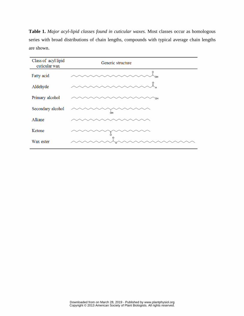
Table 1. Major acyl-lipid classes found in cuticular waxes. Most classes occur as homologous
series with broad distributions of chain lengths, compounds with typical average chain lengths
are shown.
www.plantphysiol.orgon March 28, 2019 - Published by Downloaded from Copyright © 2013 American Society of Plant Biologists. All rights reserved.

Table 2. Cuticle-associated genes discussed in this review
Gene Symbol
Gene Name Species Locus ID Description
ABCG11 ATP-binding cassette G11 A. thaliana At1G17840 ABC half transporter
ABCG13 ATP-binding cassette G13 A. thaliana At1G51460 ABC half transporter
ABCG32 ATP-binding cassette G32 A. thaliana At2G26910 ABC full transporter
BDG Bodyguard A. thaliana At1G64670 α/β-hydrolase family protein
BDG3 Bodyguard 3 A. thaliana At4G24140 Homolog of BDG
CD1 Cutin deficient 1 S. lycopersicum Solyc11G006250 Cutin synthase/hydroxyacylglycerol transesterase
CD2 Cutin deficient 2 S. lycopersicum Solyc01G091630 Homeodomain-leucine zipper iv transcription factor
CER1 Eceriferum 1 A. thaliana At1G02205 Involved in alkane formation
CER10 Eceriferum 10 A. thaliana At3G55360 Enoyl-CoA reductase
CER2 Eceriferum 2 A. thaliana At4G24510 Required for C28 to C30 elongation of fatty acids
CER26 Eceriferum 26 A. thaliana At4G13840 Homolog of CER2, required for elongation of fatty acids >C30
CER26-like
Eceriferum 26-like A. thaliana At3G23840 Homolog of CER2 and CER26
CER3 Eceriferum 3 A. thaliana At5G57800 Involved in alkane formation
CER4 Eceriferum 1 A. thaliana At4G33790 VLCFA-CoA by fatty acyl-coenzyme A
CER5/ ABCG12
Eceriferum 5/ATP-binding cassette G12
A. thaliana At1G51500 ABC half transporter
CER6 Eceriferum 6 A. thaliana At1G68530 β-ketoacyl-CoA synthase
CER7 Eceriferum 7 A. thaliana At3G60500 Exosomal exoribonuclease
CER9 Eceriferum 9 A. thaliana At4G34100 Putative e3 ubiquitin ligase
CFL1 Curly flag leaf 1 O. sativa Os02G31140 WW-domain containing protein
CYP77A6 CYP77A6 A. thaliana At3G10570 CYP77A subfamily of cytochrome p450
CYP86A4 CYP86A4 A. thaliana At1G01600 CYP86A subfamily of cytochrome p450
DCR Defective in cuticular ridges
A. thaliana At5G23940 BAHD acyltransferase
FDH Fiddlehead A. thaliana At2G26250 β-ketoacyl-CoA synthase
GPAT6 Glycerol-3-phosphate sn-2-acyltransferase 6
A. thaliana At2G38110 Bifunctional glycerol-3-phosphate sn-2-acyltransferase/phosphatase
HDG1 Homeodomain glabrous 1 A. thaliana At3G61150 Homeodomain-leucine zipper iv transcription factor
HTH Hothead A. thaliana At1G72970 Glucose-methanol-choline oxidoreductase family protein
IRG1 Inhibitor of rust germ tube differentiation 1
M. truncatula Medtr5G014400 C2H2 zinc finger transcription factor
KCR1 β-ketoacyl-CoA reductase 1 A. thaliana At1G67730 β-ketoacyl-CoA reductase
LACS1 Long-chain acyl-CoA synthase 1
A. thaliana At2G47240 Long-chain acyl-CoA synthase
LACS2 Long-chain acyl-CoA synthase 2
A. thaliana At1G49430 Long-chain acyl-CoA synthase
LACS3 Long-chain acyl-CoA synthase 3
A. thaliana At1G64400 Long-chain acyl-CoA synthase
LCR Lacerata A. thaliana At2G45970 CYP86A subfamily of cytochrome p450
LTL1 Li-tolerant lipase 1 A. thaliana At3G04290 Homolog of CD1
www.plantphysiol.orgon March 28, 2019 - Published by Downloaded from Copyright © 2013 American Society of Plant Biologists. All rights reserved.

LTPG1 Glycosylphosphatidylinositol-anchored lipid transfer protein 1
A. thaliana At1G27950 GPI-anchored lipid transfer protein
LTPG2 Glycosylphosphatidylinositol-anchored lipid transfer protein 2
A. thaliana At3G43720 GPI-anchored lipid transfer protein
MAH1 Mid-chain alkane hydroxylase 1
A. thaliana At1G57750 CYP96A subfamily of cytochrome p450
MYB106 Myb domain protein 106 A. thaliana At3G01140 Myb transcription factor
MYB16 Myb domain protein 16 A. thaliana At5G15310 Myb transcription factor
MYB30 Myb domain protein 30 A. thaliana At3G28910 Myb transcription factor
MYB41 Myb domain protein 41 A. thaliana At4G28110 Myb transcription factor
MYB96 Myb domain protein 96 A. thaliana At5G62470 Myb transcription factor
OCL1 Outer cell layer 1 Z. mays GRMZM2G026643
Homeodomain-leucine zipper iv transcription factor
PAS2 Pasticcino 2 A. thaliana At5G10480 β-hydroxyacyl-CoA dehydratase
SHN2 Shine 2 A. thaliana At5G11190 Homolog of WIN1/SHN1
WIN1/ SHN1
Wax inducer 1/Shine 1 A. thaliana At1G15360 AP2-domain containing transcription factor
WSD1 Wax synthase/acyl-CoA:diacylglycerol acyltransferase 1
A. thaliana At5G37300 Wax synthase/acyl-CoA:diacylglycerol acyltransferase family protein
WXP1 Wax production 1 M. truncatula Medtr5G062700 AP2-domain containing transcription factor
- - A. thaliana At5G33370 Homolog of CD1
www.plantphysiol.orgon March 28, 2019 - Published by Downloaded from Copyright © 2013 American Society of Plant Biologists. All rights reserved.

Figure 1
Epicuticular Wax Crystals
Epicuticular Wax FilmCuticle Proper
Cutin
PolysaccharidesCuticular Layer
PolysaccharideCell Wall
CuticleIntracuticular Wax
Epidermal CellEpidermal Cell
A
B C
D
EpidermalCell
Cuticle
Cuticular Layer
PolysaccharideCell Wall
Cuticle Proper
Figure 1. Plant cuticle structure. A. Schematic diagram highlighting the major structural features of the cuticle and underlying epidermal cell layer. Not drawn to scale. B. Scanning electron micrograph (SEM) image of an Arabidopsis leaf epidermis and overlying cuticle, seen in cross section. Scale bar, 5μm (image courtesy of Dr. Lacey Samuels). C. Transmission electron micrograph (TEM) image of an Arabidopsis stem epidermal cell wall and cuticle. Scale bar, 500 nm (image courtesy of Dr. Christiane Nawrath). D. Light microscopy image showing the cuticle of a mature green stage tomato fruit stained with Sudan Red and the polysaccharide cell walls stained with Alcian Blue. Scale bar, 50 μm.
www.plantphysiol.orgon March 28, 2019 - Published by Downloaded from Copyright © 2013 American Society of Plant Biologists. All rights reserved.

PolysaccharideCell Wall
Cuticular layer
Cuticle proper
Endoplasmic Reticulum
C16/C18-CoA16-OH
C16-CoA
10,16-diOHC16-CoA
10,16-diOHC16-Glycerol
(2-MHG)
VLC Acyl-CoA
VLC Fatty Acid
VLC 1˚Alcohol
VLCAldehyde
VLC Alkane
VLC2˚ Alcohol
VLCKetoneWax Esters
FAEMalonyl-
CoACoA + CO2
CER4CER2,CER26
CER26-likeLACS1THS
WSD1
MAH1CER1CER3
MAH1GPAT6
Glycerol
Cutin
CD1(Cutin Synthase)
CYP77A6CYP86A4
ABCG11 ABCG32
CER6
CER10PAS2
KCR1
Waxes
C16/C18Fatty acid
(From Plastid)
LACS1LACS2
O
OHO
O
OHO
O
OH
OH
Cutin Wax
ABCG11/ABCG12
LTPG
Apoplastic LTPs?
Cuticular Wax
Figure 2
Figure 2. Cutin and wax biosynthetic pathways. Genes (blue text) are described in the review. Red text denotes compound classes that are typically observed in cuticular wax mixtures.
www.plantphysiol.orgon March 28, 2019 - Published by Downloaded from Copyright © 2013 American Society of Plant Biologists. All rights reserved.

O
HOOH
OHO
HOOH
O
HO
OOH
O
HOOH
OH
OH
A
B=
O
HOOH
OH
Linear
Branched
O
OHO
HO
Figure 3
Figure 3. Typical cutin monomers and polymeric structure. A. Some typical C16 and C18-fatty acid derived cutin monomers. From top to bottom: 10,16-dihydroxyhexadecanoic acid, 16-hydroxyhexadeca-noic acid, 9,10-epoxyoctadecanoic acid, 9,10,18-trihydroxyoctadecanoic acid and octadeca-cis-6, cis-9-diene-1,18-dioate, the major cutin monomer of Arabidopsis stems and leaves. B. Linear and branched domains made possible by different ester linkages of 10,16-dihydroxyhexadecanoic acid, depicted schematically as indicated.
www.plantphysiol.orgon March 28, 2019 - Published by Downloaded from Copyright © 2013 American Society of Plant Biologists. All rights reserved.
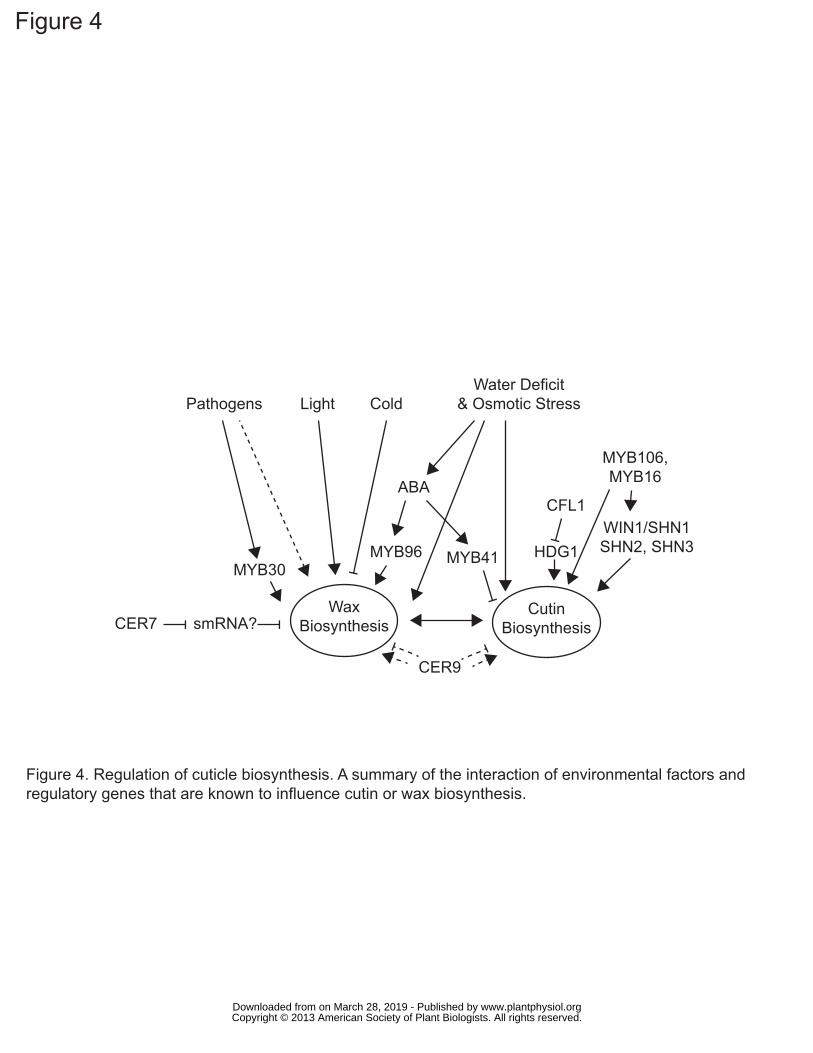
ABA
LightPathogens ColdWater Deficit
& Osmotic Stress
MYB96 MYB41MYB30
CFL1
HDG1WIN1/SHN1SHN2, SHN3
MYB106,MYB16
CER7
CER9
smRNA?Wax
BiosynthesisCutin
Biosynthesis
Figure 4
Figure 4. Regulation of cuticle biosynthesis. A summary of the interaction of environmental factors and regulatory genes that are known to influence cutin or wax biosynthesis.
www.plantphysiol.orgon March 28, 2019 - Published by Downloaded from Copyright © 2013 American Society of Plant Biologists. All rights reserved.