1 powerpoint lecture outlines to accompany holes human anatomy and physiology eleventh edition shier...
TRANSCRIPT

1
PowerPoint Lecture Outlines to accompany
Hole’s HumanAnatomy and Physiology
Eleventh Edition
Shier Butler Lewis
Chapter
7
Copyright © The McGraw-Hill Companies, Inc. Permission required for reproduction or display.

2
Chapter 7Skeletal System
Bone Classification
• Long Bones• Short Bones• Flat Bones• Irregular Bones• Sesamoid (Round) Bones

3
Parts of a Long Bone• epiphysis
• distal• proximal
• diaphysis• compact bone• spongy bone• articular cartilage• periosteum• endosteum• medullary cavity• trabeculae• marrow
• red• yellow

4
Compact and Spongy Bone
5
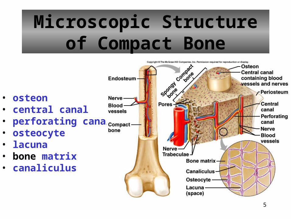
Microscopic Structureof Compact Bone
• osteon• central canal• perforating canal• osteocyte• lacuna• bone matrix• canaliculus
6
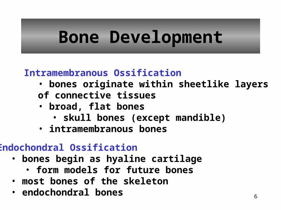
Bone Development
Intramembranous Ossification• bones originate within sheetlike layers of connective tissues• broad, flat bones
• skull bones (except mandible)• intramembranous bones
Endochondral Ossification• bones begin as hyaline cartilage
• form models for future bones• most bones of the skeleton• endochondral bones

7
Endochondral Ossification
• hyaline cartilage model• primary ossification center• secondary ossification centers
• epiphyseal plate• osteoblasts vs. osteoclasts
8
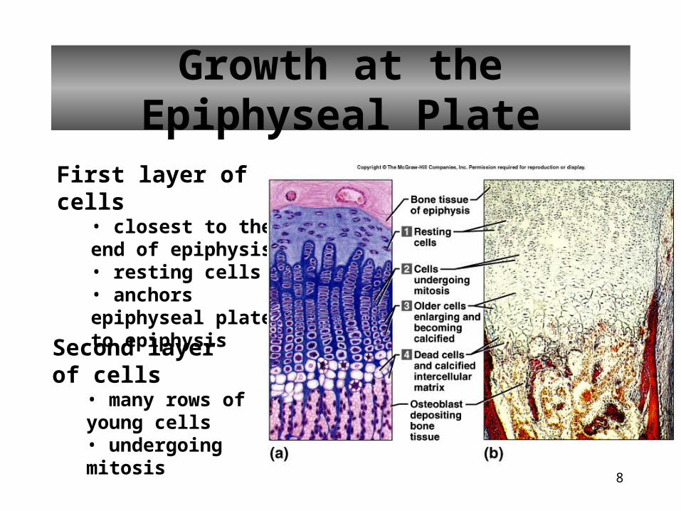
Growth at the Epiphyseal Plate
First layer of cells• closest to the end of epiphysis• resting cells• anchors epiphyseal plate to epiphysis
Second layer of cells• many rows of young cells• undergoing mitosis

9
Growth at the Epiphyseal Plate
Third layer of cells• older cells• left behind when new cells appear• cells enlarging and becoming calcified
Fourth layer of cells• thin• dead cells• calcified extracellular matrix

10
Homeostasis of Bone Tissue
•Bone Resorption – action of osteoclasts and parathyroid hormone•Bone Deposition – action of osteoblasts and calcitonin

11
Factors Affecting Bone Development, Growth, and
Repair
• Deficiency of Vitamin A – retards bone development• Deficiency of Vitamin C – results in fragile bones • Deficiency of Vitamin D – rickets, osteomalacia• Insufficient Growth Hormone – dwarfism• Excessive Growth Hormone – gigantism, acromegaly • Insufficient Thyroid Hormone – delays bone growth• Sex Hormones – promote bone formation; stimulate ossification of epiphyseal plates• Physical Stress – stimulates bone growth

12
Bone Function
• Support, Movement & Protection• gives shape to head, etc.• supports body’s weight• protects lungs, etc.• bones and muscles interact when limbs or body parts move
• Blood Cell Formation• hematopoiesis• red marrow
• Inorganic Salt Storage• calcium • phosphate• magnesium• sodium• potassium

13
Skeletal Organization
Axial Skeleton• head • neck • trunk
Appendicular Skeleton• upper limbs• lower limbs• pectoral girdle• pelvic girdle

14
Skeletal Organization

15
Cranium
Frontal (1)• forehead• roof of nasal cavity• roofs of orbits• frontal sinuses• supraorbital foramen• coronal suture

16
Cranium
Parietal (2)• side walls of cranium• roof of cranium• sagittal suture

17
Cranium
Occipital (1)• back of skull• base of cranium• foramen magnum• occipital condyles• lambdoid suture

18
Cranium
Temporal (2)• side walls of cranium• floor of cranium• floors and sides of orbits• squamous suture• external acoustic meatus• mandibular fossa• mastoid process• styloid process• zygomatic process

19
Cranium
Sphenoid (1)• base of cranium• sides of skull• floors and sides of orbits• sella turcica• sphenoidal sinuses

20
Cranium
Ethmoid (1)• roof and walls of nasal cavity• floor of cranium• wall of orbits• cribiform plates• perpendicular plate• superior and middle nasal conchae• ethmoidal sinuses• crista galli

21
Facial Skeleton
Maxillary (2)• upper jaw• anterior roof of mouth• floors of orbits• sides of nasal cavity• floors of nasal cavity• alveolar processes• maxillary sinuses• palatine process

22
Facial Skeleton
23
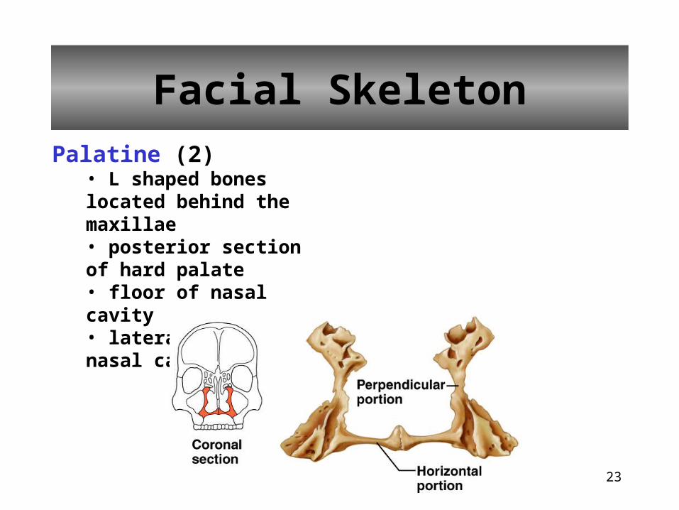
Facial Skeleton
Palatine (2)• L shaped bones located behind the maxillae• posterior section of hard palate• floor of nasal cavity• lateral walls of nasal cavity

24
Facial Skeleton
Zygomatic (2) • prominences of cheeks• lateral walls of orbits• floors of orbits• temporal process

25
Facial Skeleton
Lacrimal (2)• medial walls of orbits• groove from orbit to nasal cavity
Nasal (2)• bridge of nose

26
Facial Skeleton
Vomer (1)• inferior portion of nasal septum

27
Facial Skeleton
Inferior Nasal Conchae (2)• extend from lateral walls of nasal cavity

28
Facial Skeleton
Mandible (1)• lower jaw• body• ramus• mandibular condyle• coronoid process• alveolar process• mandibular foramen• mental foramen

29
Infantile Skull
Fontanels – fibrous membranes
30
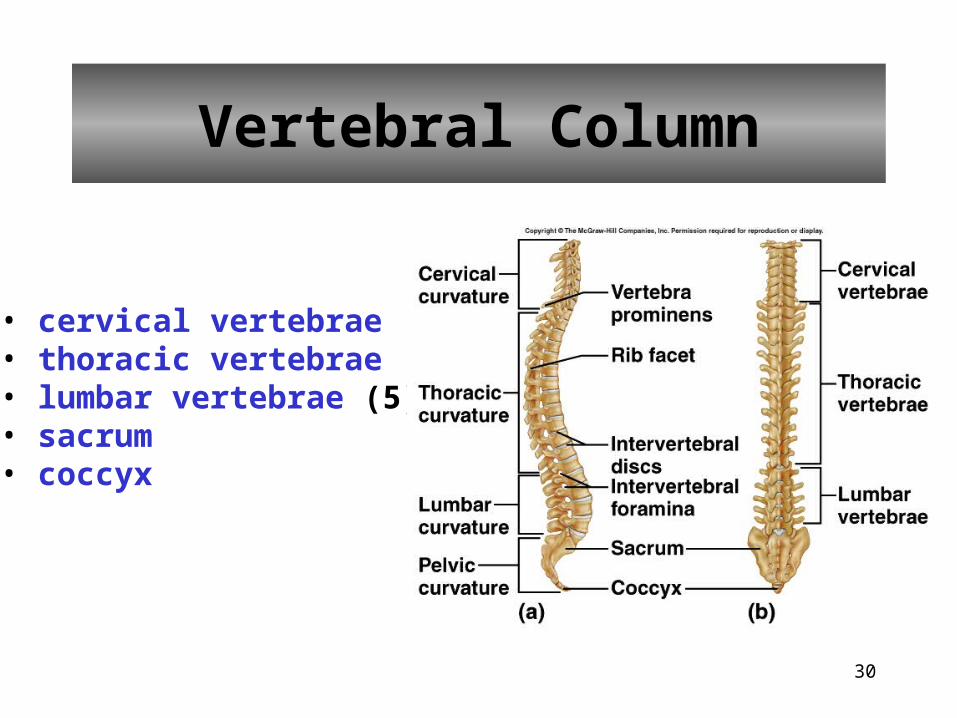
Vertebral Column
• cervical vertebrae (7)• thoracic vertebrae (12)• lumbar vertebrae (5)• sacrum • coccyx

31
Vertebral Column
• cervical curvature• thoracic curvature• lumbar curvature• sacral curvature• rib facets• vertebra prominens• intervertebral discs• intervertebral foramina

32
Cervical Vertebrae
• Atlas – 1st; supports head• Axis – 2nd; dens pivots to turn head• transverse foramina• bifid spinous processes• vertebral prominens – useful landmark
33
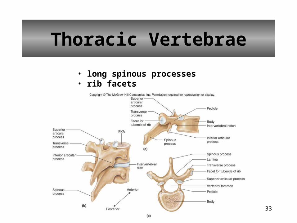
Thoracic Vertebrae
• long spinous processes• rib facets

34
Lumbar Vertebrae
• large bodies• thick, short spinous processes
35
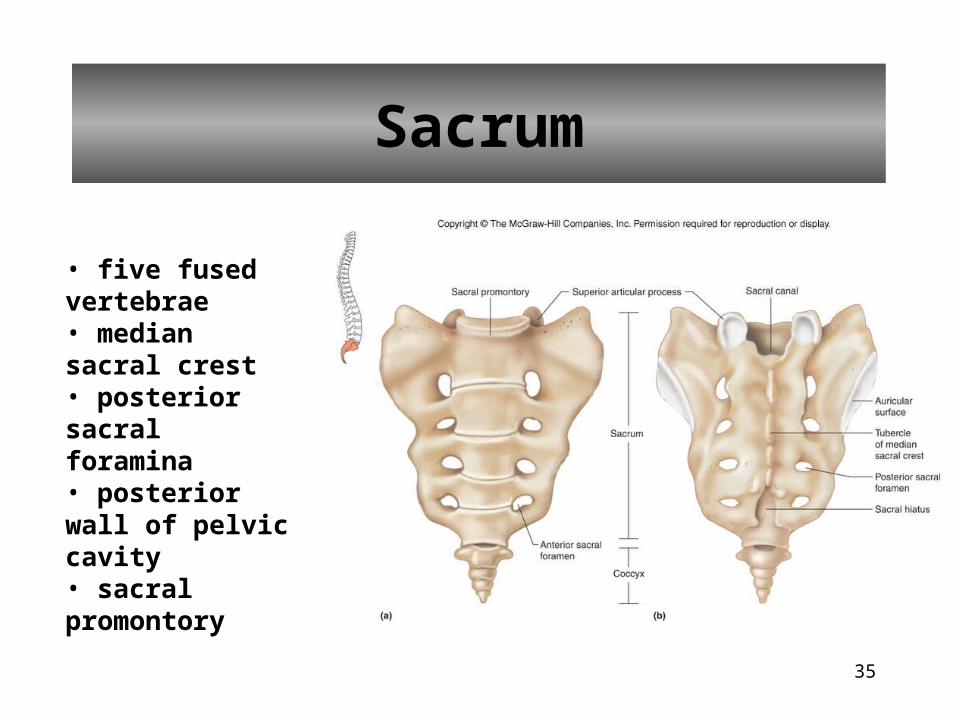
Sacrum
• five fused vertebrae• median sacral crest• posterior sacral foramina• posterior wall of pelvic cavity• sacral promontory

36
Coccyx
• tailbone• four fused vertebrae

37
Thoracic Cage
• Ribs• Sternum• Thoracic vertebrae• Costal cartilages• Supports shoulder girdleand upper limbs• Protects viscera• Role in breathing

38
Ribs
• True ribs (7)• False ribs (5)
• floating (2)
39
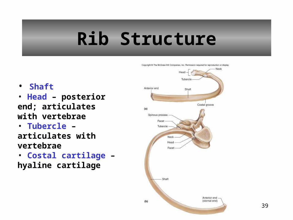
Rib Structure
• Shaft• Head – posterior end; articulates with vertebrae• Tubercle – articulates with vertebrae• Costal cartilage – hyaline cartilage

40
Sternum
• Manubrium• Body• Xiphoid process

41
Pectoral Girdle
• shoulder girdle • clavicles• scapulae• supports upper limbs

42
Clavicles
• articulate with manubrium• articulate with scapulae (acromion process)

43
Scapulae
• spine• supraspinous fossa• infraspinous fossa
• acromion process• coracoid process• glenoid cavity

44
Upper Limb
• Humerus• Radius• Ulna• Carpals• Metacarpals• Phalanges

45
Humerus
• head• greater tubercle• lesser tubercle• anatomical neck• surgical neck• deltoid tuberosity• capitulum• trochlea• coronoid fossa• olecranon fossa

46
Radius
• lateral forearm bone• head• radial tuberosity• styloid process

47
Ulna
• medial forearm bone• trochlear notch• olecranon process• coronoid process• styloid process

48
Wrist and Hand
• Carpals (16)• trapezium• trapezoid• capitate• scaphoid• pisiform• triquetrum• hamate• lunate
• Metacarpals (10)
• Phalanges (28)• proximal phalanx• middle phalanx• distal phalanx

49
Pelvic Girdle
• Coxae (2)• supports trunk of body• protects viscera

50
Coxae
• hip bones•acetabulum
• ilium• iliac crest• iliac spines• greater sciatic notch
• ischium• ischial spines• lesser sciatic notch• ischial tuberosity
• pubis• obturator foramen• symphysis pubis• pubic arch

51
Greater and Lesser Pelves
Greater Pelvis• lumbar vertebrae posteriorly• iliac bones laterally• abdominal wall anteriorly
Lesser Pelvis• sacrum and coccyx posteriorly• lower ilium, ischium, and pubis bones laterally and anteriorly

52
Male and Female Pelves
Female• iliac bones more flared• broader hips• pubic arch angle greater• more distance between ischial spines and ischial tuberosities• sacral curvature shorter and flatter• lighter bones

53
Lower Limb
• Femur• Patella• Tibia• Fibula• Tarsals• Metatarsals• Phalanges

54
Femur
• longest bone of body• head• fovea capitis• neck• greater trochanter• lesser trochanter• linea aspera• condyles• epicondyles

55
Patella
• kneecap• anterior surface of knee• flat sesamoid bone located in a tendon

56
Tibia
• shin bone• medial to fibula• condyles• tibial tuberosity• anterior crest• medial malleolus

57
Fibula
• lateral to tibia• long, slender• head• lateral malleolus• does not bear any body weight

58
Ankle and Foot
• Tarsals (14)• calcaneus• talus• navicular• cuboid• lateral cuneiform• intermediate cuneiform• medial cuneiform
• Metatarsals (10)
• Phalanges (28)• proximal• middle• distal

59
Ankle and Foot

60
Life-Span Changes
• decrease in height at about age 30• calcium levels fall• bones become brittle• osteoclasts outnumber osteoblasts• spongy bone weakens before compact bone• bone loss rapid in menopausal women• hip fractures common• vertebral compression fractures common

61
Clinical Application
Types of Fractures
• green stick• fissured• comminuted• transverse• oblique• spiral