1 opioid receptor
TRANSCRIPT

Neuron, Vol. 11, 903-913, November, 1993, Copyright 0 1993 by Cell Press
Cloning and Pharmacological Characterization o f a Rat ~1 Op ioid Receptor
Robert C. Thompson, Alfred Mansour, Huda Akil, and Stanley 1. Watson Mental Health Research Institute University of Michigan Ann Arbor, Michigan 48109-0720
Summary
We have isolated a rat cDNA clone that displays 75% amino acid homology with the mouse 6 and rat K opioid receptors. The cDNA (designated pRMuR-12) encodes a protein of 398 amino acids comprising, in part, seven hydrophobic domains similar to those described for other C protein-linked receptors. Data from binding assays conducted with COS-1 cells transiently transfected with a CMV mammal ian expression vector containing the full coding region of pRMuR-12 demonstrated p receptor selectivity. In situ hybridization mRNA analysis revealed an mRNA distribution in rat brain that corresponds well to the distribution of binding sites labeled with p-selec- tive ligands. Based upon these observations, we con- clude that pRMuR-12 encodes a p opioid receptor.
Introduction
The existence of multiple forms or subtypes of opioid receptors was first suggested by Martin and colleagues (Gilbert and Martin, 1976; Martin et al., 1976), using the chronic spinal dog preparation, and has since been supported by numerous behavioral, pharmacological, and receptor binding studies (Gillan and Kosterlitz, 1982; Wood, 1982; Goldstein and James, 1984; Herling and Woods, 1984; James and Goldstein, 1984). Opioid receptors have been divided into at least three main classes: K, 6, and p. These receptors have unique phar- macological profiles, anatomical distributions, and functions in several species, including man (Wood, 1982; Simon, 1991; Lutz and Pfister, 1992; Mansour and Watson, 1993). Several pharmacological agents as well as various members of the three endogenous opioid peptide families (prodynorphin, proenkephalin, and proopiomelanocortin) have been shown to interact with these receptors in distinctive and well-described manners. K receptors have been shown to exist in diverse regions of the CNS; they are particularly en- riched in the cortex, striatum, and hypothalamus (Man- sour et al., 1987; Neck et al., 1988; Unterwald et al., 1991) and are thought to mediate many of the actions of the dynorphin peptide family in addition to those of benzomorphans. These actions include the modu- lation of drinking, water balance, food intake, gut mo- tility, temperature control, and various endocrine func- tions (Morley and Levine, 1983; Leander et al., 1985; lyengar et al., 1986; Manzanares et al., 1990). 6 recep- tors are also found throughout the CNS, particularly in forebrain areas such as the striatum and cerebral
cortex, and have been shown to bind enkephalin-like peptides (Mansour et al., 1988; Wood, 1988). 6 recep- tors are also thought to modulate several hormonal systems and to mediate analgesia.
The p receptors represent the third member of the opioid receptor family.These receptors bind morphine- like drugs and several endogenous opioid peptides, including P-endorphin (derived from proopiomelano- cortin) and several members of both the enkephalin and the dynorphin opioid peptide families. These re- ceptors have been localized in many regions of the CNS, including the striatum (striatal patches), thalamus, nucleus tractus solitarius, and spinal cord (Mansour et al., 1987; McLean et al., 1986; Temple and Zukin, 1987; Wood, 1988; Mansour and Watson, 1993). p receptors appear to mediate the opiate phenomena classically associated with morphine and heroin administration, including analgesia, opiate dependence, cardiovascu- lar and respiratory depression, and several neuroen- docrine effects. Pasternak and co-workers (Pasternak and Snyder, 1975; Wolozin and Pasternak, 1981; Pas- ternak and Wood, 1986; Pasternak, 1993) and Rothman and co-workers (Rothman et al., 1987) have suggested that p receptors can be subdivided into two classes, ~1 and ~2. ~1 receptors are thought to have high affin- ity for both morphine and enkephalins. In contrast, ~2 receptors selectively interact with morphine-like compounds and DAMGO ([D-AlaZ,N-MePhe4,Gly-o15]en- kephalin). These studies further suggest that ~1 and ~2 are functionally distinct: ~1 receptors, unlike ~2 receptors, apparently mediate analgesia but not respi- ratory depression or physical dependence (Pasternak, 1993).
Owing to the importance of opioid receptors in opi- oid pharmacology and physiology, many researchers have attempted to purify these proteins with the aim of ultimately describing the molecular sequence of these receptors. In one such attempt, Schofield re- ported the isolation of a protein (opioid-binding cell adhesion molecule) apparently involved with an opi- oid receptor (Schofield et al., 1989). This protein has been shown to be a member of the immunoglobulin superfamily of cell adhesion molecules but lacks trans- membrane domains, a feature assumed necessary for signal transduction. Using an expression cloning strat- egy, Xie et al. (1992) detailed the isolation and pharma- cological identification of a G protein-coupled pro- tein with opioid receptor binding activity. This protein demonstrated several key features of opioid recep- tors: opioid binding and stereospecificity; however, the affinity (Ko) for pharmacologically relevant agents was low and not selective. The biochemical purifica- tion of the p receptor proteins has been reported by several groups (Simon et al., 1975; Bidlack and Abood, 1980; Ruegg et al., 1980; Simonds et al., -1980; Cho et al., 1981, 1986; Howells et al., 1982; Chow and Zukin, 1983; Demoliou-Mason and Barnard, 1984; Maneckjee

Neuron 904
et al., 1985; Ueda et al., 1987). Many of these reports suggest that this protein is approximately 40-60 kd. Additionally, this receptor can be functionally cou- pled to G proteins (Koski and Klee, 1981; Barchfeld and Medzihradsky, 1984; Hsia et al., 1984; Puttfarchen et al., 1988; Attali et al., 1989). However, the low abun- dance of the protein and its apparently high hydro- phobicity have proved to be major stumbling blocks for the sequencing of sufficiently long peptide frag- ments necessary for the molecular cloning of this re- ceptor.
Kieffer et al. (1992) and Evans et al. (1992) reported the isolation and pharmacological characterization of the first high affinity opioid receptor, the 6 opioid receptor, using an expression cloning strategy. This receptor was shown to contain seven hydrophobic domains characteristic of membrane receptors known to transduce extracellular signals (e.g., ligand binding) via G proteins to the intracellular environment. The cloned receptors exhibited high affinity and selectiv- ity for 8 opioid ligands, and Evans et al. (1992) showed that activation of their receptor with selective 8 ago- nists could decrease CAMP levels in transfected COS cells. The mouse S receptors were found to be highly homologous to several members of the G protein- coupled receptor family, particularlythesomatostatin receptors. The cloning of the mouse 6 receptors facili- tated the cloning of other members of the opioid re- ceptor family. Recently, Yasuda and co-workers de- scribed the isolation of a mouse K receptor cDNA that had been isolated as a member of the related somatostatin receptor family (Yasuda et al., 1993). In- dependently, our laboratory, using DNA fragments homologous to the mouse 6 opioid receptor and low stringency cDNA library screening, isolated and phar- macologically characterized a rat K receptor cDNA (Meng et al., 1993). The rat and mouse K receptors contain seven transmembrane domains and are nega- tivelycoupled toadenylatecyclase via G proteins. This report describes the cloning and characterization of a homologous seven transmembrane domain protein whose pharmacological profile and mRNA localiza- tion suggest that it is a rat t.r receptor.
Results
Isolation and Characterization of Rat p Receptor cDNA Clones Using random primed cDNA from rat olfactory bulb and oligonucleotide primers (based upon transmem- branedomains III,V,VI,andVIIofthemouse6opioid receptor mRNA [Evans et al., 1992; Kieffer et al., 1992]), amplified fragments were generated by the polymerase chain reaction (PCR) and subcloned. One clone, p5A-1, contained a DNA sequence that was 74% homologous at the nucleic acid level to the mouse8 opioid receptor mRNA sequence between transmembrane regions III and VI. The p5A-1 sequence was also similar (59% ho- mology at the nucleic acid level) to several species of somatostatin receptor mRNAs. A rat olfactory bulb
hZAPll cDNA librarywas plated and screened with the radiolabeled DNA insert from p5A-1. Twelve positive clones were obtained. Restriction mapping and South- ern blot analysis were performed on rescued plasmid DNA. One clone, designated pRMuR-12, demonstrated a strong hybridization signal on Southern blots and contained a cDNA insert of approximately2.0 kb. Vari- ous restriction enzyme digestion fragment of pRMuR- 12 were subcloned into Bluescript and completely se- quenced in both orientations. DNA sequence analysis revealed that pRMuR-12 contained a full open reading frame with 74% homology with the mouse 8 and rat K opioid receptor proteins.
DNA and Protein Structure Analysis An EcoRl-Hindlll fragment isolated form pRMuR-12 was shown to contain the entire protein coding region of this cDNA and was used for all subsequent experi- ments. The complete DNA sequence contained an open reading frame encoding a protein of 398 amino acids comprising, in part, seven hydrophobic domains. GenBank data base searches using both the amino acid and the nucleic acid sequences revealed that the pRMuR-12 sequence was novel and highly homolo- gous to the mouse 6 opioid receptor (74% similar and 59.5% identical at the amino acid level) and the so- matostatin receptors (60.5% similar and 39.5% identi- cal at the amino acid level). As can be seen in Figure 1, the regions of greatest homology were found in the hydrophobic domains, particularly domains I, II, III, VI, and VII. The regions of greatest divergence included the amino and carboxyl termini of the pro- tein, the region surrounding and including hydropho- bic domain IV, and the region between hydrophobic domains V and VI. In addition to the seven hydropho- bic domains classically associated with G protein- coupled receptors, the open reading frame contains several other features of this receptor family, includ- ing potential N-linked glycosylation sites (five at amino acids 9,31, 38,46, and 53), palmitoylation sites (two at amino acids 346 and 351), and three phosphorylation sites (protein kinase C phosphorylation at amino acid 363, calcium/calmodulin-dependent kinase phosphory- lation at amino acid 261, and CAMP-dependent protein kinase [protein kinase A] phosphorylation at amino acid 279).
Transient Transfection of CMV-pRMuR DNA into COS-1 Cells Initial binding assays performed on COS-1 cells trans- fected with the pCMV-neo (a gift from Dr. Michael Uhler, University of Michigan) expression vector con- taining the full open reading frame of pRMuR-12 dem- onstrated the opioid nature of the encoded receptor. [3H]DAMG0 bound with high affinity (Ko = 1.2 nM), whereas the selective K agonist [3H]U69,593 (n-(5a, 7a;8(3)-(+)-N-methyl-N-[7-(l-pyrroIidinyl)-l-oxaspiro-(4,5) dec-8-yl lbenzeneacetamide) and the selective 8 ago- nist [3H]DPDPE (cyclic (o-penicillamine*, o-penicilla- mines) enkephalin) did not. To characterize this clone

Rat F Opioid Receptor Cloning 905
KU Pat LEAETRPLP. ......................... ........ K Rat ......................................... ..... D Mouse PI .................. .....................
Figure 1. Amino Acids Comparison between the Rat B, Rat K, and Mouse S Opioid Receptor Sequences
The deduced amino acid sequence of the rat p receptor (Mu Rat) is compared with the rat K (K Rat) and mouse 6 (D Mouse) receptors. Amino acids conserved in all three receptors are shown in bold. Putative transmembrane domains are under- lined. The positions of potential N-linked glycosylation sites are indicated by closed arrows. Consensus recognition sites for pro- tein kinase C (asterisks), protein kinase A (open circle), and pal- mitoylation (open arrowheads) are also shown.
further, [3H]DAMG0 (1.6 nM) was used as a labeling ligand, and various IL-, 6-, and K-selective ligands and nonselective ligands were tested in competition stud- ies (Table 1). Several classic p-selective ligands had high affinities for the expressed receptor (morphine, K, = 7.14 nM; levorphanol, Ki = 0.78 nM), whereas 6 (DSLET [(D-Ser2, LeuS)enkephalin-Thr], K, = 541.63 nM; DPDPE, K, = 4,100.OO nM) and K (U50,488 [(trans)-3,4- dichloro-N-methyl-N-[2-(l-pyrrolidinyl)cyclohexyl] ben- zene acetamide methanesulfonate], K, = 1464.00 nM; U69,593, Ki = 1910.00 nM) ligands did not. DADLE (o-Ala-o-Leu-enkephalin; Ki = 6.56 nM) and the syn- thetic dynorphin (l-8) analog E2078 ((N-methyl-Tyrl, N-a-methyl-Arg7,0-Leu8) dynorphin A-(1-8) ethylamide; Ki = 2.87 nM) were also potent in displacing [3H]DAMG0.
Several antagonists were also tested. Naloxone (Ki = 2.74 nM), naltrexone (K, = 0.36 nM), CTAP (D-Pen-Cys- Tyr-D-Trp-Arg-Thr-Pen-Thr-NH?; Ki = 0.21), and nBNl (nor-binaltrophine HCI; Ki = 3.90) all displaced [3H]DAMG0 binding with high affinity. The expressed receptor also bound several nonselective opioid li- gands such as (-)bremazocine (K, = 0.05 nM) and EKC (ethylketoclazocine; Ki = 1.28 nM) with high affinity. Furthermore, ligand binding to the transiently ex- pressed receptor clone was stereospecific. (-)Brema-
Table 1. Pharmacological Profile of the Cloned Rat p Receptor
K, (nM)
p ligands DAMGO Morphine Levorphanol
S ligands DADLE DPDPE DSLET
K ligands E2078 U50,488 U69,593
Nonselective ligands EKC (-)Bremazocine
Enantiomers (+)Bremazocine Dextrorphan
Antagonists CTAP Naloxone Naltrexone nBNl
1.57 7.14 0.78
6.56 4100.00
541.63
2.87 1464.00 1910.00
I .2a 0.05
>5000.0 >5000.0
0.21 2.74 0.36 3.90
Using [‘HIDAMCO (1.6 nM) as the labeling ligand, competition studies were performed on membrane preparations from COS-1 cells transiently transfected with pCMV-neo containing the full open reading frame of pRMuR-12. See text for description of ligands and abbreviations.
zocine and levorphanol bound with high affinity, but their enantiomers, (+)bremazocine and dextorphan, respectively, bound with low affinity.
Northern Blot Analysis of p Receptor mRN.4 Figure 2 show results from total RNA and poly(A) RNA from rat medullalpons as well as poly(A) RNA from humanSY5Ycells(6~gforSY5Yand10~gformedulla/ pons) electrophoresed on 16% formaldehyde-agar- ose (1%) gels hybridized with cRNA probes comple- mentary to p5A-1. Blots were hybridized and washed at high stringency. Lane 1 represents a short photo- graphic exposure of the SY5Y mRNA lane (lane 2). Lanes2and3arefroma44 hrautoradiogramdetecting a single mRNA species in the rat medulla/pans mRNA sample and one prominent mRNA species in the hu- man SY5Y cellular mRNA sample. Although less clear, analysis of total RNA from rat medulla/pans tissue (lane 4) identified a p receptor RNA species that comi- grated with the band seen in the poly(A) RNA (lane 3). These mRNAs are approximately 12,000-14,000 and 14,000-16,000 nucleotides in length, respectively.
Genomic Southern Blot Analysis Figure 3 show results from analysis of rat genomic DNA (20 pg per restriction enzyme diges#t) digested with restriction enzymes, electrophoresed on 0.8% agarose gels, and hybridized with random hexamer-

Neuron 906
123 4
2.37- I
0.24-
Figure 2. Northern Blot Analysis of Rat p Receptor mRNA in Rat Medulla/Ports and SY5Y Cells
Poly(A) RNA (6 pg for SY5Y [lane 1 and 21 and IO ug for medulla/ pons [lane 31) and total RNA (20 ug for medulla/pans [lane 41) was electrophoresed on formaldehyde-agarose (1%) gels, trans- ferred to nylon membrane, and hybridized (overnight) at 6OT in 50% formamide, 1 x HYB buffer. cRNA probes complementary to p5A-1 (see text) were used at approximately 2 x IO6 cpms of cRNA per ml of hybridization solution. Blots were washed in progressively decreasing SSC solutions (final concentrations of 0.1 x SSC, 0.5% SDS) at 72T for 2 hr. Lane 1 represents a short photographic exposure of the SY5Y mRNA in lane 2. Lanes 2 and 3 represent exposures generated using two intensifying screens at -7OT for 44 hr. Lane 4 represents an exposures using two intensifying screens at -70°C for 7 days. Molecular weights were determined using an RNA ladder (BRL).
primed 2.0 kb cDNA from pMuR-12 as a probe under high stringency conditions. Several DNA bands were detected. Two bands were detected in the BamHl and Pstl lanes, consistent with the presence of these re- striction endonuclease siteswithin thecDNAsequence. Multiple hybridization-positive bands were detected in the Hindlll and EcoRl lanes. There were no internal EcoRl sites and only one Hindlll site found within the cDNA probe sequence. The presence of introns separating the 2.0 kb cDNA sequence into multiple exons would be consistent with the observed hybrid- ization pattern in these two lanes. The presence of two hybridization-positive bands in the BamHl and Pstl lanes suggests that there is one p receptor gene in the rat genome.
Distribution of the p Receptor mRNA In the olfactory bulb, from which the p receptorcDNA clone was derived, p receptor mRNA was localized to the internal granular and glomerular layers.
Neocortical areas such as the parietal and temporal cortices did not demonstrate the same receptor distri- bution, as might be expected from receptor binding
-10 -5
-3
-2
-1
Figure 3. Cenomic Southern Blot of Rat or Receptor
Rat genomic DNA (20 ug per restriction enzyme digest) was di- gested with the indicated enzymes. Random hexamer priming of the full 2.0 kb cDNA from pRMuR-I2 was used as a probe. Blots were washed in decreasing SSC solutions (final concentra- tions of 0.1 x SSC, 0.5% SDS) at 60°C for 30 min. Exposures were performed using two intensifying screens at -70°C for 48 hr. Molecular weights were determined using an DNA ladder (BRL).
studies. A thin layer of cells expressing the p receptor mRNA was observed in the deepest extents of layer VI, and no cells were detectable in layers I and IV, as was demonstrated with receptor binding. Low levels of l.r receptor mRNA could be detected in the piriform and cingulate cortex and the deep layer of the entorhi- nal cortex. v receptor mRNA was expressed at high lev- els in the striatal patches and the subcallosal streakven- tral to the corpus callosum (Figure 4A). The f.r striatal patches were most prominent in the rostra1 and lateral extents of the caudate-putamen and extended ventrally into the nucleus accumbens, where theywere the most dense in the medial and ventral shell. Specific hybrid- ization was also observed in the striatal matrix; how- ever, expression levels were comparatively reduced. More laterally, the cells of the medial septum ex- pressed high levels of u receptor mRNA.
Scattered cells expressing high levels of f.r receptor mRNA were seen in the bed nucleus stria terminalis

Rat n Opioid Receptor Cloning 907
Figure 4. Dark-Field Autoradiograms Demonstrating the Distri- bution of p Receptor mRNA at Three Levels of the Rat Brain (A), (B), and (C) are sections through three levels of the brain. p receptor mRNA is expressed in the striatal patches of the cau- date-putamen (CPU) and nucleus accumbens (ACB), subcallosal streak, olfactory tubercle, several nuclei of the thalamus, includ- ing the medial habenula (Hb), the medial dorsal (MD), and the central (CM) nuclei, hippocampus (HPC), dentate gyrus, basolat- eral amygdala, medial amygdala (Me), locus ceruleus (LC) and parabrachial nucleus (PB).
and the medial preoptic area. In the medial preoptic
area, the highest density of cellsexpressing p receptor mRNA were observed in the anterior ventral division in the anterior medial preoptic and more posteriorly in the medial preoptic nucleus itself.
Scattered cells expressing f.t receptor mRNA were also seen in the globus and ventral pallidum, regions
demonstrating a low level of ~1 receptor binding and enkephalinergic projections.
Of the diencephalic structures expressing the TV re- ceptor mRNA (Figure 4B), highest levels are observed in several thalamic nuclei, including the medial ha- benula, dorsomedial, ventrolateral, central, reuniens, and medial geniculate nuclei of the thalamus. Bycom- parison, the vast majority of hypothalamic nuclei, in- cluding the paraventricular, periventricular, supraop- tic, suprachiasmatic, ventromedial, and dorsomedial, did not appear to have cells that expressed t.t opioid receptor mRNA. Scattered cells expressing moderate levels of p receptor mRNA were seen in the lateral hypothalamus and the mammillary nuclei.
In the hippocampus (Figure 4B), j.r receptor mRNA expression was observed in the pyramidal cell layer of the hippocampal formation and the granular cells of the dentate gyrus. As can be seen from the high magnification image of CA2 cells in Figure 5A, p recep- tor mRNA expression was heterogeneous in the pyra- midal cell layer; either not all cells expressed this gene or the cells expressed it at markedly reduced levels. It is unclear presentlywhat the functional significance of this heterogeneity may be.
Several amygdaloid nuclei expressed the u receptor mRNA, including the medial, basolateral, and cortical nuclei (Figure 48). Levels were highest in the medial,
posterior medial cortical amygdala, and intercalated nuclei of the basolateral amygdala.
In the midbrain (data not shown), or receptor mRNA was observed in the ventral and lateral periaquaductal gray, a region known to subserve a role in pain and analgesia. j.r receptor mRNA expression did extend to the pontine central gray. Low levels of w receptor mRNA were also observed in scattered cells of the ventral tegmental area and substantia nigra, pars com- pacta.
Cells expressing p receptor mRNA were also local- ized in the superior and inferior colliculi. Within the superior colliculus, p receptor mRNA was primarily found in the large cells of the intermediate gray layer and in the inferior colliculus, cells of both the external cortex and the central nucleus express p receptor mRNA.
The large cells of the red nucleus and the rostral, central, and lateral interpeduncular nuclei expressed kt receptor mRNA. As can be seen in Figure 5B, the highest density of cells expressing u receptor mRNA were in the rostra1 division, with scattered cells in the central division. The levels of p receptor mRNA ex- pression in the lateral interpeduncular nucleus was low compared with other subdivisions.
Several brain stem nuclei expressed ).I receptor mRNA. Highest levels were observed in the locus cer- uleus and parabrachial nuclei (Figure 4C; Figure 5C). This distribution was consistent with the ability of ).I opioid drugs to modulate norepinephrine release and to affect the levels of behavioral arousal. Scattered u-expressing cells were observed in the sensory and motor trigeminal, medial cerebellar, vestibular, and

NC3JPXl 908
Figure 5. Dark-Field Photomicrographs of In Situ Hybridization Showing Cells Expressing p Opioid Receptor mRNA in the CA2 Field of the Hippocampus, lnterpeduncular Nucleus, and Locus Ceruleus Note The high levels of expression in select cells of CA2 and in the rostra1 subdivision of the interpeduncular nucleus (IPR). LC, locus ceruleus. Bar, 100 pm.
facial nuclei, in the rostra1 olive, and in widely distrib- uted cells of the recticular pontine. No p receptor expression was found in the hypoglossal and cochiear nuclei.
Scattered cells of the raphe magnus and median raphe also expressed p receptor mRNA, but the levels
of expression were far higher in the median raphe. The nucleus of the solitary tract also demonstrated high levels of u receptor mRNA and has been shown to be associated with cardiovascular, gustatory, and respiratory functions.
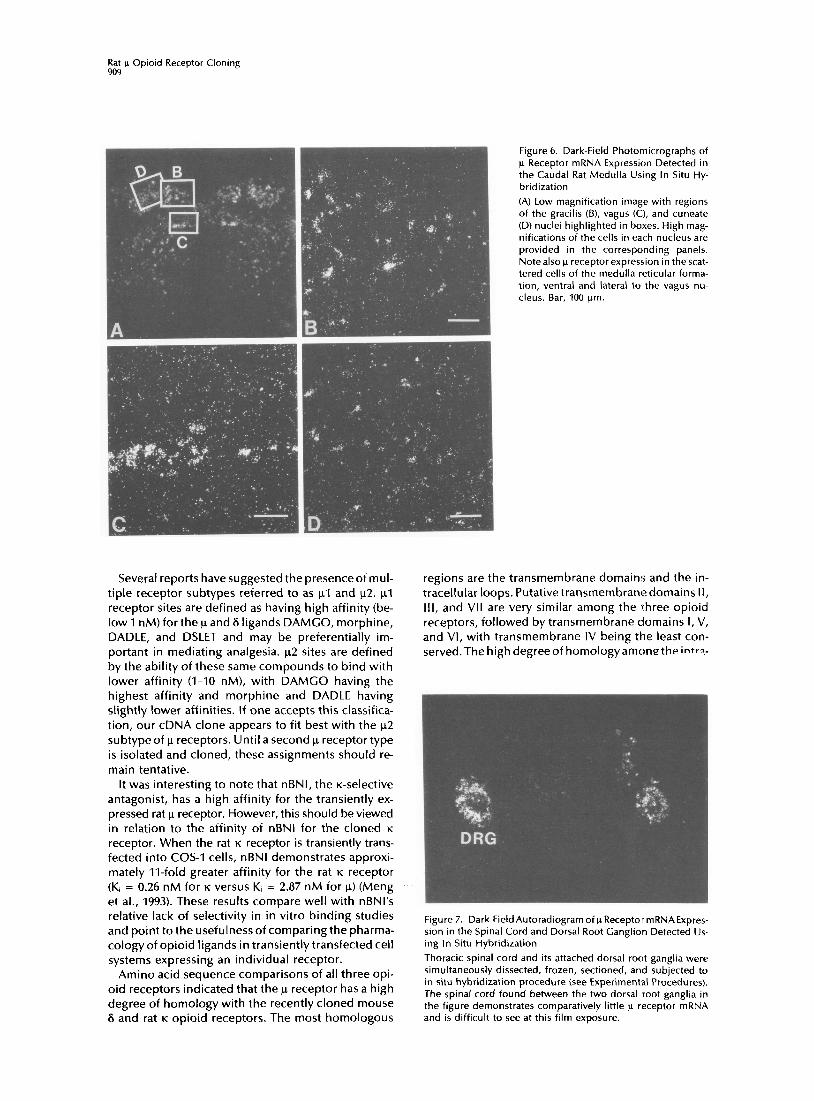
Moderate to high levels of p receptor expression were also observed in cells of the cuneate, gracilis, and vagus nuclei (Figure 6). Figure 6A shows a low magnification in situ hybridization image of the u re- ceptor mRNA distribution in the medulla, with the gracilis, vagus, and cuneate nuclei highlighted in boxes. As can also be seen from Figure 6A, the large cells of the medulla reticular nucleus also expressed u recep- tor mRNA.
In the spinal cord, the highest levels of opioid recep- tor binding were found in layers I and II, and only very low levels of u receptor mRNA could be found in the spinal cord. However, as can be seen in the coronal section of spinal cord in which the dorsal root ganglia were also transected, high levels of p receptor mRNA were found in the cells of the dorsal root gan- glia that project to layers I and II of the dorsal horn (Figure 7).
To control for potential cross-hybridization between this u receptor cDNA sequence and other closely re- lated receptors (K and 6 opioid receptors and somato- statin receptors), cRNA probes directed to different regions of the pRMuR-12 protein (both to transmem- brane domains Ill-VI and to the 5’ untranslated/N- terminus regiomwere used. In both cases, cRNA probes revealed indistinguishable mRNA distributions.
Discussion
The results of the present study demonstrate that we have cloned a novel member of the G protein-cou- pled receptor family that can be identified as a rat j.r opioid receptor. This conclusion is based on the following observations: First, the cloned receptor in- teracts with several classic opioid ligands, such as morphine, naltrexone, naloxone, levorphanol, and bremazocine, and their relative affinities are consis- tent with its opioid nature. Second, binding data show that the protein encoded by pRMuR-12 has a high affinity for the u agonist DAMGO and the u antagonist CTAP, whereas it has very low affinity for K-selective U50,488 and U69,593 and S-selective DPDPE. Third, the receptor exhibits the predicted stereospecificity, recognizing (-)bremazocine and levorphanol with af- finities several orders of magnitude higher than the affinities for their respective enantiomers, (+)brema- zocine and dextrorphan. Fourth, the rank orders of agonist and antagonist binding are consistent with previously described in vivo pharmacology reported for p receptors. Finally, the anatomical distribution of the mRNA coding for this receptor is in agreement with the distribution of u receptor binding as defined with pharmacological ligands. Taken together, these results provide compelling evidence that the present clone encodes a rat u opioid receptor.
Rat p Opioid Receptor Cloning 909
Several reports have suggested the presenceof mul- tiple receptor subtypes referred to as ~1 and ~2. ~1 receptor sites are defined as having high affinity (be- low 1 nM) for the p and 6 ligands DAMGO, morphine, DADLE, and DSLET and may be preferentially im- portant in mediating analgesia. ~2 sites are defined by the ability of these same compounds to bind with lower affinity (I-IO nM), with DAMGO having the highest affinity and morphine and DADLE having slightly lower affinities. If one accepts this classifica- tion, our cDNA clone appears to fit best with the ~2 subtype of p receptors. Until a second p receptor type is isolated and cloned, these assignments should re- main tentative.
It was interesting to note that nBNI, the K-selective antagonist, has a high affinity for the transiently ex- pressed rat p receptor. However, this should be viewed in relation to the affinity of nBNl for the cloned K
receptor. When the rat K receptor is transiently trans- fected into COS-1 cells, nBNl demonstrates approxi- mately II-fold greater affinity for the rat K receptor (Ki = 0.26 nM for K versus K, = 2.87 nM for p) (Meng et al., 1993). These results compare well with nBNl’s relative lack of selectivity in in vitro binding studies and point to the usefulness of comparing the pharma- cology of opioid ligands in transiently transfected cell systems expressing an individual receptor.
Amino acid sequence comparisons of all three opi- oid receptors indicated that the p receptor has a high degree of homology with the recently cloned mouse 6 and rat K opioid receptors. The most homologous
Figure 6. Dark-Field Photomicrographs of p Receptor mRNA Expression Detected in the Caudal Rat Medulla Using In Situ Hy- bridization
(A) Low magnification image with regions of the gracilis (B), vagus (C:I, and cuneate (D) nuclei highlighted in boxes. High mag- nifications of the cells in each nucleus are provided in the corresponding panels. Notealso p receptor expression in the scat- tered cells of the medulla reticular forma- tion, ventral and lateral to the vagus nu- cleus. Bar, 100 pm.
regions are the transmembrane domains and the in- tracellular loops. Putative transmembrane domains II, III, and VII are very similar among the three opioid receptors, followed by transmembrane domains I, V, and VI, with transmembrane IV being the least con- served.The highdegreeof homologyamonetheintr+
Figure 7. Dark-Field Autoradiogram of p Receptor mRNAExpres- sion in the Spinal Cord and Dorsal Root Ganglion Detected Us- ing In Situ Hybridization
Thoracic spinal cord and its attached dorsal root ganglia were simultaneously dissected, frozen, sectioned, and subjected to in situ hybridization procedure (see Experimental Procedures). The spinal cord found between the two dorsal root ganglia in the figure demonstrates comparatively little p receptor mRNA and is difficult to see at this film exposure.

NellrOn 910
cellular loops of the 6, xl, and p receptors suggests that the receptors may share similar second messen- ger systems, since the intracellular loops are thought to be critical for the coupling of this receptor family to G proteins (Clark et al., 1989). Additionally, many of the potential posttranslational features thought to be involved in the regulation of other receptors also appear to be conserved in opioid receptors. In addi- tion to the general presence of N-terminal glycosyla- tion sites, p receptor phosphorylation sites at amino acid279(protein kinaseA)andaminoacid363(protein kinase C) and a palmitoylation site at amino acid 346 appear to be conserved across opioid receptor pro- teins. One of these sites, Th?“, in the p receptor corresponds to a similar phosphorylation site in the B-adrenergic receptor. In the B-adrenergic receptor, this phosphorylation site is thought to be critical in agonist-induced desensitization mediated via G, acti- vation and elevation of CAMP levels, wherebythis site is phosphorylated, causing the dissocation of the re- ceptor from the G protein (Clark et al., 1989). This site is completely conserved among the three known classes of opioid receptors despite the fact that opioid recep- tors inhibit the generation of CAMP. Further studies will be needed to investigate the precise role, if any, of this conserved and putative phosphorylation site in opioid receptors.
Regions of divergence include the amino and car- boxy1 termini of the protein, as well as extracellular loops II and III. This sequence heterogeneity in these receptor domains is particularly striking and may be useful not only for discriminating opioid from non- opioid receptors (e.g., opioid from somatostatin), but also for discriminating opioid receptor types (K from 6 from p), as several of these regions are thought to be on the extracellular surface of the protein and may be involved in ligand selectivity. The notion that such extracellular domains may play an important role in determining ligand binding and ligand selectivity for peptide receptors was elegantly demonstrated in a series of studies on luteinizing hormone and follicle- stimulating hormone receptors (Braun et al., 1991; Roche et al., 1992).
Rat u receptor mRNA detected by Northern blot analysis is shown in Figure 2. Analysis of total RNA from many regions of the rat brain generated weak signals but indicated thatthe medulla/pans contained the greatest amount of p receptor mRNA and was cho- sen for poly(A) mRNA purification. A single large mo- lecular weight mRNA of approximately 12,000-14,000 nucleotides was detected in this poly(A) RNA from the medulltipons. Human SY5Y cellular mRNA was also analyzed because of the presence of p receptors in this cell line (as well as S receptors). It appears that a prominent mRNA of approximately 14,000-16,000 nucleotides is detected in this human cell line in addi- tion to several other smaller mRNA species. It is un- clear at this time whether these additional mRNAs are u receptor forms or represent nonspecific hybridiza- tion, even though these blots were hybridized at high
stringency. We are currently using DNA fragments from different regions of the rat p receptor cDNA to determine whether these fragments identify the same mRNA species in this human cell line. It is interesting to note that in the g-expressing cell line NC108 five to six mRNA species were detected, although it appears that only two mRNAs are found in brain tissue.
Genomic Southern blot analysis of rat DNA using thefull cDNA isolated from pRMuR-12 (approximately 2.0 kb) indicates several interesting features of the structure of the rat u receptor gene. First, the hybrid- ization pattern in the BamHl and Pstl digests is con- sistant with the hypothesis that a single p receptor gene exists in the rat genome. Second, there appears to be several introns separating the 2.0 kb cDNA. This is deduced from the multiple hybridization-positive bands in the EcoRl and Hindlll digest lanes seen in Figure3.Thereare noEcoRl sitesandonlyone Hindlll site in the 2.0 kb cDNA fragment used as a hybridiza- tion probe, although several hybridization-positive bands are detected. Approximately, three to five bands are detected in the EcoRl lane, and five bands are detected in the Hindlll lane,although onecannot rule out the possibilitythat some of these bands represent incomplete enzyme digests. In isolating cDNA clones encoding the rat p receptor, one unusual cDNA clone appeared to contain an intron near the transmem- brane IV location. Whether this cDNA clone contains an authentic intron or represents a cloning artifact is currently being evaluated. Interestingly, the mouse S opioid receptor gene has an intron in a very similar location (Evans, personal communication). Isolation and characterization of rat genomic clones will ad- dress the complete structure of the ).I receptor gene.
~1 receptor mRNA is expressed in many regions and nuclei throughout the CNS. The high level of pRMuR- 12 mRNA in theolfactory bulb, striatal patches, subcal- losal streak, thalamus, medial preoptic area, amygdala, interpeduncular, locus cerulus, parabrachial, cuneate, gracilis, and vagus nuclei, as well as in the nucleus of the solitarytract, isconsistentwith the known p-binding site distribution (Mansour et al., 1987; Mansour and Watson, 1993; McLean et al., 1986; Temple and Zukin, 1987) and the behavioral and pharmacological effects thought to be mediated by these receptors, including analgesia, respiration, cardiovascular effects, and hor- monal regulation. The visualization of p opioid recep- tor mRNA in the locus cerulus, ventral tegmental area, and substantia nigra, pars compacta is consistent with the ability of p ligands to modulate presynaptic nor- epinephrine and dopamine release and may be criti- cal in behavioral arousal and reward systems.
In situ hybridization results shown in Figure 7 sug- gest that while opioid receptor binding is localized predominantly in layers I and II of the spinal cord (Besse, 1991), b receptor mRNA is found predominantly in the dorsal root ganglia. This suggests that the vast majority of u receptors are likely transported from their site of synthesis in the dorsal root ganglia and are localized in presynaptic terminals in layers I and II

Rat p Opioid Receptor Cloning 911
of the spinal cord. This agrees well with lesion results (Besse et al., 1990), in which dorsal rhizotomy produced a 76% decrease in p receptor binding in the spinal cord. Low p receptor mRNA expression is also found in the dorsal horns of the spinal cord itself, consistent with the presence of postsynaptic receptor sites.
During the preparation of this paper, Chen et al. (1993) reported the cloning and pharmacological char- acterization of a w receptor also isolated from rat brain. The DNA sequence reported in this paper appears to be almost identical to the our b receptor cDNA sequence, differing in the translation of only 1 amino acid residue (valine for isoleucine substitution in trans- membrane region V). These authors also report ago- nist and antagonist binding profiles for their p recep- tor expressed in COS-7 cells. Many similarities are evident; however, they do not report binding for mor- phine. Additionally, these authors demonstrate that their p receptor is functionally coupled to the inhibi- tion of adenylate cyclase in the transiently transfected cells.
In summary, we have reported the isolation and characterization of a rat receptor cDNA whose phar- macological profile and anatomical distribution are consistent with the previously characterized p recep- tor. RNA analysis demonstrated that the rat and possi- bly the human p receptor mRNAs are large molecular weight species. Genomic DNA analysis in the rat sug- gests that the rat ~1 receptor gene is found in a single copy with several introns. The availability of cloned opioid receptorswill greatlyenhanceour understand- ing of the functions of these receptors in vivo, as well as provide for the systematic development of more selective agonists and antagonists. Developing ligands that can discriminate between various members of the opioid receptor family as well as between sub- classes within an opioid receptor group could pro- duce reagents that induce analgesia without many of the side effects (e.g., tolerance and dependence).
Experimental Procedures
Isolation of Opioid Receptor Fragment Oligonucleotide primers complementary to regions encoding transmembranedomains III,V,VI,andVII ofthemouseSopioid receptor DNA sequence were designed and used in PCR. Pri- mer 1 (TM 3), AAGAATTCTCAACATCATGAGCCTGCACCCCTACCCTAC; primer 2 (TM S), AAGAATTCCCAACCTGCTCCCGATCCTCAT- CATC; primer 3 (TM 6), AAAAGCTTCCACACCATGACGAAGAT- CTGCATCGC; primer 4 CTM 7), AAAACCTTITAACGGGAACGT- CGGGTAGGTCAGGCGGCAGCCCCACC. The PCR DNA tem- platewas first strand cDNA from rat olfactory bulb mRNA gener- ated with random hexamers using the Promega RiboClone cDNA Synthesis system. The PCR products were subcloned into a T-tailed vector system (Novagen) and sequenced using Sequenase (USB). Upon DNA sequencing, one amplified DNA fragment (432 bp) was shown to be 74% homologous to the NC108 8 opioid recep- tor mRNA from transmembrane regions III-VI. The DNA insert from this clone (designated p5A-1) was used for screening cDNA libraries and as a template to synthesize cRNA probes used for in situ hybridization.
Library Screening and Sequencing A rat olfactory bulb ZQAPII cDNA library was purchased and
plated according to the manufacturer’s recommendations (Stra- tagene). Approximately 3.2 x 10⁶ cDNA clones, were plated, and replicate nitrocellulose membranes were screened with a 32P-radiolabeled random-primed DNA insert from p5A-1 (432 bp). Prehybridization and hybridization conditions were 50% for- mamide, 1 x hybridization buffer (5x SSC, 50 mM KHP04 [pH 7.01, 1 x Denhardt’s solution, 0.1% PP04, 0.1% SDS, 200 pg/ml yeast RNA) at 37OC for 16-18 hr. Twelve positive clones were obtained and plaque the purified. DNA inserts were rescued into plasmid using the Ml3 helper phage Exassist (Stratagene). Restriction mapping and Southern blot analysiswere performed on rescued plasmid DNA. One clone, designated pRMuR-12, demonstrated a strong hybridization signal on Southern blot analysis and contained a cDNA insert of approximately 2.0 kb. Various restriction enzyme digestion fragment of clone pRMuR- 12 were subcloned into Bluescript and completely sequenced in both orientations. An EcoRI-Hindlll fragment was shown to contain the entire open reading frame of this cDNA. DNA se- quence data were analyzed by the GCG package (Genetics Com- puter Group, 1991). The DNA sequence of pRMuR-12 has been submitted to CenBank under accession number L22455.
Expression of Receptor and Binding Assay Initial binding assays performed on COS-1 cells transfected with the pCMV-neo expression vector containing the full open read- ing frame of pRMuR-12 documented the opioid nature of the encoded receptor. For binding assays, 25 pg of plasmid DNA was transfected into each 100 mm dish of COS-1 cells using the method of Chen and Okayama (1987). Receptor binding of the membrane preparation of the transfected cells was performed according to Goldstein and Naidu (1989). [‘H]IJ69,593 (58 Ci/ mmol; NEN), [3H]DAMG0 (55 Ci/mmol; NEN), and [SH]DPDPE (34.3 Cilmmol; NEN) were used in the characterization of the receptor. Nonspecific binding was defined as the binding of radie ligand in the presence of 1 PM levorphanol. PH]DAMGO (1.6 nM) was used to label the receptor in competition studies. All assays were conducted in 50 mM Tris buffer (pH 7.4) at room tempera- ture. Receptor binding results were analyzed with the LIGAND program (Munson and Rodbard, 1980).
Northern and Southern Blot Analyses RNA was isolated using standard guanidinium thiocyanate ex- tractions with subsequent poly(A) RNA chromatography using Fastrack (Invitrogen) kits. Poly(A) RNA (6 pg for ‘SYSY and 10 pg for medulltipons) was electrophoresed on 16% formaldehyde- agarose (1%) gels, transferred to nylon membrane (Nytran, Schlei- cher & Schuell, Inc.), UV cross-linked, prehybridized (2 hr), and hybridized (overnight) at 60°C in 50% formamide, 1 x HYB buffer (5x SSC, 50 mM NaP04 [pH 7.41, 0.1% NaPPOd, 5x Denhardt’s solution, 0.5% SDS). cRNA probes complementary to pSA-1 (see text) were generated using standard transcription methods and T7 RNA polymerase. Approximately, 2 x IO6 cpms of cRNA were used per ml of hybridization solution. Blots were washed in pro- gressively decreasing SSC solutions, with final concentrations of 0.1 x SSC, 0.5% SDS, at 72°C for 2 hr. DNA was isolated from rat liver tissue, crushed on liquid N2, resuspended in proteinase K buffer (50 mM Tris [pH 7.51, 0.5% SDS, 5 mM IiDTA), digested for 3 hr, and gently extracted several times with phenol prior to RNAase treatment. These nucleic acid samples were reextracted with phenol-chloroform and ethanol precipitated. DNA was di- gested overnightwith the indicated enzymes under manufacturer’s recommended conditions. These digests were emlectrophoresed on 1 x Tris borate EDTA, 1% agarose gels, acid nicked (30 min, 0.25 M HCI), denatured (2 x 30 min; 0.5 M NaOH, 1.5 M NaCI), neutralized (2 x 30 min; 0.5 M Tris [pH 7.41, 1.5 M NaCI), and transferred to nylon membranes (Nytran, Schleicher & Schuell, Inc.)inlOx SSCovernight. BlotswereUVcross-linkedand baked at80°C undervacuum for 1.5 hr, prehybridized (21 hr), and hybrid- ized at 37OC in 50% formamide, 1 x HYB buffer (see above) over- night. Random hexamer priming of the full 2.0~ kb cDNA from pRMuR-12 was used as a probe at a concentration of 1.0 x IO6 cpm per ml of hybridization buffer. Blots were washed in de-

NeUrO” 912
creasing SSC solutions (final concentrations of 0.1 x SSC, 0.5% SDS) at 60°C for 30 min.
In Situ Hybridization Adult male Sprague-Dawley rats (20-25 g; Charles River) were sacrificed by decapitation. The brains were then dissected and frozen in liquid isopentane (-3OOC) for 30 s and transferred to dry ice. Frozen brains were stored at -80°C until sectioning on a Bright cryostat (15 urn). Brain sections were thaw mounted on precleaned and polylysine-subbed microscope sl idesand stored at -80°C.
Frozen brain sections were directly removed from storage at -80°C and placed into 4% formaldehyde for 60 min (22°C) prior to processing for in situ hybridization (Mansour et al., 1990). Fol lowing three 5 min rinses in 2x SSC (300 m M NaCI, 30 m M sodium citrate [pH 7.2]), sections were treated with proteinase K (1 Kg/ml in 100 m M Tris [pH 8.01, 50 m M EDTA) for 10 min at 37OC. Slides were then rinsed in water, fol lowed by 0.1 M tr iethanolamine (pH 8.0), and treated with a mixture of 0.1 M tr iethanolamine (pH 8.0) and acetic anhydride (400:1, v/v) with stirring for 10 min. The sections were then rinsed in water, dehy- drated through graded alcohols, and al lowed to air dry.
Rat brain sections were hybridized with [3SS]UTP- and [3SS]CTP- labeled riboprobes generated to the pRMuR-12 clone. The TV re- ceptor cRNA was generated from a PCR fragment subcloned into pCEM4Z (designated p5A-1) and corresponds to t ransmembrane regions III-VI of the receptor. As p5A-1 was produced using mouse 8 opioid receptor mRNA primers, we also used a 759 bp EcoRI-BamHI cDNA fragment derived from the 5’ region of the pRMuR-12 cDNA clone. These two probes generated indistin- guishable mRNA distributions. cRNA probe was diluted in hy- bridization buffer(75% formamide, 10% dextran sulfate, 3x SSC, 50 m M Na2P0, [pH 7.4],1 x Denhardt’s solution, 0.1 Fgiml yeast tRNA, 10 m M dithiothreitoi) to give a final concentration of 1 x IO6 to 2 x IO6 cpmi30 ~1. Diluted probe was appl ied to coronal (40 ul) and horizontal (100 ul) sections. Glass coverslips were placed over brain sections to keep cRNA probes and hybridiza- tion buffer in contact with tissue.
Slides were then transferred to chambers containing 50% for- mamide and hybridized at 55”C, overnight. The slides were then rinsed in 2x SSC (5 min) and treated with RNAase A (200 ugi ml in 100 m M Tris [pH 8.01, 0.5 M NaCI) for 60 min at 37OC. Subsequently, the sections were rinsed in 2x SSC for 5 min (22OC) and 0.1 x SSC for 60 min (65°C). Fol lowing the low salt wash, sections were rinsed in water, and dehydrated through graded alcohols, and air dried. Sections were apposed to Kodak XAR-5 X-ray film for 1-11 days or d ipped in NTB2 film emulsion and developed 3-14 days later.
Acknowledgments
The authors would like to thank Dr. James Woods for providing the unlabeled opioid compounds used in this study and Drs. Bianca Ruzicka and Charles A. Fox for their critical reading and helpful discussion of this manuscript. This work was supported by NIDA Grant ROI DA02265 to H. A. and S. J. W., by Markey Grant (from the Lucille P. Markey Charitable Trust) #88-46 to H. A. and S. J. W., and by Gut Center Grant P30-AM34933 to H. A. and S. J. W.
The costs of publication of this article were defrayed in part by the payment of page charges. This article must therefore be hereby marked “advert isement” in accordance with 18 USC Sec- tion 1734 solely to indicate this fact.
Received July 22, 1993; revised August 25, 1993.
References
Attali, B., Saya, D., and Vogel, Z. (1989). Kappa-opiate agonists inhibit adenylate cyclase and produce heterologous desensitiza- tion in rat spinal chord. J. Neurochem. 5.2, 360-369.
Barchfeld, C., and Medzihradsky, F. (1984). Receptor-mediated
stimulation of brain GTPase by opiates in normal and dependent rats. Biochem. Biophys. Res. Commun. 127, 641-648.
Besse, D., Lombard, M. C.,Zajac, J. M., Roques, B. P., and Besson, J. M. (1990). Pre and postsynaptic distribution of p, S, and K opioid receptors in the superficial layers of the cervical dorsal horn of the rat spinal cord. Brain Res. 521, 15-22.
Beese, D., Lombard, M. C., and Besson, J. M. (1991). Autoradio- graphic distribution of p, 6, and K opioid binding sites in the superficial dorsal horn, over the rostrocaudal axis of the rat spi- nal cord. Brain Res. 548, 287-291.
Bidlack, J. M., and Abood, L. C. (1980). Solubilization of the opiate receptor. Life Sci. 27, 331-340.
Braun, T., Schofieid, P. R., and Sprengel, R. (1991). Amino- terminal leucine-rich repeats in gonadotropin receptors deter- mine hormone selectivity. EMBO J. 70, 1885-1890.
Chen, C., and Okayama, H. (1987). High efficiency transforma- tion of mammal ian cells by plasmid DNA. Mol. Cell. Biol. 7,2745- 2752.
Chen, Y., Mestek, A., Liu, J., Huriey,J. A., and Yu, L. (1993). Molec- ular cloning and functional expression of a p-opioid receptor from rat brain. Mol. Pharmacol. 44, 8-12.
Cho,T. M., Yamato, C., Cho,J. S., and Loh, Ii. H. (1981). Solubiliza- tion of membrane bound opiate receptors from rat brain. Life Sci. 28, 2651-2657.
Cho, T. M., Hasegawa, J.-l., Ge, B.-L., and Loh, H. H. (1986). Purifi- cation to apparent homogenei ty of a p-type opioid receptor from rat brain. Proc. Natl. Acad. Sci. USA 83, 4138-4142.
Chow, T., and Zukin, R. S. (1983). Solubilization and preliminary characterization of mu and kappa opiate receptor subtypes from rat brain. Mol. Pharmacol. 24, 203-212.
Clark, R. B., Fr iedman, J., Dixon, R. A., and Strader, C. D. (1989). Identification of a specific site required for rapid heterologous desensitization of the beta-adrenergic receptor by cAMP-depen- dent protein kinase. Mol. Pharmacol. 36, 343-348.
Demoliou-Mason, C., and Barnard, E. A. (1984). Solubilization of high yield of opioid receptors retaining high-affinity delta, mu and kappa binding sites. FEBS Lett. 170, 378-382.
Evans, C. J., Keith, D. E., Morrison, H., Magendro, K., and Ed- wards, R. H. (1992). Cloning of a delta opioid receptor by func- tional expression. Science 258,1952-1955.
Genetics Computer Group (1991). Program Manual for the CCC Package (Madison, Wisconsin: University of Wisconsin Press).
Gilbert, P. E., and Martin, W. R. (1976). The effects of morphine- and nalorphine-like drugs in the nondependent , morphine- dependent and cyclazocine-dependent chronic spinal dog. J. Pharmacol. Exp. The:. 798, 66-82. Gillan, M. C. C., and Kosterlitz, H. W. (1982). Spectrum of the mu, delta, and kappa binding sites in homogenates of rat brain. Br. J. Pharmacol. 77, 461-469.
Goldstein, A., and James, I. F. (1984). Site-directed alkylation of multiple opioid receptors. II. Pharmacological selectivity. Mol. Pharmacol. 25, 343-348.
Goldstein, A., and Naidu, A. (1989). Multiple opioid receptors: l igand selectivity profiles and binding site signatures, Mol. Phar- macol. 36, 265-272.
Herling, S., and Woods, J. H. (1984). Discriminative stimulus ef- fects of narcotics: evidence for multiple receptor-mediated ac- tions. Life Sci. 28, 1571-1584.
Howells, R. D., Cioannini, T. L., Hilier, J. M., and Simon, E. J. (1982). Solubilization and characterization of active binding sites from mammal ian brain. J. Pharmacol. Exp. Ther. 222, 629-634.
Hsia, J. A., Moss, J., Hewlett, E. L., and Vaughan, M. (1984). ADP- ribosylation of adenylate cyclase by pertussis toxin. effects on inhibitory agonist binding. J. Biol. Chem. 259, 1086-1090.
lyengar, S., Kim, H. S., and Wood, P. L. (1986). Effects of K opiate agonists on neurochemical and neuroendocrine indices: evi- dence for K receptor subtypes. Life Sci. 39, 637-644.
James, I. F., and Goldstein, A. (1984). Site-directed alkylation of

Rat p Opioid Receptor Cloning 913
multipleopioid receptors. I. Binding selectivity. Mol. Pharmacol. 25, 337-342.
Kieffer, B. L.,Befort, K.,Caveriaux-Ruff,C.,and Hirth, C.C.(1992). The&opioid receptor: isolation of acDNA by expression cloning and pharmacological characterization. Proc. Natl. Acad. Sci. USA 89, 1204812052.
Koski, G., and Klee, W. (1981). Opiates inhibit adenylate cyclase by stimulating CTP hydrolysis. Proc. Natl. Acad. Sci. USA 78, 4185-4189.
Leander, J. D., Zerbr, R. L., and Hart, j. C. (1985). Diuresis and suppression of vasopressin by kappa opioids: comparison with mu and delta opioids and clonidine. J. Pharmacol. Exp. Ther. 234, 463-469.
Lutz, R. A., and Pfister, H. P. (1992). Opioid receptors and their pharmacological profiles. J. Receptor Res. 72, 267-286.
Maneckjee, R., Zukin, R. S., Archer, S., Michael, J., and Osei- Gyimah, P. (1985). Purification and characterization of the p opi- ate receptor from rat brain using affinity chromatography. Proc. Natl. Acad. Sci. USA 82, 594-598.
Mansour, A., and Watson, S. J. (1993). Anatomical Distribution of Opioid Receptors in Mammalians: An Overview (Berlin: Springer-Verlag).
Mansour, A., Khachaturin, H., Lewis, M. E., Akil, H., and Watson, S. J. (1987). Autoradiographic differentiation of mu, delta, and kappaopioid receptors in the rat forebrain and midbrain. J. Neu- roscience 7, 2445-2464.
Mansour, A., Khachaturian, H., Lewis, M. E.,Akil, H.,and Watson, S. J. (1988). Anatomy of CNS opioid receptors. Trends Neurosci. 11, 308-314.
Manzanares, I., Lookingland, K. J., and Moore, K. E. (1990). Kappa-opioid-receptor-mediated regulation of alpha-melano- cyte-stimulating hormone secretion and tuberohypophyseal dopaminergic neuronal activity. Neuroendocrinology 52, 200- 205.
Martin, W. R., Eades, C. C., Thompson, J. A., Huppler, R. E., and Gilbert, P. E. (1976). The effects of morphine- and nalorphine-like drugs in the nondependent and morphine-dependent chronic spinal dog. J. Pharmacol. Exp. Ther. 797, 517-532.
McLean, S., Rothman, R. B., and Herkenham, M. (1986). Autora- diographic localization of p-and S-opiate receptors in the fore- brain of the rat. Brain Res. 378, 49-60.
Meng, F., Xie, G.-X., Thompson, R. C., Mansour, A., Watson, S. J., and Akil, H. (1993). Cloning and pharmacological character- ization of a rat kappaopioid receptor. Proc. Natl. Acad. Sci. USA, in press. Morley, J. E., and Levine, A. S. (1983). Involvement of dynrophin and the K opioid receptor in feeding. Peptides 4, 797-800.
Munson, P. J., and Rodbard, D. (1980). LICAND: a versatile com- puterized approach for characterization of ligand-binding sys- tems. Anal. Biochem. 707, 220-239.
Neck, B., Rajpara, A., O’Connor, L. H., and Cicero, T. J. (1988). Autoradiography of (3H]U-69593 binding sites in rat brain: evi- dence for kappa opioid receptor subtypes. Eur. J. Pharmacol. 154, 27-34.
Pasternak, G. W. (1993). Pharmcological mechanisms of opioid analgesics. Clin. Neuropharmacol. 76, 1-18.
Pasternak, G. W., and Snyder, S. H. (1975). Identification of novel high affinity opiate receptor binding in rat brain. Nature 253, 563-565.
Pasternak, G. W., and Wood, P. L. (1986). Multiple R receptors. Life Sci. 38, 1889-1898.
Puttfarchen, P. S., Werling, L. L., and Cox, B. M. (1988). Effects of chronic morphine exposure on opioid inhibition of adenylate cyclase in 7315~ cell membranes: a useful model for the study of tolerance at n opioid receptors. Mol. Pharmacol. 33,520-527.
Roche, P. C., Ryan, R. J., and McCormick, D. J. (1992). Identifica- tion of hormone-binding regions of the luteinizing hormone/ human chorionic gonadotropin receptor using synthetic pep-
tides. Endocrinology 737, 268-274.
Rothman, R. B., Jacobson, A. E., Rice, K. C., and Herkenham, M. (1987). Autoradiographic evidence for two classes of p opioid binding sites in rat brain using [‘251]FK33824. Peptides 8, 1015- 1021.
Ruegg, U. T., Hiller, J. M., and Simon, E. J. (1980). Solubilization of an active opiate receptor from Bufo marinus. Eur. J. Pharmacol. 64, 367-368.
Schofield, P. R., McFarland, K. C., Hayflick, J. S., Wilcox, J. N., Cho, T. M., Roy, S., Lee, N. M., Loh, H. H., and Seeburg, P. H. (1989). Molecular characterization of a new immunoglobulin su- perfamily protein with potential roles in opioid binding and cell contact. EMBO J. 8, 489-495.
Simon, E. J. (1991). Opioid receptors and endogenous opioid peptides. Medicinal Res. Rev. 77, 357-374.
Simon, E. J., Hiller, J. M., and Edelman, I. (1975). Solubilization of a stereospecific opiate-macromolecular complex from rat brain. Science 790, 389-390. Simonds, W. F., Koski, C., Streaty, R. A., Hjelmeland, L. M., and Klee, W. A. (1980). Solubilization of active opiate receptors. Proc. Natl. Acad. Sci. USA 77, 4623-4627.
Temple, A., and Zukin, R. S. (1987). Neuroanatomical patterns of the p, 8, and K opioid receptors of rat brain as determined by quantitative in vitro autoradiography. Proc. INatl. Acad. Sci. USA 84, 4308-4312.
Ueda, H., Harada, H., Misawa, H., Nozaki, M., and Takagi, H. (1987). Purified opioid p-receptor is of a different molecular size than 8- and x-receptors. Neurosci. Lett. 75, 339-344.
Unterwald, E. M., Knapp, C., and Zukin, R. S. (1091). Neuroana- tomical localization of kappa 1 and kappa 2 opioid receptors in rat and guinea pig brain. Brain Res. 562, 57-65.
Wolozin, B. L., and Pasternak, C. W. (1981). Classjfication of mul- tiple morphine and enkephalin binding sites in the central ner- vous system. Proc. Natl. Acad. Sci. USA 78, 6181-6185.
Wood, P. L. (1982). Multiple opiate receptors: support for unique mu, delta, and kappa sites. Neuropharmacology 27, 487-497.
Wood, P. L. (1988).Thesignificanceof multiple CNS opioid recep- tor types: a review of critical considerations relating to technical details and anatomy in the study of central opioid actions. Pep- tides 7, 49-55.
Xie, Y. B., Wang, H., and Segaloff, D. L. (1990). Extracellular do- main of lutropinlchoriogonadotropin receptor expressed in transfected cells binds choriogonadotropin with high affinity. J. Biol. Chem. 265, 21411-21414.
Xie, G.-X., Miyajima, A., and Goldstein, A. (1992). Expression clon- ing of cDNA encoding a seven-helix receptor from human pla- centa with affinity for opioid ligands. Proc. Natl. Acad. Sci. USA 89, 4124-4128.
Yasuda, K., Raynor, K., Kong, H., Breder, C. D., Reisene, T., and Bell, C. I. (1993). Cloning and functional comparison of kappa and delta opioid receptors from mouse brain. Proc. Natl. Acad. Sci. USA, in press.