1 from molecules to medicine - mikeblaber.orgmikeblaber.org/2011_lss.pdf · from molecules to...
TRANSCRIPT
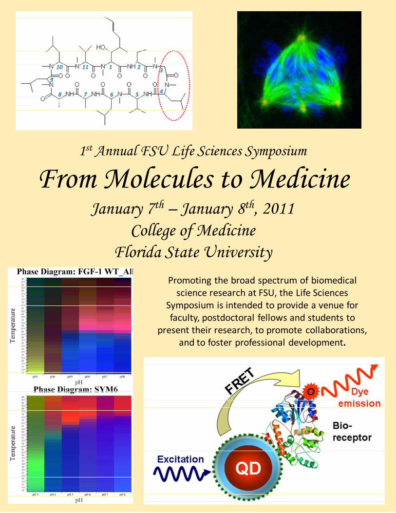
1st Annual FSU Life Sciences Symposium
From Molecules to MedicineJanuary 7th – January 8th, 2011
College of MedicineFlorida State University
Promoting the broad spectrum of biomedical science research at FSU, the Life Sciences
Symposium is intended to provide a venue for faculty, postdoctoral fellows and students to
present their research, to promote collaborations, and to foster professional development.
2
MESSAGE FROM THE PROGRAM CHAIR
The idea for this symposium grew from the tremendous changes that have occurred over the past several years at FSU in the area of life sciences. Physical infrastructure has been the most obvious change, with new science buildings in the College of Medicine, and departments of Biological Science, Psychology and Chemistry all springing up over the past several years. Less obvious, but perhaps more importantly, life sciences research is being pursued by a wide variety of investigators, and in departments that you might not normally associate with such
research. New approaches, as well as solutions to old problems, can emerge from collaborative interactions between such faculty, postdocs, students and staff – but, only if we can successfully raise awareness of this wide diversity of life sciences research. That is the goal for this symposium. I would like to thank the Department of Biomedical Sciences, the Office of Research, the symposium steering committee, the administrative staff of the department of Biomedical Sciences, and our corporate and university sponsors for their support in making the symposium actually happen. I would like to thank everyone in attendance; hopefully, the symposium will prove useful. Dr. Michael Blaber Chair, 2011 FSU Life Sciences Symposium Organizing Committee College of Medicine, Florida State University
Welcome! The Department of Biomedical Sciences is pleased to sponsor the inaugural 2011 FSU Life Sciences Symposium "From Molecules to Medicine". Our hope is to highlight the accomplishments of biomedical science researchers at FSU. We wish to bring together faculty, postdoctoral fellows, graduate students and staff to listen and learn from each other and from outside speakers. We hope to generate an atmosphere of excitement about the life sciences at FSU, and a view of a future in which we have a shared vision amidst a growing
atmosphere of collaboration. Most of all we hope that all of you will leave here with a feeling of camaraderie and community. The organizing committee was selected to represent the diversity among the FSU Life Sciences community. I commend them for the extraordinary program they have assembled. I look forward to an exciting two-day symposium during which I expect both to learn a great deal and to make many new friends. I trust you will do the same. Dr. Richard Nowakowski Chair, Biomedical Sciences College of Medicine, Florida State University

3
2011 FSU Life Sciences Symposium
OFFICIAL PROGRAM
MESSAGE FROM THE PROGRAM CHAIR……………………………… PAGE 2
SITE MAP ……………………………………………………………………. PAGE 4
MEETING AT A GLANCE…………………………………………………... PAGE 5-6
ORAL SESSION ABSTRACTS……………………………………………. PAGE 7-20
POSTER SESSION ABSTRACTS……………………………………….... PAGE 21-45
REGISTERED ATTENDEES………………………………………………. PAGE 46-51
REGISTERED VENDORS…………………………………………………. PAGE 52
ATTENDEE AFFILIATION…………………………………………………. PAGE 52
NOTES………………………………………………………………..……… PAGE 53-54
SPONSORS…………………………………………………………………. PAGE 55
CREDITS…………………………………………………………………….. PAGE 56
4
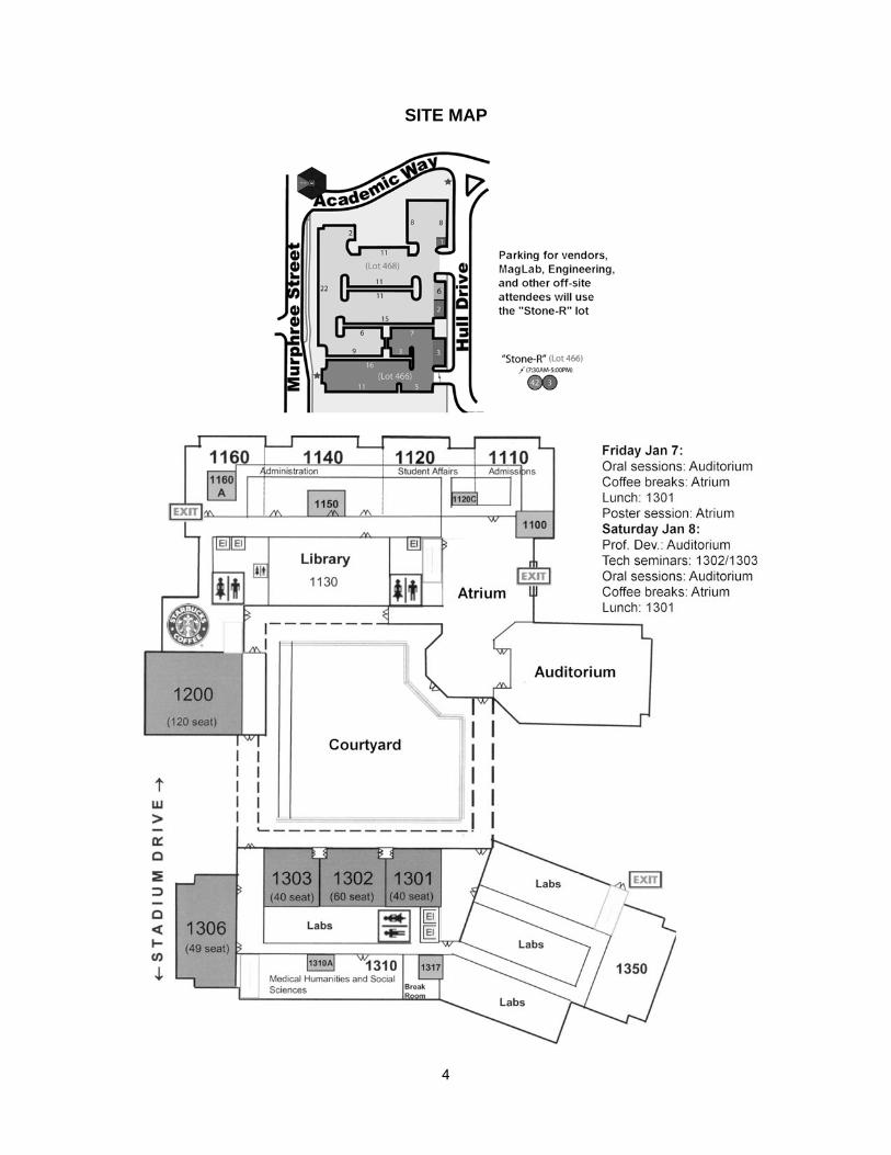
SITE MAP

5
MEETING AT A GLANCE
Day 1 – Friday Jan 7 2011
8:30 – 8:45 Welcome 8:45 Morning Session I (Auditorium) Session chairs: Scott Stagg and Hedi Mattoussi 8:45 – 9:30 Michael Dustin, NYU medical school How Killer T Lymphocytes Lock onto Tumor Cells 9:30 – 10:00 Joe Schlenoff, FSU Dept. Chemistry & Biochemistry Cells in Tune with Surfaces 10:00 – 10:30 Geoffrey Strouse, FSU Dept. Chemistry & Biochemistry
Nanomaterials to Treat Genetic Disorders
10:30 Break/coffee (Atrium) 10:45 – 11:15 Anant Paravastu, FSU Dept. Chemical & Biomed. Eng.
Solid State NMR Structural Characterization of Disease Related and Designer Protein Self-Assembly
11:15 – 11:45 Tim Cross, FSU Dept. Chemistry & Biochemistry Opportunities, Challenges and Urgency for the Characterization of Membrane Protein Drug Targets
11:45 – 12:15 Scott Stagg, FSU Dept. Chemistry & Biochemistry/Institute of Molecular Biophys. Structure and Heterogeneity of the COPII Coat
12:15 - 1:15 Lunch (Box lunch in 1301 for registered attendees) 1:30 Afternoon Session I (Auditorium) Session chairs: Hengli Tang and Tim Megraw 1:30-2:15 Val Sheffield, HHMI/ University of Iowa
Human Genetics of Bardet-Biedel Syndrom (BBS) 2:15-2:45 David Houle, FSU Biological Science
Phenomics: the Next Challenge 2:45-3:15 Jonathan Dennis, FSU Biological Science/Institute of Molecular Biophysics
The Regulatory Organization of the Human Genome
3:15 Break/coffee (Atrium) Group Photo – Atrium 3:30 – 4:00 Beth Stroupe, FSU Biological Science/Institute of Molecular Biophysics
Structure and Activity in Bacterial Sulfite Reductase 4:00 – 4:30 Brian Chadwick, FSU Biological Science
Exploring the Role of Macrosatellites in Genome Biology and Disease Susceptibility
4:30 – 5:00 David Gilbert, FSU Biological Science SPACE AND TIME IN THE NUCLEUS: Replication Timing Reflects the Stable Alterations in 3D Chromosome Organization During Development and Disease
5:00 – 5:30 Tim Megraw, FSU Dept. Biomedical Sciences Cell Biology of MCPH: a Neural Stem Cell Disease
5:30 – 7:00 Posers/vendors/Wine & Cheese refreshments (Atrium)

6
Day 2 – Saturday Jan 8 2011
Concurrent Sessions 8:30 – 9:20 9:25 – 10:15
Room 1400
Jeff W. Garis, FSU Career Center C.V./resume Preparation
Jeff W. Garis, FSU Career Center Interview Skills
Room 1302
Joel Silfies, Nikon Instruments Inc. Detection, Resolution and Imaging Beyond the Abbe Diffraction Limit
Juergen von der Heiden, Hunt Optics and Imagine CARS and SHG: Multiphoton Excitation without Fluorophores!
Room 1303
Tim Sackos and Manju R. Sethi, Thermo Scientific The Future of QPCR: Best Practices, Standardization, and the MIQE Guidelines
Kirsteen Maclean, Genomics Division, Thermo Scientific Adavances in qPCR and PCR Assays
10:15 Break/coffee (Atrium) 10:30 Morning Session II (Auditorium) Session chair: Mike Blaber 10:30 – 11:15 Peter Sarnow, Stanford U MicroRNAs in Viral Hepatitis 11:15 – 11:45 Branko Stefanovic, FSU Dept. of Biomedical Sciences
Progress towards Discovery of Antifibrotic Drugs 11:45 – 12:15 Yoichi Kato, FSU Dept. of Biomedical Sciences
Notch a Victory over Diseases 12:15 - 1:30 Lunch (Box lunch in 1301 for registered attendees) 1:30 Afternoon Session II (Auditorium) Session chair: Richard Hyson 1:30-2:15 Stephen Dalton, UGA
Cardiovascular Progenitors Derived from Human Pluripotent Cells 2:15-2:45 Karen Berkley, FSU Dept. of Psychology/Program in Neuroscience
Neural mechanisms of Pelvic Pain: Lessons from Translational Research on Endometriosis
2:45-3:15 Carlos Bolaños, FSU Dept. of Psychology/Program in Neuroscience Extracellular Signal-Regulated Kinase-2 (ERK2) as a Potential Target for Drug- and Mood-Related Co-Morbid Behaviors
3:15 Break/coffee (Atrium) 3:30 – 4:00 Mohamed Kabbaj, FSU Dept. of Biomedical Sciences
Individual Differences in the Effects of Social Defeat in Rats: the HR/LR Model 4:00 – 4:30 Josh Rodefer:, FSU Dept. of Psychology/Program in Neuroscience
Neuropharmacology and Cognitive Flexibility: Implications for Neuropsychiatric Disorders
4:30 – 5:00 Michael Blaber, FSU Dept. of Biomedical Sciences Protein Engineering for Human Therapeutics
5:00 – 5:30 Roger Mercer, FSU Dept. of Biomedical Sciences Translation in Transition: Building a New Translational Science Lab at FSU
5:30 – 5:45 Closing remarks

7
ORAL SESSION ABSTRACTS
Friday Jan 7 2011 Session I Morning
HOW KILLER T LYMPHOCYTES LOCK ONTO TUMORS
Michael Dustin, Maria Grazia Ruocco, Kaushik Choudhuri, Noriko Kawashima, Silvia Formenti and Sandra Demaria
Pathology, NYU School of Medicine, New York, NY, 10016
Synergy of radiation and anti-CTLA-4 blockade has been shown to induce CD8-driven tumor regression in a mouse
model of breast cancer. To understand the mechanism, we employed intravital 2-photon imaging techniques that
allow us to characterize the interactions between T cells and tumor cells in the tumor microenvironment. In order to
visualize T cells, we used a transgenic mouse line in which the green fluorescent protein was expressed under the
CXCR6 promoter. CXCR6 is a chemokine receptor expressed on effector T cells. To visualize tumor cells, we
transduced 4T1 cells with a retroviral vector expressing the cyan fluorescent protein. We analyzed tumor bearing
mice that were 1) untreated, 2) treated with radiation therapy alone, 3) treated with anti-CTLA-4 alone and 4) treated
with a combination of radiation therapy and anti-CTLA-4. We found that anti-CTLA-4 mAb (9H10) as monotherapy
increased T cell motility in vivo, preventing the formation of stable interactions between T cells and tumor cells. In
contrast, the combination of radiotherapy and anti-CTLA-4 mAb promoted arrest of T cells with tumor cells. We found
that the expression of the NKG2D ligand Rae-1 on tumor cells is increased following radiation therapy in vivo. We
identified the interaction between radiation-induced Rae-1 and NKG2D receptor expressed on T cells as an important
co-stimulatory signal that counteracts the go signal provided by anti-CTLA-4. Blocking this interaction prevented T
cell arrest in vivo and tumors continued to grow even when treated with radiation and anti-CTLA-4. Our results
suggest that Rae-1/NKG2D is a critical contributor to the formation of stable junctions between T cells and tumor
cells. The radiation induced Rae-1/NKG2D interaction is a required co-factor for 4T1 eradication and provides a
molecular mechanism underlying the effectiveness of combined radiation and anti-CTLA-4 treatment.
CELLS IN TUNE WITH SURFACES
Joe Schlenoff, Tom Keller, Jessica Martinez, Ali Lehaf and Maroun Moussallem
Chemistry & Biochemistry, FSU, Tallahassee, FL, 32306
In collaboration with the Keller group in the Department of Biological Sciences we are looking at how cells respond to
the surfaces on which they grow. To provide the most control over the surface properties, we use thin polymer films,
which we make from water-soluble polymers. Both chemical and physical properties can be systematically varied to
control cell adhesion, growth, movement and proliferation. This interdisciplinary project involves cell biology,
nanotechnology and polymer science.

8
IS THERE A PLACE FOR NANOSCIENCE IN BIOMEDICINE? THE USE OF NANOMATERIALS FOR TRACKING
AND CONTROLLING GENE DELIVERY
Geoffrey F. Strouse
Department of Chemistry and Biochemistry, Florida State University, Tallahassee, FL, 32306-4390
Nanomaterials (quantum dots, gold nanoparticles, SPIOs, lipoplexes, etc) are widely used today in biological, bio-
physical, and medical research. Recent developments include the use of NSET to track the release of a gene from a
gold nanometal to induce in-vitro targeted gene expression via a controlled delivery mechanism. In addition, the
ability to utilize the nanomaterial in-vivo as a tracer allows use of MRI methods to follow the delivery of the material
and its fate. Such advances are opening the potential to manipulate protein expression levels in-vivo and provide a
tracking modality once the nanoplex is delivered. Discussion of advances in controlled gene delivery and use of
advanced optical and spectroscopic methods to track nanomaterial delivery will be discussed.
SOLID STATE NMR STRUCTURAL CHARACTERIZATION OF DISEASE RELATED AND DESIGNER PROTEIN
SELF-ASSEMBLY
Anant Paravastu, Ashley Cormier, Serena Danting Huang, William Tay and Terrone L. Rosenberry
Chemical and Biomedical Engineering, Florida State University and Florida A&M Universtiy, Tallahassee, FL, 32310
In biology, protein self-assembly is associated with the formation of nanostructured materials (e.g., extracellular
matrices) and disease-associated plaques (e.g., amyloid deposits). Recent work on amyloid fibril forming proteins
suggests that soluble oligomeric structures play a key role in cellular toxicity. We seek an atomic level understanding
of protein self-assembly in order to design synthetic self-assembling peptides (small proteins) capable of supporting
tissue repair while avoiding formation of toxic structures. Towards this goal, we have performed solid state NMR
measurements on two designer self-assembling peptides from the literature and a stable oligomeric form of the 42-
residue variant of the Alzheimer’s β-amyloid peptide. Unlike in-vivo derived peptides, the designer self-assembling
peptides have very simple repetitive amino acid sequences designed to promote specific secondary structures. NMR
spectra unexpectedly indicate a high degree of structural order in these systems, suggesting highly ordered
supramolecular organization despite the repetitive sequences. Hydration effects on observed NMR linewidths
suggest a critical role of water in stabilizing self-assembled structures. Oligomeric β-amyloid, in contrast, seems to
have a structure that is not stabilized by water. However, the presence of water in oligomeric β-amyloid does
introduce site-dependent molecular motion that has not been observed in fibrillar β-amyloid.

9
OPPORTUNITIES, CHALLENGES AND URGENCY FOR THE CHARACTERIZATION OF MEMBRANE PROTEIN
DRUG TARGETS
Timothy A. Cross, Sharma, M., Huajun, Q., Petersen, E., Busath, D., Yi, M, Dong, H. & Zhou, H.-X
Department of Chemistry and Biochemistry, Department of Physics, Institute of Molecular Biophysics, National High
Magnetic Field Laboratory, Florida State University, Department of Physiology and Developmental Biology, Brigham
Young University
More than 50% of all drug targets are membrane proteins and yet this is the least well understood class of proteins.
They have multiple conformations generating multiple opportunities for drug development. Buth their conformation is
influenced by their environment as Anfinsen’s thermodynamic hypothesis states “that the native conformation (of a
protein) is determined by the totality of inter-atomic interactions and hence by the amino acid sequence in a given
environment.” Too often these last four words are ignored and therefore many membrane protein structures in the
Protein Data Bank are distorted and not representative of their native conformation. Today, there is a great urgency
to develop new classes of antimicrobial agents and while membrane proteins provide great opportunities the
challenge of obtaining structural characterizations of these proteins are great. A new approach using NMR
spectroscopy of lipid bilayer preparations has considerable promise. Here. I will show our first success with the
characterization of the conductance domain of M2 protein from Influenza A in a liquid crystalline lipid bilayer
environment. This protein is a proven drug target but the virus has recently mutated such that both the seasonal flu
and the swine flu pandemic were resistant to the drugs that target this essential protein for the viral life cycle.
THE STRUCTURE AND HETEROGENEITY OF THE COPII COAT
Scott Stagg, Jason O'Donnell and Nilakshee Bhattacharya
Institute of Molecular Biophysics, Florida State University, Tallahassee, FL, 32306
The COPII proteins Sar1, Sec23/24 and Sec13/31 are involved in transporting cargo from the endoplasmic reticulum
to the Golgi apparatus. In recent years several structures have been solved that elucidate the mechanisms employed
by the individual COPII proteins. In order to transport the large variety of cargo in the cell, the COPII coat must be
capable of expanding and contracting. We have used electron tomography to quantify the range of conformations
accommodated by the Sec13/31 cage. Moreover, we have reconstructed a structure of a COPII coat cage assembled
from Sec13/31 and Sec23. The assemblies form at least two geometries, and the most common size is 600 Å, similar
to what has been observed for Sec13/31. We will discuss how the orientation of Sec23 may dictate cage geometry
and orient Sar1 to participate in the fission of COPII coated vesicles in the cell.

10
Session I Afternoon
MOLECULAR MECHANISMS INVOLVED IN BARDET-BIEDL SYNDROME, A HUMAN OBESITY DISORDER
Val Sheffield, Qihong Zhang and Seongjin Seo
Pediatrics/ Medical Genetics, University of Iowa/ HHMI, Iowa City, IA, 52242
Bardet-Biedl Syndrome (BBS) is an autosomal recessive disorder resulting in obesity, polydactyly, retinal
degeneration, renal abnormalities, diabetes, hypertension, and cognitive impairment. Our laboratory has focused on
identifying genes involved in BBS, determining the functions of BBS proteins, and developing BBS animal models to
aid in the understanding of the pathophysiology of BBS-related phenotypes. Fifteen genes have been reported to be
involved in BBS. Among the known BBS proteins, seven proteins form a stable complex known as the BBSome,
which mediates vesicle trafficking to the ciliary membrane. Three BBS proteins (BBS6, BBS10, BBS12) form a
second complex with the CCT/TRiC family of group II chaperonins. This complex mediates BBSome assembly. BBS3
(ARL6), a member of Ras super family of small GTPases, is not required for BBSome assembly but controls BBSome
recruitment and ciliary entry. Many receptor proteins and signaling molecules localize to cilia, and the BBSome is
involved in transporting at least some of these proteins in and out of the cilia. In addition, the BBSome is involved in
some functions not clearly related to ciliary trafficking including leptin receptor signaling. In an effort to understand
how BBSome function is regulated, we have identified BBSome interacting proteins in vivo. This work has led to the
identification of a novel negative regulator of BBSome trafficking, which has potential implications in the treatment of
BBS phenotypes.
PHENOMICS: THE NEXT CHALLENGE
David Houle
Biological Science, Florida State University, Tallahassee, FL, 32308
A key goal of biology is to understand phenotypic characteristics, such as health, disease, and evolutionary fitness.
Phenotypic variation is produced through a complex web of interactions between genotype and environment, the
genotype-phenotype map. The map is inaccessible to study without detailed phenotypic data that allows these
interactions to be studied. Despite this need, our ability to characterize phenomes, the full set of phenotypes of an
individual, lags our ability to characterize genomes. Phenomic data will allow us to address three key phenomena: the
pleiotropic effects of genetic variation, the full range of causes of variation in phenotypic traits, and important
interactions that do not have genetic causes. I will present examples of phenomic studies using Drosophila
melanogaster as a model organism.

11
THE REGULATORY ORGANIZATION OF THE HUMAN GENOME
Jonathan Dennis and Brian Spetman
Biological Science, The Florida State University, Tallahassee, FL, 32306
Understanding the functional organization of the genome remains one of the biggest challenges in biology. A large
body of research has emerged on the modifications of chromatin and their role in gene regulation; however, the
underlying structure of chromatin is less well characterized, despite its importance as the substrate for DNA-
templated reactions. Genome-wide nuclease-sensitivity and nucleosome-position information is critically important for
understanding of genomic processes, yet this information is nonexistent across a variety of human cell lines. In order
design new diagnostics and therapies for chronic disease and fully understand chronic disease progression, we need
better descriptions of the chromatin-structural characteristics of these disease states. To address this shortcoming,
we have developed cost-effective microarray-based nuclease-sensitivity and nucleosome-position mapping platforms
to analyze chromatin structure. We have used cell lines latently infected with HIV as a model for understanding the
relationship that HIV has with the human genome. We have assayed genome-wide chromatin accessibility and have
identified significant changes within hours of HIV reactivation HIV at multiple loci throughout the human genome.
Additionally, we have the measured changes in nucleosome position and occupancy of 505 immunity-related genes.
We suggest that both large scale and local chromatin structural changes may play a significant role in the reactivation
of HIV.
CRYO-EM STRUCTURE OF THE ACTIN-TROPOMYOSIN FILAMENT
M. Elizabeth Stroupe, Duncan Sousa, Roger Craig, Larry S. Tobacman, William Lehman and M. Elizabeth Stroupe
Biological Science, Florida State University, Tallahassee, FL, 32303
Tropomyosin is a key factor in the molecular mechanisms that regulate the binding of myosin motors and numerous
cytoskeletal proteins to actin filaments in most eukaryotic cells. This regulation is achieved by the azimuthal
repositioning of tropomyosin along the thin filament in order to block or expose binding sites on actin. In regulating
muscle contraction, tropomyosin couples Ca+2 activation of troponin to myosin-binding on thin filaments. Depending
on the activation state of troponin as well the binding state of myosin, tropomyosin occupies either blocked, closed, or
open positions on actin. While the corresponding azimuthal positions of tropomyosin on actin are well known, the
changing interactions between tropomyosin and the actin filament at the amino acid level remain uncertain. In fact,
current ~20 Å-resolution 3DEM of negatively stained actin-tropomyosin cannot resolve tropomyosin’s coiled-coil
structure directly, which complicates precise fitting of high resolution striate muscle tropomyosin structures. Details
about the interactions of the remaining 40 tropomyosin isoforms found in non-muscle cells are even less certain; thus
the atomic structures of these actin-tropomyosin complexes cannot be related to their unique cellular functions. Using
native cryogenic 3DEM, we have now directly resolved and visualized the striated muscle tropomyosin on F-actin,
which will lead to the elucidation of the atomic details of this complex.

12
EXPLORING THE ROLE OF MACROSATELLITES IN GENOME BIOLOGY AND DISEASE SUSCEPTIBILITY
Brian Chadwick, Deanna Tremblay, Shawn Moseley, Andrea Horakova, Raed Rizkallah and Myra Hurt
Biological Science, Florida State University, Tallahassee, FL, 32306
Macrosatellites are among the largest tandem repeat DNA in the human genome. Each array is composed of near
identical individual repeat units of several kilobases that are organized in an uninterrupted head-to-tail arrangement
spanning 10-100’s of kb. The precise number of repeat units varies from one individual to the next and therefore the
array sizes are polymorphic in the general population. What purpose these sequences have in genome biology
remains unclear. However, their relevance to human health is clearly demonstrated by the chromosome 4
macrosatellite D4Z4 that is responsible for fascioscapulohumeral muscular dystrophy (FSHD) when the array
contracts to fewer than ten 3.3kb repeat units on a common haplotype. We are focused on exploring the role of the X-
linked macrosatellite DXZ4, an array of 20-100 3kb GC rich repeat units at Xq13. By virtue of its location on the X
chromosome, DXZ4 is hemizygous in males and exposed to the epigenetic process of X chromosome inactivation in
females, the mammalian form of dosage compensation. Gene silencing on the chosen inactive X chromosome (Xi) is
achieved by repackaging the DNA into facultative heterochromatin. However, DXZ4 does not conform to this new
chromatin arrangement, and instead adopts a euchromatic conformation bound by the zinc finger proteins CTCF and
YY1. Strikingly, chromatin changes observed at DXZ4 on the Xi closely resemble those seen at the contracted D4Z4
array in FSHD patients.
SPACE AND TIME IN THE NUCLEUS: REPLICATION TIMING REFLECTS STABLE ALTERATIONS IN 3D
CHROMOSOME ORGANIZATION DURING DEVELOPMENT AND DISEASE
David Gilbert
Biological Science, FSU, Tallahassee, FL, 32306
All eukaryotic cells replicate segments of their genomes in a defined temporal sequence. In multicellular organisms,
at least half the genome is subject to changes in this temporal sequence during development. We find that this
temporal sequence and its developmentally regulated changes are conserved in mouse and human, suggesting that
it either represents or reflects something biologically important. We recently demonstrated a remarkably strong
genome-wide correlation between replication timing and chromatin interaction maps, indicating that sequences
localized near each other replicate at similar times. This provides molecular confirmation of longstanding cytogenetic
evidence for spatial compartmentalization of early and late replicating DNA, and supports our earlier model that
replication timing is re-established in each G1-phase coincident with the anchorage of chromosomal segments at
specific locations within the nucleus (Timing Decision Point; TDP). I will review the evidence linking the replication
program to 3D chromatin architecture, discuss what such a link might mean for the mechanism and significance of a
developmentally regulated replication program. I will also discuss some of the abnormalities in the replication
program found in disease, and the potential of replication profiling to serve as a novel biomarker for the presence of
genetic and epigenetic lesions associated with disease.

13
CELL BIOLOGY OF MCPH: A NEURAL STEM CELL DISEASE
Tim Megraw
Biomedical Sciences, Florida State University, Tallahassee, FL, 32309
Primary autosomal recessive microcephaly (MCPH) is an inherited developmental disorder of the
centrosome. Afflicted individuals have reduced development of the cerebral cortex due to defective
division of the neural stem cells. Neural stem cells divide with a regulated polarity that is crucial to
the control of neurogenesis. Polarized mitosis in neural progenitors requires coordination between
polarization determinants that define the apical and basal poles of the axis early in mitosis, and
alignment of the mitotic spindle along this axis. We use mouse and Drosophila models to investigate
how specific centrosomal proteins coordinate with the polar axis of the cell to orient the spindle at
mitosis. Disruption of centrosomin (cnn) in Drosophila results in random spindle orientation in neural
stem cells. Cdk5rap2, one of the two human cnn homologs, is mutated in MCPH. Cdk5rap2 mutant
mice lose control of centriole duplication. Mutation of Drosophila cnn and mouse Cdk5rap2 produced defects in
neural stem cell division, yet apparently by different mechanisms. We present a model to explain why both defects
impact the polarity of neural stem cell divisions. In addition, we have discovered a novel conserved partner of CNN
that is required for spindle orientation and proper localization of polarizing determinants on the cell cortex. This novel
molecule may coordinate the polarization axis with the orientation of the mitotic spindle within neural stem cells.

14
Saturday Jan 8 2011 Session II Morning
DETECTION, RESOLUTION AND IMAGING BEYOND THE ABBE DIFFRACTION LIMIT
Joel S. Silfies
Product and MArketing, Nikon Instruments, Inc. , Melville, NY, 11747
We will review the concepts of object detection verses resolution. Then we will see how new techniques have been
developed to use light microscopes to resolve structures and features beyond the traditional Abbe limit of diffraction.
Specifically we will explore the new techniques of STORM - Stochastic Optical Reconstruction Microscopy and SIM -
Structured Illumination Microscopy. This talk will illustrate how these techniques work to allow super resolution
imaging and how current microscopes systems are adapted for SIM and STORM imaging.
CARS AND SHG: MULTIPHOTON EXCITATION WITHOUT FLUOROPHORES!
Juergen von der Heiden
Imaging specialist, Hunt Optics and Imaging
Second Harmonic Generation (SHG) is a label-free method of imaging certain structures in tissue using a pulsed near
infra-red (NIR) laser. The signal has a very sharp spectral peak at exactly half the wavelength of the incident light and
requires the intense photon density found at the focal plane of a multiphoton excitation system. SHG, alone and in
combination with TPEF (Two-Photon Excitation Fluorescence), offers novel opportunities for investigating the 3D
structure of macromolecules within living tissue. SHG has the advantage over TPEF of determining molecular
orientation as well as reduced toxicity. This is because the photobleaching of fluorescent dyes creates toxic free
radicals, whereas SHG uses no fluorophores and therefore shows no bleaching and associated toxicity. CARS is an
acronym for COHERENT ANTI-STOKES RAMAN SCATTERING Microscopy. It is a non-staining method for the
detection of particular types of molecules in a sample. The wavelengths of laser light and unique molecular vibration
of chemical bonds determine which types of molecules are detected with either the Non-Descanned Detectors or
Forward Detectors of the MPE system. Applications of CARS include: Lipid Domains in Model Membranes- The study
of lipid distribution in biological membranes and their role in signal transduction, membrane trafficking and disease;
In-Vivo imaing- taking advantage of the fact that CARS is a non-staining technique. There is no toxicity due to
staining; Imaging of the myelin sheath, which is a plasma membrane which wraps around an axon; Study of Multiple
Sclerosis; Obesity and Cardiovascular Disease; Skin and Cosmetic research, drug delivery and polymers.

15
THE FUTURE OF QPCR: BEST PRACTICES, STANDARDIZATION, AND THE MIQE GUIDELINES
Tim Sackos and Manju R. Sethi
Marketing, Thermo Scientific, Spring Hill, FL, 34608
This presentation is an installment from the popular Science/AAAS webinar series and is designed to raise your
understanding of quantitative polymerase chain reaction (qPCR) technology. Using the recently published MIQE
guidelines as a foundation. The purpose will not be directected at the actual process of data collection but rather the
adoption of a syandardized method of reporting the data for peer review. A panel of experts will address qPCR best
practices, with the goal of providing researchers with more consistent and reliable data.
ADAVANCES IN QPCR AND PCR ASSAYS
Kirsteen Maclean
Field Application Specialist, Thermo Fisher Scientific, Lafayette, CO, 80026
The use of quantitative real-time PCR (qPCR) is now a routine laboratory technique for the measurement of DNA or
RNA that exceeds the limitations of traditional end-point PCR methods by allowing rapid and efficient quantification of
the PCR product during each amplification cycle rather than simple detection on a gel. Important attributes for high
quality qPCR and RT-qPCR assays that must be considered during experimental design and assay selection include:
specificity, dynamic range, amplification efficiency, and assay reproducibility. In this seminar we will discuss assay
design and considerations applicable to general qPCR. Furthermore, we will demonstrate applications for the variety
of available PCR polymerases for improved fidelity, processivity in order to amplify with accuracy and speed
previously unattainable with standard PCR enzymes. Technologies for gene specific qPCR detection have been
limited by complex assay selection, lack of target sequence information, requirements for the user to individually
design gene-specific assays, and lack of target specificity. To address these concerns we also discuss the use of
Solaris™ qPCR Gene Expression Assays which represent a novel approach to gene-specific qPCR detection. The
assays are pre-designed on a genome-wide scale using a novel, tier-based algorithm to detect all variants of a given
gene and distinguish among closely related family members. Here, we describe the application of this new
probe/primer qPCR detect detection technology through examples highlighting design benefits, demonstrating the
performance of Solaris™ assays in comparison to traditional reagents, and providing specific support for the value of
these assays in common research workflows.

16
MICRORNAS IN VIRAL HEPATITIS
Peter Sarnow, Erica Machlin, Kara Norman and Selena Sagan
Microbiology & Immunology, Stanford University, STanford, CA, 94305
In general, microRNAs interact with sites residing in 3' noncoding regions in target mRNAs, leading to
posttranscriptional downregulation of mRNA expression. In contrast, liver-specific microRNA miR-122 is known to
bind at two adjacent sites that are close to the 5' end of the hepatitis C virus (HCV) RNA genome, resulting in
upregulation of viral RNA abundance. We discovered that the location of the miR-122 binding site in the viral genome
dictates it's effect on gene regulation, because insertion of the miR-122 binding site into the 3' noncoding region of a
reporter mRNA leads to downregulation of mRNA expression. Curiously, defined mutations at distinct locations in
miR-122 abolished upregulation of viral RNA, yet functioned well in microRNA-dependent downregulation of reporter
mRNA expression. This finding suggests that the HCV-miR-122 complex is distinct from RISC-miR-122 complexes
that regulate cellular target mRNAs in the liver. Extensive analyses have shown that miR-122 regulates the turnover
of HCV RNA, with only minimal effects on viral mRNA translation or rates of replication. To explore the feasibility of
antiviral intervention by targeting an oligomeric microRNA-HCV complex, it is important to understand the roles for
miR-122 in the liver. We have discovered that a major target for miR-122 is Insig1 mRNA. Insig 1 functions as an
inhibitor of liver-specific transcription factor SREBP that modulates the transcription of many genes in cholesterol
biosynthesis.
PROGRESS TOWARDS DISCOVERY OF ANTIFIBROTIC DRUGS
Branko Stefanovic, Dillon Fritz, Le Cai, Zarko Manojlovic, Azariyas Challa and Lela Stefanovic
Biomedical sci, FSU, Tallahassee, FL, 32306
• Fibrosis is a major health problem affecting 30% of world population. It is characterized by excessive synthesis of
type I collagen in parenchymal organs. There is no cure for fibrosis and development of antifibrotic drugs targeting
collagen has been hampered by the lack of knowledge of the regulatory steps specific for type I collagen. We have
discovered that synthesis of type I collagen critically depends on the interaction between the two molecules; the 5’
stem-loop structure (5’SL) present in collagen mRNAs and the protein LARP6. 5’SL is a unique sequence element
found only in collagen mRNAs. LARP6 binds 5’SL with high specificity and affinity and this binding is critical for high
expression of type I collagen. Knock down of LARP6 decreased collagen expression from human lung and
scleroderma fibroblasts. Fibroblasts from knock-in mice in which the 5’SL was mutated in the context of endogenous
collagen α1(I) gene produced.

17
NOTCH A VICTORY OVER DISEASES
Yoichi Kato
Biomedical Sciences, Florida State University, Tallahassee, FL, 32306-4300
The Notch signaling pathway is an evolutionally conserved intracellular signaling pathway and regulates downstream
responses, such as cell-fate specification, progenitor cell maintenance, boundary formation, cell proliferation and
apoptosis. Importantly, dysfunction of this pathway leads to a variety of human diseases including cancer formation
and vascular diseases. Recently, we identified B-cell leukemia/lymphoma 6 (BCL6), a transcriptional repressor, as a
Notch-associated factor by an interaction-based screen. BCL6 was originally found as a gene responsible for B-cell
non-Hodgkin lymphoma. In our study, BCL6 has been shown as an inhibitory factor of Notch signaling in patterning of
left-right body axis during embryonic development. To inhibit the Notch activity, BCL6 prevents the intracellular
domain of Notch1 receptor from recruiting Mastermind-like 1, a co-activator, into the transcriptional complex on the
promoters of Notch-target genes to activate Notch-mediated transcription. Our data also suggest that this regulatory
mechanism of Notch signaling by BCL6 is important for the differentiation of neuron as well as the protection from
apoptosis. In my presentation, I will discuss the importance of this regulatory mechanism of Notch signaling in the
biological processes and possible relevance to human diseases.
Session II Afternoon
CARDIOVASCULAR PROGENITORS DERIVED FROM HUMAN PLURIPOTENT CELLS
Stephen Dalton
Department of Biochemistry and Molecular Biology, University of Georgia, Athens, GA, 30602
Human pluripotent cells represent a valuable tool for the development of cell-based therapies because they can be
easily expanded and differentiated into a wide-range of cell types. We have taken advantage of these properties by
developing approaches for the generation of cardiovascular lineages, using human pluripotent cells as a starting
point. This presentation will focus on the generation of a specific type of cardiovascular progenitor found in the pro-
epicardium, a mesoderm-derived tissue that plays critical roles in cardiac development, homeostasis and repair.
These findings have major implications for the utilization of pluripotent cell-derived cardiovascular progenitors for cell
therapeutic applications and drug discovery.

18
NEURAL MECHANISMS OF PELVIC PAIN: LESSONS FROM TRANSLATIONAL RESEARCH ON
ENDOMETRIOSIS
Karen J. Berkley
Neuroscience/Psychology, Florida State University, Tallahassee, FL, 32306-4301
Endometriosis (endo) is an estrogen-dependent disorder defined by extrauteral growths of endometrial tissue.
Symptoms range from severe dysmenorrhea to chronic pelvic pains that co-occur with other pain disorders. How the
growths relate to pain is unknown (no correlation), and the pain is difficult to alleviate without using hormones that
often produce intolerable side effects, or surgery that fails to help. Studies with a rat model provide clues for
mechanisms & treatments. Growths in the model & women recruit sensory & sympathetic nerve branches that sprout
from nearby fibers, creating an interaction between growths & CNS. The new fibers are affected by estradiol &
become sensitized. This peripherally-dynamic condition produces local & remote CNS sensitization & affects CNS
estrogen receptors. Thus, the dynamic & estradiol-responsive nervous system is directly involved in endo, allowing
generation of pains that can become independent of the growths. Stimulated by patient reports, other studies show
that cannabinoid receptors are located on neurons innervating the growths, and that treatment with cannabinoids
alleviates endo-induced pains. Overall, the findings support a change in focus from pathology to pain, acknowledging
that the origin of pain is the CNS. This change encourages a multi-therapeutic approach to treatment and recognition
that translational research that includes basic scientists, clinicians & patients can bring about productive advances.
EXTRACELLULAR SIGNAL-REGULATED KINASE-2 AS A POTENTIAL TARGET FOR DRUG- AND MOOD-
RELATED CO-MORBID BEHAVIORS
Carlos Bolanos
Psychology, Florida State University, Tallahassee, FL, 32306
Substance abuse and major depressive disorders often co-occur, yet the neurobiological mechanisms underlying this
comorbidity are poorly understood. Neurotrophic factors and their signaling pathways have been implicated in the
regulation of mood and the neurobiological adaptations in response to stress and drugs of abuse. Chronic exposure
to cocaine or unpredictable stress increases the activity of extracellular signal-regulated kinase (ERK 1/2) in the
ventral tegmental area (VTA), an important substrate for motivation, drug reward, and stress. However, the functional
significance of changes in ERK activity in this brain region is unknown. Using viral-mediated gene transfer to alter
ERK2 activity within the rat VTA, we show that overexpressing ERK2 increases preference for environments
previously paired with cocaine and also increases sensitivity to stressful stimuli. In contrast, blocking ERK2 activity in
the VTA blunts cocaine preference, whereas it induces an antidepressant-like phenotype. Together, these results
highlight a role for ERK signaling in the VTA as a key mediator of responsiveness to drugs of abuse and stressful
stimuli, and point to the possibility that ERK2 activation in this brain region may mimic comorbid conditions such as
substance abuse and major depressive disorder.

19
INDIVIDUAL DIFFERENCES IN THE EFFECTS OF SOCIAL DEFEAT IN RATS:THE HR/LR MODEL
Mohamed Kabbaj
Biomedical Sciences, Florida State University, Tallahassee, FL, 32306
Chronic social stress has been linked to anxiety and depression disorders in humans. The mechanism behind this
process, as well as the variable nature of these stress-induced pathologies, has not yet been clearly elucidated.
Social defeat in rodents is a model of social stress that centers on intraspecies conflict and provides an ethological
and ecological method to study the relation between chronic stress and depression without the complication of
habituation. In order to examine the role that individual differences might play in stress-induced disorders we used
naïve male Sprague-Dawley rats classified as either high (HR) or low (LR) responders according to their locomotor
activity in a novel environment. During the meeting we will present data showing how social defeat affects HR and LR
rats’ drug addiction, anxiety, depression and learning and memory. We will also present some data on the
neurobiological bases that may underlie some of the behavioral changes seen after social defeat in HR and LR rats.
NEUROPHARMACOLOGY AND COGNITIVE FLEXIBILITY: IMPLICATIONS FOR NEUROPSYCHIATRIC
DISORDERS
Joshua Rodefer
Psychology & Neuroscience, FSU, Tallahassee, FL, 32306
Cognitive impairment is one of the most functionally debilitating aspects of neuropsychiatric and neurodegenerative
disorders, such as schizophrenia and Alzheimer’s disease. Yet this problem persists, remaining an unmet need
because no effective pharmacotherapies approved for use currently exist. There is emerging evidence that selective
novel drug treatments improve cognition in both human and nonhuman species and this has sparked interest in the
development of new rodent models to evaluate neuropsychological traits. The investigation of novel antipsychotics
and nicotinic compounds (among others) suggests these compounds mediate improvement in attention, learning and
working memory. When compared with existing pharmacotherapies, these specific drugs may represent unique
targets for the treatment of neuropsychiatric and neurodegenerative disorders that feature cognitive impairment as a
key symptom.

20
PROTEIN ENGINEERING FOR HUMAN THERAPEUTICS
Michael Blaber and Jihun Lee
Biomedical Sciences, Florida State University, Tallahassee, FL, 32306-4300
Protein therapeutics target over 200 human diseases and although still an overall minority, they are the fastest-
growing category of new drug approvals. Proteins as therapeutics present new challenges in comparison to
traditional small molecules due to their ability to adopt alternative conformations – which can result in altered function,
solubility, aggregation and immunogenicity. Maintaining protein integrity during all stages of production,
transportation, storage, reconstitution and delivery has typically been approached by formulation additives. However,
since the underlying basis of conformational change is thermodynamic stability, the opportunity exists to modulate
this property by specific mutation. I will describe an interaction between protein thermostability and buried free-
cysteine residues that can substantially affect the in vitro functional half-life of fibroblast growth factor-1 (FGF-1).
Furthermore, this interaction can be modulated by specific mutagenesis to produce a set of FGF-1 mutants with a
wide-range of functional half-lives. The results suggest a protein design strategy whereby "second-generation" forms
of protein therapeutics might have specifically-tailored functional half-lives to accomplish specific therapeutic goals.
TRANSLATION IN TRANSITION: BUILDING A NEW TRANSLATIONAL SCIENCE LAB AT FSU
Roger Mercer
CoM Division of Research, Florida State University, Tall, FL, 32306
The FSU College of Medicine has created a Translational Science Laboratory (TSL) that will build scientific programs
that focus on the study of human diseases – particularly neurodegenerative diseases and cancer – with the goal of
translating basic science discoveries into treatment of disease. The TSL will provide support for projects arising in the
labs of investigators in the basic sciences from across the campus and throughout the region and will support
projects originating with clinicians in the College’s Clinical Research Network as it comes fully online. The TSL will
provide instrumentation, expertise and services that will enable biomarker discovery and validation, high end protein
identification and characterization, differential proteomic analysis, and small molecule identification and quantitation
including metabolomic analyses. In addition to providing turnkey services for these analyses, the TSL will also
emphasize training of students, postdoctoral fellows and faculty to enable researchers to conduct their own
investigations using state-of-the-art analytical technology. This presentation will address the mission of the TSL,
outline implementation plans and timing, discuss the rollout of capabilities in the areas discussed above, and provide
a look at the first major instrument to be installed – a nanoelectrospray LC –MS/MS system with high resolution and
mass accuracy, suitable for protein identification as well as differential proteomic analysis.

21
POSTER SESSION ABSTRACTS
A SINGLE-CONSTRUCT APTAMER-NP CONJUGATE FOR THE SENSITIVE OPTICAL DETECTION OF ATP
Rachel Armstrong and Geoffrey Strouse
Chemistry and Biochemistry, Florida State University, Tallahassee, FL, 32304
Aptamer beacon systems have recently become popular due to their high target selectivity, functionalizability, and
wide-range of applications. In the biomedical field, they are utilizable as detection assays, binding agents, molecular
delivery systems, etc. Here we report a highly sensitive (Kd = 76nM) hairpin aptamer-gold nanoparticle conjugate for
the optical detection of ATP with a low minimum detection limit of ~0.5 nM. Utilizing NSET (nanometal surface energy
transfer) between the nanoparticle and a fluorescent dye, this aptamer beacon system is able to detect and measure
ATP concentration based upon the conformational change of the hairpin aptamer when ATP-bound. In the ATP-
unbound state, a fluorescent-quenching (“off” state) is observed, whereas a fluorescent “on” state is observed in the
presence of ATP. This aptamer beacon system shows promise in biomedical monitoring systems due to its high
sensitivity and its potential for simultaneous multi-target detection, as it incorporates multi-functionalizable
nanoparticles.
INTERACTION OF RESVERATROL AND THE ALZHEIMER’S AΒ PEPTIDE
Efrosini Artikis, Bradley R. Groveman and Ewa A. Bienkiewicz
Biomedical Sciences, Florida State University, Tallahassee, FL, 32306
The Alzheimer’s amyloid β (Aβ) peptide exists in multiple association states, including monomeric, oligomeric and
fibrillar. Recent evidence shows that this structural diversity translates into different degrees of Aβ neurotoxicity, with
the oligomeric state thought to be the most harmful. Consequently, any factors that modify the Aβ association state
have a potential to affect the neuronal survival. Direct binding of small molecules to the various forms of the amyloid β
perturbs the relative distribution of the amyloid states, thus impacting the resulting neurotoxicity. The mechanisms
involved in this process are still not fully understood and appear to involve more than one Aβ self-association
pathway. Resveratrol, a stilbene derivative and a potent antioxidant present in red wine shown to have a
neuroprotective effect, is being investigated in this study. This molecule is of special interest, as it has been shown to
play a role in the newly-proposed “off-pathway” mechanism of the Aβ self-association and neurotoxic species
formation. In this study, the resveratrol/Aβ interaction and the corresponding Aβ conformational states are being
investigated using circular dichroism (CD) spectroscopy, fluorescence, and Western blot approaches. The resulting
findings provide new understanding of the alternative pathways involved in Aβ neurotoxicity, which may ultimately
serve as a gateway to development of novel approaches halting progression of the Alzheimer’s disease.
22
NEURAL TOPOGRAPHY OF A LEARNED SEQUENTIAL BEHAVIOR
Mark Basista, Wei Wu, Richard Bertram and Frank Johnson
Psychology, Florida State University, Tallahassee, FL, 32306
Male zebra finches learn a specific sequence of syllables (a song) during a sensitive period of juvenile development.
Although two distinct regions of songbird pre-motor cortex can drive vocalization (LMAN and HVC), the learned adult
syllable pattern is encoded only in HVC. Here, we used a transection technique to investigate the neural mapping of
the adult vocal pattern in HVC. Adult males first received bilateral ablation of LMAN, so that all subsequent singing
was driven exclusively by HVC. Birds then received either rostro-caudal or medio-lateral transection of HVC
bilaterally. We found that rostro-caudal transection of HVC had no effect on vocal patterning (N=3), while medio-
lateral transection severely disrupted vocal patterning (N=3) or resulted in no singing at all (N=2). Interestingly, partial
transection (~50%) along the medio-lateral axis of HVC produced selective loss of only one or two syllables from the
vocal pattern, as well as shifts in spectral features of other notes (N=3). These data indicate that the learned syllable
sequence is produced by streams of neural activity that are oriented along the rostro-caudal axis of HVC, and
suggest that specific syllables may be encoded by localized clusters of neurons within HVC.
CO-EXPRESSION OF ∆FOSB IN NEURONAL NITRIC OXIDE SYNTHASE-EXPRESSING CELLS OF THE
NUCLEUS ACCUMBENS AND MEDIAL PREOPTIC AREA
Genevieve Bell, Genevieve A. Bell, Renu Bhatt and Elaine M. Hull
Psychology, Florida State University, Tallahassee, FL, 32306
The transcription factor ∆FosB functions in reinforcing rewarding behaviors, including drug addiction and natural
rewards such as sexual behavior. Repeated sexual experience increases ∆FosB immunoreactive (ir) positive cells in
the nucleus accumbens (NAc). However, little is known regarding the role of ∆FosB in the medial preoptic area
(MPOA), an area essential for the regulation of male sexual behavior. Recent studies in our lab have shown that
∆FosB-ir cells in the MPOA increases significantly after the first sexual experience, with smaller increases elicited
after multiple experiences. Nitric oxide (NO), also expressed in the MPOA, facilitates sexual behavior. Given that
∆FosB is expressed in the MPOA after sexual experience, we investigated whether it is co-expressed in NO-
containing cells that facilitate sexual behavior. Adult male rats were given one sexual experience. 18-24 hours after
ejaculation, animals were deeply anesthetized, perfused and 40 µm tissue sections collected from the NAc and
MPOA. Using double-label immunohistochemistry, sections were immuno-labeled for FosB (1:500; Santa Cruz
Biotechnology) and neuronal nitric oxide synthase (nNOS 1:1000; Invitrogen). ∆FosB was significantly increased in
the MPOA following one sexual experience; however, FosB/nNOS co-expression was barely observed in the MPOA
and NAc. These findings suggest that any actions of ∆FosB on sexual behavior may not be mediated by changes in
nNOS-expressing cells of the MPOA.

23
ENDOMETRIOSIS (ENDO) IN THE RAT: SYMPATHETIC AND SENSITIZED SENSORY INNERVATION OF THE
ECTOPIC GROWTHS DEVELOPS IN PARALLEL WITH ENDO-INDUCED VAGINAL HYPERALGESIA
Karen Berkley, Stacy McAllister, Kristina McGinty, Anthony Herzog and Natalia Dmitrieva
Psychology/Neuroscience, FSU, Tallahassee, FL, 32306
Endometriosis is a painful disorder defined by extrauteral endometrial growths. How the growths contribute to pain
symptoms is poorly understood. A rat model of endometriosis is created by autotransplanting on abdominal arteries
pieces of uterus (ENDO), which form vascularized and innervated cysts. ENDO induces vaginal hyperalgesia (i.e.,
dyspareunia, common in women with endo). Here we tested the hypothesis that the innervation of the cysts
contributes to hyperalgesia symptoms by examining the relationship between the developmental time courses of the
vaginal hyperalgesia, the innervation, and neurogenic activity in the sensory C-fibers innervating the cysts after
ENDO surgery. Four studies were done. 1. Rudimentary sensory and sympathetic innervation appeared in the cysts
by TWO WEEKS after ENDO surgery, increasing dramatically in density by 4 wks. 2. Neurogenic activity became
significant by THREE WEEKS after ENDO surgery, remaining elevated thereafter. Significant vaginal hyperalgesia
appeared FOUR WEEKS after ENDO surgery, increased during the next 4 wks, then stabilized. Removing the cysts
before innervation was in place prevented hyperalgesia from developing. Cyst removal after innervation was in place
eliminated the hyperalgesia that had developed. These findings strongly support the hypothesis that sensitization of
C-fiber innervation of the ectopic growths (cysts) in endometriosis contributes to the development of ENDO-
associated vaginal hyperalgesia.
STRUCTURE OF THE SEC13/31 + SEC23 COPII COAT CAGE
Nilakshee Bhattacharya, Jason O’Donnell and Scott M. Stagg
Institute of Molecular Biophysica, FSU, Tallahassee, FL, 32306
COPII coated vesicles are responsible for packaging and transporting newly synthesized proteins from the
endoplasmic reticulum to the Golgi apparatus. The COPII coat consists of Sec13/31, Sec23/24, and Sar1. Mutation in
these coat protein cause medical conditions like Anderson disease, chylomicron retention disease and cranio-
lenticulo-sutural dysplasia, which highlights the biological relevance of the coat proteins. Previously we solved two
different COPII structures (Stagg et. Al., Nature 2006 and Stagg et. Al., Cell 2008) that suggest that the hinge region
formed by the four heterotetramer can direct cage expansion to accommodate cargo of various sizes. Recently a
tubular structure of Sec 13/31 solved where the tubules were formed by the concatenation of individual sec13/31
cage (O’Donell et. Al, J. STruc. Biol.). Earlier, we hypothesized that the distribution of Sec23/24 dictates the geometry
of the COPII coat. We now show that Sec23 by itself influences the outer geometry of the cage. We have
reconstructed a structure of a COPII coat cage assembled from Sec13/31 and Sec23. The assemblies form at least
two geometries, and the most common size is 600 Å, similar to what has been observed for Sec13/3123 density
forming an inner core. We will discuss how the orientation of Sec23 may dictate cage geometry and orient Sar1 to
participate in the fission of COPII coated vesicles in the cell.

24
STRUCTURAL ELEMENTS AND THEIR ROLE IN THE CELLULAR PRION PROTEIN N-TERMINAL DOMAIN
Julia T. Bourg, Efrosini Artikis, Bradley R. Groveman, Laura Taubner, Byron Caughey and Ewa A. Bienkiewicz
Biomedical Sciences, Florida State University, Tallahassee, FL, 32306
The prion protein is a “Jekyll and Hyde” molecule. It can adopt two distinct conformations that translate into dual and
strikingly opposite fates of this protein. In stark contrast to a form notoriously known for its pathology, the cellular,
non-pathological prion protein (PrP) has been implicated in a number of critically important cellular functions that
include signal transduction, neuroprotection, and angiogenesis. While much research has focused on the PrP
pathology, the molecular mechanisms involving the cellular prion protein remain elusive and are the focus of this
study. The functional significance of the PrP N-terminal domain (N-PrP) presents a conundrum since this domain has
thus far been classified as “largely unstructured”. This study tests a hypothesis that the prion N-terminal domain,
although lacking a well-defined architecture, contains “flickering” structural elements that facilitate PrP function(s) in
the cell. A multifaceted biophysical analysis of N-PrP and its fragments is being carried out with the goal to
understand this domain’s structural elements and their role in biomolecular interactions. Since this research revolves
around aspects of protein structure, physiological function, and macromolecular recognition, the resulting findings
also provide new insights into the fundamental knowledge involving “intrinsically disordered”, but functionally
important proteins and their domains.
A DROSOPHILA MODEL FOR ADENOVIRUS E1A ONCOPROTEIN PATHOGENESIS
Christina Brown and Timothy Megraw
Biomedical Science, FSU College of Medicine, Tallahassee, FL, 32304
The DNA tumor viruses share in common the ability to transform vertebrate cells through the action of virus-encoded
tumor oncoproteins that interfere with normal cell physiology. We created a genetic model to dissect the
pathogenesis and tumorigenesis produced by Adenovirus-5 (Ad-5), using the GAL-4 system to control the tissue-
specific expression of the Ad-5 oncoprotein E1A in D. melanogaster during development. Expression of E1A in the
eye resulted in a ‘glassy’ eye phenotype that is amenable to genetic modifier screens, which was then used to identify
individual gene mutations that modify (suppress or enhance) the eye phenotype, allowing the discovery of potential
interacting proteins and elucidation of the pathways involved in cancer and other aspects of E1A pathogenesis.
Screening of 400 hairpin RNAi “TRiP” lines and 200 deficiency mutations on the second chromosome has yielded 15
potential genes that interact with the E1A pathway. The novel genes that were found to enhance the E1A phenotype
are reported here. Mutations in the domains that elicit known E1A functions are under investigation. Cytology and
biochemistry will be implemented to elucidate how these genes promote or suppress pathogenesis of E1A. This
Drosophila model for E1A pathogenesis offers a promising way for efficient screening of genes that interact in the
E1A pathway, allowing further investigation of how the DNA tumor viruses lead to tumor growth.

25
DIFFERENCES IN ASTRAL VS ANASTRAL MITOSIS REVEALED BY GENETIC AND PROTEOMIC APPROACHES
Oscar Cabrera, Timothy Megraw, Violaine Mottier-Pavie and Roger Mercer
Biomedical Sciences, Florida State University College of Medicine, Tallahassee, FL, 32306
Centrosomes are the major microtubule-organizing centers in animal cells, and contribute dominantly to spindle
assembly at mitosis. Centrosomin (Cnn) is a core centrosome component that recruits protein components including
γ-tubulin and other regulators of spindle assembly to centrosomes. In cnn mutants, cells assemble mitotic spindles,
divide, and complete zygotic development through deployment of an alternative, “centrosome-free” or anastral
pathway, producing viable adults. Using genetic and proteomic approaches, we used 2D-Difference Gel
Electrophoresis (2D-DIGE) between wild type and cnn mutant brains and embryos, and identified at least four
proteins that are differentially expressed or modified. As a complementary approach, we used mass spectrometric
global proteome analysis from brain and embryo lysates, and identified proteins unique to both wild type and cnn, as
well as proteins with significant up or down regulation. As an alternative approach, we used a genetic screen and
isolated 22 mutants that are synthetic lethal with cnn. At least one of these mutants is required for proper spindle
assembly in cnn mutant cells but not in wild type cells. Together, the two approaches will provide an understanding of
the differences between astral and anastral division, paving the way toward a mechanistic understanding of these two
pathways for mitotic spindle assembly in vivo. In future studies these finding may lead to new means to halt cell
division in therapeutic settings.
THE EFFECT OF TESTOSTERONE IN DEPRESSIVE-LIKE BEHAVIORS
Nicole Carrier and Mohamed Kabbaj
Biomedical Science, Florida State University, Tallahassee, FL, 32304
Sex differences in anxiety and depression, where females are more than twice as likely as men to suffer, are widely
accepted. In the open-field and social interaction tests for anxiety-like behaviors, we found the levels of estrogen in
females to have no effect. Similarly, we found that anxiety-like behaviors were not dependent upon estrus cycle stage
in the female. These results prompted us to test the hypothesis that testosterone may be associated with decreased
anxiety and depressive-like behaviors in males. In a preliminary study, we found that male rats that were castrated
and implanted with a placebo pellet exhibited significant signs of anhedonia and depressive-like symptoms and these
deficits were reversed with testosterone replacement. The anti-depressive mechanisms of testosterone, including the
possible effects on neurotrophic factors, are currently being investigated.

26
VIMENTIN FILAMENTS BIND AND STABILIZE COLLAGEN MRNAS
Azariyas Challa, Branko Stefanovic and Lela Stefanovic
Biomedical Sciences, Florida State University, Tallahassee, FL, 32306
Excessive collagen production is the hallmark of all fibroproliferative disorders. The 5’ stem-loop (5’SL) in the 5’ UTR
of collagen α(I) and α2(I) mRNAs is the key element regulating stability and translation of collagen mRNAs. LARP6 is
the RNA binding protein which exhibits high affinity binding to the 5’SL of collagen α(I) and α2(I) mRNAs. Here we
report that vimentin filaments associate with type I collagen mRNAs in a 5’SL and LARP6 dependent manner. This
association is needed for stabilization of type I collagen mRNAs. Vimentin knockout mouse fibroblasts produce
reduced amount of collagen due to decreased stability of mRNAs encoding type I collagen. Disruption of vimentin
filaments using drugs or expressing a dominant negative intermediate filament markedly reduced production of type I
collagen. This was also primarily due to decreased stability of collagen mRNAs. Our RNA-FISH experiments showed
that collagen I mRNAs colocalize with vimentin filaments. SiRNA knock down of LARP6 abrogated the interaction of
type I collagen mRNAs with vimentin filaments. We also found that LARP6 interacts and colocalizes with vimentin
filaments. We mapped the domain of LARP6 needed for interaction with vimentin to be the La-domain. We conclude
that vimentin intermediate filaments may play a key role in the development of tissue fibrosis by stabilizing type I
collagen mRNAs that subsequently contributes to increased collagen synthesis. This finding will serve as a basis for
targeting vimentin in the development of novel anti-fibrotic therapies.
A NOVEL CENTROSOMIN PARTNER REGULATES SPINDLE ORIENTATION AND CELL POLARITY IN NEURAL
STEM CELLS
Jieyan Chen and Timothy Megraw
Biomedical Sciences, Florida State University, Tallahassee, FL, 32306
Asymmetric division is a crucial aspect of stem cell division to regulate cell differentiation during development. During
neurogenesis in Drosophila melanogaster, asymmetric mitosis of the neuroblast gives rise to a renewed daughter
neuroblast and one differentiated ganglion mother cell (GMC), while asymmetric division in sensory organ precursor
cells (SOPs) produce specified cells of the external sensory organ. To ensure proper asymmetric division, fate
determinants in the cell must be appropriately polarized, and then the mitotic spindle must orient correctly along the
polar axis in order to accomplish polar division. We have identified a novel protein that is required for proper spindle
orientation and cell polarization. Homozygous mutants exhibit doubled bristles and sockets on the notum, indicating
defects in SOP asymmetric division. Additionally, cell fate determinants were mislocalized in asymmetrically dividing
mutant larval neuroblasts, and there is a delay in spindle orientation along the apical-basal axis. These effects on
asymmetric division may involve the centrosome as we have identified a physical interaction between this novel
protein and Centrosomin, a centrosome core component. However, astral microtubules, are not overtly disrupted in
mutant neuroblasts. Other phenotypes include wing defects and maternal effect lethality. We will present a model for
how this novel molecule links coordination of activities at the centrosome to polarization of stem cells.
27
SCARECROW PROMOTES ROOT MERISTEM ACTIVITY BY SUPPRESSING SUGAR SIGNALING
Hongchang Cui, Yueling Hao and Jiao Sima
Biological Science, Florida State University, Tallahassee, FL, 32306
SHR and SCR are key regulators of stem cell renewal and radial patterning in the Arabidopsis root. There is evidence
that SHR and SCR control the two processes through distinct mechanisms, but the molecular basis has been
unknown. To dissect the SHR/SCR developmental pathway, we have determined genome-wide locations of SHR
targets using a ChIP/chip method. Among the top-ranked targets we identified not only genes that are involved in
development but also genes that respond to stresses. This observation has lead us to the unexpected finding that
SCR also plays a critical role in modulating glucose signaling through a pathway that involves ABI4 and ABA2 but not
HXK1. Interestingly, the stem cell and short root phenotype, but not the radial pattern defect, of scr mutant was
partially rescued by the glucose insensitive mutant aba2, suggesting that one mechanism by which SCR promotes
root growth is by suppressing glucose signaling.
CELL FATE SPECIFICATION, REPROGRAMMING AND SUGAR SIGNALING IN PLANTS
Hongchang Cui, Jiao Sima and Yueling Hao
Biological Science, Florida State University, Tallahassee, FL, 32306
A fundamental question that our lab is interested in is how different cell types in multi-cellular organisms are
generated from a single zygotic cell. We are dissecting the mechanisms that control cell fate specification in plants
using the Arabidopsis root as a model system and a variety of methods ranging from molecular genetics to genomics
and epigenomics. The Arabidopsis root is particularly useful for high throughput studies, because individual cell types
can be isolated by the fluorescence-assisted cell sorting technique. We are particularly interested in plants because
they have the potential to regenerate new individual plants from differentiated cells. Our recent studies showed that
sugar signaling also plays a role in stem cell renewal and we are studying two proteins that appear to coordinate
plant development and sugar signaling.

28
COMPUTATIONAL MODEL OF MICROCIRCUIT DYNAMICS UNDERLYING BIRD SONG IN THE ZEBRA FINCH
Arij Daou, Richard Bertram, Wei Wu and Frank Johnson
Mathematics, Neuroscience, Florida State University, Tallahassee, FL, 32304
Sequences of motor activity are fundamental elements of animal and human behavior. One of the touchstone
questions in neuroscience is how the brain learns and generates these complex sequences; this question entails
understanding of the underlying complex neural circuitry responsible for producing these patterns. Like humans,
songbirds learn to produce highly stereotyped complex sequences of vocal gestures; their songs. This renders the
songbird an excellent model system for studying sequential behavior and complex learned patterns. The learned
song pattern is generated by the HVC, a telencephalic nucleus that is in some ways analogous to pre-motor cortical
regions in mammals. The HVC contains three neural populations: neurons that project to the telencephalic motor
output for song (RA), neurons that project to the avian striatum (Area X), and interneurons. These three populations
are interconnected, with specific patterns of excitatory and inhibitory connectivity. We have developed a simple ionic
current-based computational model that replicates this neural architecture, with the goal of understanding the
mechanism for the rhythmic firing patterns that occur in all three neural populations during singing. The mathematical
model reproduces these patterns, and shows how the sequence of activity can be stored and directed by the specific
excitatory and inhibitory connections between these three types of neurons within the HVC microcircuit. (Supported
by NIH, DC002035)
ENDOMETRIOSIS (ENDO)-INDUCED HYPERALGESIA IN THE RAT: CONTRIBUTION OF NERVE GROWTH
FACTOR (NGF) AND TRKA IN DORSAL ROOT GANGLION (DRG) NEURONS INNERVATING THE ECTOPIC
GROWTHS.
Natalia Dmitrieva, Anthony J. Herzog, Kristina A. McGinty, Stacy L. McAllister and Karen J. Berkley
Psychology, FSU, Tallahassee, FL, 32306
Endometriosis is a painful disorder defined by extrauteral endometrial growths. How the growths contribute to pain
symptoms is poorly understood. A rat model of endometriosis is created by autotransplanting on abdominal arteries
pieces of uterus (ENDO). ENDO, but not surgical control induces vaginal hyperalgesia. We recently showed that
sensitized activity in C-fiber innervation of the cysts contributes to the development of ENDO-associated vaginal
hyperalgesia. Here, we tested the hypothesis that increased expression of NGF and its receptor trkA contribute to this
sensitization. The study was done using single- and double-labeling immunohistochemistry to assess expression of
NGF and trkA in T9-T10 DRGs harvested from rats 1 - 6 wks after ENDO surgery and from control rats. (The T9-T10
DRGs contain the sensory neurons whose axons sprout to innervate the cysts.) RESULTS: The expression in DRG
neurons of NGF, trkA or both antibodies began to be greater than the expression in control rats by 1 wk after ENDO
surgery, increased further by 2 wks after ENDO surgery, and then stabilized. This time course is similar to the
development of the cysts’ sensory (and sympathetic) innervation (Berkley et al., this meeting). CONCLUSION: These
results support the hypothesis that upregulation of NGF and its receptor trkA contribute both to the sensitization of
afferent neurons whose axons sprout to innervate the cysts and to the accompanying hyperalgesia that develops
after ENDO surgery.

29
INDIVIDUAL DIFFERENCES IN SOCIAL DEFEAT-INDUCED DEPRESSIVE-LIKE BEHAVIOR
Florian Duclot, Fiona Hollis, Michael J. Darcy and Mohamed Kabbaj
Biomedical Sciences, Florida State University, Tallahassee, FL, 32306
Depression is a major neuropsychiatric disorder deeply affecting mood and behavior, and exhibiting marked
variations between patients in response to treatments, with some individuals remaining resistant. Such individual
variations in depressive states can also be found in rodents. Indeed, according to their locomotor activity in response
to novelty, naïve male rats can be classified as either high (HR) or low responders (LR), predicting subsequent
differences in behavioral sensitivity to stress, anxiety and depression. Social defeat is a model of psychosocial stress
inducing, in rodents, severe alterations in stress responsiveness, social behaviors, and specific changes in gene
expression together with variations of histone acetylation in a time-, tissue- and histone-specific manner. Interestingly,
these histone modifications follow a distinct pattern between HR and LR rats, suggesting an epigenetic contribution to
individual differences in depressive-like symptoms. In order to identify the mechanisms directing these individual
differences, male Sprague-Dawley rats were socially defeated for one or four consecutive days by a Long-Evans
resident male, and sensitivity to defeat was measured in HR and LR rats using several behavioral paradigms,
followed by an analysis of epigenetic factors in depression-related brain areas. By providing behavioral and molecular
clues, these data strengthen the importance of considering individual differences in depression-related investigations.
TITIN KINASE INTERACTION WITH HAX1 IN NONMUSCLE CELLS
Augustine Felix, Peter Cavnar and Sudhakara Pattabhi
CHEMISTRY & BIOCHEMISTRY, FLORIDA STATE UNIVERSITY, TALLAHASSEE, FL, 32306
In striated muscles, the large multifunctional elastic protein titin plays important roles in assembling and maintaining
the contractile sarcomere structure and as a mechanosensor that signals changes in protein turnover and gene
expression. Our lab discovered that isoforms of titin exist in nonmuscle cell structures including stress fibers, where
the cellular titin (c-titin) may play similar structural and signaling roles. C-titin, like striated muscle titin, contains a
kinase domain near its C-terminus. Yeast two hybrid (Y2H) screening of a HeLa cell cDNA library revealed that the c-
titin kinase domain interacts with several overlapping clones of the C-terminal end of Hax1. Hax1 is a ubiquitously
expressed multifunctional protein that interacts with a variety of other proteins and plays a variety of roles in cells
from modulating apoptosis to regulating the actin cytoskeleton formation. Additional Y2H, alanine mutagenesis, and
GST-Pull Down analyses showed that a beta-strand cap region of the titin kinase domain interacts with a short highly
conserved region of Hax1 near a possible phosphorylation site. Western blot analysis using an anti-
phosphoserine/threonine antibody demonstrated that the titin kinase domain phosphorylates Hax1 in vitro.
Immunolocalization revealed that some titin kinase domain and Hax1 colocalize in lamellipodia, where the interaction
between the titin kinase domain and Hax1 may regulate actin cytoskeleton formation and cell migration.

30
CHECKPOINT KINASE DEPENDENT TRANSCRIPTIONAL REPRESSION OF HISTONE GENES UPON
REPLICATION INHIBITION IN BUDDING YEAST
Ugander Reddy Gajjalaiahvari, Akash Gunjan and Johanna Paik
Biomedical sciences, Florida state university, Tallahassee, FL, 32306
In eukaryotic cells DNA replication and histone synthesis are tightly coordinated with each other. Interference with
replication leads to downregulation of the histone transcripts. In budding yeast this process is mainly regulated by
transcriptional repression and is dependent on the checkpoint kinases Mec1, Tel1 and Rad53. To further understand
the molecular mechanisms underlying this regulation we are exploring the potential downstream targets of these
checkpoint kinases. We propose that the checkpoint kinases stabilize Hpc2, a subunit of the histone regulatory
complex Hir which is required for histone gene repression.
BINDING AND ACTIVATION OF P90 RIBOSOMAL S6 KINASE (RSKS) BY ORF45 ARE REQUIRED FOR KSHV
LYTIC REPLICATION AND EPISOME VIRAL GENOME MAINTENANCE
Bishi Fu, Ersheng Kuang, Xiaojuan Li, Denis Avey, Corey Saraceni and Fanxiu Zhu
Department of Biological Science, FSU, Tallahassee, FL, 32306
KSHV immediate-early and tegument protein ORF45 is in the forefront of coping with the host cellular environment
because of its unique expression. We have shown that ORF45 causes sustained activation of ERK and p90
ribosomal S6 kinases (RSKs) through formation of complexes with RSKs. We have identified the critical region of
KSHV ORF45 that is involved in RSK interaction and activation. We found that the loosely conserved ORF45s of γ-2
herpesviruses share the core RSK-binding motif and all activate RSK to different extents. Alanine scanning
mutagenesis revealed that a single F66A point mutation abolished binding of ORF45 to RSK, and consequently its
ability to activate the kinases. We introduced the mutation into the BAC36 genome, producing BAC-45F66A. We also
repaired the mutation and obtained a revertant BAC-45res-A66F. We transfected the BAC DNAs and obtained cells
that all carry latent episomal KSHV genomes. When the cells were treated under different conditions to induce lytic
replication, we found that the BAC-45F66A behaved similarly as the ORF45-null BAC-stop45 and produced 5-10 fold
fewer progeny viruses than the wild type and the revertant. When the cells were continuously passaged, we found
that the BAC-45F66A and BAC-stop45 lost their episomal genomes much more rapidly than the wild type and the
revertant. These results indicate that the ORF45/RSK axis plays critical roles in not only lytic replication but also
episomal viral genome maintenance of KSHV.

31
ORF45 OF KAPOSI SARCOMA-ASSOCIATED HERPESVIRUS ACTS AS A MOLECULAR SCAFFOLD FOR
CELLULAR AND VIRAL PROTEINS DURING LYTIC REPLICATION PHASE
Joseph Gillen, Fayi Wu, Andre Irsigler, Rani Dhanarajan and Fanxiu Zhu
Department of Biological Science, Florida State University, Tallahassee, FL, 32304
ORF45 of Kaposi’s sarcoma-associated herpesvirus (KSHV) is gamma herpesvirus-specific, immediate-early, and
tegument protein. Our genetic studies have revealed its crucial roles in both early and late stages of KSHV infection.
To characterize the ORF45-containing complexes, we immunoprecipitated (IP) 35S-labeled TPA-induced BCBL1 cell
lysates with a panel of monoclonal antibodies. In addition to the previously identified RSK1/2 proteins, we found
several other copurified proteins. The ORF45 and associated cellular and viral proteins were immunoaffinity purified
with IgG-conjugated magnetic Dynabeads. The identities of these proteins were revealed by LC-MS/MS and
confirmed by western blot. Among them, ORF33 is an essential and highly homologous viral tegument protein
present in all herpesviruses; USP7 is a cellular deubiquitination enzyme known to interact with p53 and MDM2. We
mapped the ORF33-binding domain to the very C-terminal end of ORF45 and also confirmed binding of USP7 to
ORF45 and mapped the binding domain to the aa210-237 region of ORF45. Further experiments revealed that the
prominent cellular proteins and ORF33 can bind to ORF45 independently and that ORF45 is required for the
assembly of ORF33, RSK, ERK, and USP7 into a larger complex. In conclusion, ORF45 forms diverse complexes
with viral and cellular proteins during the lytic cycle. Further dissection of these complexes will be essential for
understanding the critical roles of ORF45 in KSHV life cycle.
THE TYPE I TGFSS RECEPTOR ALK-1 FUNCTIONS IN TGFSS-MEDIATED SIGNALING IN ANGIOENESIS
James Hawker, Iain Duffy, Richard Jarvis, David Erichsen, January Watters and Matthew Salay
Chem and Biochem, Physiology, Science and Math, FSU, Texas A&M, St. Leo Univ, Tallahassee, FL, 32306
Activin-Like Kinase 1 (ALK1, TSR1), a type I transforming growth factor (TGFß) family receptor found in endothelial
cells is involved in angiogenesis (new blood vessel formation). Control of angiogenesis is critical in diseases such as
tumor fornation as tumors can induce their own blood supply. We show that expression of ALK-1 but not the
ubiquitous ALK 5 type I receptor increased dramatically in endothelial cells after tube formation stimulated by TGFß +
fibroblast growth factor (FGF) in serum-free medium on collagen gels. FGF-2 and TGFß also both upregulate cell
surface expression of ALK1 and downregulate expression of ALK5 in human microvascular endothelial cells
(HMVEC). This suggests a critical role for the ALK-1 receptor in endothelial tube formation. Furthermore we show
that TGFß signals through a complex of ALK 1, the ALK signaling protein Smad1, and the accessory receptor
endoglin. Three ligands, TGFß, activin, and bone morphogenetic protein 9 (BMP-9) have been shown to bind ALK-1.
We present evidence that endogenous activin binds ALK 1 and can be displaced by exogenous TGFß. Antisense
RNA that targets both ALK 1 and ALK 5 disrupts tube formation. To explore the roles of each receptor type in more
detail we are generating kinase defective and kinase deletion mutants of ALK-1 and ALK-5 to test their roles in
angiogenesis and growth factor signal transduction. We hope to dissect the role of ALK-1 and TGFß family ligands in
angiogenesis of tumors.

32
OXYTOCIN INDUCES AN ACUTE PROLACTIN RESPONSE THAT ACTS IN THE BRAIN TO INDUCE A
PROLACTIN SECRETORY RHYTHM
Cleyde Helena, Ruth Cristancho-Gordo, Arturo E. Gonzalez-Iglesias, Joël Tabak, Richard Bertram and Marc E.
Freeman
Biological Science, Florida State University, Tallahassee, FL, 32306
We have previously shown that a single intravenous injection of oxytocin (OT) in ovariectomized rats can induce a
prolactin (PRL) secretory rhythm similar to that initiated by mating or artificial cervical stimulation. This consists of an
early morning surge in PRL release followed by an early evening surge, repeated for up to 12 days. We hypothesized
that OT releases PRL, and that peripheral PRL crosses into the central nervous system to trigger the PRL secretory
rhythm. To test this, we injected OT intravenously into OVX rats and collected blood samples to monitor the timing of
the OT-evoked PRL release from lactotrophs. We found that OT induced a biphasic PRL response, with first an acute
and sharp increase 5 min after the OT injection and a delayed and more prolonged elevation after 6 h. We then
injected OT peripherally while a PRL antagonist was infused into the lateral ventricle. The OT injection induced a
rhythmic release of PRL, but the blockage of the PRL action in the brain at the time of the injection prevented the
initiation of the rhythmic PRL surges, even after clearance of the drug. Taken together, our results suggest that the
peripheral injection of OT acts directly on pituitary lactotrophs to stimulate PRL release, which is transported into the
brain where it acts to trigger the OT-induced circadian PRL secretory rhythm.
BEHAVIORAL AND MOLECULAR INVESTIGATIONS INTO SUSCEPTIBILITY TO DEPRESSION IN RATS
Fiona Hollis and Mohamed Kabbaj
Biomedical Sciences, Florida State University, Tallahassee, FL, 32301
Stress has been linked with such psychological disorders as major depression and post-traumatic stress disorder
(PTSD). There are individual differences in the response to stress where some appear more vulnerable to its effects
than others. As the most common stressors are psychological in nature, we investigated individual vulnerabilities in
depressive-like behavior following repeated social defeat stress. Social defeat in rodents is a model of social stress
that centers on intraspecies conflict and provides an ethological and ecological method to study the relation between
chronic stress and depression without the complication of habituation. As evidence for individual variation, we were
able to classify an outbred population of male Sprague-Dawley rats as either susceptible or unsusceptible to the
effects of repeated social defeat using the social approach and avoidance test. We compared the behavior of these
two groups to non-defeated controls to assess for the presence of depressive-like symptoms. We found that rats that
scored as “susceptible” in the social approach and avoidance test showed several other depressive-like changes in
behavior, while those that scored as “unsusceptible” appeared similar to non-defeated controls. As the mechanisms
behind such variation in behavior have yet to be elucidated, we are exploring epigenetic mechanism underlying these
phenotypes.
33
IDENTIFICATION OF RK6 INHIBITOR IN RAT BRAIN
Sara Jackson and Michael Blaber
Biomedical Science, Florida State University, Tallahassee, FL, 32304
The human kallikrein 6 (KLK6) homologue, rat myelencephalon-specific protease (MSP), has been reported to be
abundantly expressed in the CNS and has been implicated in demyelinating disease, like multiple sclerosis (MS). In
addition, the inhibition of KLK6 in experimental models of MS has resulted in reduced severity of symptoms. The
regulation of this protein, therefore, is of particular interest to the medical community. Our goal is to identify the major
CNS-specific inhibitor of rat kallikrein 6 (rK6) and characterize a human homologue. Purified rat brain homogenate
was ‘spiked’ with recombinant rK6 to promote inhibitor/KLK6 complex formation and then analyzed on SDS-PAGE.
An α-MSP polyclonal antibody was used to probe and identify rK6 in two high molecular mass bands. The presence
of these bands suggests a complex formation between rK6 and another protein. Since the proteins in the two higher
mass bands indicate stability to SDS, it is possible that the rK6 is in complex with a member of the serpin (serine
protease inhibitor) family. This project will investigate the identity of the protein in complex with rK6 further using co-
immunoprecipitation techniques and mass spectrometry. The identification of proteins that associate with MSP is
useful in understanding the regulation of this protein in inflammatory CNS disease.
HISTONE GENE DOSAGE MODULATES DNA REPAIR VIA THE HOMOLOGOUS RECOMBINATION PATHWAY
Dun Liang and Akash Gunjan
Biomedical Sciences, Florida State University , Tallahassee, FL, 32306
In eukaryotes, multiple genes encode histones proteins that package genomic DNA and regulate its accessibility. Due
to their positive charge, histones can also bind non-specifically to the negatively charged DNA and affect its
metabolism, including DNA repair. We have investigated the effect of altering histone gene dosage on DNA damage
and repair in the budding yeast Saccharomyces cerevisiae. An increase in histone gene dosage resulted in enhanced
DNA damage sensitivity, while a reduction in histone H3 and H4 gene dosage resulted in resistance to DNA
damaging agents, even in mutants defective in the DNA damage checkpoint. This effect is due to an increase in the
efficiency of repair via the Homologous Recombination (HR) pathway upon a reduction in histone gene dosage. Cells
with reduced histone gene dosage experience greater histone loss around a DNA Double Strand Break (DSB), while
the recruitment of HR factors is concomitantly enhanced. We propose that histones compete with the HR machinery
and reduce its efficiency. Our findings may have major implications for DNA repair, genomic stability carcinogenesis
and aging in human cells that have dozens of histone genes.

34
TURN STRUCTURE IN THE TRANSITION STATE OF FIBROBLAST GROWTH FACTOR-1
Liam Longo, Liam Longo, Jihun Lee, Winston Lee and Michael Blaber
Biomedical Sciences, Florida State University, Tallahassee, FL, 32304
The ability of a protein to perform its biological function hinges on successful attainment of the native conformation.
Many of the fundamental protein folds exhibit tertiary structural symmetry, implying that a symmetric architecture is
compatible with a foldable polypeptide. However, little is known about the role of tertiary structural symmetry in
protein folding. To better understand the interaction between protein folding and symmetry, Fibroblast Growth Factor-
1, a β-trefoil protein with a pseudo threefold axis of symmetry, was subjected to a phi-value analysis of its 11 turn
regions. Analysis of kinetic and equilibrium data revealed that the large majority of mutants satisfied the assumptions
of two-state folding and were analyzed further to extract structural information about the transition state ensemble.
The resulting phi-values were polarized with nearly all positions being either folded or unfolding in the transition state
ensemble. Turns related by tertiary structural symmetry did not fold concurrently despite the gross structural
similarities of their environment. Eight of the 10 analyzed turns exhibited native-like structure or intermediate structure
in the transition state ensemble, underpinning the structural importance of turns for the folding of a globular protein.
Interestingly, the only two turns identified as being fully denatured-like in the transition state ensemble were those
closest to the termini, suggesting that closure of the termini happens late in the folding pathway. The results of this
study clearly indicate that tertiary structural symmetry in the native-state conformation is not retained in the transition
state. Instead, the hallmark symmetry of this β-trefoil is established after the barrier crossing event.
DEVELOPMENTAL EXPOSURE TO A SEROTONIN AGONIST PRODUCES SUBSEQUENT BEHAVIORAL AND
NEUROCHEMICAL CHANGES IN THE ADULT MALE PRAIRIE VOLE
Melissa M. Martin, Yan Liu and Zuoxin Wang
Neuroscience, Florida State University, Tallahassee, FL, 32301
It has been previously shown that 5-HT acts as a developmental signal in the. In rat pups, 5-HT manipulation during
early development induces abnormal development in various brain regions with an associated increase in abnormal
behaviors. The present study was conducted on the socially monogamous prairie vole (Microtus ochrogaster).
Previous studies have shown that the complex social behaviors displayed by prairie voles are regulated by a variety
of neurotransmitters, and thus this rodent model may provide an excellent opportunity to study neurochemical
mechanisms underlying a complex psychiatric disorder, such as autism. In this study, prairie voles received perinatal
injections of either saline or 5-MT daily (1mg/kg). Subjects were then behaviorally tested at approximately PND 80.
Compared to saline controls, 5-MT-treated males spent less time in side-by-side contact with a conspecific male in a
social affiliation test. Further, 5-MT-treated males entered the open arms fewer times and spent less time there in an
elevated plus maze test and spent more time in the corners of an open field test. These data suggest that
developmental exposure to 5-MT impaired social affiliation and increased anxiety-like behavior in male prairie voles.
Interestingly, 5-MT males had fewer 5-HT immunoreactive neurons in the dorsal raphe nucleus of the brain compared
to saline controls, suggesting that such altered 5-HT system may play a role influencing social and anxiety-like
behaviors.

35
A NOVEL ROLE OF RNA HELICASE A IN TRANSLATION REGULATION OF TYPE I COLLAGEN
Zarko Manojlovic and Branko Stefanovic
Biomedical Sciences, Florida State University, Tallahassee, FL, 32303
Type I collagen is the most exuberant polypeptide in mammalians, composed of a heterotrimer of two α1 and one α2
polypeptides. Type I collagen contains a 75 nucleotides (nt) from the 5’-terminal 7-methylguanine cap, a discrete
stem-loop (5’SL) structure that resembles the PCE-like region described in many retroviruses as a regulatory
mechanism enhancing translation efficiency. For translation to progress to elongation, numerous initiation factors
cooperate to ensure the unwinding of the secondary structure in the 5’UTR. The complexity of the 5’ UTR of the type I
collagen makes collagen mRNAs poor substrates for translation. To establish a 100 fold increase of type I collagen
synthesis during fibrogenic proliferation a coordination between RHA and Larp6, a collagen 5’SL binding protein is
required. This makes RHA an important regulatory molecule in stimulating fibrogenesis. Our data reveals a novel role
of RHA with cooperative binding to Larp6 as an enhancer required in translation of type I collagen mRNAs during
fibrogenesis. This is the first description of the role of RHA in synthesis of extracellular matrix proteins and may have
profound implications to the future development of antifibrotic drugs.
ENDOMETRIOSIS (ENDO) IN THE RAT: INDIVIDUAL DIFFERENCES IN SENSORY AND SYMPATHETIC
INNERVATION OF THE ECTOPIC GROWTHS CORRELATES WITH INDIVIDUAL DIFFERENCES IN THE
SEVERITY OF HYPERALGESIA
Stacy McAllister, Kristina McGinty, Anthony Herzog, Natalia Dmitrieva and Karen Berkley
Psychology/Neuroscience, FSU, Tallahassee, FL, 32306
Endometriosis is a painful disorder defined by extrauteral endometrial growths. How the growths contribute to pain
symptoms is poorly understood. A rat model involves autotransplanting on abdominal arteries pieces of uterus
(ENDO), which form vascularized cysts. ENDO induces vaginal hyperalgesia (i.e., dyspareunia, common in women
with endo). Vaginal hyperalgesia in the rat develops by 4 wks after ENDO surgery, increasing and stabilizing by 8
wks. The cysts become innervated by branches of sensory and sympathetic fibers that sprout from nearby nerves. In
support of the hypothesis that these fibers contribute to the hyperalgesia is that the development of innervation after
ENDO surgery parallels the development of the hyperalgesia. Another aspect of the developing hyperalgesia is that
there are large individual differences in hyperalgesic severity 4 wks post-ENDO. If the hypothesis that the innervation
contributes to the hyperalgesia is correct, then the density of this innervation should correlate with the hyperalgesic
severity at this time. Here we report pilot data from 6 rats showing that the correlation between the sensory
innervation and hyperlagesia was r=0.73, between the sympathetic innervation and hyperalgesia was r=0.77, and
that a significant correlation was obtained for the total sensory+sympathetic innervation, r=0.81, p<0.05. These data
not only support the hypothesis but suggest further that sensory-sympathetic coupling may be a predominant
mechanism.

36
TRANSCRIPTIONAL REGULATION OF SEXUAL BEHAVIOR IN MALE RATS: EFFECTS OF ∆FOSB AND ∆JUND
IN THE MPOA
Jenna A. McHenry, Chris L. Robison, Vincent Vialou, Carlos Bolaños-Guzmán, Eric J. Nestler and Elaine M. Hull
Psychology, Florida State University, Tallahassee, FL, 32306
Previous sexual experience sensitizes male rats to sexually-relevant stimuli and facilitates copulatory efficiency. In
the nucleus accumbens (NAc), repeated sexual behavior increases the expression of the transcription factor ∆FosB
to a greater extent than a single sexual experience and overexpression of ∆FosB in the NAc facilitates sexual
behavior. We recently found the opposite result of sexual experience on ∆FosB in a different brain area, the medial
preoptic area (MPOA), an integral site for the regulation of male sexual behavior. We theorize that in the MPOA up-
regulation of ∆FosB may inhibit, and down-regulation may facilitate, male sexual behavior. In the present study,
adeno-associated virus (AAV) vectors for ∆FosB (AAV-∆FosB), ∆JunD (AAV-∆JunD), or a control expressing only
green fluorescent protein (AAV-GFP) were microinjected bilaterally into the MPOA of sexually naïve male rats. ∆FosB
heterodimerizes with JunD to form an activator protein-1 transcription factor (AP-1); ∆JunD is a dominant negative
antagonist of such transcription. Copulatory testing revealed that administration of AAV-∆JunD in the MPOA
facilitated sexual function, whereas AAV-∆FosB impaired sexual function, compared to animals that received AAV-
GFP. Additionally, ∆FosB animals displayed increased levels of aggression toward stimulus females during
copulation testing, compared to both AAV-GFP and AAV-∆JunD males. These data support the hypothesis that
∆FosB in the MPOA impairs male sexual behavior.
INTRAGENIC TRANSCRIPTION IN RESPONSE TO STRESSFUL DNA REPLICATION
Kelly McKnight and Yanchang Wang
Biomedical Science, Florida State University College of Medicine, Tallahassee, FL, 32303
The function of a given gene can be regulated at multiple levels and one of the most important is at the transcriptional
level. Regulation can occur by the increase or decrease of mRNA levels or by the use of alternative promoters to
generate different gene products from a single gene with distinct functions. The use of an alternative promoter can
allow intragenic transcription, whereby the initiation of transcription occurs within the coding region of a gene. Our
data indicates a previously uncharacterized example of intragenic transcription in response to stressful DNA
replication. Interestingly, when DNA synthesis is blocked with the drug Hydroxyurea (HU), we observe two shorter
forms of Ase1 protein, a spindle midzone component. We demonstrated that the smaller Ase1 protein is a result of
intragenic transcription because we verified the expression of a shorter ASE1 mRNA. We also found that the
presence of the short forms of Ase1 depends on the S-phase checkpoint, which is essential for cell cycle arrest and
the integrity of replication forks in response to DNA synthesis block. We propose that a group of genes is subjected to
intragenic transcription as part of the S-phase response and that this may be due to the change in chromatin
structure. This uncharacterized regulatory mechanism for gene function may be crucial for genome integrity, and
higher eukaryotes may use a similar mechanism to respond to stressful DNA synthesis.

37
DESIGN OF MULTIDENTATE CATECHOL- AND POLYETHYLENE GLYCOL- DERIVATIZED OLIGOMERS FOR
BIOCOMPATIBLE IRON OXIDE NANOPARTICLES
Hyon Bin Na, Goutam Palui, Xin Ji and Hedi Mattoussi
Department of Chemistry and Biochemistry, Florida State University, Tallahassee, FL, 32306
Iron oxide nanoparticles (NPs) offer great promises for use in biomedical applications, including magnetic resonance
imaging and magnetic separation bioassays. Effective synthetic strategies, based on high temperature reaction of
organometallic precursors, have been developed to prepare high quality iron oxide NPs that are mainly hydrophobic.
Cap exchange with hydrophilic ligands is a reliable strategy to transfer them to buffer media and to endow them with
biocompatibility. It has recently been reported that catechol derivatives, such as dopamine and L-3,4-
dihydroxyphenylalanine, exhibit strong affinity to metal oxides nanoparticles. We demonstrate the design and
preparation of a new set of hydrophilic catecol-derivatized oligomeric ligands. These ligands consist of a short
oligomer backbone, several dopamine anchoring groups, several hydrophilic poly(ethylene glycol) segments, and
reactive lateral groups for coupling to biological receptors. We found that rapid ligand exchange of hydrophobic iron
oxide NPs with these multidentate ligands takes place and the resulting NPs were easily dispersed in buffer media
with greatly enhanced colloidal stability. We will describe the synthesis routes for these ligands, where the density of
anchoring groups, intrinsic ligand structure and the type of end reactive groups can be tuned, and apply them to cap
and transfer iron oxide NPs to buffer media. We will also outline a few demonstrations using these NPs for contract
imaging.
UNDERSTANDING THE ROLE OF THE NONSTRUCTURAL PROTEIN NS5A IN THE LIFE CYCLE OF HEPATITIS
C VIRUS
Anita Nag
Biological Science, Florida State University, Tallahassee, FL, 32306
Hepatitis C virus (HCV), which affects 170 million people worldwide, is a major cause of primary hepatocellular
carcinoma. HCV is also associated with higher risk of non-Hodgkin lymphoma and multiple myeloma, a type of blood
cancer that affects plasma cells. Since there is no available vaccine and current forms of treatment are not effective
due to the presence of several genotypes, and due to resistance issues, there is an urgent search for new anti-HCV
drugs. The HCV life cycle depends on both viral and cellular proteins. The viral life cycle is defined by two major
steps: first, the replication of the viral RNA which involves translation of the viral RNA to produce viral proteins which
also help in production of new viral RNAs. Second, viral assembly where RNA is packaged and viral particles are
assembled before being released through the cellular surface. For RNA viruses, such as HCV, high mutation rates
and great diversity makes cellular factors a better target for developing anti-HCV drugs. Cellular protein cyclophilinA
(CyPA) interacts with viral nonstructural protein NS5A and is susceptible to anti-HCV drug CsA. NS5A is instrumental
to both steps of viral life cycle, replication and assembly, by yet-unknown mechanism. Using an engineered virus that
carries an exsogenous tag on NS5A, NS5A associated complexes are isolated and identified through mass
spectroscopy. These factors are individually studied for their roles in HCV infection.

38
MELATONIN HAS NEUROPROTECTIVE ACTIONS IN A CELLULAR MODEL OF ALZHEIMER’S DISEASE
Gina O'Neal-Moffitt, Anushi Patel, Chuanhai Cao, Patrick Bradshaw, Xioyang Lin and James Olcese
Program in Neuroscience, Florida State University College of Medicine, Tallahassee, FL, 32306
Some important pathological elements of Alzheimer’s disease (AD) include cerebral plaques dense in β-amyloid
peptide (Aβ), neurofibrillary tangles, inflammation and oxidative damage. In AD, dysfunctional mitochondria release
reactive oxygen species that cause oxidative stress. The neurohormone melatonin (MEL) has frequently been
reported to exert anti-β-amyloid aggregation, antioxidant, and anti-inflammatory actions in various in vitro and animal
models including transgenic mice expressing the mutated human Swedish amyloid precursor protein (sweAPP). (cf
Olcese et al. 2009) In the current study, we utilized wild-type murine neuroblastoma (N2a-wt) cells as compared to
N2a cells stably transfected with the sweAPP gene (N2a-sweAPP), which leads to neurotoxic levels of Aβ fragments
in human AD. These Aβ fragments can cause alterations in specific gene expression, some of which likely contribute
to neurodegeneration. We employed gene microarrays, qPCR, immunoblotting, enzyme assays and mitochondrial
studies to examine N2a-sweAPP cells after exposure to either vehicle or physiological and pharmacological levels of
the receptor ligands Iodo-MEL and MEL. Evaluation of gene and protein expression of standard anti-oxidant markers
such as superoxide dismutase, catalase and glutathione peroxidase indicate that I-MEL reduces oxidative stress and
assessment of respiratory function and reactive oxygen species after I-MEL treatment demonstrate restored
mitochondrial function.
POLYETHYLENE GLYCOL-BASED MULTIFUNCTIONAL OLIGOMERS FOR INTERFACING GOLD AND
SEMICONDUCTOR NANOCRYSTALS WITH BIOLOGICAL MEDIA
Goutam Palui, Hyon Bin Na and Hedi Mattoussi
Department of Chemistry and Biochemistry, Florida State University, Tallahassee, FL, 32306
Inorganic nanocrystals, such as semiconductor quantum dots (QDs) and Au nanoparticles, exhibit unique size-
dependent photophysical properties. They are very attractive for use in sensor design and live cell imaging.
Interfacing these inorganic nanoparticles with biology involves the combination of basic chemistry with biomolecular
engineering. In general, high quality QDs synthesized from organometallic precursors, are dispersible in hydrophobic
solvents and AuNPs are often made via citrate-reduction. Thus, post-synthetic surface modification is required to
render these nanocrystals stable in aqueous media and biologically compatible. We have previously developed a set
of molecular scale ligands based on bi-dentate and multidentate dihydrolipoic acid (DHLA) motifs, and each was
made of a strong anchoring head, a tunable poly(ethylene glycol) segment and a terminal functional group; the latter
promoted biocompatibility of the nanocrystals. Recently, we have developed a new set of compact multifunctional
ligands that contain each an oligomer coupled to several copies of a short poly(ethylene glycol) (PEG)-appended
thioctic acid (TA) or DHLA. Here the insertion of several PEG segments promotes water solubility, while TA and
DHLA groups provide multidentate anchoring onto Au and ZnS-overcoated semiconductor QDs, respectively. We will
discuss the ligand design, preparation, capping of the nanocrystals, coupling to target biomolecules to design some
specific biological sensor.

39
THE LONG-TERM EFFECT OF NICOTINE ON CRYSTALLIZED SONG IN THE ADULT MALE ZEBRA FINCH
William Perry and Susanne Cappendijk
Biomedical Sciences, Florida State University, Tallahassee, FL, 32306-4300
The zebra finch (Taeniopygia guttata) is a well-known animal model to study cognitive processes using the natural
occurring song pattern as a parameter for synaptic plasticity. The song pattern of the male zebra finch crystallizes
approx. 120 days after hatching, and remains constant for the entirety of the animal’s life. In this study, we examine
the effect of nicotine on the quality of the crystallized song of adult male zebra finches. Sixteen mature (>120 days
old) male birds were selected at random, and housed individually in recording cages to monitor song production. After
baseline recording and control injections were made, nicotine or saline was given. Our results show that the
crystallized song pattern in nicotine-exposed animals is significantly degraded, in which the overall structure of the
song remains constant, but the finer features vanish from the spectrogram beginning approximately three weeks
following the cessation of the nicotine treatment, and continuing for two months until the end of the experiment. Age
matching controls did not show this effect. These results indicate a long-term change in the crystallized song, which is
caused by the 7-day administration of nicotine. We propose two hypotheses: 1) nicotine interferes with auditory
feedback pathways and creates a form of deafness in the animals 2) a genetic change in the song nuclei occurs, in
which case injection of nicotine after a latency period would reverse the song deterioration.
THE REGION-SPECIFIC REGULATION OF NEURONAL DEVELOPMENT IN THE MEDULLARY DORSAL HORN
Melissa Pflueger, S. Lin and X.M. Yu
Neuroscience-BioMed, FSU, Tallahassee, FL, 32308
Neurons located in the Vc (subnucleus caudalis) relay noxious input from the skin and/or deep tissues to higher brain
centers and has therefore been designated as the “medullary dorsal horn”. Recent work in our lab has demonstrated
numerous developmental differences in dissociated cultures of Vc and DH (spinal dorsal horn) neurons. When
compared with DH neurons, the growth of neurons located in the Vc was significantly slower: the number of primary
processes was fewer and the total length of processes of Vc neurons was shorter. Furthermore we found that small
(non-protein) ninhydrin-reacting molecules purified from DH-conditioned medium promoted neuronal growth; whereas
the corresponding ninhydrin-reacting molecules from Vc-conditioned medium inhibited neuronal growth. In order to
better understand the mechanisms involved in the delayed development of Vc cultures, immunocytochemical studies
of Vc and DH dissociated cultures at Day 7 were performed using co-labeling with Hoechst 33342 and Tuj1, NeuN,
MAP2, NK-1R or GFAP antibodies. When compared with DH neurons, significantly more mature Vc neurons,
detected with NeuN or MAP2 antibody, showed abnormal nuclear staining, but no difference was found in young
neurons. Taken together these data strongly suggest that through locally released factors the development and
function of Vc neurons can be region-specifically regulated. Thus a novel insight is provided for investigating
neurodegenerative disease and facial pain.

40
SUBUNIT-SPECIFIC DIFFERENCES IN NMDA RECEPTOR FUNCTION AT EXCITATORY SYNAPSES ONTO
LAYER 5 PYRAMIDAL NEURONS IN SOMATOSENSORY BARREL CORTEX
Jyotsna Pilli and Sanjay Kumar
Biomedical Sciences, FSU, Tallahassee, FL, 32306-4300
NMDA receptors (NMDARs) are implicated in diverse cellular phenomena ranging from learning and memory to
inflammation and pain. Contrary to common assumptions, subunit composition of synaptically expressed NMDARs on
excitatory neurons appears to be input-specific. To test the generality of these observations, we assayed NMDAR
subunit composition in individual layer 5 pyramidal neurons in the somatosensory barrel cortex. We stimulated in
VPM of the thalamus or striatum to activate TC afferents and in layer I to activate fibers from the primary whisker
motor cortex (M1) / posterior medial nucleus (POM) (Petreanu et al, 2009) designated IC. Typical decay time
constants (τ) for NMDAR-mediated EPSCs in barrel cortex (~20 ms) were significantly faster than those observed in
the frontal cortex (~75 ms) suggesting a preponderance of NR2A at barrel cortex synapses. However, decay time
constants and half-widths (HW) for normalized IC responses (at +30 mV in aCSF) were significantly larger than those
for TC responses (τIC: 18.6 ± 1.8 vs τTC: 13.4 ± 1.4 ms; HWIC: 16.9 ± 2.1 vs HWTC: 11.4 ± 1.2 ms; p < 0.05 for
both, t-test, n = 12 cells), while rise times (RT) were similar at both positive and negative holding potentials (RTTC:
1.8 ± 0.2 vs RTIC: 2.3 ± 0.3 ms at +30 mV; RTTC: 2.1 ± 0.3 vs RTIC: 2.2 ± 0.3 ms at -70 mV; p > 0.8). Thus while TC
and IC inputs most likely contain the NR2A subunit that might be responsible for speeding-up responses.
SLK19-MEDIATED CENTROMERE COHESION ALLOW FOR FAITHFUL CHROMOSOME SEGREGATION
Daniel Richmond and Yanchang Wang
Biomedical Sciences, Florida State University, Tallahassee, FL, 32304
Before chromosome segregation, kinetochores (KT) are attached by spindle microtubules (MT) emanating from
opposite spindle poles to establish bipolar attachment. Defects in the KT-MT interaction cause chromosome mis-
segregation and aneuploidy, a hallmark of cancer. The bipolar attachment generates force on chromosomes which is
counter-acted by the cohesin complex at the centromeric region. Interestingly, a ‘breathing’ phenomenon occurs
when the sister centromeres separate and come back together, suggesting a unique role for centromere cohesin. The
mechanism of how and why centromeric breathing occurs and its potential role in the KT-MT interaction remains
unclear. Slk19 is a protein that is located at the kinetochore where it likely has a role in KT-MT attachments due to its
genetic interaction with spindle assembly checkpoint proteins. Our preliminary data which uses various genetic and
biochemical techniques show that Slk19 provides peri-centromeric-specific cohesion which allows for faithful KT-MT
interactions. Additionally, we show that there is an interaction between separate cohesin complexes and this
interaction is nearly abolished in slk19 mutants. Currently, we are testing the novel hypothesis that Slk19 can bridge
separate cohesin molecules around the centromere to provide a centromeric-specific holding force to aid
chromosome bipolar attachment.

41
DUAL-MODALITY QUANTUM DOT-MRI CONTRAST AGENTS FOR IN-VITRO AND IN-VIVO DELIVERY
Christopher Ridel, Jens Rosenburg, Sam Grant and Geoffrey Strouse
Chemistry, Florida State University, Tallahassee, FL, 32304
Quantum Dots (QDs) have attracted huge attention over the past two decades with untold numbers of applications.
Herein, we report the use of QDs as a platform for delivery of MRI contrast agents in cell tissues as well as live
animal. InP/ZnS quantum dots were synthesized via CEM microwave technologies and water-solubilized via ligand
exchange with a short peptide sequence. Through a simple condensation reaction, the chelating ring of the contrast
agent was appended to the QD-peptide construct. Finally, the lanthanide was incubated with the QD-peptide-
chelating ring complex to form the final, dual-modality system. The system was tested in solution, in which T1, T2 and
T2* contrast measurements were observed. Injection into the ventricles of a rat brain displayed T1 24 hours later.
This QD-MRI system shows promise for enhancing contrast delivery within live animal systems.
DIFFERENCES IN DELTA-FOSB PROTEIN ACROSS BRAIN REGIONS AFTER CHRONIC COCAINE
ADMINISTRATION
Christopher Robison, Jenna McHenry, Timothy Arrant and Elaine Hull
Psychology, Florida State University, Tallahassee, FL, 32306
The buildup of the transcription factor ∆FosB occurs in the NAc after exposure to euphoriant drugs and natural
rewards such as sex. Our lab has previously shown that the MPOA displays a different pattern of ∆FosB expression
following sexual reward. Here, ∆FosB induction in the MPOA was greater following the first sexual experience than
after repeated sexual experiences and this induction was transient. The current study tests whether this pattern was
specific to sexual behavior or generalized to DAergic stimuli. We administered repeated injections of cocaine or
saline to rats with a schedule identical to that of the sex study and measured ∆FosB protein and locomotor activity.
Cocaine induced a similar but nonsignificant pattern of ∆FosB expression in the MPOA, suggesting that a DAergic
mechanism may exist, but that the MPOA DA response to cocaine is insufficient to produce the same magnitude of
response (there are relatively few DA transporters in the MPOA for cocaine to inhibit). Additionally, the NAc displayed
an abnormal ∆FosB expression pattern - acute induction was robust, while chronic accumulation was minimal. The
level of ∆FosB expression matched the pattern of drug-induced locomotor activity, which has also been correlated
with ∆FosB expression by other groups. These data suggest a temporal pattern of ∆FosB expression that is both
region- and stimulus-specific. Additionally, stimulus schedule may play an important role in regulating its long-term
buildup.

42
COFACTOR-INDEPENDENT MUTANT (COFIM) SCREENING: PROOF-OF-CONCEPT STUDIES WITH
CYCLOPHILIN A AND HCV RECEPTORS
Jason Robotham, Feng Yang, Henry Grise, Rachael Kenworthy, Stephen Frausto, Timothy M. Logan, Ewa
Bienkiewicz and Hengli Tang
Biological Science, Florida State University, Tallhassee, FL, 32303
Successful completion of the life cycle of a virus depends not only on the function of proteins encoded by the virus
but also on cellular cofactors. Since the advent of genome-wide small interfering RNA screening, large numbers of
cellular cofactors important for viral infection have been discovered. Here we report a new, unbiased genetic
approach to the identification of the viral target through which a cellular cofactor functions to exert its effect on viral
infection. Cell lines expressing small hairpin RNAs targeting cellular cofactors are first established and tested for viral
inhibition. Viral escape mutants are then selected to isolate variants that can infect the knockdown cells lines which
are refractory to infection by the wildtype virus. Mutations responsible for the escape phenotype are then identified
and characterized using reverse genetics. In proof-of-concept studies, we chose two groups of clinically relevant
cellular cofactors for HCV: CyPA and entry receptors claudin-1 and SR-BI. For CyPA, we isolated a mutant virus that
replicated higher in CyPA-knockdown cells than in the control cells. The mutations responsible for the reduced CyPA-
dependence mapped to a proline-rich region of NS5A. For the second group of cofactors, selection of viral mutants
in cell lines with individual receptor knockdown produced escape mutants that showed general reduced dependence
on all four receptors, albeit to different levels. Taken together, we believe that CoFIM screen can complement
genome-wide siRNA projects to identify viral targets of cellular cofactors in the absence of small molecule
compounds or prior knowledge of protein function.

43
DISSOCIABLE PRO-COGNITIVE CONTRIBUTIONS OF SOCIAL INTERACTION AND ENVIRONMENTAL
ENRICHMENT ON COGNITIVE FLEXIBILITY IN DEVELOPING MALE RATS
Samantha Saland and Joshua Rodefer
Psychology & Neuroscience, Florida State University, Tallahassee, FL, 32306
Pro-cognitive benefits of environmental enrichment provide insight into the neural substrates by which cognitive
dysfunction develops. While enhancing environmental novelty and complexity during development has been
demonstrated to be effective in a wide array of behavioral assays assessing cognitive functioning, the ambiguity of
currently used enrichment paradigms encourages the need to identify potentially dissociable pro-cognitive
contributions of enrichment components. As such, we examined the effects of two of these components—social
interaction and environmental complexity—on cognitive performance in a well-validated rodent model of cognitive
flexibility. We administered phencyclidine to male rats using a regimen that we have demonstrated to be reliable and
effective in the production enduring deficits in cognitive flexibility. Animals experienced one of four rearing conditions
where animals were socially isolated (SI) or pair-housed (PH) in either an impoverished (IE) or enriched (EE)
environment. After a drug washout period, animals were tested on novel object recognition (NOR) learning and
attentional set-shifting (SS) discrimination problems to assess the effects of rearing condition on behavioral
impairment. Results indicate that regardless of social exposure, daily access to a running wheel in a novel and
complex environment was sufficient to attuenuate PCP-induced cognitive impairment in both NOR and SS
procedures.
CHROMATIN REGULATORY CONTROL OF TLR7-STIMULATED INNATE IMMUNE RESPONSE
Brittany Sexton, Justin Fincher, Steve Miller and Jonathan H. Dennis
Biological Science, Florida State University, Tallahassee, FL, 32303
In human cells, three meters of DNA is organized within a five micrometers nucleus. The cell is able to compensate
for this length of DNA by packaging it into chromatin. This organization into chromatin, affects all DNA-templated
processes. One well-characterized gene that is regulated through changes in chromatin structure is IFN beta, an
inflammatory protein that is produced when cells are stimulated by viral pathogens. Such a mechanism could possibly
be generalized to other genes in the inflammatory response. Pattern recognition receptors are a family of proteins
essential for the innate immune response to pathogens. TLR7 is a receptor that recognizes molecular patterns
associated with viral infections. We sought to investigate if TLR7 stimulation exerts its regulatory effects through
changes in chromatin structure. After treating human macrophage-like cell lines with the TLR7 agonist imiquimod,
chromatin was prepared to determine nuclease sensitivity as a readout for higher order chromatin structure, using an
innovative application of whole genome tiling microarrays. This work provides the first insights into the chromatin
regulatory control of the TLR7-mediated innate immune response. We expect that these descriptions of chromatin
structure remodeling will help describe TLR7-mediated gene expression, and furthermore will provide a platform for
future studies involving the regulation of chromatin in immunity and inflammation.
44
AURORA KINASE IPL1 COORDINATES SPINDLE POLE BODY REDUPLICATION WITH CHROMOSOME
SEGREGATION IN YEAST MEIOSIS
Katelan Shirk, Hui Jin, Thomas H Giddings, Martin Avey, Mark Winey and Hong-Guo Yu
Cellular and Molecular Biology, Florida State University, Tallahassee, FL, 32303
During meiotic interphase I, the microtubule-organizing center, called the spindle pole body (SPB) in yeast, is
duplicated when chromosomes replicate. A pair of sister SPBs establishes a bipolar spindle that facilitates the
segregation of homologs in meiosis I. To form two independent spindles for sister-chromatid separation in meiosis II,
SPBs are reduplicated at interphase II where DNA replication is absent. Here we report that the Aurora kinase Ipl1,
which protects sister-chromatid cohesion, is also required for maintaining sister-SPB cohesion in yeast meiosis.
Premature loss of cohesion leads to SPB overreduplication and the formation of multipolar spindles. The Polo-like
kinase Cdc5 promotes SPB separation, is necessary for SPB reduplication, and interacts antagonistically with Ipl1 at
the SPB. Finally, we present evidence that meiotic cohesin plays a role in regulating SPB cohesion and reduplication.
Our data suggest that a shared regulatory mechanism coordinates SPB dynamics with chromosome segregation
during yeast meiosis.
POSTTRANSLATIONAL REGULATION OF CORE HISTONE PROTEINS IN HUMAN CELLS
Rakesh Singh and Akash Gunjan
Dept of Biomedical Sciences, Florida State University, TALLAHASSEE, FL, 32306
Histones are required by the cells to package the replicating DNA during S phase of the cell cycle. However, histone
levels must be tightly regulated so as to maintain proper stoichiometry with the amount of DNA available. Thus,
histone genes are mainly expressed during S phase. Until recently, histones were considered to be extremely stable
proteins. Using the budding yeast as a model system, we have previously shown that the essential DNA damage
checkpoint kinase Rad53 iregulates histone protein levels by targeting excess histones for degradation. Since the
basic regulatory mechanisms are highly conserved among eukaryotes, it is likely that a similar mechanism for the
regulation of histone protein levels exists in higher organisms including humans. Chk2 is the mammalian homolog of
Rad53 and it is known to be the major DNA damage checkpoint kinase along with Chk1. It is possible that like its
yeast counterpart, Chk2 may be playing a role in regulating histone levels as well. We will present evidence to
support the hypothesis that like budding yeast, histone levels are regulated posttranslationally in human cells.This
pathway is very important in maintaining genomic integrity as excess histone dose is not only cytotoxic but can also
lead to genomic instability which is a hallmark of carcinogenesis.

45
GAS CHROMATOGRAPHY-MASS SPECTROMETRY METABOLOMIC AND 13C-ISOTOPE TRACER
EXPERIMENTS APPLIED TO GOLD NANOPARTICLE TRANSFECTED HEK293 CELLS
David Skelton, Megan Muroski, Geoffrey Strouse and Timothy Logan
Chemistry and Biochemistry, Florida State University, Tallahassee, FL, 32306
Nanoparticles are emerging in the field of gene therapy as vehicles for the delivery of proteins and/or nucleic acid.
Gold is an attractive nanoparticle for such purposes due to its ease of synthesis, FDA approval for human usage, and
unique imaging potential. While nanoparticle synthesis, ligand conjugation, and ligand release have been studied, the
cellular metabolic processes associated with nanoparticle transfection remain poorly understood. We performed
experiments to determine the metabolic changes that occur during nanoparticle uptake using gas chromatography-
mass spectrometry to establish metabolic profiles of human embryonic kidney (HEK) 293 cells pre- and post-
transfection with 6 nanometer gold nanoparticles. Principal component analysis clearly distinguished between the
control and transfected samples. The metabolic pathways which were most affected in terms of metabolite
concentrations were determined using the loadings plot and further investigated by observing the label accumulation
of their corresponding metabolites following incubation with U-13C-glucose and 1-13C-glucose to assess the relative
flux through these pathways. This study shows that significant changes to the cellular metabolome occur following
uptake of gold nanoparticles and that these changes are observed in glucose metabolizing pathways.
THE PROTECTIVE ROLE OF VITAMIN B12 IN ALZHEIMER’S DISEASE
Patrice C. Williams, Bradley R. Groveman, Julia T. Bourg, Melissa Pflueger, Xiao-Qian Fang, Shuangxiu Lin, Xian-
Min Yu, and Ewa A. Bienkiewicz
Biomedical Sciences, Florida State University, Tallahassee, FL, 32306
The Alzheimer's disease (AD) is a fatal neurodegenerative disorder that afflicts millions of people world-wide. A
hallmark, and a putative cause, of AD is the self-association of the amyloid-β (Aβ) peptide that leads to formation of
toxic Aβ species and insoluble plaques within AD patients’ brains. Vitamin B12 (B12) has been postulated to play an
important role in the pathology of AD, but the mechanism of this involvement has not been elucidated. This study
tested a hypothesis that a direct binding between B12 and Aβ impacts the Aβ peptide self-association, affecting the
Alzheimer's disease pathology. A biophysical approach utilizing surface plasmon resonance (SPR) and fluorescence
was used to characterize the interaction of Aβ with B12. The protective role of B12 was tested in hippocampal tissue
culture, demonstrating physiological relevance and efficacy of B12 in alleviating the neurotoxicity inflicted by Aβ. The
specific questions asked were: (1) What are the characteristics of the B12 interaction with the Aβ peptide? (2) Does
the B12 binding to a monomeric Aβ prevent formation of toxic Aβ species? Conversely, can B12 dissociate the Aβ
fiber and shift the Aβ species equilibrium towards the non-toxic, monomeric Aβ form? and (3) Does B12 alleviate the
Aβ neurotoxic effects in the hippocampal neurons? This study characterized the neuroprotective B12/Aβ interaction
on a molecular level and provided insights into potential B12-based therapeutics for Alzheimer’s disease.

46
REGISTERED ATTENDEES
NAME Lara Al-Hariri POSITION Graduate Student DEPARTMENT Chemistry INSTITUTION FSU EMAIL [email protected] NAME Rachel Armstrong POSITION Graduate Student DEPARTMENT Chemistry and Biochemistry INSTITUTION FSU EMAIL [email protected] NAME Efrosini Artikis POSITION Undergraduate Student DEPARTMENT Biomedical Science INSTITUTION FSU EMAIL [email protected] NAME Mark Basista POSITION Graduate Student DEPARTMENT Neuroscience- Psychology INSTITUTION FSU EMAIL [email protected] NAME Genevieve Bell POSITION Graduate Student DEPARTMENT Psychology INSTITUTION FSU EMAIL [email protected] NAME Karen J Berkley POSITION Faculty DEPARTMENT Neuroscience/Psychology INSTITUTION FSU EMAIL [email protected] NAME Nilakshee Bhattacharya POSITION Postdoc DEPARTMENT Institute of Molecular Biophysics INSTITUTION FSU EMAIL [email protected] NAME Ewa 'Ava' Bienkiewicz POSITION Faculty DEPARTMENT Biomedical Sciences INSTITUTION FSU EMAIL [email protected] NAME Kerem Bingol POSITION Graduate Student DEPARTMENT Molecular Biophysics INSTITUTION Molecular Biophysics EMAIL [email protected] NAME Michael Blaber POSITION Faculty DEPARTMENT Biomedical Sciences INSTITUTION FSU EMAIL [email protected]
NAME Carlos A. Bolanos POSITION Faculty DEPARTMENT Psychology INSTITUTION FSU EMAIL [email protected] NAME Julia Bourg POSITION Undergraduate Student DEPARTMENT Physics INSTITUTION FSU EMAIL [email protected] NAME Christina Brown POSITION Graduate Student DEPARTMENT Biomedical Sciences INSTITUTION FSU College of Medicine EMAIL [email protected] NAME David Brown POSITION Graduate Student DEPARTMENT Neuroscience INSTITUTION FSU EMAIL [email protected] NAME Oscar Cabrera POSITION Graduate Student DEPARTMENT Biomedical Sciences INSTITUTION FSU EMAIL [email protected] NAME Susanne Cappendijk POSITION Faculty DEPARTMENT Biomedical Sciences INSTITUTION College of Medicine EMAIL [email protected] NAME Nicole Carrier POSITION Graduate Student DEPARTMENT Biomedical Science INSTITUTION FSU EMAIL [email protected] NAME Ted Chaffin POSITION Faculty DEPARTMENT University Libraries INSTITUTION FSU EMAIL [email protected] NAME Azariyas Challa POSITION Graduate Student DEPARTMENT Biomedical sciences INSTITUTION FSU EMAIL [email protected] NAME Jieyan Chen POSITION Graduate Student DEPARTMENT College of Medicine INSTITUTION FSU EMAIL [email protected]

47
NAME Sukhee Cho POSITION Graduate Student DEPARTMENT Biological science INSTITUTION FSU EMAIL [email protected] NAME John Corthell POSITION Graduate Student DEPARTMENT Biology INSTITUTION FSU EMAIL [email protected] NAME Timothy Cross POSITION Faculty DEPARTMENT Chemistry and Biochemistry INSTITUTION FSU EMAIL [email protected] NAME Stephen Dalton POSITION Faculty DEPARTMENT Biochemistry and Molecular Biology INSTITUTION University of Georgia EMAIL [email protected] NAME Arij Daou POSITION Graduate Student DEPARTMENT Mathematics INSTITUTION FSU EMAIL [email protected] NAME A Gib DeBusk POSITION Faculty DEPARTMENT Biological Science INSTITUTION FSU (retired) EMAIL [email protected] NAME Bryan Deering POSITION Undergraduate Student DEPARTMENT Psychology INSTITUTION FSU EMAIL [email protected] NAME Natalia Dmitrieva POSITION Faculty DEPARTMENT Psychology INSTITUTION FSU EMAIL [email protected] NAME Amanda Dossat POSITION Graduate Student DEPARTMENT Psychology INSTITUTION FSU EMAIL [email protected] NAME Florian Duclot POSITION Postdoc DEPARTMENT Biomedical Sciences INSTITUTION FSU EMAIL [email protected]
NAME Iain Duffy POSITION Faculty DEPARTMENT MATH AND SCIENCES INSTITUTION SAINT LEO UNIVERSITY EMAIL [email protected] NAME Michael Dustin POSITION Faculty DEPARTMENT Pathology INSTITUTION New York University EMAIL [email protected] NAME Sumana Dutta POSITION Postdoc DEPARTMENT Chemistry and Biochemistry INSTITUTION FSU EMAIL [email protected] NAME Ivana Elakovic POSITION Staff DEPARTMENT Biomedical Sciences INSTITUTION College of Medicine EMAIL [email protected] NAME Kevin Elliott POSITION Graduate Student DEPARTMENT Psychology INSTITUTION FSU EMAIL [email protected] NAME Debra Ann Fadool POSITION Faculty DEPARTMENT Biological Sci./Prog. in Neurosci. INSTITUTION FSU EMAIL [email protected] NAME Augustine Felix POSITION Graduate Student DEPARTMENT Chemistry & Biochemistry INSTITUTION FSU EMAIL [email protected] NAME Jean-francois Frantz POSITION Postdoc DEPARTMENT Biochemistry INSTITUTION NHMFL EMAIL [email protected] NAME Bishi Fu POSITION Postdoc DEPARTMENT Department of Biological Science INSTITUTION FSU EMAIL [email protected] NAME Ugander Reddy Gajjalaiahvari POSITION Graduate Student DEPARTMENT Biomedical Sciences INSTITUTION FSU EMAIL [email protected]

48
NAME Gail Galasko POSITION Faculty DEPARTMENT Biomedical Sciences INSTITUTION FSU EMAIL [email protected] NAME Jeff Garis POSITION Director DEPARTMENT Career Center INSTITUTION FSU EMAIL [email protected] NAME Ramy Ghostine POSITION Graduate Student DEPARTMENT Chemistry INSTITUTION FSU EMAIL [email protected] NAME David Gilbert POSITION Faculty DEPARTMENT Biological Science INSTITUTION FSU EMAIL [email protected] NAME Joseph Gillen POSITION Graduate Student DEPARTMENT Biological Sciences INSTITUTION FSU EMAIL [email protected] NAME Marelis Z. Gonzalez POSITION Undergraduate Student DEPARTMENT Biology INSTITUTION University of Puerto Rico- Arecibo EMAIL [email protected] NAME Jessica Gordon POSITION Undergraduate Student DEPARTMENT Chemistry INSTITUTION FSU EMAIL [email protected] NAME Haifa Hariri POSITION Graduate Student DEPARTMENT Chemistry and Biochemistry INSTITUTION FSU EMAIL [email protected] NAME James Hawker POSITION Faculty DEPARTMENT Chemistry and Biochemistry INSTITUTION FSU EMAIL [email protected] NAME Cleyde Helena POSITION Postdoc DEPARTMENT Biological Sciences INSTITUTION FSU EMAIL [email protected]
NAME Fiona Hollis POSITION Graduate Student DEPARTMENT Biomedical Sciences INSTITUTION FSU EMAIL [email protected] NAME David Houle POSITION Faculty DEPARTMENT Biolgocial Sciences INSTITUTION FSU EMAIL [email protected] NAME Elaine Hull POSITION Faculty DEPARTMENT Psychology and Neuroscience INSTITUTION FSU EMAIL [email protected] NAME Rick Hyson POSITION Faculty DEPARTMENT Neuroscience INSTITUTION FSU EMAIL [email protected] NAME Sara Jackson POSITION Graduate Student DEPARTMENT Biomedical Science INSTITUTION FSU EMAIL [email protected] NAME Yoichi Kato POSITION Faculty DEPARTMENT Biomedical Sciences INSTITUTION FSU College of Medicine EMAIL [email protected] NAME Brett Kirkland POSITION Postdoc DEPARTMENT Biomedical Engineering INSTITUTION FSU EMAIL [email protected] NAME Anna Kozlova POSITION Graduate Student DEPARTMENT Molecular Biophysics INSTITUTION FSU EMAIL [email protected] NAME Ali Lehaf POSITION Graduate Student DEPARTMENT Chemistry INSTITUTION FSU EMAIL [email protected] NAME Cathy W. Levenson POSITION Faculty DEPARTMENT BMS INSTITUTION FSU EMAIL [email protected]

49
NAME Gregory Loney POSITION Graduate Student DEPARTMENT Psychology/Neuroscience INSTITUTION FSU EMAIL [email protected] NAME Liam Longo POSITION Graduate Student DEPARTMENT Biomedical Sciences INSTITUTION FSU EMAIL [email protected] NAME Xiaohui Man POSITION Postdoc DEPARTMENT Biological Science INSTITUTION College of medicine EMAIL [email protected] NAME Zarko Manojlovic POSITION Graduate Student DEPARTMENT Biomedical Sciences INSTITUTION FSU EMAIL [email protected] NAME Vedrana Marin POSITION Postdoc DEPARTMENT Biomedical Sciences INSTITUTION FSU EMAIL [email protected] NAME Marie Markarian POSITION Graduate Student DEPARTMENT Chemistry and Biochemistry INSTITUTION FSU EMAIL [email protected] NAME Steven Marks POSITION Postdoc DEPARTMENT Biomedical Sciences INSTITUTION FSU College of Medicine EMAIL [email protected] NAME Melissa Martin POSITION Graduate Student DEPARTMENT Neuroscience INSTITUTION FSU EMAIL [email protected] NAME Jessica Martinez POSITION Graduate Student DEPARTMENT Biological Sciences INSTITUTION FSU EMAIL [email protected] NAME Melissa Masicampo POSITION Graduate Student DEPARTMENT psychology INSTITUTION FSU EMAIL [email protected]
NAME Hedi Mattoussi POSITION Faculty DEPARTMENT Chemistry and Biochemistry INSTITUTION FSU EMAIL [email protected] NAME Stacy McAllister POSITION Graduate Student DEPARTMENT Psychology/Neuroscience INSTITUTION FSU EMAIL [email protected] NAME Jenna A. McHenry POSITION Graduate Student DEPARTMENT Psychology INSTITUTION FSU EMAIL [email protected] NAME Kelly McKnight POSITION Graduate Student DEPARTMENT Biomedical Science INSTITUTION FSU College of Medicine EMAIL [email protected] NAME Roger Mercer POSITION Faculty DEPARTMENT COM Division of Research INSTITUTION FSU EMAIL [email protected] NAME Tim Megraw POSITION Faculty DEPARTMENT Biomedical Sciences INSTITUTION FSU EMAIL [email protected] NAME Yimin Miao POSITION Graduate Student DEPARTMENT Chemistry INSTITUTION FSU EMAIL [email protected] NAME Hyon Bin Na POSITION Postdoc DEPARTMENT Chemistry and Biochemistry INSTITUTION FSU EMAIL [email protected] NAME Anita Nag POSITION Postdoc DEPARTMENT Biological Science INSTITUTION FSU EMAIL [email protected] NAME Richard Nowakowski POSITION Professor and Chair DEPARTMENT Biomedical Sciences INSTITUTION FSU EMAIL [email protected]

50
NAME Gina O'Neal-Moffitt POSITION Graduate Student DEPARTMENT Neuroscience/BMS INSTITUTION FSU EMAIL [email protected] NAME Anant Paravastu POSITION Faculty DEPARTMENT Chemical and Biomed. Engineering INSTITUTION FSU and Florida A&M University EMAIL [email protected] NAME Sudhakara Rao Pattabhi POSITION Graduate Student DEPARTMENT biology INSTITUTION FSU EMAIL [email protected] NAME Joana Paulino POSITION Graduate Student DEPARTMENT Institute of Molecular Biophysics INSTITUTION FSU EMAIL [email protected] NAME Melissa Pflueger POSITION Graduate Student DEPARTMENT Neuroscience-BioMed INSTITUTION FSU EMAIL [email protected] NAME Jyotsna Pilli POSITION Postdoc DEPARTMENT Biomedical Sciences INSTITUTION FSU EMAIL [email protected] NAME Cheryl Pye POSITION Staff DEPARTMENT Biological Science INSTITUTION FSU EMAIL [email protected] NAME Jamie Quinn POSITION Graduate Student DEPARTMENT Psychology INSTITUTION FSU EMAIL [email protected] NAME Sotirios Raptis POSITION Postdoc DEPARTMENT IMB INSTITUTION FSU EMAIL [email protected] NAME Daniel Richmond POSITION Graduate Student DEPARTMENT Biomedical Sciences INSTITUTION FSU College of Medicine EMAIL [email protected]
NAME Christopher D Ridel POSITION Graduate Student DEPARTMENT Chemistry INSTITUTION FSU EMAIL [email protected] NAME Christopher Robison POSITION Graduate Student DEPARTMENT Psychology INSTITUTION FSU EMAIL [email protected] NAME Jason Robotham POSITION Postdoc DEPARTMENT Biological Science INSTITUTION FSU EMAIL [email protected] NAME Joshua Rodefer POSITION Faculty DEPARTMENT Psychology & Neuroscience INSTITUTION FSU EMAIL [email protected] NAME Nelson Roque POSITION Undergraduate Student DEPARTMENT Psychology INSTITUTION FSU EMAIL [email protected] NAME Layal Rouhana POSITION Graduate Student DEPARTMENT Chemistry and Biochemistry INSTITUTION FSU EMAIL [email protected] NAME Samantha Saland POSITION Graduate Student DEPARTMENT Psychology & Neuroscience INSTITUTION FSU EMAIL [email protected] NAME Peter Sarnow POSITION Faculty DEPARTMENT Microbiology & Immunology INSTITUTION Stanford University EMAIL [email protected] NAME Joe Schlenoff POSITION Faculty DEPARTMENT Chemistry & Biochemistry INSTITUTION FSU EMAIL [email protected] NAME Rachael Sebasovich POSITION Undergraduate Student DEPARTMENT Human Sciences and Psychology INSTITUTION FSU EMAIL [email protected]

51
NAME Rohit Seth POSITION Postdoc DEPARTMENT Biomedical Science INSTITUTION FSU EMAIL [email protected] NAME Katelan Shirk POSITION Graduate Student DEPARTMENT Cellular and Molecular Biology INSTITUTION FSU EMAIL [email protected] NAME Rabih Shamoun POSITION Graduate Student DEPARTMENT Chemistry INSTITUTION FSU EMAIL [email protected] NAME Val C. Sheffield POSITION Faculty DEPARTMENT Pediatrics/ Medical Genetics INSTITUTION Univ. Iowa College of Med./ HHMI EMAIL [email protected] NAME Ariel Simonton POSITION Graduate Student DEPARTMENT Biological Science - Neuroscience INSTITUTION FSU EMAIL [email protected] NAME Rakesh Kumar Singh POSITION Postdoc DEPARTMENT Biomedical Sciences INSTITUTION FSU EMAIL [email protected] NAME David Skelton POSITION Graduate Student DEPARTMENT Chemistry and Biochemistry INSTITUTION FSU EMAIL [email protected] NAME Adam Smith POSITION Graduate Student DEPARTMENT Psychology & Program in Neurosci. INSTITUTION FSU EMAIL [email protected] NAME Thayumanasamy Somasundaram POSITION Faculty DEPARTMENT Institute of Molecular Biophysics INSTITUTION FSU EMAIL [email protected] NAME Likai Song POSITION Faculty DEPARTMENT NHMFL INSTITUTION FSU EMAIL [email protected]
NAME Branko Stefanovic POSITION Faculty DEPARTMENT Biomedical Sciences INSTITUTION FSU EMAIL [email protected] NAME Geoffrey F. Strouse POSITION Faculty DEPARTMENT Chemistry and Biochemistry INSTITUTION FSU EMAIL [email protected] NAME Hengli Tang POSITION Faculty DEPARTMENT Biological Science INSTITUTION FSU EMAIL [email protected] NAME Patricio Velez POSITION Postdoc DEPARTMENT Biological Science INSTITUTION FSU EMAIL [email protected] NAME Hao Wang POSITION Graduate Student DEPARTMENT Biomedical Sciences INSTITUTION COM,FSU EMAIL [email protected] NAME Yanchang Wang POSITION Faculty DEPARTMENT Biomedical Sciences INSTITUTION FSU EMAIL [email protected] NAME Zuoxin Wang POSITION Faculty DEPARTMENT Psychology INSTITUTION FSU EMAIL [email protected] NAME Tim Waters POSITION Undergraduate Student DEPARTMENT Biological Science INSTITUTION FSU EMAIL [email protected] NAME January Watters POSITION Undergraduate Student DEPARTMENT Math and Sciences INSTITUTION Saint Leo University EMAIL [email protected]

52
REGISTERED VENDORS
NAME Tanya Beaver INSTITUTION Thermo Fisher Scientific EMAIL [email protected] NAME: Hunt Optics & Imaging INSTITUTION Hunt Optics & Imaging EMAIL [email protected] NAME Kirsteen MacLean INSTITUTION Thermo Fisher Scientific EMAIL [email protected] NAME Tim Sackos INSTITUTION Thermo Scientific EMAIL [email protected]
NAME Joel S. Silfies INSTITUTION Nikon Instruments Inc EMAIL [email protected] NAME Ric Villani INSTITUTION Nikon Instruments Inc. EMAIL [email protected] NAME John Zentmeyer INSTITUTION Nikon EMAIL [email protected]
ATTENDEE AFFILIATION
• Department of Chemistry & Biochemistry, FSU • Department of Biomedical Sciences, FSU • Department of Psychology, FSU • Department of Physics, FSU • Department of Biological Science, FSU • Department of Mathematics, FSU • Department of Chemical and Biomedical Engineering, FSU • National High Magnetic Field Laboratory, FSU • Program in Neuroscience, FSU • Institute of Molecular Biophysics, FSU • University Libraries, FSU • Career Center, FSU • Department of Math and Sciences, Saint Leo University • Department of Pathology, New York University • Department of Microbiology and Immunology, Stanford University • Department of Pediatrics/Medical Genetics, University of Iowa/HHMI • Department of Biochemistry and Molecular Biology, University of Georgia • Thermo Fisher Scientific • Nikon Instruments • Hunt Optics & Imaging

53
NOTES:

54
NOTES:

55
SPONSORS:

Steering Committee:Michael Blaber (Chair) – Biomedical SciencesTimothy Megraw – Biomedical SciencesHedi Mattoussi – Chemistry & BiochemistryHengli Tang – Biological ScienceScott Stagg – Chemistry & Biochemistry/Institute
of Molecular BiophysicsRichard Hyson – Psychology & Neuroscience
Admin Support:Faye Brown – Administrative Coordinator,
Biomedical SciencesTiffany McNabb – Administrative
Coordinator, Biomedical SciencesDaettia Butler – Clerical/Administrative
Assistant, Biomedical SciencesWeb Page Design:
Wendi Cannon – College of MedicineCover Design:
Daettia Butler