1. fff techuologies,fff~scaffold mnuf. 2. scaffold … · laser directed guided writing(ldgw) of...
TRANSCRIPT

1. FFF Techuologies,FFF~Scaffold Mnuf.
2. Scaffold Manuf. Technologies
3. Non-degradation Scaffold
4.BONE Tissue Eng. Scaffolds
5. 3-D cell Assembled
6. Laser Directed Guided Writing(LDGW) of
cell
Prof. Yongnian Yan

BONE
Tissue Scaffolds
( Degradable )
Prof. Yongnian Yan

Scaffold poly (L-lactic acid)
Tricalcium Phosphate
Developed in CLRF, Tsinghua University
Figure by Tsinghua University, CLRF&CBM

Implant bone Tissue Scaffold
Dog
Figure by Tsinghua University, CLRF&CBM Prof. Yongnian Yan

No Scaffold
Dog
Figure by Tsinghua University, CLRF&CBM Prof. Yongnian Yan

1. FFF Techuologies,FFF~Scaffold Mnuf.
2. Scaffold Manuf. Technologies
3. Non-degradation Scaffold
4.BONE Tissue Eng. Scaffolds
5. 3-D cell Assembled
6. Laser Directed Guided Writing(LDGW)
of cell
Prof. Yongnian Yan

Deeply integrating manufacturing
science with life science and cell
biology, viewing cells and the extra-
cellular materials as assemble units
we propose the 3D controlled
assembling by FFF to manufacture the
Analogy Tissue Precursors (ATPs).
Prof. Yongnian Yan

The 3D structure with
the
characteristic of living
and metabolism
Analogy Tissue Precursor ——
Prof. Yongnian Yan

Illustration of the organism
manufacturing
Figure by Tsinghua University, CLRF&CBM

Two photos removed for copyright reasons.
Bioplotter Cell printer
Vladimir Mironov, et al.,”Organ printing :
computer-aided jet-based 3D tissue
engineering”,Biotechnology,Vol.21 No.4,
April 2003
Landers&Mulhaupt(2000)
EnvisionTec
Prof. Yongnian Yan
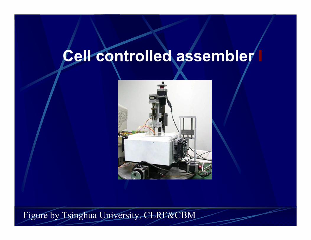
Cell controlled assembler I
Figure by Tsinghua University, CLRF&CBM

core parts of assembler
Odd nozzle extruding machine
of Cell assembler I
Multi-nozzle extruding machine
of Cell assembler II
Figure by Tsinghua University, CLRF&CBM

The table listed forming parameters
Extrusion cavity volume (ml) 1
Nozzle diameter (um) 200
Scanning speed (mm/s) 20
Extrusion frequency (Hz) 79
Material concentration (%) 5
Cross linker concentration
(%)
6
Lattice size (mm) 0.8

The experimented cellscartilage cell
fibroblast cell
hepatocyte cell
endothelium cell
myocardiac cell
hepatocyte + fibroblast
The experimented materialsgelatin
sodium alginate
chitosan

2mm 2mm
Figure by Tsinghua University, CLRF&CBM

3D structure with
hepatocyte/gelatin/sodium alginate
2mm5mm
Figure by Tsinghua University, CLRF&CBM
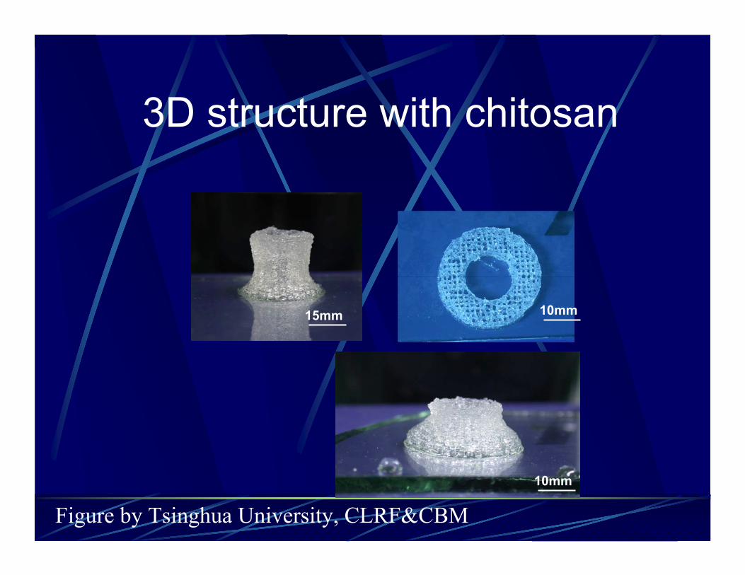
3D structure with chitosan
10mm15mm
10mm
Figure by Tsinghua University, CLRF&CBM

Hepatic cells initially resided in
the micro-environment provided
by the 3D formed structure and
presented large and round
shape
Confocal laser scanning (CLS)
image of the hepatocytes
One week after in vitro culture,
stained with propidium iodide
(PI,sigma USA)
Figure by Tsinghua University, CLRF&CBM

Figure by Tsinghua
University, CLRF&CBM
Image of histological section after two weeks
in vitro, hepatic cells were still surviving and
proliferating vigorously everywhere in the 3D
structure, the long sinusoids were observed
in many fields, as shown in picture.

Figure by
Tsinghua University,
CLRF&CBM
LSC images of the hepatocytes after three weeks culture.
a) LSC observation with both PI staining and FITC-conjugation.
b) Negative control.
The cells displayed positive for albumin antigen-antibody reaction,
and negative for the negative control of abnormal rabbit serum.
Arrows indicate the duct-like structures were formed.

0
5
10
15
1 2 3 4 5 6 7 8
Culture time (weeks)
Fu
nct
ion
sGOT(10 IU/L)
Cr(µmol/L)
Ur(mmol/L)
TG(0.1 mmol/L)
Glu(mmol/L)
ALB(g/L)
The amounts of albumin secretory and urea synthesis
increase during 8 weeks culture.
The amounts of albumin and the urea were in relative low
level at the first 3 weeks, then increase in 3 to 6 weeks.
After 6 weeks, the amounts kept in high level consistently.
It indicates that hepatocytes perform liver-specific function
in the network block.
Figure by Tsinghua
University, CLRF&CBM

Forming process:

1. FFF Techuologies,FFF~Scaffold Mnuf.
2. Scaffold Manuf. Technologies
3. Non-degradation Scaffold
4.BONE Tissue Eng. Scaffolds
5. 3-D cell Assembled
6. Laser Directed Guided Writing(LDGW)
of cell
Prof. Yongnian Yan

Principle of Laser Guided Direct Writing LGDW
First posed by Renn, Michigan Institute of Technology, USA
Focus Lens
Laser Beam
Suspending Particles
Cavity for Suspending
Figure by Tsinghua University, CLRF&CBM Prof. Yongnian Yan

Influences of the medium on LGDW
1 flotage
2 disturb of convection
3 attenuation of the light power
Prof. Yongnian Yan

The practice system
Figure by Tsinghua University, CLRF&CBM Prof. Yongnian Yan

Laser Power 500mW girdling radius 15um
Spacing between two dots
10~15um Deposition time: 10min/dot10min/dot
Figure by Tsinghua University, CLRF&CBM Prof. Yongnian Yan