1 development of a high resolution x-ray imaging crystal spectrometer for measurement of...
TRANSCRIPT
1
Development of a High Resolution X-Ray Imaging Crystal Spectrometer for Measurement of Ion-Temperature and Rotation-Velocity Profiles in Fusion
Energy Research Plasmas
K. W. Hill, M. L. Bitter, S.D. ScottPrinceton Plasma Physics Laboratory, Princeton, NJ
S. G. LeeNFRC, Korea Basic Science Institute, Daejeon, Korea
A. Ince-Cushman, J. E. RiceMIT Plasma Science and Fusion Center, Cambridge, MA
Ch. Broennimann, E. F. EikenberrySLS, Paul Scherrer Institute, Villigen, Switzerland
R. BarnsleyQueen’s University Belfast and EFDA/JET
Visiting researcher at ITER International Team, Cadarache, France
Presented at the 16th International Toki Conference: Advanced Imaging and Plasma Diagnostics, December 5-8, 2006, Toki, Japan
2
Abstract
A high resolution imaging x-ray crystal spectrometer (XICS) is being developed for Doppler measurement of radial profiles of ion temperature, Ti, and toroidal rotation velocity on Alcator C-mod. The XICS consists of a spherically bent crystal and a 2D position sensitive x-ray detector, and provides x-ray spectra from highly charged ions from multiple sightlines through the plasma. The proof of principle of the IXCS was demonstrated by measurement of Ar XVII K spectral images at from +/- 8 cm of the plasma height in Alcator C-Mod and +/- 40 cm in NSTX. However, the time resolution was limited to values >100 ms by the ~ 400 kHz count-rate limit of the available 2D detector. A new silicon pixel array detector, the PILATUS II, with a count-rate capability of 1 MHz PER PIXEL, has been tested on C-Mod by recording spectra of Ar XVII at 3.1 keV, and should enable XICS measurements with time resolution < 10 ms. The detector test results and C-Mod XICS design and expected performance will be presented. * Supported by U.S. DoE Contract No. DE-AC02-76-CHO-3073
3
Main Points
• Proof-of-Principle of new imaging x-ray crystal spectrometer (XCS) for Ti- and rotation-profile (v) measurement previously demonstrated on NSTX, Alcator C-Mod, and TEXTOR; temporal and spectral resolution limited by the available 2d x-ray detector
• New pixelated silicon detector with better spatial resolution and 100,000 times higher count-rate capability removes limitations; detector tested on existing C-Mod spectrometer
• Imaging XCS being designed to measure full radial profiles of T i and v on C-Mod, and imaging XCS adopted for ITER
• Calculations of uncertainty in Ti and v measurements predict performance of C-Mod and ITER spectrometers
4
Imaging x-ray crystal spectrometer is versatile
• Measure time dependent profiles– Ion and electron temperature– Plasma rotation– Impurity charge-state distribution
• All plasma types– Neutral-beam injection not required
• Simple design, construction
– Single spectrometer measures full profile– Spherically bent crystal– Two-dimensional imaging x-ray detector– ASIC-based electronics and PC data acquisition
5
Spherical crystal images spectra in vertical direction
6
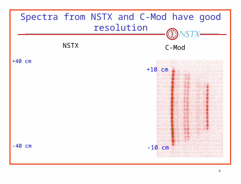
Spectra from NSTX and C-Mod have good resolution
NSTX C-Mod
+10 cm
-10 cm
+40 cm
-40 cm
7
Spatially resolved Te, Ti inferred from NSTX Ar XVII spectra
8
Lessons learned from operation of spectrometer on C-MOD and NSTX
• Initial Ar spectra with modestly high resolution obtained (2003-2005)– MWPC detector resolution not quite adequate for Ti measurement– Higher resolution detector now available will enable Ti measurement
• High signal and background count rates mitigated– 8-cm diameter crystal masked down to 6 x 2 mm2 area– Graded x-ray attenuators ~equalize count rate across radial profile; another factor of 1/8
reduction in count rate– Limited shielding against hard x rays or gamma rays added– Scattered x rays from crystal and holder reduced by apertures
• Significant count-rate limitation observed with MWPC - 400 kHz– Inherent detector count-rate limit– Pileup rejection in Time-To-Digital converter (TDC)– Throughput limitation in electronic interface– Solved by Pilatus II detector with count rate capability of 1 MHz PER PIXEL
9
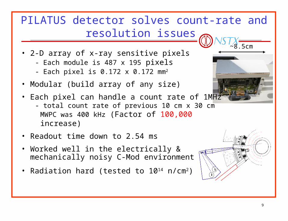
PILATUS detector solves count-rate and resolution issues
• 2-D array of x-ray sensitive pixels- Each module is 487 x 195 pixels- Each pixel is 0.172 x 0.172 mm2
• Modular (build array of any size)
• Each pixel can handle a count rate of 1MHz - total count rate of previous 10 cm x 30 cm MWPC was
400 kHz (Factor of 100,000 increase)
• Readout time down to 2.54 ms
• Worked well in the electrically & mechanically noisy C-Mod environment
• Radiation hard (tested to 1014 n/cm2)
~8.5cm
10
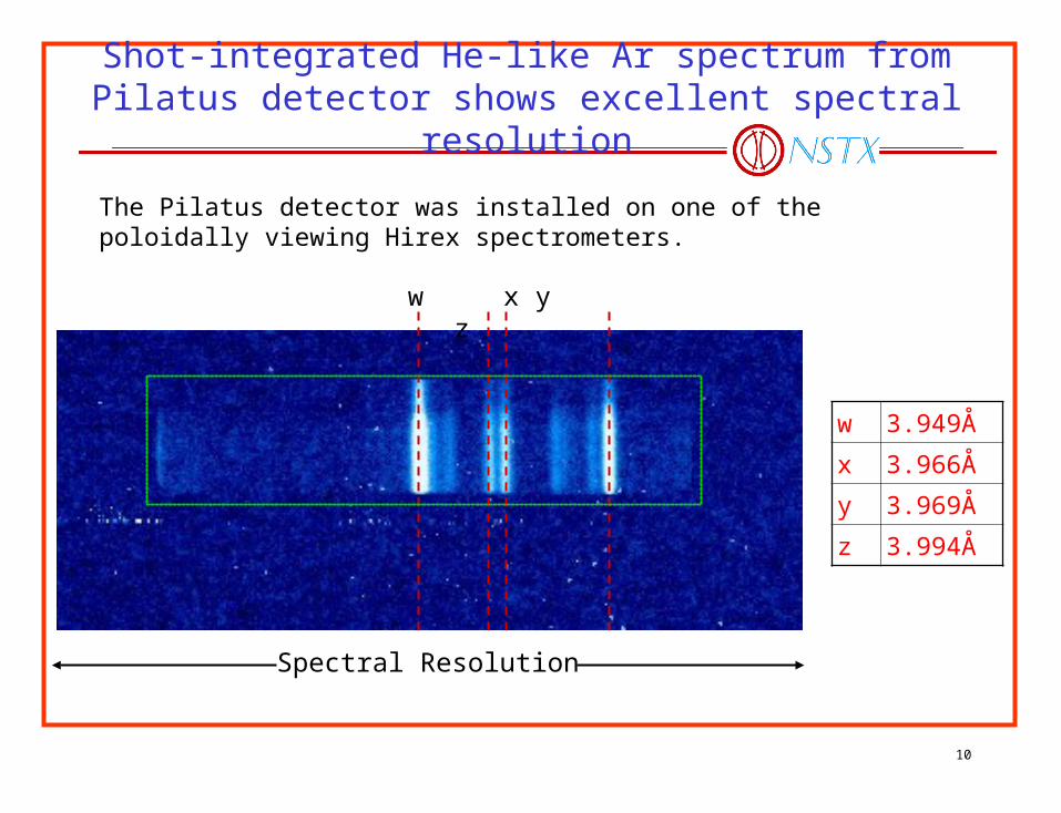
Shot-integrated He-like Ar spectrum from Pilatus detector shows excellent spectral resolution
Spectral Resolution
w x y z
w 3.949Å
x 3.966Å
y 3.969Å
z 3.994Å
The Pilatus detector was installed on one of the poloidally viewing Hirex spectrometers.
11
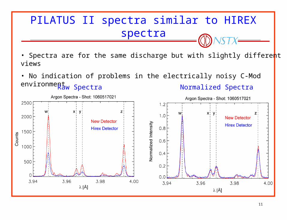
PILATUS II spectra similar to HIREX spectra
• Spectra are for the same discharge but with slightly different views
• No indication of problems in the electrically noisy C-Mod environment
Raw Spectra Normalized Spectra
12
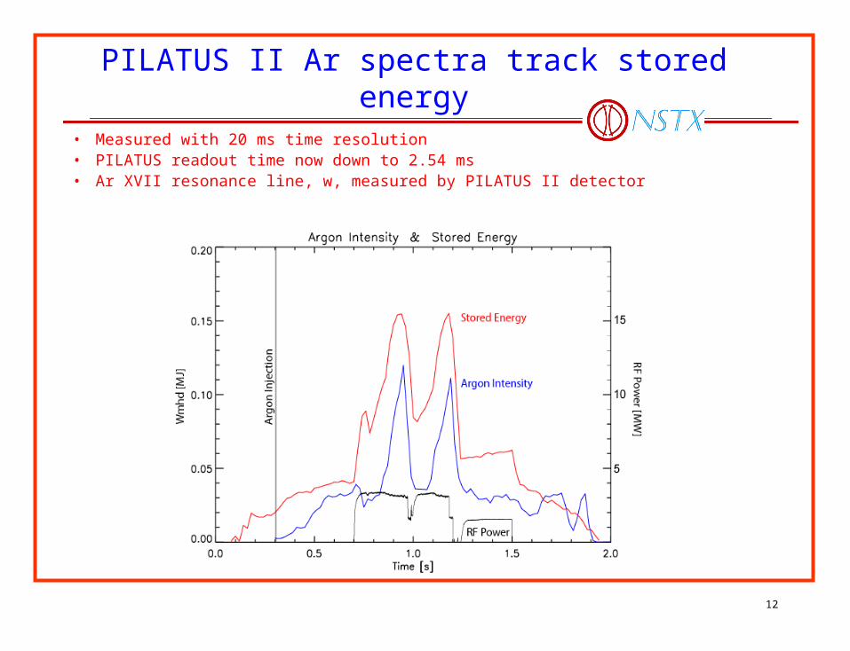
PILATUS II Ar spectra track stored energy
• Measured with 20 ms time resolution• PILATUS readout time now down to 2.54 ms• Ar XVII resonance line, w, measured by PILATUS II detector
13
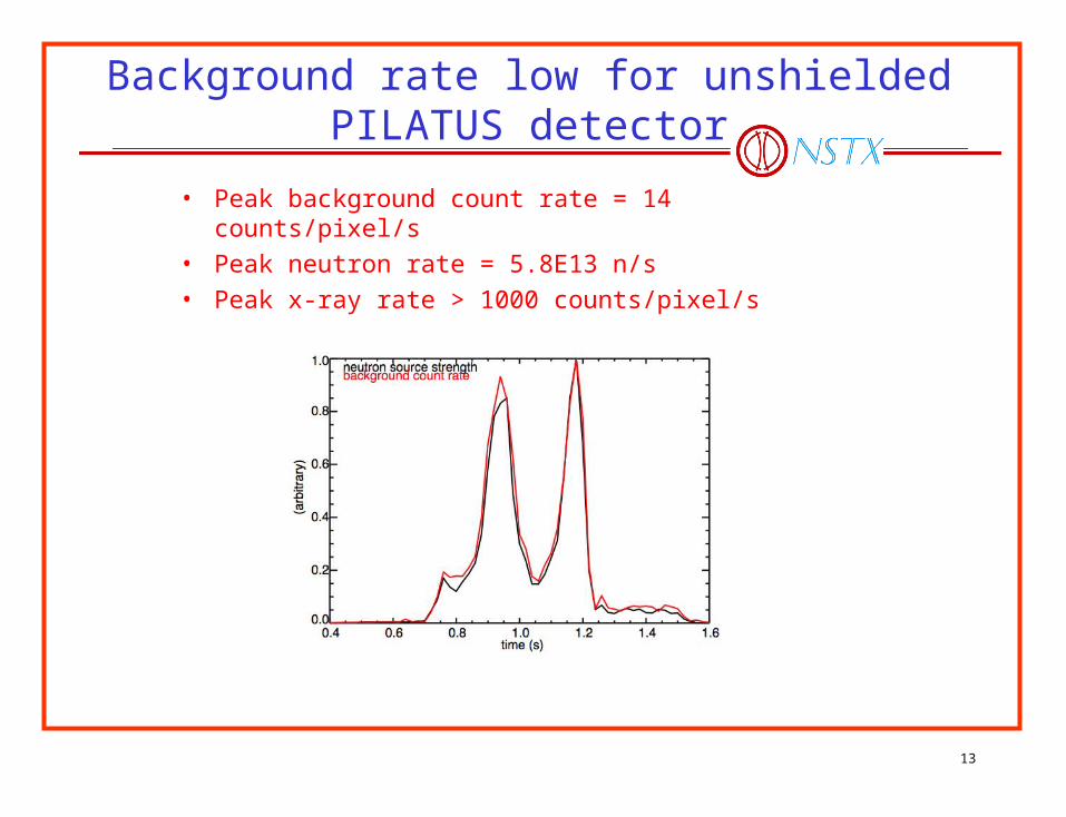
Background rate low for unshielded PILATUS detector
• Peak background count rate = 14 counts/pixel/s• Peak neutron rate = 5.8E13 n/s• Peak x-ray rate > 1000 counts/pixel/s
14
Imaging XCS configuration selected for C-Mod and ITER
• Because of the successful demonstration of the imaging XCS and the PILATUS II detector, an imaging XCS is being designed to measure full profiles of Ti and v on C-Mod, and the imaging XCS design has been selected for ITER. • On ITER the background from neutron and radiation will increase the uncertainty in measurement of the line position and width.
15
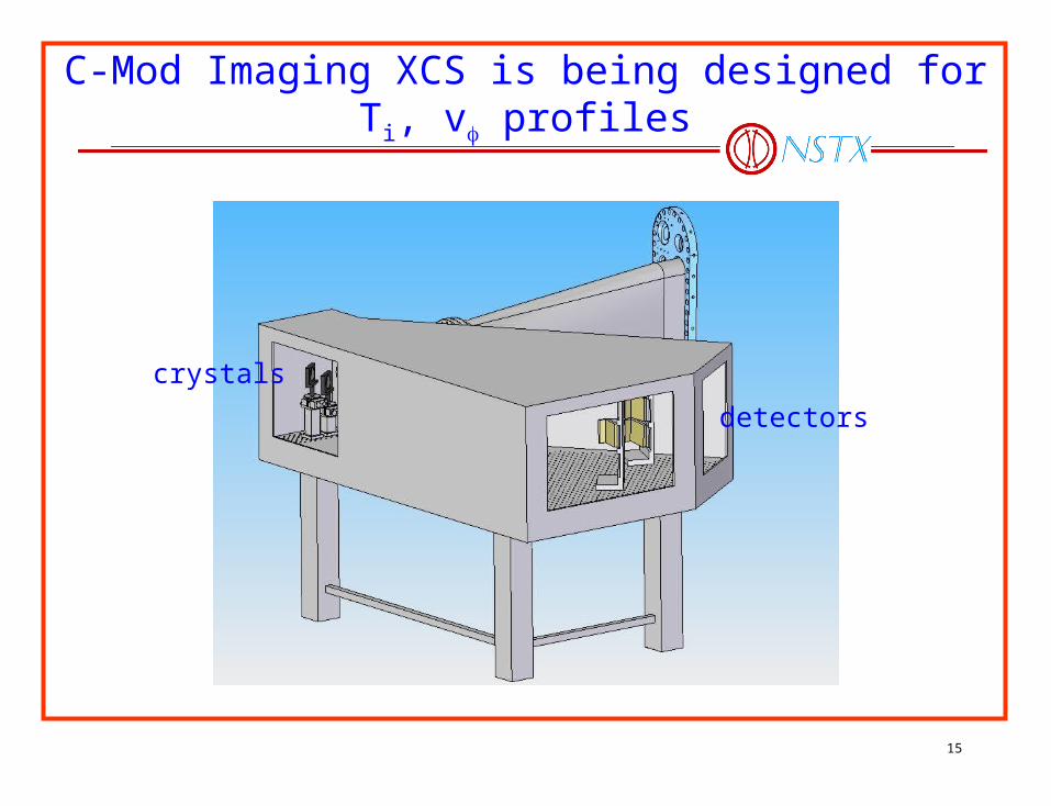
C-Mod Imaging XCS is being designed for Ti, v profiles
crystals
detectors

16
Full plasma radial view and toroidal component planned for C-Mod
• 2:1 imaging• 3 PILATUS detectors• ~30% toroidal fraction
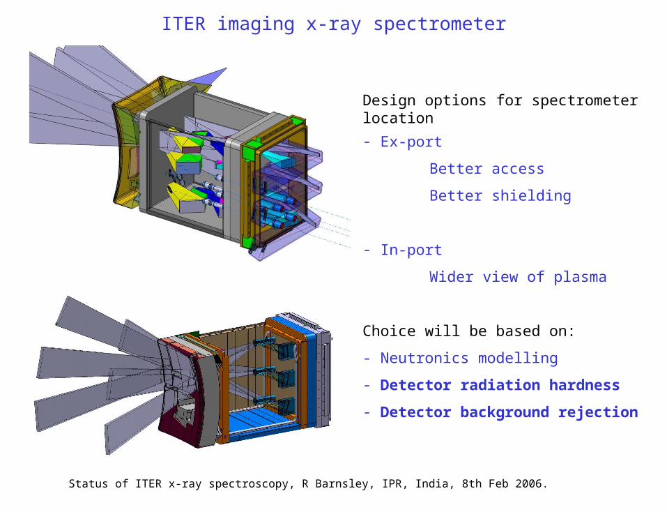
ITER imaging x-ray spectrometer
Design options for spectrometer location
- Ex-port
Better access
Better shielding
- In-port
Wider view of plasma
Choice will be based on:
- Neutronics modelling
- Detector radiation hardness
- Detector background rejection
Status of ITER x-ray spectroscopy, R Barnsley, IPR, India, 8th Feb 2006.
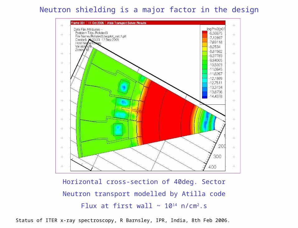
Neutron shielding is a major factor in the design
Horizontal cross-section of 40deg. Sector
Neutron transport modelled by Atilla code
Flux at first wall ~ 1014 n/cm2.s
Status of ITER x-ray spectroscopy, R Barnsley, IPR, India, 8th Feb 2006.
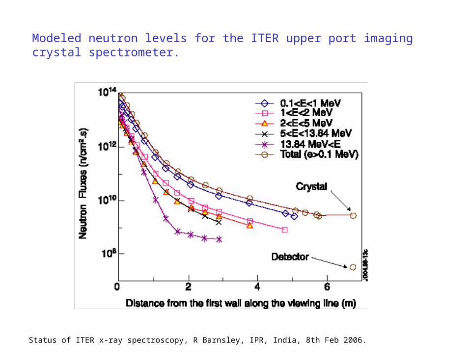
Modeled neutron levels for the ITER upper port imaging crystal spectrometer.
Status of ITER x-ray spectroscopy, R Barnsley, IPR, India, 8th Feb 2006.
20
Estimates of performance of C-Mod and ITER spectrometers
• Estimates of uncertainty in Ti measurement and minimum resolvable toroidal rotation velocity were made for C-Mod imaging spectrometer.
• On ITER, both x-ray continuum and fusion-neutron background will increase uncertainties in measurement of Ti and vtor. Numerical and analytic statistical analyses were made to quantify these increased uncertainties.
21
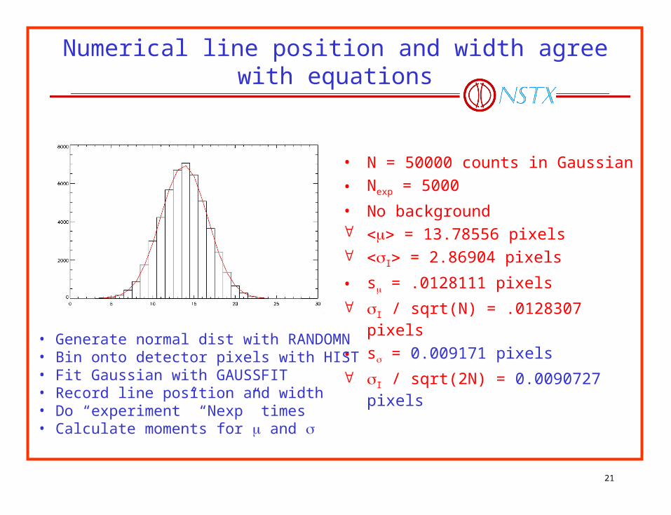
Numerical line position and width agree with equations
• N = 50000 counts in Gaussian
• Nexp = 5000
• No background = 13.78556 pixels I = 2.86904 pixels
• s = .0128111 pixels
I / sqrt(N) = .0128307 pixels
• s = 0.009171 pixels
I / sqrt(2N) = 0.0090727 pixels• Generate normal dist with RANDOMN• Bin onto detector pixels with HIST• Fit Gaussian with GAUSSFIT• Record line position and width• Do “experiment” “Nexp” times• Calculate moments for and
22
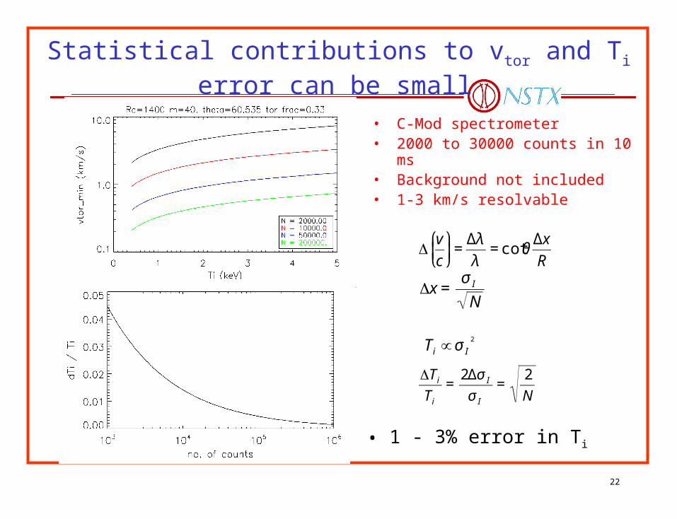
Statistical contributions to vtor and Ti error can be small
• C-Mod spectrometer• 2000 to 30000 counts in 10 ms• Background not included• 1-3 km/s resolvable
• 1 - 3% error in Ti
€
Δv
c
⎛
⎝ ⎜
⎞
⎠ ⎟=
Δλ
λ= cotθ
Δx
R
€
Δx =σ I
N
€
Ti ∝ σ I2
€
ΔTi
Ti
=2Δσ I
σ I
=2
N
23
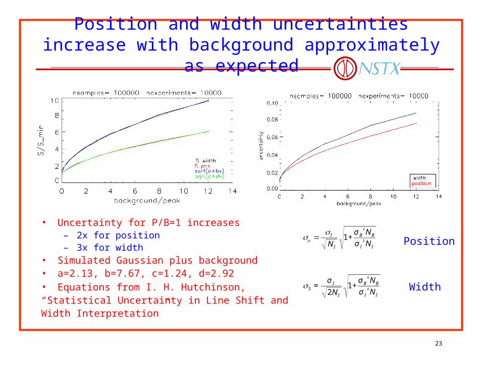
Position and width uncertainties increase with background approximately as expected
• Uncertainty for P/B=1 increases – 2x for position– 3x for width
• Simulated Gaussian plus background• a=2.13, b=7.67, c=1.24, d=2.92• Equations from I. H. Hutchinson, “Statistical Uncertainty in Line Shift andWidth Interpretation”
€
= I
N I
1+σ B
2 NB
σ I2 N I
€
S =σ I
2N I
1+σ B
4 NB
σ I4 N I
Position
Width
24
Conclusions
• New imaging x-ray spectrometer developed for Ti -, Te - and rotation-profile measurement on NSTX and Alcator C-Mod.
• Imaging concept verified on C-Mod, NSTX, and TEXTOR.
• Very small crystal area provided high count rates from C-Mod– Suggests small area crystals suitable for ITER
• Detector count-rate limit and position-resolution issues solved by PILATUS II detector.
• Imaging spectrometer being designed for C-Mod.
• Numerical and statistical analyses provide basis for estimating performance of imaging XCS on C-Mod and on ITER with neutron background.