1 da105 radiology radiation health and safety. 2 1968 – radiation control for health and safety...
TRANSCRIPT

1
DA105RADIOLOGY
RADIATION HEALTH AND SAFETY

2
• 1968 – Radiation Control for Health and Safety Act – Standardized xray equipment; required filtration, collimation, quality control programs

3
• 1981 – Consumer – Patient Radiation Health and Safety Act – Each state must monitor and regulate, in it’s Dental Practice Act who is allowed to take xrays and the training for that person

4
Radiation comes from 2 sources
• Natural Background radiation – earth, sun, atmosphere
• Artificial – Diagnostic and therapeutic, atomic testing, scientific experimentation

5
CHARACTERISTICS OF DENTAL XRAYS
Positive• Decay• Cysts• Infection• Retained roots• Foreign bodies• Tooth development• Growth irregularities

6
Negative• Can penetrate tissues
that are radiosensitive• Ionize change, or alter
tissues• Most sensitive tissue –
Growing or immature cell tissue

7Tissues which are radiosensitive FROM MOST TO LEAST
• Embryonic tissue (in Pregnant women_• Blood and bone marrow• Skin• Connective tissue• Nerve• Brain• Muscle cells• Bone• enamel

8CUMULATIVE EFFECT
• Long lasting effects from exposure which can add up to harmful amounts.
• SCATTER RADIATION – Remains in cells but the body can slough it off in 24-48 hours
• Repeated exposure does not give the body enough time to slough off the damaged cells and permanent tissue damage can occur

9
LATENT PERIOD
• The time from exposure to xray until there is observable damage

10
SHORT TERM EFFECTS
• Short term effect from high doses lead to biological damage– High Doses (nuclear accident) can cause
erythema and epilation. Dental xrays are not high dose, not possible at xray machine settings

11
LONG TERM EFFECTS
• Long term effects chronic after years of exposure. Ex. Operator hold xray in patient mouth = dermatitis and subsequent development of cancerous lesions of finger

12
OTHER LONG TERM EFFECTS
• Erythema• Epilation• Split fingernails• Blindness• sterility

13
• Any exposure to radiation conveys potential risks, even low dose such as dental xray.
• It’s important for a DA to learn critical organs because radiation safety is directed at minimizing exposure to highly sensitive organs

14CRITICAL ORGANS
• Hematopoietic – function and form of blood elements
• Thyroid gland - cancer• Breast- cancer• Salivary glands – sublingual, submandibular,
parotid

15
• Reproductive organs – male and female • Pregnancy – genetic effects, changes in
reproductive cells, leads to radiation-induced mutations
• Skin – early aging• Lens of eyes – cataracts – have patient close
eyes

16
Pregnant Patient
• Do not take x rays on pregnant patients unless emergency
• OBGYN must approve all exposures
17
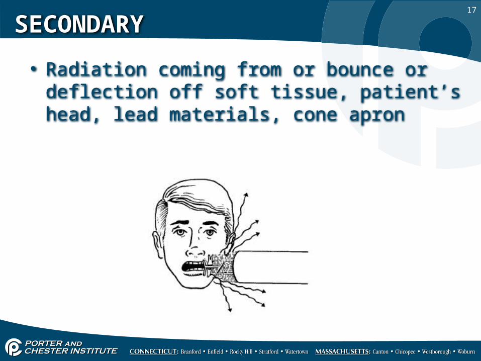
SECONDARY
• Radiation coming from or bounce or deflection off soft tissue, patient’s head, lead materials, cone apron

18
SCATTERED
• Radiation deflected by impact with matter (object); travels to all parts of body and all over the room; undesirable xrays

19
SAFETY PRECAUTIONS
• ALARA• As Low as Reasonably Achievable (National
Committee on Radiation Protection 1971• Maximum diagnostic benefit with minimum
exposure possible determine if radiation is preferable agent for making diagnosis.
• Avoid retakes/check to see if current xrays exist before taking one

20
FILTRATION
• Removes non-useful, low energy soft radiation from primary beam. Aluminum disc placed at window
• Federal Law – 1.5 mm aluminum filter at 50-70 KvP. 2.5 mm Above 70 KvP

21
COLLIMATION
• Restricts size of beam by use of lead lined PID
• One of the most important ways to minimize patient exposure
• Federal law – dental xray can be no more than 2.75 inches at patient face

22
CONTINUING EDUCATION
• Learning about new procedures and discoveries

23
CARDINAL PRINCIPLES
• 1. Use least amount of radiation that is needed
• 2. Operator never hold film for patient

24
EFFECTS OF ORAL RADIATION THERAPY
• Xerostomia dry mouth• Dysphagia difficulty in swallowing• Radiation caries caused by lack of saliva
from xerostomia• Osteoradionecrosis – death of alveolar bone• Inflammed gingiva• Mobility of teeth• Loss of appitite