1-amino-2,4 dibromoanthraquinone · 9/19/02 roc background document for...
TRANSCRIPT
FINAL
Report on Carcinogens Background Document for
1-Amino-2,4-dibromoanthraquinone
September 19, 2002
Prepared for the: U.S. Department of Health and Human Services Public Health Service National Toxicology Program Research Triangle Park, NC 27709
Prepared by: Technology Planning and Management Corporation Canterbury Hall, Suite 310 4815 Emperor Blvd Durham, NC 27703 Contract Number N01-ES-85421


9/19/02 RoC Background Document for 1-Amino-2,4-dibromoanthraquinone
Foreword
The Report on Carcinogens (RoC) is prepared in response to Section 301 of the Public Health Service Act as amended. The RoC contains a list of all substances (i) that either are known to be human carcinogens or may reasonably be anticipated to be human carcinogens; and (ii) to which a significant number of persons residing in the United States are exposed. The Secretary, Department of Health and Human Services (DHHS) has delegated responsibility for preparation of the RoC to the National Toxicology Program (NTP) who prepares the Report with assistance from other Federal health and regulatory agencies and non-government institutions.
Nominations for listing in or delisting from the RoC are reviewed by a formal process that includes a multi-phased, scientific peer review and multiple opportunities for public comment. The review groups evaluate each nomination according to specific RoC listing criteria. This Background Document was prepared to assist in the review of the nomination of 1-amino-2,4-dibromoanthraquinone. The scientific information in this document comes from publicly available, peer reviewed sources. Any interpretive conclusions, comments or statistical calculations, etc made by the authors of this document that are not contained in the original citation are identified in brackets [ ]. If any member(s) of the scientific peer review groups feel this Background Document does not adequately capture and present the relevant information they will be asked to write a commentary for this Background Document that will be included as an addendum to the document. In addition, a meeting summary that contains a brief discussion of the respective review group’s review and recommendation for the nomination will be added to the Background Document, also as an addendum.
A detailed description of the RoC nomination review process and a list of all nominations under consideration for listing in or delisting from the RoC can be obtained by accessing the NTP Home Page at http://ntp-server.niehs.nih.gov. The most recent RoC, the 9th
Edition, was published in May, 2000 and may be obtained by contacting the NIEHS Environmental Health Information Service (EHIS) at http://ehis.niehs.nih.gov (800-3153010).
i

9/19/02 RoC Background Document for 1-Amino-2,4-dibromoanthraquinone
CONTRIBUTORS
NIEHS/NTP Staff
C W Jameson, Ph.D. Head, Report on Carcinogens, Environmental Toxicology Program, NIEHS
Ruth M Lunn, Dr. P.H. Report on Carcinogens Group, Environmental Toxicology Program, NIEHS
Shawn Jeter, B.S. Report on Carcinogens Group, Environmental Toxicology Program, NIEHS
AnnaLee Sabella-Mosley Report on Carcinogens Group, Environmental Toxicology Program, NIEHS
Support to the National Toxicology Program for the preparation of this background document was provided by Technology Planning and Management Corporation
through NIEHS Contract Number NO1-ES-85421
Ronald Thomas, Ph.D., Principal Investigator
Sanford Garner, Ph.D. Co-Principal Investigator
Stanley Atwood, M.S., DABT, Senior Scientist
Susan Goldhaber, M.S. Senior Scientist
Greg Pazianos, B.S., Scientist
Ashlee Duncan, M.S. Scientist
Support staff
Angie Fralick, B.S
Tracy Saunders, B.S.
Consultants
Elizabeth Delzell, MSPH, S.D. Professor, Department of Epidemiology and International Health, University of Alabama at Birmingham, Birmingham, Alabama (contributed to Section 3)
Richard Di Giulio, Ph.D. Professor, Nicholas School of the Environment, Duke University, Durham, NC (document review)
ii

9/19/02 RoC Background Document for 1-Amino-2,4-dibromoanthraquinone
Criteria for Listing Agents, Substances or Mixtures in the Report on Carcinogens
U.S. Department of Health and Human Services National Toxicology Program
Known to be Human Carcinogens:
There is sufficient evidence of carcinogenicity from studies in humans, which indicates a causal relationship between exposure to the agent, substance or mixture and human cancer.
Reasonably Anticipated to be Human Carcinogens:
There is limited evidence of carcinogenicity from studies in humans, which indicates that causal interpretation is credible but that alternative explanations such as chance, bias or confounding factors could not adequately be excluded; or
There is sufficient evidence of carcinogenicity from studies in experimental animals which indicates there is an increased incidence of malignant and/or a combination of malignant and benign tumors: (1) in multiple species, or at multiple tissue sites, or (2) by multiple routes of exposure, or (3) to an unusual degree with regard to incidence, site or type of tumor or age at onset; or
There is less than sufficient evidence of carcinogenicity in humans or laboratory animals, however; the agent, substance or mixture belongs to a well defined, structurally-related class of substances whose members are listed in a previous Report on Carcinogens as either a known to be human carcinogen, or reasonably anticipated to be human carcinogen or there is convincing relevant information that the agent acts through mechanisms indicating it would likely cause cancer in humans.
Conclusions regarding carcinogenicity in humans or experimental animals are based on scientific judgment, with consideration given to all relevant information. Relevant information includes, but is not limited to dose response, route of exposure, chemical structure, metabolism, pharmacokinetics, sensitive sub populations, genetic effects, or other data relating to mechanism of action or factors that may be unique to a given substance. For example, there may be substances for which there is evidence of carcinogenicity in laboratory animals but there are compelling data indicating that the agent acts through mechanisms which do not operate in humans and would therefore not reasonably be anticipated to cause cancer in humans.
iii

9/19/02 RoC Background Document for 1-Amino-2,4-dibromoanthraquinone
iv

9/19/02 RoC Background Document for 1-Amino-2,4-dibromoanthraquinone
Executive Summary
Introduction 1-Amino-2,4-dibromoanthraquinone (ADBAQ) is an anthraquinone vat dye that is used in the textile industry. It was nominated by the National Institute of Environmental Health Sciences based on the results of the National Toxicology Program two-year feeding studies, which concluded that ADBAQ was carcinogenic in rats and mice.
Human Exposure Use. ADBAQ is used as a dye and as a dye intermediate. Anthraquinones are widely used as starting material for the manufacture of vat dyes, which are used in fibers and textiles, typically for cotton, wool, and cellulose acetate.
Production. ADBAQ is prepared from 1-aminoanthraquinone by bromination in dilute mineral acids and is available from two vendors in the United States.
Environmental Exposure. Because ADBAQ is not found naturally in the environment, any environmental exposure will be a result of releases from facilities where ADBAQ is produced or is used as an intermediate for production of other anthraquinone dyes. A dibromoaminoanthraquinone was found in raw wastewater from a dye manufacturing plant in four of eight samples at concentrations of 92 to 170 ppb but was not observed in final effluent.
Occupational Exposure. Dermal exposure is the main source of exposure to ADBAQ. Because ADBAQ is not very volatile, inhalation exposure likely is limited to solid particulates. No specific occupational exposure information for ADBAQ or anthraquinone dyes in general was found in current literature. Epidemiological studies, however, indicate occupational exposures to anthraquinone dyes in a New Jersey dye and resin manufacturing plant.
Human Cancer Studies Only two groups of workers exposed to anthraquinone dyes and intermediates have been investigated. All of the studies of these workers have limitations that make detection of causal relationships difficult, including small size, potential exposure to multiple agents in the workplace, and lack of quantitative exposure data. In addition, the follow-up studies lack information on nonoccupational cancer risk factors. The results of the pertinent research are not consistent. The study of Scottish workers reported small increases in esophageal and prostate cancer and found no excess of respiratory cancer (64 observed vs. 66.2 expected) or brain cancer (4/3.5). In contrast, the studies of New Jersey workers reported that work in anthraquinone dye operations was positively associated with lung cancer and with CNS tumors. In the latter investigations, the results for CNS tumors were based on a very small number of observations. The data on anthraquinone dye operations and lung cancer were more precise, and no confounding factor was identified that could explain the positive association. Thus, the increase in lung cancer in anthraquinone dye workers at the New Jersey plant may have been due to occupational
v
9/19/02 RoC Background Document for 1-Amino-2,4-dibromoanthraquinone
exposure. However, the specific agent(s) responsible for the excess have not been identified.
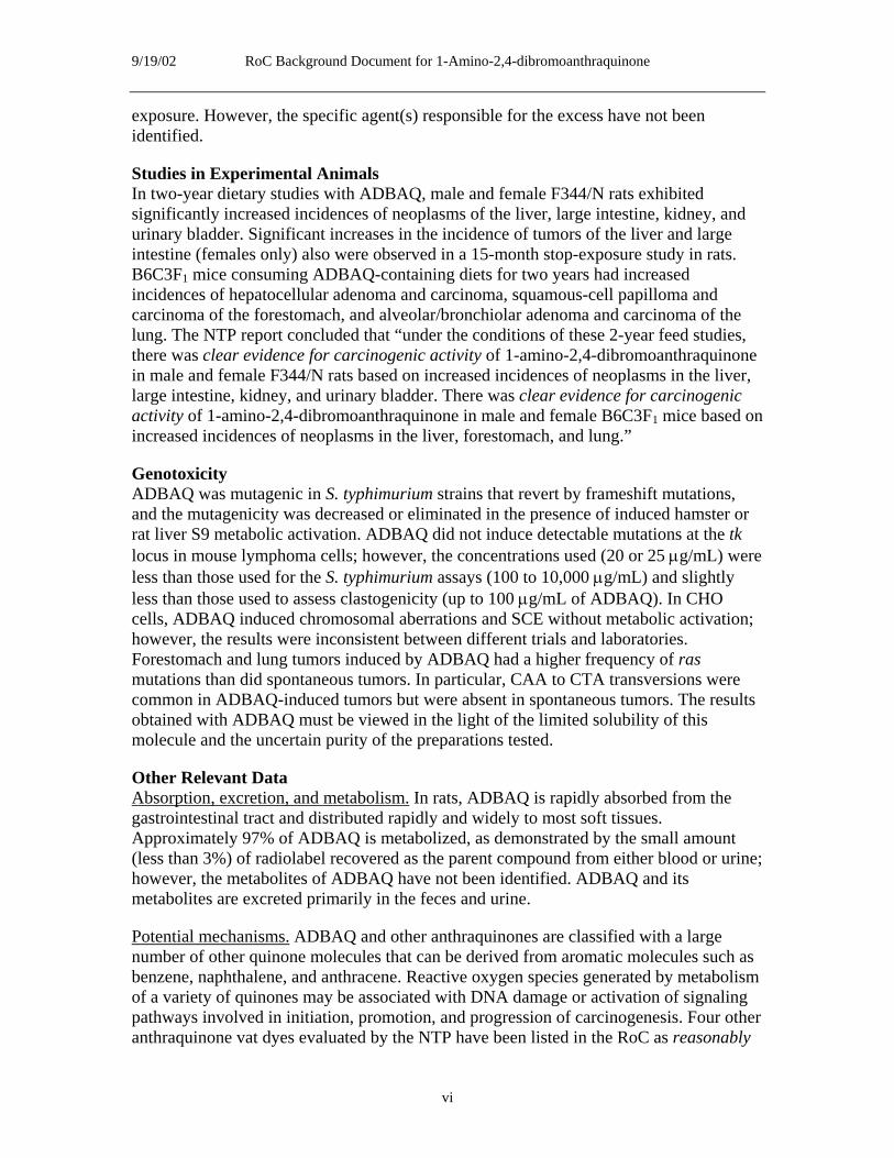
Studies in Experimental Animals In two-year dietary studies with ADBAQ, male and female F344/N rats exhibited significantly increased incidences of neoplasms of the liver, large intestine, kidney, and urinary bladder. Significant increases in the incidence of tumors of the liver and large intestine (females only) also were observed in a 15-month stop-exposure study in rats. B6C3F1 mice consuming ADBAQ-containing diets for two years had increased incidences of hepatocellular adenoma and carcinoma, squamous-cell papilloma and carcinoma of the forestomach, and alveolar/bronchiolar adenoma and carcinoma of the lung. The NTP report concluded that “under the conditions of these 2-year feed studies, there was clear evidence for carcinogenic activity of 1-amino-2,4-dibromoanthraquinone in male and female F344/N rats based on increased incidences of neoplasms in the liver, large intestine, kidney, and urinary bladder. There was clear evidence for carcinogenic activity of 1-amino-2,4-dibromoanthraquinone in male and female B6C3F1 mice based on increased incidences of neoplasms in the liver, forestomach, and lung.”
Genotoxicity ADBAQ was mutagenic in S. typhimurium strains that revert by frameshift mutations, and the mutagenicity was decreased or eliminated in the presence of induced hamster or rat liver S9 metabolic activation. ADBAQ did not induce detectable mutations at the tk locus in mouse lymphoma cells; however, the concentrations used (20 or 25 μg/mL) were less than those used for the S. typhimurium assays (100 to 10,000 μg/mL) and slightly less than those used to assess clastogenicity (up to 100 μg/mL of ADBAQ). In CHO cells, ADBAQ induced chromosomal aberrations and SCE without metabolic activation; however, the results were inconsistent between different trials and laboratories. Forestomach and lung tumors induced by ADBAQ had a higher frequency of ras mutations than did spontaneous tumors. In particular, CAA to CTA transversions were common in ADBAQ-induced tumors but were absent in spontaneous tumors. The results obtained with ADBAQ must be viewed in the light of the limited solubility of this molecule and the uncertain purity of the preparations tested.
Other Relevant Data Absorption, excretion, and metabolism. In rats, ADBAQ is rapidly absorbed from the gastrointestinal tract and distributed rapidly and widely to most soft tissues. Approximately 97% of ADBAQ is metabolized, as demonstrated by the small amount (less than 3%) of radiolabel recovered as the parent compound from either blood or urine; however, the metabolites of ADBAQ have not been identified. ADBAQ and its metabolites are excreted primarily in the feces and urine.
Potential mechanisms. ADBAQ and other anthraquinones are classified with a large number of other quinone molecules that can be derived from aromatic molecules such as benzene, naphthalene, and anthracene. Reactive oxygen species generated by metabolism of a variety of quinones may be associated with DNA damage or activation of signaling pathways involved in initiation, promotion, and progression of carcinogenesis. Four other anthraquinone vat dyes evaluated by the NTP have been listed in the RoC as reasonably
vi

9/19/02 RoC Background Document for 1-Amino-2,4-dibromoanthraquinone
anticipated to be human carcinogens. In addition, a high percentage (36/80) of phenolic anthraquinones have been reported to be mutagenic in Salmonella.
vii

9/19/02 RoC Background Document for 1-Amino-2,4-dibromoanthraquinone
viii

9/19/02 RoC Background Document for 1-Amino-2,4-dibromoanthraquinone
Table of Contents
Executive Summary ........................................................................................................................ v 1 Introduction............................................................................................................................... 1
1.1 Chemical identification .............................................................................................. 1 1.2 Physical-chemical properties ..................................................................................... 1
2 Human Exposure....................................................................................................................... 5 2.1 Use ............................................................................................................................. 5 2.2 Production.................................................................................................................. 5 2.3 Analysis...................................................................................................................... 5 2.4 Environmental occurrence ......................................................................................... 5 2.5 Environmental fate ..................................................................................................... 5
2.5.1 Air ............................................................................................................... 5 2.5.2 Water........................................................................................................... 6 2.5.3 Soil.............................................................................................................. 6
2.6 Environmental exposure ............................................................................................ 6 2.7 Occupational exposure ............................................................................................... 6 2.8 Biological indices of exposure................................................................................... 6 2.9 Regulations ................................................................................................................ 6
3 Human Cancer Studies.............................................................................................................. 7 3.1 Human epidemiological studies ................................................................................. 7
3.1.1 Study of Scottish anthraquinone dyestuffs workers ................................... 7 3.1.2 Studies of dye and resin manufacturing workers at a plant in New
Jersey .......................................................................................................... 8 3.2 Summary .................................................................................................................. 13
4 Studies of Cancer in Experimental Animals ........................................................................... 17 4.1 Rats .......................................................................................................................... 17
4.1.1 Thirteen-week study ................................................................................. 17 4.1.2 Two-year study ......................................................................................... 18 4.1.3 Fifteen-month stop-exposure study .......................................................... 21
4.2 Mice ......................................................................................................................... 24 4.2.1 Thirteen-week study ................................................................................. 24 4.2.2 Two-year study ......................................................................................... 25
4.3 Summary .................................................................................................................. 28 5 Genotoxicity............................................................................................................................ 29
5.1 Prokaryotic systems: Salmonella typhimurium........................................................ 29 5.2 Mammalian systems................................................................................................. 29
5.2.1 Mouse lymphoma L5178YTK+/- cells ...................................................... 29 5.2.2 Chromosomal aberrations ......................................................................... 30 5.2.3 Sister chromatid exchange ........................................................................ 30
ix

9/19/02 RoC Background Document for 1-Amino-2,4-dibromoanthraquinone
5.2.4 Ras mutations in tumors ........................................................................... 31 5.3 Summary .................................................................................................................. 32
6 Other Relevant Data................................................................................................................ 33 6.1 Absorption, distribution, metabolism, and excretion ............................................... 33
6.1.1 Absorption ................................................................................................ 33 6.1.2 Distribution and metabolism .................................................................... 33 6.1.3 Excretion................................................................................................... 33
6.2 Liver focus model .................................................................................................... 34 6.3 Carcinogenicity and mutagenicity of other quinones and anthraquinones .............. 35 6.4 Summary .................................................................................................................. 41
7 References............................................................................................................................... 43 Appendix A: NTP TR 383 (1996). Toxicology and Carcinogenesis Studies of 1-Amino
2,4-Dibromoanthraquinone in F344/N Rats and B6C3F1 Mice (Feed Studies). PP A-1 – A-86. .................................................................................................................................... 47
Appendix B: Tissues examined for histopathology in mice and rats in the NTP bioassays of ADBAQ.............................................................................................................................. 49
List of Tables
Table 1-1. Physical and chemical properties of ADBAQ ............................................................... 2 Table 1-2. Anthraquinone and some anthraquinone-based compounds ......................................... 3 Table 3-1. Human cancer studies on anthraquinone dye workers ................................................ 14 Table 4-1. Survival and body weight in F344/N rats following dietary exposure to
ADBAQ for 13 weeks............................................................................................................. 18 Table 4-2. Survival, body weight, and tumor incidence in F344/N rats following dietary
exposure to ADBAQ for two years......................................................................................... 20 Table 4-3. Survival, body weight, and tumor incidences in F344/N rats in the 15-month
stop-exposure study of ADBAQ (20,000 ppm) ...................................................................... 23 Table 4-4. Occurrence of altered hepatocellular foci in female F344/N rats following
dietary exposure to ADBAQ for 15 months ........................................................................... 24 Table 4-5. Survival and body wieght in B6C3F1 mice following dietary exposure to
ADBAQ for 13 weeks............................................................................................................. 25 Table 4-6. Survival, body weight, and tumor incidence in B6C3F1 mice following dietary
exposure to ADBAQ for two years......................................................................................... 27 Table 5-1. Mutagenicity of ADBAQ in S. typhimuriuma ............................................................. 29 Table 5-2. Induction of chromosomal aberrations and SCE in CHO cells by ADBAQa ............. 31 Table 5-3. Frequency and spectra of ras mutations in forestomach and lung tumors from
B6C3F1 mice exposed to ADBAQ for two yearsa .................................................................. 32 Table 6-1. Occurrence of altered hepatocellular foci neonatal rats following dietary
exposure to ADBAQ for 75 or 300 daysa ............................................................................... 34
x

9/19/02 RoC Background Document for 1-Amino-2,4-dibromoanthraquinone
Table 6-2. Carcinogenicity and mutagenicity of anthraquinone and some anthraquinonederived dyes (data from NTP and IARC) ............................................................................... 37
Table 6-3. Genotoxicity of some anthraquinone dyesa ................................................................. 40
List of Figures
Figure 1-1. Structure of ADBAQ.................................................................................................... 1 Figure 4-1. Exposure protocol for the two-year study of ADBAQ in F344/N rats ...................... 19 Figure 4-2. 15 Month stop exposure study ................................................................................... 21 Figure 4-3. Exposure protocol for the two-year study of ADBAQ in B6C3F1 mice.................... 26
xi

9/19/02 RoC Background Document for 1-Amino-2,4-dibromoanthraquinone
xii

9/19/02 RoC Background Document for 1-Amino-2,4-dibromoanthraquinone
1 Introduction
1-Amino-2,4-dibromoanthraquinone (ADBAQ) is an anthraquinone vat dye that is used in the textile industry and is a member of a class of insoluble dyes that are impregnated into textile fibers. ADBAQ was nominated by the National Institute of Environmental Health Sciences (NIEHS) for possible listing in the Report on Carcinogens (RoC) based on a National Toxicology Program (NTP) bioassay (TR-383) reporting clear evidence of carcinogenicity in rats and mice. The NTP studied the carcinogenicity of ADBAQ as part of a class study with five other anthraquinone-derived dyes with representative and diverse structures, for the purpose of predicting the carcinogenicity of chemicals in this class. The conclusions from the NTP’s two-year feeding study of ADBAQ were that there was clear evidence of carcinogenic activity in male and female F344/N rats (neoplasms in the liver, large intestine, kidney, and urinary bladder) and in male and female B6C3F1 mice (neoplasms in the liver, forestomach, and lung).
1.1 Chemical identification ADBAQ (C14H7Br2NO2, mol wt 381.02, CASRN 81-49-2) is a substituted anthraquinone with a single primary amine located in the 1-position and two bromine atoms located ortho and para to the amino group. Synonyms for ADBAQ include 1-amino-2,4-dibromo9,10-anthracenedione; 1-amino-2,4-dibromanthrachinon; anthraquinone, 1-amino-2,4dibromo-; and 2,4-dibromo-1-anthraquinonylamine. Its RTECS number is CB5500000. The structure of ADBAQ is shown in Figure 1-1.
ONH2
OBr
Br
Figure 1-1. Structure of ADBAQ
1.2 Physical-chemical properties ADBAQ is a red powder at room temperature and melts at 226°C. It is sparingly soluble in ether and benzene; soluble in hot nitrobenzene, chloroform, acetic acid, pyridine, and concentrated sulfuric acid; and insoluble in water. It is sensitive to long-term air and light exposure. The physical and chemical properties of ADBAQ are summarized in Table 1-1.
1

9/19/02 RoC Background Document for 1-Amino-2,4-dibromoanthraquinone
Table 1-1. Physical and chemical properties of ADBAQ Property Information Reference Molecular weight 381.02 ChemFinder 2001 Color red ChemFinder 2001 Odor odorless NTP Chemical Repository 2001 Physical state powder ChemFinder 2001 Melting point (°C) 226 ChemFinder 2001 Flash point (°C) > 200 (> 392°F) NTP Chemical Repository 2001 Solubility (at 23oC):
water acetone DMSO 95% ethanol toluene
< 0.1 g/100 mL < 1 mg/mL 1–10 mg/mL < 1 mg/mL < 1 mg/mL
ChemFinder 2001 NTP Chemical Repository 2001 NTP Chemical Repository 2001 NTP Chemical Repository 2001 NTP Chemical Repository 2001
Anthraquinone (TR-494) and the following six other anthraquinone-based compounds (identified in Table 1-2) have been tested in two-year bioassays by the NTP or have been listed in the RoC: 2-aminoanthraquinone (TR-144, RoC), 1-amino-2methylanthraquinone (TR-111, RoC), 1,8-dihydroxyanthraquinone (danthron) (RoC), 2methyl-1-nitroanthraquinone (TR-029), 1,4,5,8-tetraaminoanthraquinone (disperse blue 1) (TR-299, RoC), and emodin (TR-493). Carcinogenicity and genetic toxicology data for these seven molecules are summarized in Table 6-2. Other anthraquinone-derived compounds that are listed in the NTP database but have not been tested in two-year bioassays include the following: chrysophanic acid, CASRN 481-74-3; rhein, CASRN 478-43-3; 1,8-dihydroxy-4,5-dinitroanthraquinone, CASRN 81-55-0; C.I. disperse blue 27, CASRN 15791-78-3; C.I. disperse red 60, CASRN 17418-58-5; disperse blue 7, CASRN 3179-90-6; D&C violet no. 2, CASRN 81-48-1; D&C green 5, CASRN 440390-1; carminic acid, CASRN 1260-17-9; and vat blue 4, CASRN 81-77-6.
2

O
O
H2N
O
H3C
O NH2
OOH OH
O
H3C
O
O
NO2
O
O
NH2 NH2
NH2NH2
OOH OH
H3C OH
O
9/19/02 RoC Background Document for 1-Amino-2,4-dibromoanthraquinone
Table 1-2. Anthraquinone and some anthraquinone-based compounds Compound name Cas # Chemical formula Molecular weight Structure
Anthraquinone 84-65-1 C14H8O2 208.22 O
2-Amino 117-79-3 C14H9NO2 223.23 anthraquinone
1-Amino-2methylanthraquinone
82-28-0 C15H11NO2 237.26
O
1,8-Dihydroxyanthraquinone (danthron)
117-10-2 C14H8O4 240.22
2-Methyl-1-nitroanthraquinone
129-15-7 C15H9NO4 267.24
1,4,5,8Tetraaminoanthraquinone (disperse blue 1)
2475-45-8 C14H12N4O2 268.27
Emodin 518-82-1 C15H10O5 270.24
3

9/19/02 RoC Background Document for 1-Amino-2,4-dibromoanthraquinone
4

9/19/02 RoC Background Document for 1-Amino-2,4-dibromoanthraquinone
2 Human Exposure
2.1 Use ADBAQ is used as a dye and as a dye intermediate (NTP Chemical Repository 2001). Anthraquinones are widely used as starting material for the manufacture of vat dyes. Vat dyes are a class of water-insoluble dyes that can easily be reduced to a water-soluble and usually colorless form. In this form, they can readily be impregnated into fibers and textiles. Oxidation then produces an insoluble colored form that is remarkably fast to washing, light, and chemicals. Vat dyes typically are used for cotton, wool, and cellulose acetate.
2.2 Production ADBAQ is prepared from 1-aminoanthraquinone by bromination in dilute mineral acids (HSDB 2000). ADBAQ was found to be available from two vendors in the United States: Pfaltz and Bauer, in Connecticut, and Alfa Aesar, in Massachusetts (ChemFinder 2001). U.S. production of vat dyes totaled 14 million kilograms (30.8 million pounds) in 1991 (NTP 1996).
2.3 Analysis ADBAQ has been analyzed in various samples by gas chromatography/flame ionization detection and gas chromatography/mass spectrometry (Games and Hites 1977). Recently, several other analytical methods have been described for detecting and quantifying anthraquinone-type dyes (liquid chromatography/nuclear magnetic resonance, liquid chromatography/mass spectrometry, and high-performance liquid chromatography [HPLC]); however, ADBAQ has not been analyzed by these methods (Preiss et al. 2000, Novotná et al. 1999).
2.4 Environmental occurrence ADBAQ is not known to be naturally formed. Production of ADBAQ and its subsequent use as an intermediate in the production of anthraquinone dyes may result in releases into the environment (HSDB 2000). No information was found in the U.S. Environmental Protection Agency’s Toxic Release Inventory regarding ADBAQ (TRI 2001).
2.5 Environmental fate ADBAQ is sensitive to long-term exposure to light and air (NTP Chemical Repository 2001). Although ADBAQ was found in raw wastewater of a dye manufacturing plant, it was not present in the final effluent (effluent into the Cooper River, which flows into Charleston Bay of the Atlantic Ocean) (Games and Hites 1977). This may indicate that ADBAQ was either biodegraded or adsorbed to sludge during treatment (HSDB 2000).
2.5.1 Air
Based on estimated vapor pressure, ADBAQ should exist as a solid particulate in the ambient atmosphere. Particulate-phase ADBAQ would be removed from the atmosphere by dry deposition (HSDB 2000).
5

9/19/02 RoC Background Document for 1-Amino-2,4-dibromoanthraquinone
2.5.2 Water
ADBAQ may adsorb to suspended solids and sediment in water. The amino group in ADBAQ also may bind covalently with active sites in sediment and particulate matter. It is not expected to volatilize from water surfaces. The estimated partition coefficients and a recommended regression-derived equation were used to calculate a bioconcentration factor of 6,400 for ADBAQ. This suggests that ADBAQ will bioconcentrate in aquatic organisms (HSDB 2000).
2.5.3 Soil
ADBAQ is expected to be immobile in soil; its mobility may be limited because the amino group may bind to active sites in the soil. Volatilization is not expected to occur from either moist or dry soil surfaces (HSDB 2000).
2.6 Environmental exposure Because ADBAQ is not found naturally in the environment, any environmental exposure will be a result of releases from facilities where ADBAQ is produced or is used as an intermediate for production of other anthraquinone dyes. A dibromoaminoanthraquinone was found in raw wastewater from a dye manufacturing plant in four of eight samples at concentrations of 92 to 170 ppb but was not observed in final effluent (HSDB 2000).
2.7 Occupational exposure Dermal exposure is the main source of exposure to ADBAQ. Because ADBAQ is not very volatile, inhalation exposure likely is limited to solid particulates. Ikeda et al. (1977) found that workers exposed to dyes (as evidenced by diazo-positive metabolites in their urine) had many dye stains on their hands, wrists, and forearms. Analyses of the air in the workroom environments were not conducted.
No specific occupational exposure information for ADBAQ or anthraquinone dyes in general were found in current literature. Epidemiological studies, however, indicate potential occupational exposures to anthraquinone dyes in a New Jersey dye and resin manufacturing plant. A cohort study followed more than 3,000 workers who may have been exposed to anthraquinone dyes (Sathiakumar and Delzell 2000). Results of this study are described in Section 3.
2.8 Biological indices of exposure No indices of ADBAQ exposure in humans or animal models were found in current literature.
2.9 Regulations No regulations specifically for ADBAQ were identified.
6

9/19/02 RoC Background Document for 1-Amino-2,4-dibromoanthraquinone
3 Human Cancer Studies
No study has evaluated the relationship between human cancer and specific exposure to ADBAQ; however, exposure to anthraquinone dyes as a class has been studied. Since the late 1880s, the dye manufacturing industry has used a large number of anthraquinones and aromatic amines (arylamines) to synthesize industrial colorants and has used nitro aromatic amines to make hair dyes. Relatively little is known about the human carcinogenicity of anthraquinone dyes. The NTP classifies 2-aminoanthraquinone, 1amino-2-methylanthraquinone, 1,8-dihydroxyanthraquinone, and 1,4,5,8tetraaminoanthraquinone as reasonably anticipated to be human carcinogens. The International Agency for Research on Cancer (IARC) lists 1,8-dihydroxyanthraquinone, 2-methyl-1-nitroanthraquinone, and 1,4,5,8-tetraaminoanthraquinone as possibly carcinogenic to humans (Group 2B) and considers 2-aminoanthraquinone and 1-amino-2methylanthraquinone not classifiable as to carcinogenicity to humans (Group 3). Neither the NTP nor IARC has evaluated the carcinogenicity of anthraquinone dyes as a chemical category or anthraquinone dye manufacturing as an exposure circumstance.
3.1 Human epidemiological studies Data on the carcinogenicity to humans of dyes and chemicals used in synthesizing dyes come mainly from studies of workers in manufacturing plants. These groups of workers typically were exposed to many different chemicals and classes of chemicals, often making it difficult to attribute observed carcinogenic effects to a single agent. Only five epidemiologic studies of workers potentially exposed to anthraquinone dyes and anthraquinone dye intermediates are available. One of these investigations evaluated mortality among workers at a plant in Scotland that mainly made anthraquinone dyes (Gardiner et al. 1982). Each of the other four studies pertains to the same group of workers at a plant in New Jersey (Barbone et al. 1992, 1994, Delzell et al. 1989, Sathiakumar and Delzell 2000). The New Jersey plant’s manufacturing operations were more diverse than those of the plant in Scotland and included the production of anthraquinone, anthraquinone dye intermediates and dyes, epichlorohydrin, azo dye intermediates and dyes, epoxy resins, and a variety of additives.
3.1.1 Study of Scottish anthraquinone dyestuffs workers
Gardiner et al. (1982) evaluated mortality from various causes among 1,975 men employed at an anthraquinone dyestuffs plant for at least six months between 1956 and the end of 1965. Personnel records were used to identify all subjects who had worked in jobs that entailed potential exposure to substituted anthraquinones. The study excluded men who had worked exclusively in clerical, accounting, and personnel management positions. Vital status and cause of death as of the end of June 1980 were determined for about 97% of the subjects through the use of plant records and data from the National Records Offices of Edinburgh and Newcastle upon Tyne. Analyses compared workers’ mortality rates with those of the general male population of Scotland in 1975, adjusting for age and using the standardized mortality ratio (SMR) (ratio of observed to expected numbers of deaths, multiplied by 100) as the measure of association. An SMR of 100 implies that the mortality rate of workers was the same as that of the general population, after analytic adjustment for any difference in the age distributions of these two groups.
7

9/19/02 RoC Background Document for 1-Amino-2,4-dibromoanthraquinone
Gardiner et al. (1982) found that workers at the plant had about 23% fewer deaths from all causes combined than expected on the basis of general population rates (470 observed vs. 611.5 expected deaths, SMR = 76.9). Workers also had fewer deaths than expected from cerebrovascular disease (36/66.5, SMR = 54.1), cardiovascular disease (188/235.6, SMR = 79.8), and all cancers combined (129/149.7, SMR = 86.2). There were at least 5 observed or expected deaths for eight specific types of cancer. Among these, there were more deaths than expected for cancer of the esophagus (6/4.3) and fewer deaths than expected for cancer of the stomach (14/18.1), intestinal tract (7/11.0), pancreas (4/6.8), bladder (4/5.6), respiratory system (64/66.2), prostate (4/8.0), and rectum (6/6.8). None of these differences was statistically significant.
The mortality rate for all cancer combined was higher than expected for workers in some areas of the plant. Gardiner et al. (1982) did not present detailed results for specific cancers by plant area but noted statisitcally significant excesses of cancer of the esophagus in the engineering department (all deaths from this cancer were among men in this department) and cancer of the prostate in one particular process area. Workers’ respiratory cancer rates were not statistically significantly greater than expected in any plant area. Work activities in the engineering department were not thoroughly described but presumably included maintenance operations. Gardiner et al. (1982) indicated that engineering workers constituted 45% of the study group, that they worked mainly in proximity to plant process areas, and that their plant exposures probably were similar to those of process workers. The authors could not interpret their results for prostate cancer. They stated that cadmium, a suspected cause of prostate cancer, was not used at the plant.
Gardiner et al. (1982) stated that their research did not provide any evidence of “an abnormal pattern of mortality” in the group of anthraquinone dye workers studied. Because of its methodologic weaknesses, the study provided little evidence either for or against the hypothesis that anthraquinones cause cancer in humans. Limitations were the small numbers of deaths for most specific types of cancer, the lack of quantitative estimates of exposure to anthraquinones or other chemicals, and the lack of data on nonoccupational factors. Moreover, the very low SMR for all causes of death combined, especially in older age groups, which probably contained a high proportion of inactive workers, suggests that ascertainment of deaths in the study group may have been incomplete or that the use of comparison rates derived from a single year (1975) was inappropriate. If so, all SMRs may have been underestimated.
3.1.2 Studies of dye and resin manufacturing workers at a plant in New Jersey
Delzell et al. (1989) conducted a retrospective follow-up study of mortality from 1952 through 1985 among 2,642 white men who had been employed for at least six months at a dye and resin plant in New Jersey. Barbone and colleagues carried out nested case-control studies of lung cancer (Barbone et al. 1992) and central nervous system (CNS) tumors (Barbone et al. 1994) occurring among these workers. In a later investigation, Sathiakumar and Delzell (2000) expanded the original retrospective follow-up study (Delzell et al. 1989) to include 3,266 male and female workers of all races and evaluated their mortality patterns during the period 1952 through 1995.
8

9/19/02 RoC Background Document for 1-Amino-2,4-dibromoanthraquinone
The manufacturing operations of the New Jersey plant took place in three major areas. Operations in the “south dye area” (anthraquinone dye/epichlorohydrin area) included anthraquinone production (1952 to 1983), anthraquinone dye production (1952 to 1983), anthraquinone dye intermediates production (1952 to 1983), formulation of anthraquinone dyes (1952 to late 1980s), and epichlorohydrin production (1961 to 1965). The “north dye area” produced azo dyes and intermediates from 1959 to the late 1980s. The plastics and additives area made epoxy resins, fabric softeners, water-repellent agents, optical brighteners, and various additives from 1959 to the late 1980s. Raw materials and intermediates used in each area included many chemicals, some of which are known carcinogens (e.g., benzene, benzidine, ethylene oxide) or suspected carcinogens (e.g., epichlorohydrin, formaldehyde, dimethyl sulfate). Chemicals used in the anthraquinone dye/epichlorohydrin (AQ/ECH) area included anthracene, anthraquinones, anthraquinone sulfonates, anthraquinone intermediates, aniline, substituted aniline, benzene, nitrobenzene, chlorobenzenes, chlorotoluenes, pyridine, tetrachloroethylene, chlorine, ammonia, arsenic acid, methanol, sulfuric acid, and mercury.
3.1.2.1 Original retrospective follow-up study
The first investigation of workers at the New Jersey plant used company personnel records to identify all white men with at least six months of employment as of the end of 1984 and to develop information on each subject’s jobs at the facility (Delzell et al. 1989). Plant records and linkages with several national and state databases established the vital status of 96% of the 2,642 eligible subjects as of the end of 1985. Death certificates provided information on deceased subjects’ causes of death. Comparisons of workers’ mortality rates with those for the white male general population of the United States yielded observed and expected numbers of deaths for the overall study group and subgroups of hourly workers classified by the process area(s) in which they had worked for at least one year.
The overall study group had 357 deaths from all causes combined, compared with 440 expected deaths (SMR = 81, 95% confidence interval [CI] = 73 to 90), and 106 cancer deaths, compared with 97 expected cancer deaths (SMR = 109, 95% CI = 89 to 132). There were more deaths from CNS cancer than expected (5/3.3), not including the deaths of two employees from this cancer while living in Europe, and there were 3 deaths each from bladder cancer (expected = 2.3) and kidney cancer (expected = 2.5). The paper did not include results for other specific types of cancer for the overall study group.
The study included 588 hourly employees who had worked in the AQ/ECH area for at least one year. These men had fewer deaths than expected from all causes combined (65/97) and from cancer (20/22). For all specific types of cancer, observed and expected numbers of deaths were under 10 and were too small to yield statistically stable results. Hourly AQ/ECH workers had 6 lung cancer deaths (expected = 8). Although only 44 workers in this area had been employed in epichlorohydrin operations, and most of these also had worked longer in anthraquinone dye operations, this group had statistically significantly more lung cancer deaths than expected (4/0.91, P = 0.03).
9

9/19/02 RoC Background Document for 1-Amino-2,4-dibromoanthraquinone
Hourly maintenance workers (N = 514), many of whom were employed throughout the plant, had more deaths than expected from all cancers combined (37/25, SMR = 148, 95% CI = 103 to 202), from lung cancer (18/9.2, SMR = 196, 95% CI = 116 to 309), and from liver cancer (3/0.46).
The investigators concluded that the excess of lung cancer among maintenance workers could have been due to an unidentified occupational exposure. They noted that the association they observed between epichlorohydrin and lung cancer had not been found in research on other, larger groups of workers exposed to this chemical.
The study was limited by the small number of subjects and of deaths in the AQ/ECH work group. Other weaknesses were the absence of quantitative exposure estimates or data on nonoccupational risk factors. However, even if such data had been available, the study would have had little power to assess dose response, confounding, or interaction for specific forms of cancer.
3.1.2.2 Case-control study of lung cancer.
Barbone et al. (1992) conducted a nested case-control study of lung cancer that included as cases 51 white male employees of the New Jersey plant who had died of or developed primary lung cancer by October 1988. Controls were 102 individually matched employees chosen randomly from among other white male employees who were born in the same year and were alive as of the case subject’s death or diagnosis date. Plant job records identified each process area and building in which a subject had carried out production-related work. The AQ/ECH area consisted of five buildings with the following operations: (1) anthraquinone production, (2) production of anthraquinone dye intermediates, (3) anthraquinone dye synthesis, (4) anthraquinone dye standardization, and (5) epichlorohydrin production. Interviews with long-term employees (not subjects) provided more detailed work history information on building-specific work, as well as an ordinal score for subjects’ potential exposure to asbestos and epichlorohydrin. Interviews with subjects or family members furnished data on smoking and on jobs and exposures outside the New Jersey plant and were completed for 69% of cases and 74% of controls. Plant medical records, available for 67% of cases and 68% of controls, supplied information on smoking and exposures to chemicals that had been reported to the plant medical department. The odds ratio (OR) (the ratio of the rates of lung cancer in subjects exposed and not exposed to a given factor) was used as the measure of association.
As expected, smoking was positively and consistently associated with lung cancer. Lung cancer also was positively associated with ever having worked in the AQ/ECH area (OR = 2.4, 95% CI = 1.1 to 5.2), based on 21 exposed cases and 24 exposed controls. This association was limited to subjects who had worked in the area for at least 10 years (smoking-adjusted OR = 4.6, 95% CI = 0.9 to 23), but there was no trend of increasing OR with increasing time spent in the area. Lung cancer was positively associated with employment in each of the five building-specific operations in the area, but the association was statistically significant only for anthraquinone production (OR = 12, 95% CI = 1.4 to 99) and was of borderline statistical significance for anthraquinone dye standardization (OR = 3.3, 95% CI = 1.0 to 11). The positive association between work in the AQ/ECH area and lung cancer did not appear to be due to exposure to asbestos or
10

9/19/02 RoC Background Document for 1-Amino-2,4-dibromoanthraquinone
epichlorohydrin or to smoking. Results for other process areas were not striking or statistically significant.
Workers in the AQ/ECH area potentially were exposed to chlorine and to epichlorohydrin, in addition to anthraquinone dyes and intermediates. The study reported a positive association between acute exposure to chlorine and lung cancer (OR = 5.7, 95% CI = 1.1 to 30). Adjustment for smoking raised the OR for chlorine to 27, but these results were based on only 6 exposed cases and 3 exposed controls. Chlorine was encountered in the AQ/ECH area by 3 of the 6 chlorine-exposed case subjects and 2 of the 3 chlorine-exposed control subjects. A positive [but not statistically significant] association between lung cancer and exposure to epichlorohydrin (OR = 1.7, 95% CI = 0.7 to 4.1) was reported, based on 12 exposed cases and 18 exposed controls. The latter association was restricted to workers with short-term and relatively low exposure to epichlorohydrin. Only 3 of the 21 case subjects in the AQ/ECH area had spent any appreciable time in epichlorohydrin operations.
Barbone et al. (1992) noted that the positive association between lung cancer and anthraquinone dye operations could have been due to occupational exposure (to anthraquinone or other chemicals used in the area) or to chance. The study lacked data on cumulative exposure to anthraquinones. If anthraquinones cause lung cancer, the lack of an association between duration of employment in the various anthraquinone areas and lung cancer in this study could have been due to poor correlation between duration of work in the areas and amount of exposure. The study also did not have quantitative or comprehensive data on exposure to chlorine and had only crude estimates of epichlorohydrin exposure. Thus, the impact of these agents on the association between anthraquinone work areas and lung cancer may not have been accurately assessed. Barbone et al. (1992) noted that the only other data available on human exposure to anthraquinones, the study by Gardiner et al. (1982), did not find an excess of lung cancer among workers with potential exposure.
3.1.2.3 Case-control study of central nervous system tumors
The case-control study of CNS tumors included as cases 11 white male employees who were known to have died of or developed a primary CNS tumor by October 1988 (7 with astrocytoma or glioblastoma, 2 with meningioma, and 2 with a benign tumor) (Barbone et al. 1994). Controls were 44 individually matched subjects chosen randomly from among other white male employees who were born in the same year and were alive as of the case subject’s death or diagnosis date. Procedures for obtaining and classifying work history data, estimating exposure to epichlorohydrin, conducting interviews, and obtaining data from plant medical records were similar to those used in the case-control study of lung cancer (Barbone et al. 1992). Interviews, completed for 73% of cases and 84% of controls, asked about non-plant jobs and occupational exposures, smoking habits, and history of head trauma, irradiation to the head, epilepsy, and use of antiepileptic drugs.
The occurrence of CNS tumors was statistically significantly and positively associated with anthraquinone dye intermediate production, but only 3 case subjects and no controls had worked in this process area. Positive associations also were found with work in the epoxy resin process area and in the azo dye area and with estimated exposure to
11

9/19/02 RoC Background Document for 1-Amino-2,4-dibromoanthraquinone
epichlorohydrin. Barbone et al. (1994) reported that the association with anthraquinone dye intermediate production was “nonindependent” of the association with exposure to epichlorohydrin and concluded that the small study size precluded firm conclusions about the causes of CNS tumors in the plant workforce. All of the results were based on small numbers of exposed subjects and were quite imprecise. The study also did not include quantitative estimates of exposure to anthraquinone or other chemicals and was not able to evaluate anthraquinone dose response or to adequately assess the possibility that the association between anthraquinone dye intermediate production and CNS tumors was confounded by the other positive associations observed, or vice versa.
3.1.2.4 Retrospective follow-up study update
Sathiakumar and Delzell (2000) expanded and updated the original study by Delzell et al. (1989), adding women and nonwhite men to the study group and extending follow-up through the end of 1995. The expanded study compared the mortality rates for 3,266 employees with the age-, race-, gender-, and time-period-specific rates for the general population of New Jersey.
The total study group had 728 observed and 809.8 expected deaths from all causes combined (SMR = 90, 95% CI = 83 to 97), 225 observed and 231.7 expected total cancer deaths (SMR = 97, 95% CI = 85 to 111), 89 observed and 73.2 expected lung cancer deaths (SMR = 122, 95% CI = 98 to 150), and 8 observed and 6.0 expected CNS cancer deaths (SMR = 134, 95% CI = 58 to 264). White male employees who had worked in the AQ/ECH area (N = 842) had an excess of lung cancer, based on 32 observed and 19.1 expected deaths (SMR = 168, 95% CI = 115 to 237), and had 3 deaths from CNS cancer (1.5 expected, SMR = 205, 95% CI = 42 to 598). Maintenance workers at the plant also had more deaths from lung cancer than expected (40/26.2, SMR = 153, 95% CI = 109 to 208).
The lung cancer excess among workers in the AQ/ECH area was not consistently larger in subgroups with long duration of employment (5 years or more) or long potential induction time in the area (20 years or more); however, small numbers limited the conclusions that could be drawn from these subgroup analyses. In analyses that compared the lung cancer mortality rate of AQ/ECH area workers with that of other plant employees, adjusting for age, calendar time, and employment in maintenance operations, the OR was 1.7 (95% CI = 1.1 to 2.6) for the AQ/ECH area, confirming the increased lung cancer mortality for this area seen in the SMR analysis.
This study, like the study of the same workforce by Barbone et al. (1992), found a positive association between employment in anthraquinone dye operations and lung cancer. There was no discernible trend of increasing lung cancer SMR or OR with increasing length of employment in anthraquinone dye operations, but this could be explained by poor correlation between duration of employment and cumulative exposure to anthraquinones or other chemicals used in the area. Sathiakumar and Delzell (2000) also found an increase in CNS cancer among workers potentially exposed to anthraquinones, based on small numbers, consistent with the report by Barbone et al. (1994).
12

9/19/02 RoC Background Document for 1-Amino-2,4-dibromoanthraquinone
Compared with the previous follow-up study of this group of workers, the update had 72% more person-years and twice as many observed deaths. The median length of follow-up was 27 years, and 22% of the workers were deceased. However, numbers of deaths from specific types of cancer were small in the work-area-specific analyses, and the update lacked direct exposure estimates and data on nonoccupational risk factors. These limitations impede the detection of causal associations.
3.2 Summary Only two groups of workers exposed to anthraquinone dyes and intermediates have been investigated. All of the studies of these workers have limitations that make detection of causal relationships difficult, including small size, potential exposure to multiple agents in the workplace, and lack of quantitative exposure data. In addition, the follow-up studies lack information on nonoccupational cancer risk factors. The results of the pertinent research are not consistent. The study of Scottish workers reported small increases in esophageal and prostate cancer and found no excess of respiratory cancer (64 observed vs. 66.2 expected) or brain cancer (4/3.5). In contrast, the studies of New Jersey workers reported that work in anthraquinone dye operations was positively associated with lung cancer and with CNS tumors. In the latter investigations, the results for CNS tumors were based on a very small number of observations. The data on anthraquinone dye operations and lung cancer were more precise, and no confounding factor was identified that could explain the positive association. Thus, the increase in lung cancer in anthraquinone dye workers at the New Jersey plant may have been due to occupational exposure. However, the specific agent(s) responsible for the excess have not been identified. Table 3-1 summarizes the human cancer studies on anthraquinone dye workers.
13

9/19/02 RoC Background Document for 1-Amino-2,4-dibromoanthraquinone
Table 3-1. Human cancer studies on anthraquinone dye workers
Reference Study design Population Exposure Effects Comments
Gardiner et Cohort study 1,975 men employed at Potential exposure to SMR (observed/expected deaths) Smoking and alcohol al. 1982 anthraquinone dyestuff substituted anthraquinones all deaths 76.9 (470/611.5) consumption were not Scotland plant for at least 6
months from 1956–1965, was assessed from personnel records.
all cancers 86.2 (129/149.7) esophagus 139.5 (6/4.3)
ascertained.
followed up until June stomach 77.3 (14/18.1) 1980 intestinal tract 63.6 (7/11.0)
pancreas 58.8 (4/6.8) bladder 71.4 (4/5.6) respiratory 96.7 (64/66.2) prostate 50.0 (4/8.0) rectum 88.2 (6/6.8)
Delzell et al. Retrospective 2,642 white men Exposure was asssessed Observed/expected Workers in the AQ/ECH 1989 New Jersey, USA
cohort study employed at a dye and resin plant for at least 6 months from 1952–1985 588 hourly workers employed in the AQ/ECH area for at least one year
from personnel records. Three manufacturing areas: AQ/ECH area: anthraquinone, AQ dye and AQ dye intermediates, and ECH production and formulation of AQ dyes North dye area: azo dyes and intermediates Plastics and additive area
AQ/ECH area workers: all deaths 65/97 all cancers 20/22 lung cancer 6/8 Epichlorohydrin workers: (1961– 1965) (N = 44) lung cancer 4/0.91; P = 0.03
area also were exposed to other chemicals, including aniline- and benzene-related compounds, tetrachloroethylene, chlorine, sulfuric acid, and mercury. Nonoccupational risk factors were not acertained.
14

9/19/02 RoC Background Document for 1-Amino-2,4-dibromoanthraquinone
Reference Study design Population Exposure Effects Comments
Barbone et al. Nested case- Cases: 51 white male The AQ/ECH area consisted OR (95% CI); no. of exposed cases ORs were computed 1992 New Jersey, USA
control study (Delzell et al. 1989) Lung cancer
employees who died of or developed lung cancer by 10/1988 Controls: 102 employees randomly selected and matched for age and alive as of case’s death or diagnosis date
of 5 buildings: (1) AQ production (2) production of AQ intermediates (3) AQ dye synthesis (4) AQ dye standardization (5) ECH production
Worked in AQ/ECH area: ever 2.4 (1.1–5.2); 21 > 10 years 4.6 (0.9–23) Building in AQ/ECH area: AQ production 12 (1.4–99); 6 AQ dye/standardization 3.3 (1.0–11); 8 ECH production:
with and without adjustment for smoking and other job exposures. Only smoking proved to be a confounder and was kept in analyses if it modified the strength or direction of an association.
3 exposed cases, 0 exposed controls; OR not calculated
Lung cancer was positively associated with chlorine exposure.
Barbone et al. 1994 New Jersey, USA
Nested case-control study (Delzell et al. 1989) CNS tumors
Cases: 11 white males who died of or developed a primary CNS tumor by 10/1988 Controls: 44 employees matched for age and
Same as Delzell et al. (1989) and Barbone et al. (1992)
OR (95% CI); no. of exposed cases AQ dye intermediates: production ∞ (1.7–∞); 3 routine potential exposure to ECH 4.2 (0.7–26); 4
Three of four case subjects with potential exposure to ECH also worked in either the AQ dye intermediates or azo dyes areas.
alive as of the case’s death or diagnosis date
Sathiakumar Cohort study 3,266 employees (men Same as Delzell et al. SMR (95% CI); observed/expected Nonoccupational risk and Delzell 2000 New Jersey, USA
Expansion of Delzell et al. (1989)
and women) at a dye and resin plant, followed through 1995 728 deaths
(1989) Entire cohort: all deaths 90 (83–97); 728/809.8 all cancers 97 (85–111); 225/231.7 South dye area, white males
factors (including smoking) were not ascertained.
(N = 842): lung cancer 168 (115–237); 32/19.1
15

9/19/02 RoC Background Document for 1-Amino-2,4-dibromoanthraquinone
16

9/19/02 RoC Background Document for 1-Amino-2,4-dibromoanthraquinone
4 Studies of Cancer in Experimental Animals
The NTP (1996) carried out studies in male and female rats and mice in which ADBAQ was administered in the diet continuously for either 13 weeks or two years. In addition, the NTP conducted a 15-month high-dose stop-exposure study in rats (9 months of exposure to ADBAQ followed by 6 months on a control diet). Data from the 13-week studies in rats and mice were reported by Fleischman et al. (1986), and results from the two-year studies in rats were reported by Harada et al. (1989) and Maronpot et al. (1989a). The results of the NTP studies and additional experiments by Maronpot et al. (1989b) are summarized below.
The NTP obtained two lots of ADBAQ for the studies and confirmed the identity and purity of each lot by chemical analysis. Fleischman et al. (1986) reported that the study material was technical-grade ADBAQ of 80% to 85% purity, determined by HPLC. The major impurities were identified as anthraquinone (9.3%) and either 2-aminodibromoanthraquinone or a different 1-aminodibromoanthraquinone (4.7%). The same material was used for the first 2 months of the two-year studies. The study material used for the remaining 22 months of the two-year studies was found to be approximately 97% pure, with six unidentified impurities.
Little information is available on the potential carcinogenicity of the three contaminants identified in the initial ADBAQ sample. Anthraquinone has not been reviewed in an IARC monograph. It has been the subject of an NTP rodent bioassay (TR-494), which concluded that there was some evidence of carcinogenic activity in male F344/N rats (kidney and urinary bladder), clear evidence of carcinogenic activity in female F344/N rats (liver, kidney, and urinary bladder), and clear evidence of carcinogenic activity in male and female B6C3F1 mice (liver). All of the reported sites for anthraquinone-induced neoplastic lesions also are target sites for ADBAQ. No information was available on the potential carcinogenicity of the two other contaminants.
4.1 Rats The NTP (1996) conducted three studies of ADBAQ in rats: a 13-week study, a two-year (104-week) study, and a 15-month stop-exposure study. Male and female F344/N rats were administered ADBAQ in their diet at concentrations ranging from 1,000 to 50,000 ppm.
4.1.1 Thirteen-week study
Groups of F344/N rats (10 of each sex) were fed diets containing ADBAQ at a concentration of 0, 2,500, 5,000, 10,000, 25,000, or 50,000 ppm for 13 weeks. After the 13-week exposure, necropsies were performed, in which tissues were collected and processed for histologic examination (a complete list of tissues and organs examined is included in Appendix B). Survival to study termination and mean final body weight (b.w.) as a percent of mean control body weight are shown in Table 4-1. The nonneoplastic lesions of the liver included basophilic foci, clear-cell foci, eosinophilic foci, cytomegaly, bile duct hyperplasia, inflammation, fibrosis, necrotizing cholangitis, vacuolar degeneration, and pigmentation. The incidence of hepatocellular cytomegaly
17

9/19/02 RoC Background Document for 1-Amino-2,4-dibromoanthraquinone
(hypertrophy) was significantly increased (P < 0.01, Fisher’s exact test) in both male and female rats at 25,000 and 50,000 ppm and in female rats at 10,000 ppm. The incidences of inflammation around bile ducts and bile duct hyperplasia also were significantly increased (P < 0.01, Fisher’s exact test) in male and female rats in the 25,000- and 50,000-ppm groups. The kidney lesions included renal tubule pigmentation (in both males and females in all exposure groups) and hyaline droplet accumulation (in males only in the 2,500-, 5,000-, and 10,000-ppm groups). Data for other exposure-related lesions in all groups are summarized in the NTP report (Appendix A, p. A-26, Table 4).
Table 4-1. Survival and body weight in F344/N rats following dietary exposure to ADBAQ for 13 weeks
End point
Exposure concentration (ppm)
0 2,500 5,000 10,000 25,000 50,000
Males Survival to study termination Final mean b.w. ± SDa (g) % of control mean b.w.
0 358 ± 3
100
10/10 325 ± 3
91**
9/10 328 ± 3
92**
10/10 310 ± 3
86**
10/10 232 ± 3
65**
7/10 164 ± 6
46**
Females Survival to study termination Final mean b.w. ± SD (g) % of control mean b.w.
10/10 211 ± 3
100
10/10 197 ± 3
93**
10/10 188 ± 3
89**
10/10 185 ± 2
88**
10/10 159 ± 2
75**
9/10 130 ± 4
61** aSD = standard deviation. **Significantly different (P ≤ 0.01) from the control group by Williams’s or Dunnett’s test.
Fleischman et al. (1986) reported results from the NTP’s 13-week toxicology studies of ADBAQ in Fischer 344/N rats. Both male and female rats in the 25,000- and 50,000-ppm groups were emaciated and lethargic, and their body weights were significantly lower (P < 0.01, Williams’s or Dunnett’s test) than those of controls. Pathological findings of nonneoplastic lesions of the liver and kidney were the same as those reported in NTP (1996). The uterus was atrophied in females fed ADBAQ at a concentration of 10,000 ppm or higher, and the thymus was atrophied in males at 5,000 ppm or higher and in females at 25,000 ppm or higher. A consistent finding in both sexes was anemia, with a significant reduction (P < 0.01, Student’s t test) in the number of red blood cells at all concentrations tested. The authors concluded that ADBAQ was “markedly toxic in rats.”
4.1.2 Two-year study
Dietary concentrations of ADBAQ for the two-year study in rats were based on the results of the 13-week study. Exposure-related lesions of the liver, kidney, and spleen were present mainly in the 25,000- and 50,000-ppm groups in the 13-week studies. Exposure levels of 0, 2,000, 5,000, and 10,000 ppm were selected for the two-year study. Although the 10,000-ppm exposure level could be considered slightly high, based on body-weight effects (Table 4-1), this dietary concentration was predicted not to adversely affect the health or survival of the animals because it did not cause liver toxicity in the
18

Concentration Duration (sample size)
0 ppm 9 months (N = 10) (control) 15 months (N = 10)
24 months (103–104 weeks) (N = 50)
2,000 ppm 9 months (N = 10) 24 months (103–104 weeks) (N = 40)
5,000 ppm 9 months (N = 10) 24 months (103–104 weeks) (N = 60)
10,000 ppm 9 months (N = 10) 15 months (N = 10) 24 months (103–104 weeks) (N = 50)
9/19/02 RoC Background Document for 1-Amino-2,4-dibromoanthraquinone
13-week study. A higher exposure level (20,000 ppm) was chosen for the stop-exposure study.
Male and female F344/N rats were obtained at approximately six weeks of age and quarantined for 12 to 14 days (males) or 9 days (females) before the study began. Groups of rats (70 of each sex) received ADBAQ in the diet at a concentration of 0, 5,000, or 10,000 ppm, and 50 rats of each sex received ADBAQ at 2,000 ppm. A subset from each exposure level (10 of each sex) was designated for an interim evaluation after 9 months. Additional subsets from the control and 10,000-ppm groups (10 of each sex) were evaluated after 15 months. Major tissues were collected for histological examination. The exposure protocol is summarized in Figure 4-1.
Shaded bars indicate ADBAQ-containing diet, and open bars indicate control diet. The total length of each bar indicates the time to study termination and the sacrifice of animals in that group.
Figure 4-1. Exposure protocol for the two-year study of ADBAQ in F344/N rats
Survival to study termination, final mean body weight as a percentage of mean control body weight, and neoplastic lesion incidence at two years are summarized in Table 4-2. Neoplastic lesions were observed in the liver, large intestine, kidney, urinary bladder, and forestomach. Neoplasms of the liver included both single and multiple adenomas and single and multiple carcinomas. The large intestine contained single and multiple adenomatous polyps and single and multiple carcinomas. Renal lesions included renal tubule adenoma and carcinoma, and urinary bladder lesions included transitional-cell papilloma and carcinoma. Lesions of the forestomach included squamous-cell papilloma and carcinoma, but the increased incidences did not reach statistical significance. Significant increases were seen at all exposure levels for tumors of the liver, large intestine, and kidney. The incidence of urinary bladder tumors also was significantly increased in high-dose males and mid- and high-dose females.
19

9/19/02 RoC Background Document for 1-Amino-2,4-dibromoanthraquinone
Table 4-2. Survival, body weight, and tumor incidence in F344/N rats following dietary exposure to ADBAQ for two years
Exposure concentration (ppm)
End point 0 2,000 5,000 10,000
Males Survival to study termination Final mean b.w. (g) (% of control mean b.w.)
26/50 406 (100)
24/40 349 (86)
21/60 341 (84)
10/50*** 283 (70)
Neoplastic lesions (two-year data only) Liver
Adenoma Carcinoma Adenoma or carcinoma
N = 50 1 1 2
N = 40 20*** 12*** 25***
N = 59 40*** 55*** 57***
N = 50 34*** 46*** 47***
Large intestine Adenomatous polypCarcinoma
N = 50 0 0
N = 40 13**
1
N = 59 51** 11**
N = 50 40** 17**
Kidney (renal tubule) Adenoma Carcinoma Adenoma or carcinoma
N = 50 2 0 2
N = 40 10**
0 10**
N = 59 11*
2 13**
N = 50 14***
1 15***
Urinary bladder (transitional cell) PapillomaCarcinoma Papilloma or carcinoma
N = 50 0 0 0
N = 38 1 0 1
N = 58 2 1 3
N = 50 8** 4*
12*** Females Survival to study termination 38/50 32/40 38/60 12/50*** Final mean b.w. (% of control mean b.w.) 362 (100) 290 (80) 234 (65) 194 (54)
Neoplastic lesions (two-year data only) Liver N = 50 N = 40 N = 60 N = 48
Adenoma 0 28*** 47*** 29*** Carcinoma 0 12*** 57*** 45*** Adenoma or carcinoma 0 33*** 59*** 47***
Large intestine N = 50 N = 40 N = 60 N = 49 Adenomatous polyp 0 28** 53** 43** Carcinoma 0 2 21** 8**
Kidney (renal tubule) N = 50 N = 40 N = 60 N = 48 Adenoma 0 3* 16*** 16*** Carcinoma 0 0 0 2 Adenoma or carcinoma 0 3* 16*** 16***
Urinary bladder (transitional cell) N = 50 N = 40 N = 60 N = 46 Papilloma 0 2 7* 9**
20

9/19/02 RoC Background Document for 1-Amino-2,4-dibromoanthraquinone
Exposure concentration (ppm)
End point 0 2,000 5,000 10,000 Carcinoma 0 0 8** 16*** Papilloma or carcinoma 0 2 17*** 26***
*Significantly different (P ≤ 0.05) from the control group by the logistic regression test. **Significantly different (P ≤ 0.01) from the control group by the logistic regression test. ***Significantly different (P ≤ 0.001) from the control group by lifetable pairwise comparisons (survival
data) or the logistic regression test (tumor data).
Hepatocellular, large intestine, kidney, and bladder tumors also were found in rats that received ADBAQ at 10,000 ppm for 15 months (interim sacrifice). The incidences of hepatocellular adenoma and carcinoma in 10/10 male and 9/10 female rats were significantly greater (P < 0.01, Fisher’s exact test) than in controls. A similar incidence of liver tumors was seen in the 15-month stop-exposure study (discussed below).
4.1.3 Fifteen-month stop-exposure study
The NTP conducted a stop-exposure study to evaluate the potential for progression or regression of ADBAQ-induced lesions. Male and female F344/N rats were obtained at approximately six weeks of age and quarantined for 12 to 14 days (males) or 9 days (females) before the study began. Groups of rats (10 of each sex) were fed a diet containing ADBAQ at a concentration of 20,000 ppm and were sacrificed after 9 months (9-month exposure group). Additional groups (10 of each sex) were fed a diet containing ADBAQ at 20,000 ppm for 9 months, followed by the control diet for 6 months (9-month stop-exposure group). The final group (20 of each sex) received only the diet containing ADBAQ at 20,000 ppm for 15 months (15-month exposure group). The exposure protocol for the stop-exposure studies is summarized in Figure 4-2.
Concentration Duration (sample size)
0 ppm 9 months (N = 10)
(control) 15 months (N = 10)
20,000 ppm 9 months (N = 10)
9 months (N = 10)
15 months (N = 20)
Shaded bars indicate ADBAQ-containing diet, and open bars indicate control diet. The total length of each bar indicates the time to study termination and the sacrifice of animals in that group.
Figure 4-2. 15 Month stop exposure study
21

9/19/02 RoC Background Document for 1-Amino-2,4-dibromoanthraquinone
Survival to study termination, final mean body weight as a percentage of mean control body weight, and neoplastic lesion incidence are summarized in Table 4-3. Neoplastic lesions were found in the liver of male and female rats at the 15-month evaluation in both the 9-month stop-exposure group and the 15-month exposure group. The large intestines contained adenomatous polyps of the rectum in exposed males and females; however, the tumor incidence was statistically significantly increased (P < 0.05, Fisher’s exact test) only in males in the 15-month exposure group and females in the 9-month stop-exposure group. The incidences of tumors of the kidney and urinary bladder were not statistically significantly increased, but the numerical increases in these tumors are concordant with those observed in the two-year bioassay (see Table 4-2).
22

9/19/02 RoC Background Document for 1-Amino-2,4-dibromoanthraquinone
Table 4-3. Survival, body weight, and tumor incidences in F344/N rats in the 15-month stop-exposure study of ADBAQ (20,000 ppm)
9-month evaluation 15-month evaluation
End point Controla 9-mo.
exposure Controla 9-mo. stop-exposure
15-mo. exposure
Males Survival to study termination Final mean b.w. (g) % of control mean b.w.
70/70 453 100b
30/30 364 80
57/60 484 100
7/10 388 80b
17/20 364 75
Neoplastic lesions N = 10 N = 10 N = 10 N = 10 N = 20
Liver (hepatocellular adenoma or carcinoma)
Large intestine, rectum (adenomatous polyp)
Kidney (renal tubule adenoma) Urinary bladder:
squamous-cell carcinoma transitional epithelial papilloma transitional epithelial carcinoma
0
0
0
0 0 0
2
0c
0
0d
1d
0d
0
0
0
0 0 0
9**
3
3
0 0 0
20**
7*
2
1e
3e
1e
Females Survival to study termination Final mean b.w. (g) % of mean control b.w.
69/70 258 100b
29/30 211 82
59/60 328 100
10/10 258 79b
12/19 220 67
Neoplastic lesions N = 10 N = 10 N = 10 N = 10 N = 18
Liver (hepatocellular adenoma or carcinoma)
Large intestine, rectum (adenomatous polyp)
Kidney (renal tubule adenoma) Urinary bladder:
squamous-cell carcinoma transitional epithelial papilloma transitional epithelial carcinoma
0
0
0
0 0 0
2
0
0
0 0 0
0
0
0
0 0 0
8**
5*
3
1 0 0
16**
3f
2f
4 1g
1 aControl animals are those from the two-year study. bMean body weights are for all animals at 37 weeks in control and 15-month exposure groups. cA single adenomatous polyp of the colon was observed in this group. dN = 9; eN = 19; fN = 17. gA single squamous-cell papilloma of the urinary bladder also was observed in this group.
*Significantly different (P ≤ 0.05) from the control group by Fisher’s exact test.
**Significantly different (P ≤ 0.01) from the control group by Fisher’s exact test.
23

9/19/02 RoC Background Document for 1-Amino-2,4-dibromoanthraquinone
Maronpot et al. (1989a) analyzed the incidence of hepatic foci that either stained positive for γ-glutamyl transpeptidase (GGT) or were identified as altered hepatocellular foci (AHF) in hematoxylin and eosin–stained sections of livers from female F344/N rats fed ADBAQ at a concentration of 10,000 or 20,000 ppm for 15 months (in the NTP two-year and stop-exposure studies). The number of foci per cubic centimeter and the focus volume fraction (percent of liver volume occupied by foci) were significantly increased in both the low- and high-dose groups, and the mean focus volume was significantly increased in the high-dose group (Table 4-4). The authors concluded, however, that increases in AHF in the two-year carcinogenicity study were insufficient evidence of hepatocarcinogenicity in the absence of a liver tumor response.
Table 4-4. Occurrence of altered hepatocellular foci in female F344/N rats following dietary exposure to ADBAQ for 15 months Exposure concentration (ppm) (N = 10)
Foci/cm3
(mean ± SE) Volume fraction (mean % ± SE)
Focus volume (mean mm3 ± SE)
0 61.27 ± 37.6 0.19 ± 0.16 0.010 ± 0.006 10,000 1,005.76 ± 254.7** 2.70 ± 0.57* 0.024 ± 0.007 20,000 1,217.53 ± 287.0b** 5.88 ± 0.79*** 0.072 ± 0.021**
Source: Maronpot et al. 1989a. *Significantly different (P < 0.05) from the control group (test not specified). **Significantly different (P < 0.01) from the control group (test not specified). ***Significantly different (P < 0.001) from the control group (test not specified).
Harada et al. (1989) performed qualitative and quantitative assessment of altered hepatocellular foci in histological material from the NTP two-year and 15-month stop-exposure studies of ADBAQ in rats. Liver samples collected at 9-, 15-, and 24-month intervals were included in the analysis. The authors separated both basophilic and eosinophilic AHF into common and atypical types. No exposure-related changes in number, size, or volume fraction were found for commonly occurring AHF. However, these parameters were significantly increased for atypical AHF and were correlated with the level and duration of exposure to ADBAQ in both male and female rats. The numbers of AHF in the two higher-dose groups (10,000 and 20,000 ppm) tended to decrease over time, and this trend correlated with the occurrence of liver tumors in the animals selected for AHF analysis.
4.2 Mice The NTP (1996) conducted two studies of ADBAQ in male and female B6C3F1 mice: a 13-week study and a two-year (104-week) study.
4.2.1 Thirteen-week study
Groups of B6C3F1 mice (10 of each sex) were fed diets containing ADBAQ at a concentration of 0, 2,500, 5,000, 10,000, 25,000, or 50,000 ppm for 13 weeks. After the 13-week exposure, necropsies were performed, in which tissues of all animals were collected, weighed, and processed for histologic examination (a complete list of tissues
24

9/19/02 RoC Background Document for 1-Amino-2,4-dibromoanthraquinone
and organs examined is included in Appendix B). Survival to study termination and final mean body weight as percent of control mean body weight are summarized in Table 4-5. Nonneoplastic lesions of the liver were observed, including pigmentation in males (5/10 at 2,500 ppm and 8/10 to 10/10 at 10,000 ppm or higher) and females (1/10 at 25,000 and 50,000 ppm) and centrilobular hypertrophy in males only (8/10 to 10/10 at 10,000 ppm or higher). Data for exposure-related nonneoplastic lesions in all groups are summarized in the NTP report (Appendix A, p. A-57, Table 20).
Table 4-5. Survival and body weight in B6C3F1 mice following dietary exposure to ADBAQ for 13 weeks
Exposure concentration (ppm)
End point 0 2,500 5,000 10,000 25,000 50,000
Males Survival to study termination Final mean b.w. ± SD (g) % of control mean b.w.
10/10 30.5 ± 0.6
100
10/10 30.6 ± 0.6
100
9/10 30.7 ± 0.7
101
10/10 32.1 ± 0.4
105
9/10 30.9 ± 0.6
101
10/10 31.5 ± 0.4
103
Females Survival to study termination Final mean b.w. ± SD (g) % of control mean b.w.
10/10 24.0 ± 0.2
100
10/10 24.7 ± 0.6
103
10/10 25.0 ± 0.6
104
9/10 25.0 ± 0.4
104
10/10 23.6 ± 0.3
98
10/10 24.7 ± 0.4
103
Fleischman et al. (1986) also reported the results from the NTP’s 13-week study of ADBAQ in B6C3F1 mice. The effects of exposure to ADBAQ for 13 weeks were less pronounced in mice than in rats. Body weight was not affected in either sex at any exposure level (Table 4-5). Dose-related lesions of the liver were observed in male mice and included centrilobular glycogen depletion at 10,000 ppm or higher and pigmentation at all exposure levels. The authors concluded that “at comparable doses, ADBAQ was considered to be markedly toxic in rats and of minimal, non-life-threatening toxicity in mice.”
4.2.2 Two-year study
Dietary concentrations of ADBAQ for the two-year study in mice were based on the results of the 13-week study, specifically, on the frequency and severity of centrilobular hypertrophy of the liver observed in male mice. Because only lesions of mild severity were observed in groups exposed to ADBAQ at 10,000 or 25,000 ppm, and lesions of moderate severity were observed in the 50,000-ppm group, exposure concentrations for the two-year study were set at 0, 10,000, and 20,000 ppm. Other considerations included consistency among males and females (as females could have been given higher exposure concentrations) and correspondence to the concentrations selected for the stop-exposure study in rats (see Appendix A, p. A-57).
Male and female B6C3F1 mice were obtained at five weeks of age and quarantined for 15 days before the study began. Groups of mice (60 of each sex) received diets containing
25

9/19/02 RoC Background Document for 1-Amino-2,4-dibromoanthraquinone
ADBAQ at a concentration of 0, 10,000, or 20,000 ppm. A subset of 10 mice from each group was sacrificed at 15 months, and the remaining 50 mice were sacrificed at 24 months. The exposure protocol is shown in Figure 4-3.
Concentration Duration (sample size)
0 ppm 15 months (N = 10) (control) 24 months (103–104 weeks) (N = 50)
10,000 ppm 15 months (N = 10) 24 months (103–104 weeks) (N = 50)
20,000 ppm 9 months (N = 10) 24 months (103–104 weeks) (N = 50)
Shaded bars indicate ADBAQ-containing diet, and open bars indicate control diet. The total length of each bar indicates the time to study termination and the sacrifice of animals in that group.
Figure 4-3. Exposure protocol for the two-year study of ADBAQ in B6C3F1 mice
The results of the two-year study are summarized in Table 4-6. Feed consumption by exposed mice was generally similar to that of controls; however, final mean body weight was lower for exposed mice than for controls. Survival of exposed male mice was significantly lower than that of controls, but survival of exposed females was similar to that of controls. Hepatocellular adenoma or carcinoma, forestomach squamous-cell papilloma, and alveolar/bronchiolar adenoma were observed in exposed mice but not in controls at the 15-month interim sacrifice. Incidences of hepatocellular adenoma or carcinoma in high-dose males and females, squamous-cell papilloma in high-dose males and low-dose females, and alveolar/bronchiolar adenoma in high-dose and low-dose males were significantly higher than in controls. At the end of the two-year study, significant increases were observed in the incidences of heptatocellular multiple adenomas and multiple carcinomas in both male and female mice at both exposure levels. In addition, squamous-cell papilloma and carcinoma of the forestomach were significantly increased in both males and females at both exposure levels. The incidence of alveolar/bronchiolar adenoma or carcinoma of the lung also was significantly increased in both males and females.
26

9/19/02 RoC Background Document for 1-Amino-2,4-dibromoanthraquinone
Table 4-6. Survival, body weight, and tumor incidence in B6C3F1 mice following dietary exposure to ADBAQ for two years
Exposure concentration (ppm)
End point 0 10,000 20,000
Males
Survival to study termination Final mean b.w. (g) (% of control mean b.w.)
40/50 43.6 (100)
22/51*** 37.0 (85)
23/50*** 36.3 (83)
15-mo. 2-year 15-mo. 2-year
Liver (hepatocellular tumors) Adenoma Carcinoma Adenoma or carcinoma Hepatoblastoma
N = 50 10 9 18 0
N = 9 2 1 3 0
N = 51 38*** 18*
43*** 3
N = 10 4* 0 4* 0
N = 50 39*** 21** 42***
5*
Forestomach (squamous-cell tumors) PapillomaCarcinoma Papilloma or carcinoma
N = 50 0 0 0
N = 9 0 0 0
N = 51 13*** 12*** 19***
N = 10 5* 0
5*
N = 50 16*** 13*** 27***
Lung (alveolar/bronchiolar tumors) Adenoma Carcinoma Adenoma or carcinoma
N = 50 7 3 10
N = 9 3 0 3
N = 51 26***
4 28***
N = 10 5* 0 5*
N = 50 24***
1 25**
Females
Survival to study termination Final mean b.w. (g) (% of control mean b.w.)
39/50 39.2 (100)
34/50 33.6 (86)
33/50 31.9 (81)
15-mo. 2-year 15-mo. 2-year
Liver (hepatocellular tumors) Adenoma Carcinoma Adenoma or carcinoma Hepatoblastoma
N = 50 6 0 6 0
N = 10 2 0 2 0
N = 50 45*** 23*** 46***
0
N = 10 7** 1
8** 0
N = 50 49*** 27*** 50***
2
Forestomach (squamous-cell tumors) Papilloma Carcinoma Papilloma or carcinoma
N = 50 2 0 2
N = 10 4* 0
4*
N = 50 16*** 12*** 25***
N = 10 2 0 2
N = 50 27*** 11*** 34***
Lung (alveolar/bronchiolar tumors) Adenoma Carcinoma Adenoma or carcinoma
N = 50 4 0 4
N = 10 3 0 3
N = 50 17***
0 17***
N = 10 2 0 2
N = 49 13* 2
15**
*Significantly different (P < 0.05) from the control group by logistic regression test. **Significantly different (P < 0.01) from the control group by the logistic regression test. *** Significantly different (P < 0.001) from the control group by lifetable pairwise comparison (survival data) or the
logistic regression test (tumor data).
27
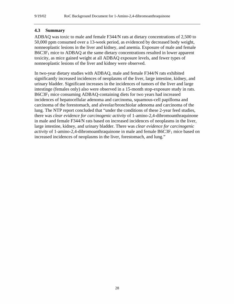
9/19/02 RoC Background Document for 1-Amino-2,4-dibromoanthraquinone
4.3 Summary ADBAQ was toxic to male and female F344/N rats at dietary concentrations of 2,500 to 50,000 ppm consumed over a 13-week period, as evidenced by decreased body weight, nonneoplastic lesions in the liver and kidney, and anemia. Exposure of male and female B6C3F1 mice to ADBAQ at the same dietary concentrations resulted in lower apparent toxicity, as mice gained weight at all ADBAQ exposure levels, and fewer types of nonneoplastic lesions of the liver and kidney were observed.
In two-year dietary studies with ADBAQ, male and female F344/N rats exhibited significantly increased incidences of neoplasms of the liver, large intestine, kidney, and urinary bladder. Significant increases in the incidences of tumors of the liver and large intestinge (females only) also were observed in a 15-month stop-exposure study in rats. B6C3F1 mice consuming ADBAQ-containing diets for two years had increased incidences of hepatocellular adenoma and carcinoma, squamous-cell papilloma and carcinoma of the forestomach, and alveolar/bronchiolar adenoma and carcinoma of the lung. The NTP report concluded that “under the conditions of these 2-year feed studies, there was clear evidence for carcinogenic activity of 1-amino-2,4-dibromoanthraquinone in male and female F344/N rats based on increased incidences of neoplasms in the liver, large intestine, kidney, and urinary bladder. There was clear evidence for carcinogenic activity of 1-amino-2,4-dibromoanthraquinone in male and female B6C3F1 mice based on increased incidences of neoplasms in the liver, forestomach, and lung.”
28

9/19/02 RoC Background Document for 1-Amino-2,4-dibromoanthraquinone
5 Genotoxicity
Limited data are available on the genotoxicity of ADBAQ. The few studies that have attempted to ascertain the mutagenicity of ADBAQ have encountered solubility problems that make the assays difficult to execute and that complicate interpretation of the results.
5.1 Prokaryotic systems: Salmonella typhimurium Salmonella typhimurium is the only prokaryote in which the mutagenicity of ADBAQ has been assessed. Several strains, including those that revert by base pair substitutions (TA100 and TA1535) and those that revert by frameshift mutations (TA98, TA1537, and TA1538), were used in the ADBAQ mutagenicity tests.
Haworth et al. (1983) showed that without rat or hamster liver microsomal (S9) metabolic activation, ADBAQ induced reverse mutation in strains TA98 and TA1537 (Table 5-1). In the presence of S9, reverse mutation was not induced in strain TA98, and strain TA1537 gave a variable response. ADBAQ was weakly mutagenic in strain TA100 (with or without S9 activation) and nonmutagenic in strain TA1535. The ADBAQ preparation used in these studies had a labeled purity of 91.7% and an analyzed purity of 79.7%. The researchers reported the presence of precipitate when ADBAQ concentrations were in the range of 333 to 10,000 μg/plate (the highest concentration tested). The NTP (1996) noted that the positive or weakly positive mutagenic responses in the Haworth et al. (1983) study were observed in strains in which reversion occurs by frameshift mutations.
Table 5-1. Mutagenicity of ADBAQ in S. typhimuriuma
Strain
Without S9 Hamster S9 Rat S9
Trial 1 Trial 2 Trial 1 Trial 2 Trial 1 Trial 2 TA100 + (weak) –/+ + (weak) –/+ –/+ –
TA1535 – – – – – –
TA1537 + + + – + –/+
TA98 + + –/+ – – –
Data were presented by Haworth et al. (1983) and NTP (1996). Trial summaries were obtained from Table G1 of NTP (1996). a+ = positive (a reproducible dose-related increase); – = negative; –/+ = equivocal (positive or weakly positive responses that were not replicated in a second trial).
5.2 Mammalian systems Rodent cells were used to determine the mutagenicity and DNA-damaging potential of ADBAQ. No reports of in vivo mammalian genotoxicity assays were found in the literature.
5.2.1 Mouse lymphoma L5178YTK+/- cells
Harrington-Brock et al. (1991) carried out a limited evaluation of the ability of ADBAQ to induce mutations at the thymidine kinase (tk) locus, but they were unable to test for
29

9/19/02 RoC Background Document for 1-Amino-2,4-dibromoanthraquinone
micronucleus formation because of ADBAQ’s limited solubility. ADBAQ was not mutagenic in the absence or presence of induced-rat liver S9 at the highest testable concentrations (25 μg/mL without S9 and 20 μg/mL with S9). The authors reported that the sample of ADBAQ was from the same batch evaluated in the NTP rodent carcinogenesis assay, and that the purity of the sample was 79.7%.
5.2.2 Chromosomal aberrations
Chinese hamster ovary (CHO) cells were exposed to various concentrations of ADBAQ and then analyzed for chromosomal aberrations (Loveday et al. 1990). The authors reported the presence of a precipitate in the flasks containing ADBAQ at 50 μg/mL (the highest concentration tested). Two separate trials without S9 metabolic activation gave equivocal results (Table 5-2). In one trial, exposure to ADBAQ at 3.02 or 10.10 μg/mL significantly increased the percentage of cells with chromosomal aberrations (Dunnett’s test, P < 0.05). In the second trial, the percentage of cells with chromosomal aberrations was not significantly increased by ADBAQ at concentrations of 1.0, 3.0, 10.0, or 30.0 μg/mL. In the presence of S9, the percentage of cells with chromosomal aberrations was not significantly increased by exposure to ADBAQ at 3.02, 10.10, or 30.20 μg/mL.
The NTP (1996) reported additional results obtained in two trials performed by Environmental Health Research and Testing, Inc. (Research Triangle Park, NC). In the absence of S9, the percentage of cells with chromosomal aberrations was significantly increased by exposure to ADBAQ at 50 μg/mL in the first trial, but increases observed at 16, 30, or 50 μg/mL in the second trial were not statistically significant. In the presence of S9, the percentage of cells with chromosomal aberrations was not significantly increased by ADBAQ at 16, 50, or 100 μg/mL. Based on these studies, the NTP (1996) concluded that ADBAQ induced chromosomal aberrations only in the absence of S9.
5.2.3 Sister chromatid exchange
Loveday et al. (1990) examined the effect of ADBAQ on the induction of sister chromatid exchange (SCE) in CHO cells. The chemical purity of ADBAQ was reported as greater than 79.7%, but the impurities were not identified. A trend test was used to describe the strength of evidence for SCE induction. Results were designated as negative, questionable, weakly positive, or positive, based on the trend test and the number of exposure levels resulting in at least a 20% increase over controls. Three exposure levels were used for each trial, and at least two had to produce a 20% increase for the result to be considered positive. In the presence or absence of induced-rat liver S9, ADBAQ induced SCE in a dose-dependent manner. In the absence of S9, ADBAQ induced at least a 20% increase in the number of SCEs at a concentration of 10 µg/mL, but not at 5 or 2.5 µg/mL. In the presence of S9, ADBAQ induced at least a 20% increase in the number of SCEs at 3.01, 10.1, or 30.1 μg/mL in trial 1 and at the highest concentration (15 μg/mL) in trial 2. The authors classified ADBAQ as weakly positive for SCE induction in the absence of S9 and positive (trial 1) or weakly positive (trial 2) in the presence of S9 (Table 5-2).
The NTP (1996) also reported results from the Environmental Health Research and Testing Laboratory, which found that ADBAQ tested at concentrations ranging from 5 to
30

9/19/02 RoC Background Document for 1-Amino-2,4-dibromoanthraquinone
50 µg/mL induced SCE in the absence of S9 (results were positive in one trial and weakly positive in another). In the presence of S9, ADBAQ at 5, 16, 50, or 100 μg/mL (one trial) did not induce SCE. The NTP (1996) noted that the discrepancy in SCE induction in the presence of S9 could not be explained by differences between the ADBAQ concentrations used by Loveday et al. (1990) and the NTP (1996).
Table 5-2. Induction of chromosomal aberrations and SCE in CHO cells by ADBAQa
Metabolic activation Chromosomal aberrations Sister chromatid exchanges
Loveday et al. 1990 NTP 1996b
Loveday et al. 1990 NTP 1996b
-S9 Trial 1 + + (weak)* + (weak) +
Trial 2 – + (weak) NR + (weak)
+S9 Trial 1 – – + –
Trial 2 NR NR + (weak) NR a+ = positive; – = negative; NR = not reported. bReported by the NTP (1996) as results from the Environmental Health Research and Testing Laboratory. *P < 0.05 for 50 μg/mL concentration.
5.2.4 Ras mutations in tumors
Hayashi et al. (2001) examined 8 forestomach squamous-cell papillomas, 24 squamouscell carcinomas, 19 alveolar/bronchiolar adenomas, and 4 alveolar/bronchiolar carcinomas for mutations in the H-ras and/or K-ras genes. These tumors were obtained from male and female B6C3F1 mice that were fed diets containing ADBAQ at a concentration of 10,000 or 20,000 ppm for two years (NTP 1996). Ras codons 12, 13, and 61, which are hot spots for mutations in human and rodent tumors, were examined in tumors from exposed mice, in spontaneous tumors from control mice, and in histologically normal forestomach and lung tissues. The ADBAQ-induced tumors had a higher frequency of ras mutations than did spontaneous tumors, and the mutations were found in both benign and malignant tumors (Table 5-3). The mutation spectra for the low- and high-dose groups were the same. Mutations in ras were not detected in any of the normal tissues. The predominant mutations in ADBAQ-induced tumors were CAA to CTA transversions and CAA to CGA transitions in codon 61 of K-ras (lung tumors) or H-ras (forestomach tumors). No A to T transversions at the second base were detected in spontaneous tumors. Mutations observed in codons 12 or 13 generally occurred in both ADBAQ-induced and spontaneous tumors. The authors concluded that alterations in the ras gene might be an early event in ADBAQ-induced carcinogenesis.
31

9/19/02 RoC Background Document for 1-Amino-2,4-dibromoanthraquinone
Table 5-3. Frequency and spectra of ras mutations in forestomach and lung tumors from B6C3F1 mice exposed to ADBAQ for two yearsa
Group
Total activated ras (%)
K-ras codons H-ras codon
12 13 61 61
Total Total A→T A→G A→T A→G C→A
Forestomach tumors
Controlb 4/11 (36) 3 1 0 0 0 1 0 ADBAQ 23/32 (72) 5 3 0 0 14 6 3 SCP 5/8 (63) 2 0 0 0 4 0 1 SCC 18/24 (75) 3 3 0 0 10 6 2
Lung tumors
Controlc 26/86 (30) 16 3 0 2 NT NT NT ADBAQ 16/23 (70) 7 1 6 5 NT NT NT ABA 14/19 (74) 7 1 6 3 NT NT NT ABC 2/4 (50) 0 0 0 2 NT NT NT aSCP = squamous-cell papilloma; SCC = squamous-cell carcinoma; ABA = alveolar/bronchiolar adenoma; ABC = alveolar/bronchiolar carcinoma; NT = not tested. bSpontaneous forestomach tumors in B6C3F1 mice. cStudy controls (N = 4) combined with historical spontaneous lung tumors (N = 82) of B6C3F1 mice.
5.3 Summary ADBAQ was mutagenic in S. typhimurium strains that revert by frameshift mutations, and the mutagenicity was decreased or eliminated in the presence of induced hamster or rat liver S9 metabolic activation. ADBAQ did not induce detectable mutations at the tk locus in mouse lymphoma cells; however, the concentrations used (20 or 25 μg/mL) were less than those used for the S. typhimurium assays (100 to 10,000 μg/mL) and slightly less than those used to assess clastogenicity (up to 100 μg/mL of ADBAQ). In CHO cells, ADBAQ induced chromosomal aberrations and SCE without metabolic activation; however, the results were inconsistent between different trials and laboratories. Forestomach and lung tumors induced by ADBAQ had a higher frequency of ras mutations than did spontaneous tumors. In particular, CAA to CTA transversions were common in ADBAQ-induced tumors but were absent in spontaneous tumors. The results obtained with ADBAQ must be viewed in the light of the limited solubility of this molecule and the uncertain purity of the preparations tested.
32

9/19/02 RoC Background Document for 1-Amino-2,4-dibromoanthraquinone
6 Other Relevant Data
6.1 Absorption, distribution, metabolism, and excretion The NTP (1996) reported studies on the absorption, distribution, metabolism, and excretion of ADBAQ. Male F344/N rats were administered [14C]ADBAQ orally or intravenously (i.v.), and disposition and elimination of radioactivity (parent compound and/or metabolites) was monitored for up to 72 hours. The specific location of the radioactive carbon atom(s) in the ADBAQ molecule was not identified.
6.1.1 Absorption
ADBAQ was readily absorbed from the gastrointestinal tract. The percentage of the dose absorbed was inversely proportional to dose level. When rats were administered single oral doses of 2, 23, 118, 814, or 1,473 mg/kg b.w., 90% of the lowest dose (2 mg/kg) was absorbed, but only 2% of the 814-mg/kg dose (NTP 1996).
6.1.2 Distribution and metabolism
ADBAQ was rapidly and widely distributed after i.v. administration. Radioactivity in blood was located primarily in plasma, as reflected by a whole blood:plasma ratio of 0.5. Fifteen minutes after i.v. administration (0.4 mg/kg b.w.), radioactivity was detected in most soft tissues, with the highest concentrations in excretory organs, lung, kidney, small intestine, liver, adipose tissue, and adrenal glands. At 15 minutes, tissue:blood ratios of radioactivity for these tissues were 3.0 or greater. Tissue:blood ratios declined during the 72 hours after administration, remaining greater than 1.0 only in liver and kidney at 72 hours. No quantitative data on tissue distribution were reported (NTP 1996).
Two hours after administration, only 3% of circulating radioactivity was in the form of unchanged parent compound, indicating that ADBAQ was quickly metabolized. Unchanged ADBAQ was deposited in adipose tissue, where it remained largely unmetabolized. As late as 24 hours after administration, the radioactivity remaining in adipose tissue was still unchanged ADBAQ, whereas that in liver, muscle, and skin was primarily ADBAQ metabolites (which were not identified) (NTP 1996). The metabolites of ADBAQ are unknown. Other anthraquinone-based dyes (Section 1, Table 1-2) were examined by IARC (1982) and the NTP (1978a, 1978b, 1978c, 1986), but their metabolites also are unknown. The NTP (1996) study did find that very little (less than 3%) of [14C]ADBAQ was excreted as unmetabolized ADBAQ.
6.1.3 Excretion
In the three days after administration of [14C]ADBAQ, approximately 50% of ADBAQ-associated radioactivity was recovered from feces, 15% from urine, and 6% from expired air. Consistent with extensive metabolism, less than 3% of total radioactivity excreted represented unchanged ADBAQ. Terminal half-lives of administered radioactivity in liver and kidney were estimated to be 40 and 90 hours, respectively, and the elimination half-life from adipose tissue was 11 hours.
33
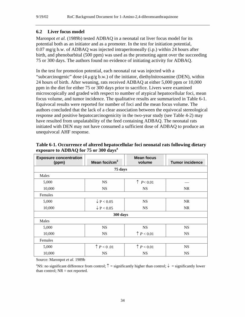
9/19/02 RoC Background Document for 1-Amino-2,4-dibromoanthraquinone
6.2 Liver focus model Maronpot et al. (1989b) tested ADBAQ in a neonatal rat liver focus model for its potential both as an initiator and as a promoter. In the test for initiation potential, 0.07 mg/g b.w. of ADBAQ was injected intraperitoneally (i.p.) within 24 hours after birth, and phenobarbital (500 ppm) was used as the promoting agent over the succeeding 75 or 300 days. The authors found no evidence of initiating activity for ADBAQ.
In the test for promotion potential, each neonatal rat was injected with a “subcarcinogenic” dose (4 μg/g b.w.) of the initiator, diethylnitrosamine (DEN), within 24 hours of birth. After weaning, rats received ADBAQ at either 5,000 ppm or 10,000 ppm in the diet for either 75 or 300 days prior to sacrifice. Livers were examined microscopically and graded with respect to number of atypical hepatocellular foci, mean focus volume, and tumor incidence. The qualitative results are summarized in Table 6-1. Equivocal results were reported for number of foci and the mean focus volume. The authors concluded that the lack of a clear association between the equivocal stereological response and positive hepatocarcinogenicity in the two-year study (see Table 4-2) may have resulted from unpalatability of the feed containing ADBAQ. The neonatal rats initiated with DEN may not have consumed a sufficient dose of ADBAQ to produce an unequivocal AHF response.
Table 6-1. Occurrence of altered hepatocellular foci neonatal rats following dietary exposure to ADBAQ for 75 or 300 daysa
Exposure concentration (ppm) Mean foci/cm3
Mean focus volume Tumor incidence
75 days Males
5,000 10,000
NS NS
↑ P< 0.01 NS
⎯
NR Females
5,000 10,000
↓ P < 0.05 ↓ P < 0.05
NS NS
NR NR
300 days Males
5,000 10,000
NS NS
NS ↑ P < 0.01
NS NS
Females 5,000 10,000
↑ P < 0 .01 NS
↑ P < 0.01 NS
NS NS
Source: Maronpot et al. 1989b aNS: no significant difference from control; ↑ = significantly higher than control; ↓ = significantly lower than control; NR = not reported.
34

9/19/02 RoC Background Document for 1-Amino-2,4-dibromoanthraquinone
6.3 Carcinogenicity and mutagenicity of other quinones and anthraquinones ADBAQ and other anthraquinones belong to a general class of quinone molecules that can be derived from aromatic molecules such as benzene, naphthalene, and anthracene. Quinone molecules can be reduced to a relatively stable hydroquinone, which usually is not associated with oxidative stress, or they may be reduced in a one-electron reduction to semiquinone free radicals that give rise to superoxide anions, hydrogen peroxide, and other reactive oxygen species (Parkinson 1996). Bolton et al. (2000) recently reviewed the role of quinones in toxicology, including carcinogenesis. Quinones may be produced from benzene, polycyclic aromatic hydrocarbons, estrogens, and catecholamines and give rise to reactive oxygen species that can damage DNA and other cellular macromolecules and activate signaling pathways. These molecular events may be associated with the initiation, promotion, and progression of carcinogenesis.
The benzoquinone metabolite of benzene has been proposed as a proximate carcinogenic product (Golding and Watson 1999); however, the IARC (1999) overall evaluation was that 1,4-benzoquinone is not classifiable as to its carcinogenicity to humans (Group 3). In a review of the role of quinoids in estrogen carcinogenesis, Bolton et al. (1998) suggested that the catechol metabolites of estrogens that are implicated in the carcinogenic effects of these steroids could be metabolized to electrophilic and redox-active quinoids that could damage DNA. Their conclusion, however, was that the ultimate carcinogenic metabolites of estrogens have not been unequivocally established.
Anthraquinones comprise a large class of diverse molecules which may be naturally occurring or synthetic in origin. Sendelbach (1989) reviewed the toxicity and carcinogenicity of anthraquinone derivatives with phenolic, amino, or nitro substitutions to the anthraquinone ring structure. The author’s general conclusion concerning amino substitutions was that addition of an amino group at either the 1- or 2-position of the anthraquinone ring appeared to consistently produce renal lesions in rats. The relevance of this conclusion to ADBAQ, which has both amino and halogen substitutions to the anthraquinone ring, is not clear. However, anthraquinone also produces renal tumors. Renal tubule adenomas and carcinomas (combined) were significantly increased (P < 0.05) by dietary exposure to ADBAQ at 5,000 ppm in female rats and at 10,000 ppm in both male and female rats (see Table 4-2).
A number of anthraquinone-derived vat dyes have been reviewed by both IARC and the NTP. ADBAQ produces tumors at some of the same sites as other anthraquinones. Table 6-2 summarizes the IARC and NTP evaluations of several anthraquinone derivatives. Information on a number of other anthraquinone derivatives at various stages of testing by the NTP can be accessed through the NTP Web site (http://ntp-server.niehs.nih.gov/main_pages/NTP_ALL_STDY_PG.html).
As described in Section 5.2.1, Harrington-Brock et al. (1991) carried out a limited evaluation of the ability of ADBAQ to induce mutations at the thymidine kinase (tk) locus of L5178/TK+/-mouse lymphoma cells, but they were unable to test for micronucleus formation because of ADBAQ’s limited solubility. An additional 15 anthraquinones were tested in the same assays. Testing of 8 of these substances was incomplete because of their limited solubility. For these 15 compounds, Table 6-3
35

9/19/02 RoC Background Document for 1-Amino-2,4-dibromoanthraquinone
summarizes the results of tests of mutagenicity at the L5178/TK+/- locus in two or three experiments and micronucleus analysis from one experiment per compound. Table 6-3 also summarizes the mutagenicity of these compounds in S. typhimurium, as reviewed by Harrington-Brock et al. (1991).
Brown (1980) reviewed the genetic effects of approximately 80 phenolic anthraquinones and reported that a high percentage of these hydroxyanthraquinones (36, or 45%) were mutagenic in S. typhimurium strains TA1537, TA1538, and TA98, which are sensitive to mutagens that cause frameshifts. A small portion (~10%) of the hydroxyanthraquinones contained either amino or halogen (chlorine or bromine) substitutions; however, the relevance of these findings to the possible mutagenicity of ADBAQ is not clear.
36

9/19/02 RoC Background Document for 1-Amino-2,4-dibromoanthraquinone
Table 6-2. Carcinogenicity and mutagenicity of anthraquinone and some anthraquinone-derived dyes (data from NTP and IARC)
Property Anthraquinone 2-Amino
anthraquinone
1-Amino-2methyl
anthraquinone
1,8-Dihydroxyanthraquinone
(danthron) 2-Methyl-1-nitroanthraquinone
1,4,5,8Tetraamino
anthraquinone (disperse blue 1) Emodin
Long-term Liver neoplasms in Hepatocellular Negative in male Hepatocellular Hemangiosarcomas Hepatocellular Uncommon carcinogenicity male and female carcinomas in male mice (NTP) adenomas and in male and female adenomas and renal tubule in mice B6C3F1 mice and female
B6C3F1 mice (NTP) Malignant hematopoietic lymphomas in high dose female mice (NTP)
Hepatocellular carcinomas and neoplastic liver nodules in female B6C3F1 mice (NTP)
carcinomas and adenomatous hyperplasia of the cecum in C3H/HeN male mice (IARC)
B6C3F1 mice (NTP)
carcinomas (combined) in male B6C3F1 mice (NTP) Alveolar/bronchiolar adenomas and carcinomas (combined) in male B6C3F1 mice (NTP)
neoplasms in male B6C3F1 mice
Long-term Renal tubule Hepatocellular Hepatocellular Adenocarcinomas Hepatocellular Transitional-cell Zymbal’s gland carcinogenicity neoplasms in male carcinomas in male carcinomas in of the colon in carcinomas in male neoplasms of the carcinomas in in rats and female F344 rats (NTP) male and female male ACI rats F344 rats (NTP) urinary bladder in female F344/N
F344/N rats Transitional
Inadequate study in female F344 rats
F344 rats (NTP) (IARC) Subcutaenous fibromas in male
male and female F344 rats (NTP)
rats
epithelial (poor survival) and female F344 Squamous-cell papillomas of the (NTP) rats (NTP) neoplasms in male urinary bladder in F344 rats (NTP) male F344/N rats Leiomyomas and
leiomyosarcomas (combined) of the urinary bladder in female F344 rats (NTP)
37

9/19/02 RoC Background Document for 1-Amino-2,4-dibromoanthraquinone
2-Amino1-Amino-2
methyl-
1,8-Dihydroxyanthraquinone 2-Methyl-1-nitro
1,4,5,8Tetraamino
anthraquinone Property Anthraquinone anthraquinone anthraquinone (danthron) anthraquinone (disperse blue 1) Emodin
Genetic toxicology
Positive for mutagenicity in S. typhimurium TA98 and TA100 with and without S9 Positive for micronucleated normochromatic erythrocytes in male and female mice (14-week dietary study) Negative for micronuclei in mouse bone marrow (i.p. injection)
Positive for chromosomal aberrations Positive for SCE Positive for mutagenicity in S. typhimurium (NTP)
Positive for chromosomal aberrations Positive for SCE Positive for mutagenicity in S. typhimurium (NTP)
Positive for chromosomal aberrations Not tested for SCE Negative for micronuclei [cells not specified] Weakly positive for mutagenicity in S. typhimurium (NTP)
Positive for chromosomal aberrations Positive for SCE Negative for reciprocal translocation in Drosophila Positive for mutagenicity in S. typhimurium
Positive for chromosomal aberrations Positive for SCE Positive for mutagenicity in mouse lymphoma cells Negative for micronuclei [cells not specified] Positive for mutagencity in S. typhimurium
Positive for mutagenicity in S. typhimurium (TA100) with S9; negative in TA98 with or without S9 Positive for chromosal aberrations Negative for micronuclei in mouse bone marrow (i.p. injection) Weakly postive for micronuclei in female mice (14-week dietary study) [cells not specified]
38

9/19/02 RoC Background Document for 1-Amino-2,4-dibromoanthraquinone
Property Anthraquinone 2-Amino
anthraquinone
1-Amino-2methyl
anthraquinone
1,8-Dihydroxyanthraquinone
(danthron) 2-Methyl-1-nitroanthraquinone
1,4,5,8Tetraamino
anthraquinone (disperse blue 1) Emodin
RoC listing or NTP evaluation
NTP: some evidence of carcinogenic activity in male F344/N rats; clear evidence of carcinogenic activity in female F344/N rats; clear evidence of carcinogenic activity in male and female B6C3F1 mice
RoC: reasonably anticipated to be a human carcinogen
RoC: reasonably anticipated to be a human carcinogen
RoC: reasonably anticipated to be a human carcinogen
NTP: found to be carcinogenic in male and female rats and mice
RoC: reasonably anticipated to be a human carcinogen
NTP: equivocal evidence of carcinogenic activity in female F344/N rats and B6C3F1 mice
IARC evaluationa
Not listed Group 3 (IARC 1987)
Group 3 (IARC 1987)
Group 2B (IARC 1990)
Group 2B (IARC 1987)
Group 2B (IARC 1990)
Not listed
aGroup 2B = possibly carcinogenic to humans, based on limited evidence of carcinogenicity in humans, less than sufficient evidence of carcinogenicity in animals. Group 3 = not classifiable as to its carcinogenicity in humans, based on inadequate or limited evidence of carcinogenicity in animals and inadequate evidence of carcinogenicity in humans.
39
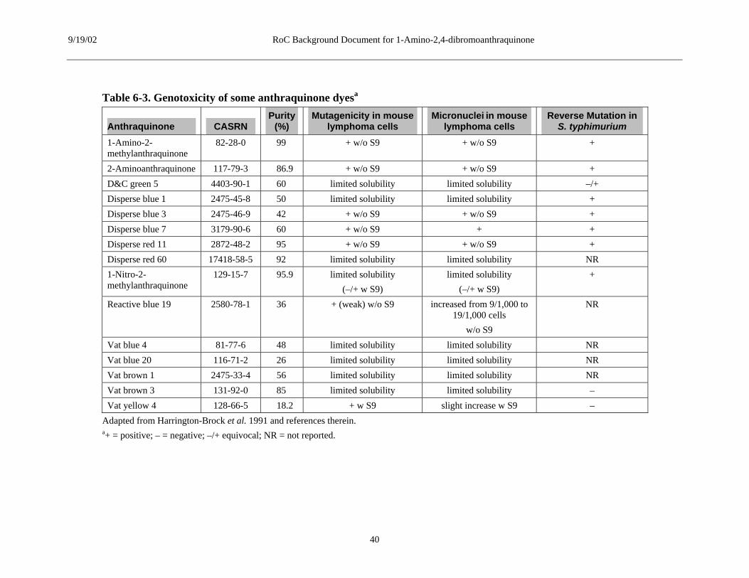
9/19/02 RoC Background Document for 1-Amino-2,4-dibromoanthraquinone
Table 6-3. Genotoxicity of some anthraquinone dyesa
Anthraquinone CASRN Purity
(%) Mutagenicity in mouse
lymphoma cells Micronuclei in mouse
lymphoma cells Reverse Mutation in
S. typhimurium 1-Amino-2methylanthraquinone
82-28-0 99 + w/o S9 + w/o S9 +
2-Aminoanthraquinone 117-79-3 86.9 + w/o S9 + w/o S9 + D&C green 5 4403-90-1 60 limited solubility limited solubility –/+ Disperse blue 1 2475-45-8 50 limited solubility limited solubility + Disperse blue 3 2475-46-9 42 + w/o S9 + w/o S9 + Disperse blue 7 3179-90-6 60 + w/o S9 + + Disperse red 11 2872-48-2 95 + w/o S9 + w/o S9 + Disperse red 60 17418-58-5 92 limited solubility limited solubility NR 1-Nitro-2methylanthraquinone
129-15-7 95.9 limited solubility (–/+ w S9)
limited solubility (–/+ w S9)
+
Reactive blue 19 2580-78-1 36 + (weak) w/o S9 increased from 9/1,000 to 19/1,000 cells
w/o S9
NR
Vat blue 4 81-77-6 48 limited solubility limited solubility NR Vat blue 20 116-71-2 26 limited solubility limited solubility NR Vat brown 1 2475-33-4 56 limited solubility limited solubility NR Vat brown 3 131-92-0 85 limited solubility limited solubility – Vat yellow 4 128-66-5 18.2 + w S9 slight increase w S9 –
Adapted from Harrington-Brock et al. 1991 and references therein. a+ = positive; – = negative; –/+ equivocal; NR = not reported.
40

9/19/02 RoC Background Document for 1-Amino-2,4-dibromoanthraquinone
6.4 Summary In rats, ADBAQ is rapidly absorbed from the gastrointestinal tract and distributed rapidly and widely to most soft tissues. Approximately 97% of ADBAQ is metabolized, as demonstrated by the small amount (less than 3%) of radiolabel recovered as the parent compound from either blood or urine; however, the metabolites of ADBAQ have not been identified. ADBAQ and its metabolites are excreted primarily in the feces and urine.
ADBAQ and other anthraquinones are classified with a large number of other quinone molecules that can be derived from aromatic molecules such as benzene, naphthalene, and anthracene. Reactive oxygen species generated by metabolism of a variety of quinones may be associated with DNA damage or activation of signaling pathways involved in initiation, promotion, and progression of carcinogenesis. Four other anthraquinone vat dyes evaluated by the NTP have been listed in the RoC as reasonably anticipated to be human carcinogens. In addition, a high percentage (36/80) of phenolic anthraquinones have been reported to be mutagenic in Salmonella.
41

9/19/02 RoC Background Document for 1-Amino-2,4-dibromoanthraquinone
42

9/19/02 RoC Background Document for 1-Amino-2,4-dibromoanthraquinone
7 References
1. Barbone, F., E. Delzell, H. Austin, and P. Cole. 1992. A case-control study of lung cancer at a dye and resin manufacturing plant. Am J Ind Med 22:835-849.
2. Barbone, F., E. Delzell, H. Austin, and P. Cole. 1994. Exposure to epichlorohydrin and central nervous system neoplasms at a resin and dye manufacturing plant. Arch Environ Health 49:355-358.
3. Bolton, J.L., E. Pisha, F. Zhang, and S. Qiu. 1998. Role of quinoids in estrogen carcinogenesis. Chem Res Toxicol 11:1113-1127.
4. Bolton, J.L., M.A. Trush, T.M. Penning, G. Dryhurst, and T.J. Monks. 2000. Role of quinones in toxicology. Chem Res Toxicol 13:135-160.
5. Brown, J.P. 1980. A review of the genetic effects of naturally occurring flavonoids, anthraquinones and related compounds. Mutat Res 75:243-277.
6. ChemFinder. 2001. 1-Amino-2,4-Dibromoanthraquinone. Available at http://www.chemfinder.camsoft.com (and search 81-49-2).
7. Delzell, E., M. Macaluso, and P. Cole. 1989. A follow-up study of workers at a dye and resin manufacturing plant. J Occup Med 31:273-278.
8. Fleischman, R.W., H.J. Esber, M. Hagopian, H.S. Lilja, and J. Huff. 1986. Thirteen-week toxicology studies of 1-amino-2,4-dibromoanthraquinone in Fischer 344/N rats and B6C3F1 mice. Toxicol Appl Pharmacol 82:389-404.
9. Games, L.M. and R.A. Hites. 1977. Composition, treatment efficiency, and environmental significance of dye manufacturing plant effluents. Anal Chem 49:1433-1440.
10. Gardiner, J.S., S.A. Walker, and A.J. MacLean. 1982. A retrospective mortality study of substituted anthraquinone dyestuffs workers. Br J Ind Med 39:355-360.
11. Golding, B.T. and W.P. Watson. 1999. Possible mechanisms of carcinogenesis after exposure to benzene. IARC Sci Publ 150:75-88.
12. Harada, T., R.R. Maronpot, R.W. Morris, and G.A. Boorman. 1989. Observations on altered hepatocellular foci in National Toxicology Program two-year carcinogenicity studies in rats. Toxicol Pathol 17:690-706; discussion 707-708.
13. Harrington-Brock, K., L. Parker, C. Doerr, M.C. Cimino, and M.M. Moore. 1991. Analysis of the genotoxicity of anthraquinone dyes in the mouse lymphoma assay. Mutagenesis 6:35-46.
14. Haworth, S., T. Lawlor, K. Mortelmans, W. Speck, and E. Zeiger. 1983. Salmonella mutagenicity test results for 250 chemicals. Environ Mutagen 5:1-142.
43

9/19/02 RoC Background Document for 1-Amino-2,4-dibromoanthraquinone
15. Hayashi, S., H.H. Hong, K. Toyoda, T.V. Ton, T.R. Devereux, R.R. Maronpot, J. Huff, and R.C. Sills. 2001. High frequency of ras mutations in forestomach and lung tumors of B6C3F1 mice exposed to 1-amino-2,4-dibromoanthraquinone for 2 years. Toxicol Pathol 29:422-429.
16. HSDB. 2000. Hazardous Substance Data Bank. Revised February 2, 2000. Last reviewed January 31, 1996. National Library of Medicine. Available at http://toxnet.nlm.nih.gov/cgi-bin/sis/htmlgen?HSDB (and search CAS No. 81-492).
17. IARC. 1982. Some Aromatic Amines, Anthraquinones and Nitroso Compounds, and Inorganic Fluorides Used in Drinking Water and Dental Preparations, Vol. 27. IARC Monographs on the Evaluation of Carcinogenic Risks to Humans. World Health Organization, International Agency for Research on Cancer, Lyon, France.
18. IARC. 1987. Overall Evaluations of Carcinogenicity: An-Updating of IARC Monographs Volumes 1 to 42, Suppl. 7. IARC Monographs on the Evaluation of Carcinogenic Risks to Humans. World Health Organization, International Agency for Research on Cancer, Lyon, France.
19. IARC. 1990. Some Flame Retardants and Textile Chemicals, and Exposures in the Textile Manufacturing Industry, Vol. 48. IARC Monographs on the Evaluation of Carcinogenic Risks to Humans. World Health Organization, International Agency for Research on Cancer, Lyon, France.
20. IARC. 1999. Re-evaluation of some organic chemical, hydrazine and hydrogen peroxide (three parts), Vol. 71. IARC Monographs on the Evaluation of Carcinogenic Risks to Humans. World Health Organization, International Agency for Research on Cancer, Lyon, France.
21. Ikeda, M., T. Watanabe, I. Hara, T. Tabuchi, S.I. Nakamura, H. Kosaka, M. Minami, and Y. Sakurai. 1977. A field survey on the health status of workers in dye-producing factories. Int Arch Occup Environ Health 39:219-235.
22. Loveday, K.S., B.E. Anderson, M.A. Resnick, and E. Zeiger. 1990. Chromosome aberration and sister chromatid exchange tests in Chinese hamster ovary cells in vitro. V: Results with 46 chemicals. Environ Mol Mutagen 16:272-303.
23. Maronpot, R.R., T. Harada, A.S. Murthy, and G.A. Boorman. 1989a. Documenting foci of hepatocellular alteration in two-year carcinogenicity studies: current practices of the National Toxicology Program. Toxicol Pathol 17:675-683; discussion 683-674.
24. Maronpot, R.R., H.C. Pitot, and C. Peraino. 1989b. Use of rat liver altered focus models for testing chemicals that have completed two-year carcinogenicity studies. Toxicol Pathol 17:651-662.
44

9/19/02 RoC Background Document for 1-Amino-2,4-dibromoanthraquinone
25. Novotná, P., V. Pacáková, Z. Bosáková, and K. Štulík. 1999. High-performance liquid chromatographic determination of some anthraquinone and naphthoquinone dyes occurring in historical textiles. J Chromatogr A 863:235-241.
26. NTP. 1978a. Bioassay of 2-Methyl-1-nitroanthraquinone for Possible Carcinogenicity. National Toxicology Program Technical Report Series TR-29.
27. NTP. 1978b. Bioassay of 1-Amino-2-methylanthraquinone for Possible Carcinogenicity. National Toxicology Program Technical Report Series TR-111.
28. NTP. 1978c. Bioassay of 2-Aminoanthraquinone for Possible Carcinogenicity. National Toxicology Program Technical Report Series TR-144.
29. NTP. 1986. Toxicology and Carcinogenesis Studies of C.I. Disperse Blue 1 (A Commercial Dye Containing Approximately 50% 1,4,5,8Tetraaminoanthraquinone, and 20% Water) (CAS No. 2475-45-8) in F344/N Rats and B6C3F1 Mice (Feed Studies). National Toxicology Program Technical Report Series No 299.
30. NTP. 1996. Toxicology and carcinogenesis studies of 1-Amino-2,4Dibromoanthraquinone in F344/N rats and B6C3F1 mice (feed studies). National Toxicology Program Technical Report Series No 383.
31. NTP Chemical Repository. 2001. 1-amino-2,4-dibromoanthraquinone. Revised August 13, 2001. National Toxicology Program. NTP Chemical Repository. Available at http://ntpserver.niehs.nih.gov/cgi/iH_Indexes/ALL_SRCH/iH_ALL_SRCH_Frames.html and search 81-49-2.
32. Parkinson, A. 1996. Biotransformation of Xenobiotics. In Casarett and Doull's Toxicology: The Basic Science of Poisons, Fifth Edition. Klaassen, C.D., M.O. Amdur and J. Doull, eds. McGraw-Hill, New York. pp. 113-186.
33. Preiss, A., U. Sänger, N. Karfich, K. Levsen, and C. Mugge. 2000. Characterization of dyes and other pollutants in the effluent of a textile company by LC/NMR and LC/MS. Anal Chem 72:992-998.
34. Sathiakumar, N. and E. Delzell. 2000. An updated mortality study of workers at a dye and resin manufacturing plant. J Occup Environ Med 42:762-771.
35. Sendelbach, L.E. 1989. A review of the toxicity and carcinogenicity of anthraquinone derivatives. Toxicology 57:227-240.
36. TRI. 2001. Toxic Release Inventory. Revised 2001. Environmental Protection Agency. Available at (http://toxnet.nlm.nih.gov/cgi-bin/sis/htmlgen?TRI).
45

9/19/02 RoC Background Document for 1-Amino-2,4-dibromoanthraquinone
46

9/19/02 RoC Background Document for 1-Amino-2,4-dibromoanthraquinone
Appendix A: NTP TR 383 (1996). Toxicology and Carcinogenesis Studies of 1-Amino-2,4Dibromoanthraquinone in F344/N Rats and B6C3F1 Mice (Feed Studies). PP A-1 – A-86.
47

9/19/02 RoC Background Document for 1-Amino-2,4-dibromoanthraquinone
Appendix B: Tissues examined for histopathology in mice and rats in the NTP bioassays of ADBAQ
49

9/19/02 RoC Background Document for 1-Amino-2,4-dibromoanthraquinone
50

9/19/02 RoC Background Document for 1-Amino-2,4-dibromoanthraquinone
Tissues examined for histopathology in mice and rats in the NTP bioassays of ADBAQ
13-Week studies 2-Year studies Stop-exposure evaluation Complete histopathologic examinations were performed on all animals that died during the study, control animals, and 50,000 ppm animals. In addition to tissue masses, gross lesions, and associated lymph nodes, the tissues examined included: adrenal gland, bone and marrow, brain, clitoral gland, epididymis, esophagus, heart, kidney, large intestine (cecum, colon, rectum), liver, lung, lymph nodes (mandibular and mesenteric), mammary gland, nose, ovary, pancreas, parathyroid gland, pituitary gland, preputial gland, prostate gland, salivary gland, seminal vesicle, skin, small intestine (duodenum, jejunum, ileum), spleen, stomach, testis, thymus, thyroid gland, trachea, urinary bladder, and uterus. The kidney, liver, spleen (rats), thymus (rats), and uterus (rats) of all other exposed animals were examined.
Complete histopathologic examinations were performed on all animals. In addition to tissue masses, gross lesions, and associated lymph nodes, the tissues examined included: adrenal gland, bone and marrow, brain, clitoral gland, epididymis, esophagus, gallbladder (mice), heart, kidney, large intestine (cecum, colon, rectum), liver, lung, lymph nodes (mandibular and mesenteric), mammary gland, nose, ovary, pancreas, parathyroid gland, pituitary gland, preputial gland, prostate gland, salivary gland, seminal vesicle, skin, small intestine (duodenum, jejunum, ileum), spleen, stomach, testis, thymus, thyroid gland, trachea, urinary bladder, and uterus.
Complete histopathologic examinations were performed on all animals. In addition to tissue masses, gross lesions, and associated lymph nodes, the tissues examined included: adrenal gland, bone and marrow, brain, clitoral gland, epididymis, esophagus, heart, kidney, large intestine (cecum, colon, rectum), liver, lung, lymph nodes (mandibular and mesenteric), mammary gland, nose, ovary, pancreas, parathyroid gland, pituitary gland, preputial gland, prostate gland, salivary gland, seminal vesicle, skin, small intestine (duodenum, jejunum, ileum), spleen, stomach, testis, thymus, thyroid gland, trachea, urinary bladder, and uterus.
51

9/19/02 RoC Background Document for 1-Amino-2,4-dibromoanthraquinone
52