0dwhuldo (6, iru&khp&rpp 7klv identification of actin as a ... fileidentification of actin...
TRANSCRIPT

Identification of actin as a direct proteomic target of berberine, using an
affinity-based chemical probe and elucidation of its modulatory role in
actin assembly
Chae-Min Yia, Jihyun Yua, Hyun-Bin Kimb, Na-Rae Leea, Sang Won Kima, Noh-Jin Leea, Jun Leea,
Jihye Seongb,c,*, Nam-Jung Kima,*, Kyung-Soo Inna,b,*
aDepartment of Fundamental Pharmaceutical Sciences, Graduate School, Kyung Hee University,
Seoul 02447, Republic of Korea
bKHU-KIST Department of Converging Science and Technology, Graduate School,
Kyung Hee University, Seoul 02447, Republic of Korea
cConvergence Center for Diagnosis Treatment Care of Dementia, Korea Institute of Science and
Technology (KIST), Seoul 02792, Republic of Korea
Table of Contents
Experimental section
Chemistry 2
Biological assay 6
Supplementary Figure S1 9
LC-MS/MS spectral data 10
1
Electronic Supplementary Material (ESI) for ChemComm.This journal is © The Royal Society of Chemistry 2017

Experimental section
Chemistry
General methods. Unless otherwise described, all commercial reagents and solvents were purchased
from commercial suppliers such as Sigma-Aldrich, and used without further purifications.
Tetrahydrofuran was distilled from sodium benzophenone ketyl and dichloromethane, acetonitrile,
triethylamine and pyridine were freshly distilled with calcium hydride. Flash column chromatography
was carried out using silica-gel 60 (230-400 mesh, Merck). Thin layer chromatography was performed
using 0.25 mm silica gel plates to monitor reactions. All reactions were performed under dry argon
atmosphere in flame-dried glassware. 1H NMR and 13C NMR spectra were recorded on a BRUKER
AVANCE 400 (400MHz), or VARIAN VNMRS 500 (500MHz) spectrometers, respectively. Chemical
shifts are provided in parts per million (ppm, ) downfield from tetramethylsilane (internal standard)
with coupling constant in hertz (Hz). Multiplicity is indicated by the following abbreviations: singlet (s),
doublet (d), doublet of doublet (dd), triplet (t), quartet (q), quintet (quin) multiplet (m) and broad (br).
Optical rotations were measured using JASCO DIP-1000 digital polarimeter at ambient temperature
using 100 nm cell of 2 mL capacity. Mass spectra and HRMS were recorded on Agilent 6530 Accurate-
Mass Q-TOF LC/MS and JEOL JMS-700. The purity of the compounds was identified by normal phase
high-pressure liquid chromatography (HPLC) is carried out either on.
Compound (2) To a solution of 1 (1.006 mmol, ref: Anticancer Agents Med. Chem., 2015, 15, 89-98.)
in DMF (10 mL) was added propargyl bromide (2.415 mmol). The reaction mixture was stirred at 80 °C
until complete consumption of the starting material on TLC. The reaction mixture was recrystallized
from diethyl ether to afford 398mg (100%) of compound 2 as a brown solid. 1H-NMR (DMSO, 300
MHz) δ 9.88 (s, 1H), 8.97 (d, 1H, J = 7.8 Hz), 8.22 (t, 1H, J = 8.8 Hz), 8.03 (dd, 1H, J = 14.8, 9.1 Hz),
7.80 (s, 1H), 7.09 (s, 1H), 6.18 (s, 2H), 5.09 (d, 2H, J = 2.4 Hz), 4.96 (t, 2H, J = 6.1 Hz), 4.09 (d, 3H, J
= 5.6 Hz), 3.62 (t, 1H, J = 2.4 Hz), 3.21 (t, 2H, J = 6.1 Hz); 13C-NMR (DMSO, 75 MHz) δ 151.2, 150.4,
148.2, 145.8, 141.2, 138.1, 133.4, 131.2, 127.0, 124.8, 122.6, 120.9, 120.8, 108.9, 106.0, 102.6, 80.3,
79.3, 61.4, 57.7, 55.8, 26.8; LR-MS (FAB) m/z 360 (M-Cl)+; HR-MS (FAB) calculated for C22H18NO4+
(M-Cl)+ 360.1236; found 360.1237.
Compound (4) To a solution of 2 (0.253 mmol), and N-[2-[2-[2-(2-azidoethoxy)ethoxy]-
ethoxy]ethyl]-biotinamide (3, 0.253 mmol) in t-BuOH (3 mL) and H2O (3 mL) were added sodium
ascorbate (0.487 mmol) and copper(II) sulfate (0.152 mmol). The reaction mixture was stirred at room
temperature until complete consumption of the starting material on TLC. Then, the solvent was
concentrated in vacuo and purified by column chromatography on silica gel (methylene chloride:
2
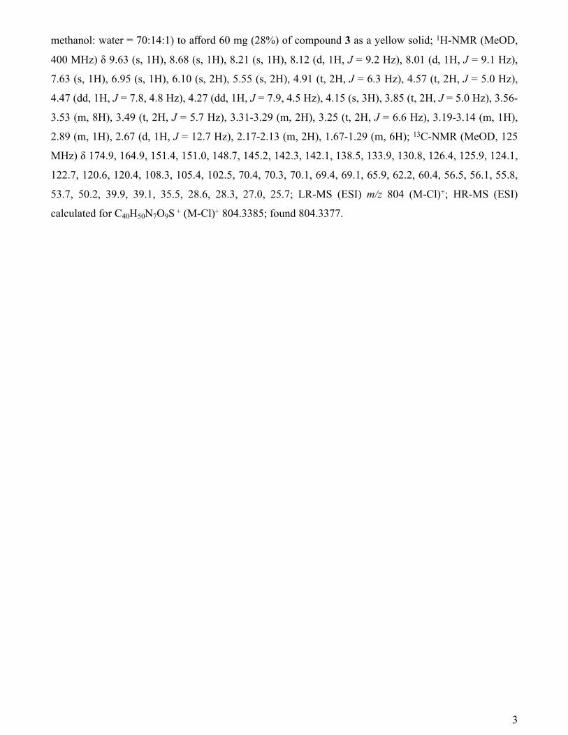
methanol: water = 70:14:1) to afford 60 mg (28%) of compound 3 as a yellow solid; 1H-NMR (MeOD,
400 MHz) δ 9.63 (s, 1H), 8.68 (s, 1H), 8.21 (s, 1H), 8.12 (d, 1H, J = 9.2 Hz), 8.01 (d, 1H, J = 9.1 Hz),
7.63 (s, 1H), 6.95 (s, 1H), 6.10 (s, 2H), 5.55 (s, 2H), 4.91 (t, 2H, J = 6.3 Hz), 4.57 (t, 2H, J = 5.0 Hz),
4.47 (dd, 1H, J = 7.8, 4.8 Hz), 4.27 (dd, 1H, J = 7.9, 4.5 Hz), 4.15 (s, 3H), 3.85 (t, 2H, J = 5.0 Hz), 3.56-
3.53 (m, 8H), 3.49 (t, 2H, J = 5.7 Hz), 3.31-3.29 (m, 2H), 3.25 (t, 2H, J = 6.6 Hz), 3.19-3.14 (m, 1H),
2.89 (m, 1H), 2.67 (d, 1H, J = 12.7 Hz), 2.17-2.13 (m, 2H), 1.67-1.29 (m, 6H); 13C-NMR (MeOD, 125
MHz) δ 174.9, 164.9, 151.4, 151.0, 148.7, 145.2, 142.3, 142.1, 138.5, 133.9, 130.8, 126.4, 125.9, 124.1,
122.7, 120.6, 120.4, 108.3, 105.4, 102.5, 70.4, 70.3, 70.1, 69.4, 69.1, 65.9, 62.2, 60.4, 56.5, 56.1, 55.8,
53.7, 50.2, 39.9, 39.1, 35.5, 28.6, 28.3, 27.0, 25.7; LR-MS (ESI) m/z 804 (M-Cl)+; HR-MS (ESI)
calculated for C40H50N7O9S + (M-Cl)+ 804.3385; found 804.3377.
3

Compound 4 (BBP)
▼ 1H-NMR (CDCl3, 400 MHz)
4
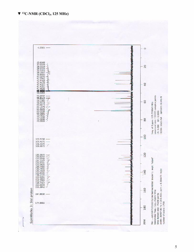
▼ 13C-NMR (CDCl3, 125 MHz)
5

Biological assay
Pull-down assay
Jurkat human leukemic T-cells (ATCC TIB-152) were maintained in RPMI medium containing 10%
FBS and penicillin/streptomycin (100 U/ml). Jurkat cells were lysed with lysis buffer (XX mM Tris-
HCl containing 0.25% NP-40 and protease inhibitor cocktail). The lysates (10 mg) were incubated with
Biotin-azide (20 μM) or Berberine biotinylated probe (BBP) (20 μM) with or without free berberine (40
μM) for 2 hours, followed by further incubation with streptavidin-resin for 30 minutes. After extensive
washing with lysis buffer 5 times, BBP-bound proteins were eluted with SDS-PAGE sample buffer and
subjected to SDS-PAGE and silver staining according to the manufacturer’s instruction (Pierce). The
band was excised and analyzed using LC-MS/MS by Yonsei Proteome Research Center (YPRC).
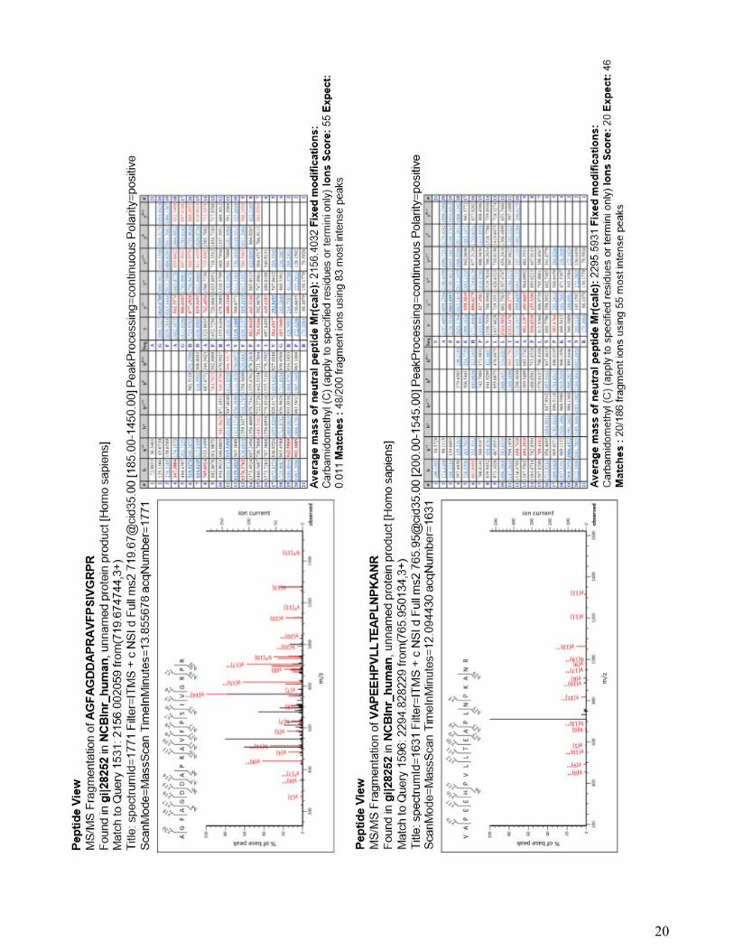
LC-MS/MS for peptide analysis
NanoLC–MS/MS analysis was performed on an agilent 1100 Series nano-LC and LTQ- mass
spectrometer (Thermo Electron, San Jose, CA). The capillary column used for LC–MS/MS analysis
(150 mm × 0.075 mm) was obtained from Proxeon (Odense M , Denmark ) and slurry packed in house
with 5 μm, 100 Å pore size Magic C18 stationary phase (Michrom Bioresources, Auburn, CA). The
mobile phase A for the LC separation was 0.1% formic acid in deionized water and the mobile phase B
was 0.1% formic acid in acetonitrile. The chromatography gradient was set up to give a linear increase
from 6 % B to 50 % B in 17 min and from 50 % B to 95 % B in 6 min and from 95 % B to 6 % B in 12
min. The flow rate was maintained at 600 nL/min after splitting. Mass spectra were acquired using data-
dependent acquisition with full mass scan (350-1800 m/z) followed by MS/MS scans. Each MS/MS
scan acquired was an average of one microscans on the LTQ. The temperature of the ion transfer tube
was controlled at 200 ℃ and the spray was 1.5.0-2.0 kV. The normalized collision energy was set at
35% for MS/MS.
Database searching
The mascot algorithm (Matrixscience, USA) was used to identify peptide sequences present in a
protein sequence database. Database search criteria were, taxonomy; homo sapiens (NCBInr database
downloaded on Mar 24 2013; 695124 sequences: 228548881 residues), fixed modification;
carboxyamidomethylated at cysteine residues, variable modification; oxidized at methionine residues,
maximum allowed missed cleavage; 2, MS tolerance; 1.2 Da, MS/MS tolerance; 0.6 Da. Only peptides
resulting from trypsin digests were considered.
6

Direct binding assay
Purified rabbit actin (13.5 μg) was reacted with BBP (20 μM) with or without free berberine (BBR, 40
μM) for 2 hours followed by dialysis using Tris-HCl (pH 8.0) buffer to remove free BBP. The samples
were further incubated with streptavidin resin for 30 minutes and washed with Tris-HCl buffer five
times. Precipitated samples were subjected to SDS-PAGE and silver staining (Pierce) according to the
instruction. To further confirm the interaction between berberine and actin Surface Plasmon Resonance
(SPR) assay was carried out.
Podosome reformation assay
Raw 264.7 cells (ATCC TIB-71) were maintained in Dulbecco’s modified Eagle’s medium (DMEM)
containing 10% FBS and penicillin/streptomycin (100 U/ml). Cells were starved for 24 hours with
serum-free DMEM, then treated with 25 μM PP2 for 30 minutes to remove podosomes. After podosome
disruption, cells were washed with phosphate buffered saline (PBS) and fed with normal complete
medium containing DMSO or BBR (100 μM). After 30 minutes of incubation, cells were washed once
with PBS and then fixed with 4% paraformaldehyde for an hour. Fixed cells were washed with PBS
once and permeabilized with 0.1% Triton-X100 for 15 min. After permeabilization, cells were washed
two or more times with PBS and stained with CytoPainter F-actin staining kit-Green Fluorescence
(Abcam) for an hour to visualize newly formed podosomes.
Actin polymerization and depolymerization Assay
In vitro actin polymerization and depolymerization assays were carried out using fluorescence-based
actin polymerization kit (Cytoskeleton, Cat. No. BK003) according to the manufacturer’s instruction.
Briefly, polymerization buffer (10X) was added to prepared pyrene-G-actin samples with indicated
concentrations of free berberine. Actin polymerization was assessed by monitoring fluorescence signal
using a fluorimeter (Tecan). To assess the depolymerizing activity of berberine, pyrene F-actin was
prepared by polymerization of G-actin for 1 hour according to the instruction. Indicated concentrations
of berberine were added to the samples and depolymerization was monitored for 90 minutes using a
fluorimeter.
Imaging of actin filaments after the treatment of berberine.
NIH3T3 cell line was cultured in DMEM supplemented with 10% fetal bovine serum (Hyclone), 2
mM L-glutamine, 1 U/ml penicillin, 100 mg/ml streptomycin, and 1 mM sodium pyruvate. For imaging
experiment, cells were plated onto 35 mm glass coverslip-bottomed dishes coated with 10 μg/ml of
7

fibronectin (Invitrogen). Cells were washed once with pre-chilled phosphate buffered saline (PBS) and
incubated with PBS containing 25 μl/ml 0.1% Triton-X100 for 10 min at 4°C. Then, the cells were
washed once with pre-warmed media and returned to the incubator with culture media with or without
100 μM berberine chloride. After 48 hours of incubation, cells were washed once with pre-warmed PBS
and then fixed with 4% paraformaldehyde. Fixed cells were washed two or more times with PBS for 10
min and permeabilized with pre-chilled 0.1%Triton-X100 for 5 min, after washed again and then,
blocked with 1% BSA (company) for 30 minutes in room temperature. After blocking, cells were
washed two or more times with PBS for 10 minutes and stained with PBS containing Alexa Fluor 594
Phalloidin (1:200)(Life technology) for 30 minutes. Images were collected by a Nikon Ti-E inverted
microscope and a cooled charge-coupled device camera using NIS software with a 560DF40 excitation
filter, a 595DRLP dichroic mirror, and a 653DF95 emission filter. A neutral-density filter was used to
control the intensity of the excitation light. Collected images were analyzed with NIS software.
8
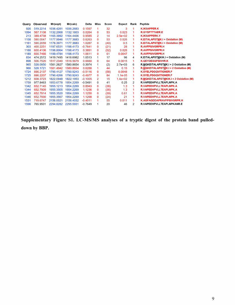
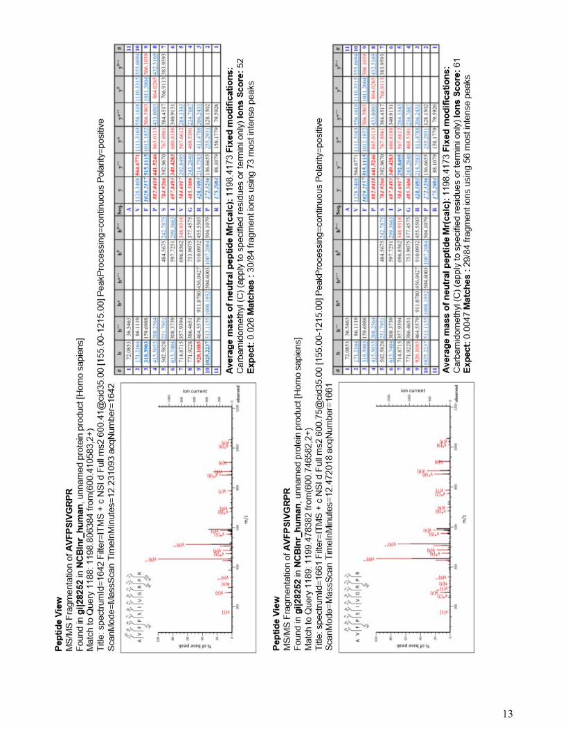
Supplementary Figure S1. LC-MS/MS analyses of a tryptic digest of the protein band pulled-
down by BBP.
9

LC-MS/MS spectral data
10

11

12
13

14

15

16

17

18

19
20