05 proteins %26 cell structure
TRANSCRIPT
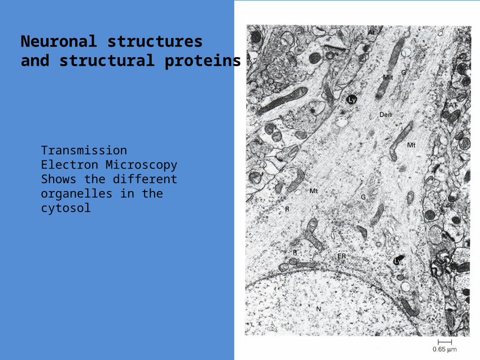
Neuronal structuresand structural proteins
Transmission Electron Microscopy Shows the different organelles in the cytosol

Golgi complexIs responsible for most posttranslational modification
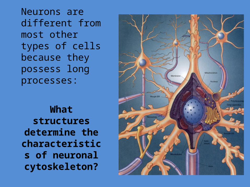
Neurons are different from most other types of cells because they possess long processes:
What structures determine the
characteristics of neuronal
cytoskeleton?

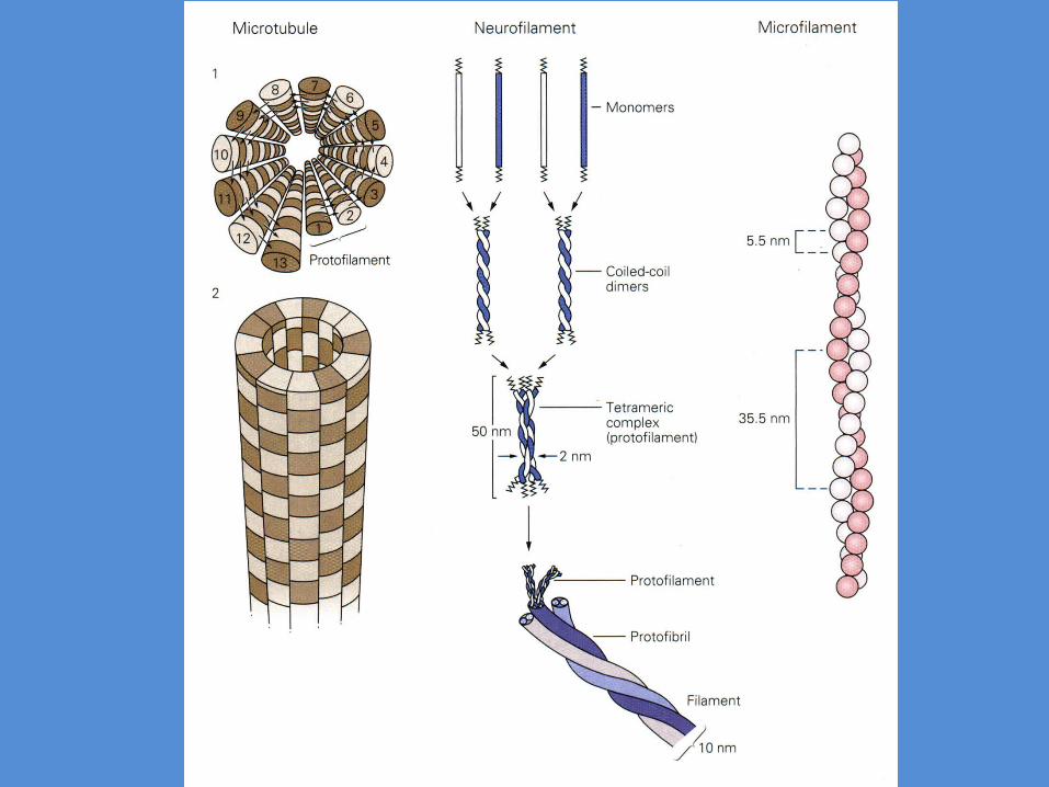
Structural protein of the neuron
• Microtubules
• Neurofilaments
• Microfilaments
Microtubules• Form long scaffold of neural processes
• 25% of total protein in neurons
• Largest diameter 25 nm
• Cylinder of 13 protofilaments – polarized
• Composed of alternating and Tubulin subunits- six genes and 20+ isoforms- grows in size catalyzed by GTPase
• The polymer is stabilized by MAPs
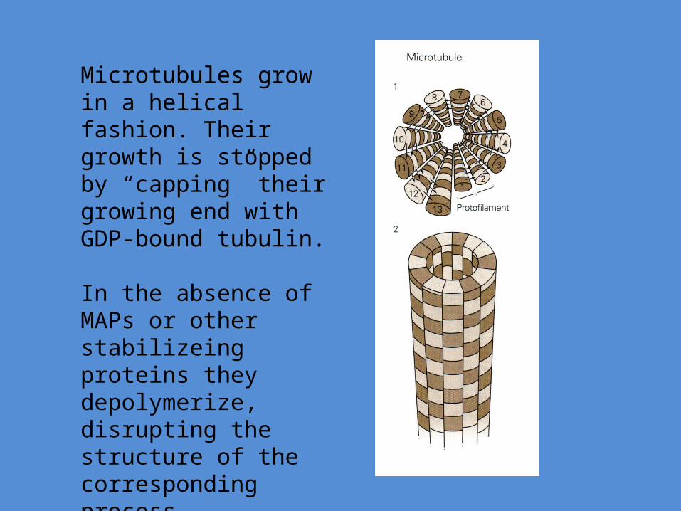
Microtubules grow in a helical fashion. Their growth is stopped by “capping” their growing end with GDP-bound tubulin.
In the absence of MAPs or other stabilizeing proteins they depolymerize, disrupting the structure of the corresponding process
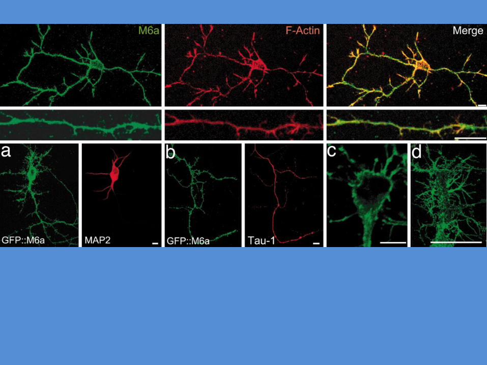
MAP2: dendritesMAP3 and tau prot: axons
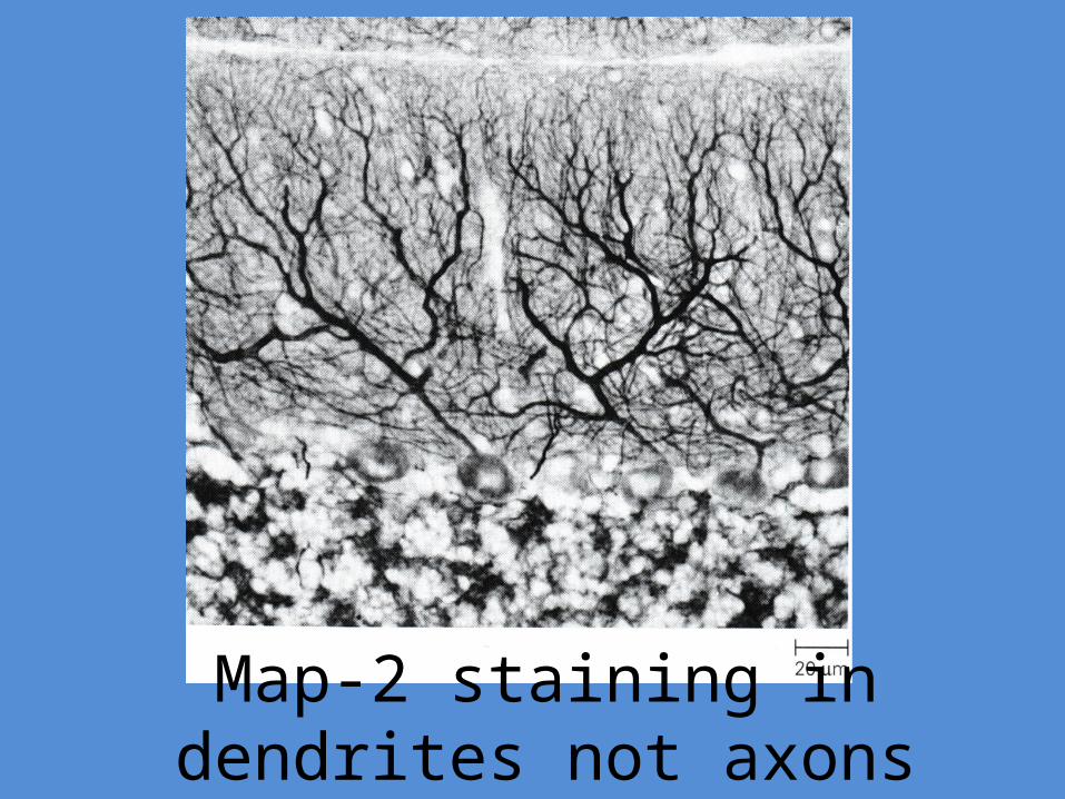
Map-2 staining in dendrites not axons

Neurofilaments
• Most abundant fibrillar protein in axons• 10nm in diameter• Most abundant in axons• Related to keratin
– Very stable – Neurofibrillary tangles


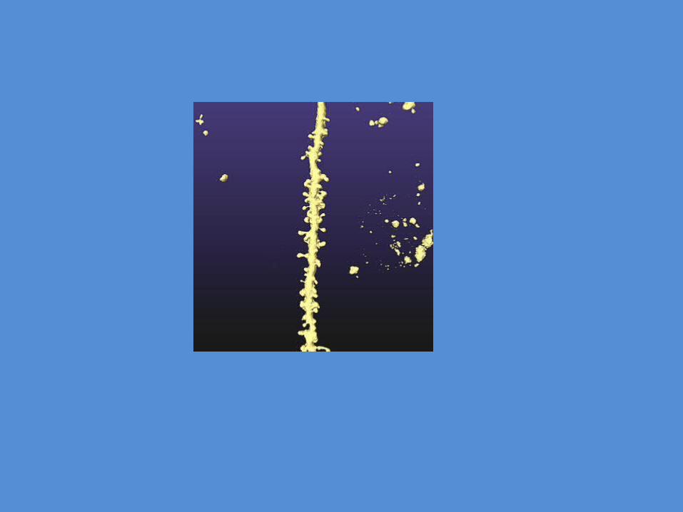
Microfilaments• 3-5 nm in diameter
– thinnest of the three elements of cytoskeleton
• Made of polarized actin monomers– highly conserved and abundant in living things
• Highly dynamic– half exists as monomers
• Many actin binding proteins secure cytoskeleton to plasma membrane
• Actin is organized in small polymers, and is localized at the periphery of neurons, where it is responsible for shaping processes like growth cones and dendritic spines.
• -actin is most abundant in the skeletal muscle
• - and -actin are present in neurons

The dynamic state of microtubules and microfilament allow the mature neuron to withdraw or extend old process and form new ones
Morphological plasticity

MicrotubuleDomains in the axon

Microtubules and actin filaments are the TRACK along which proteins and organelles are moved by molecular motors
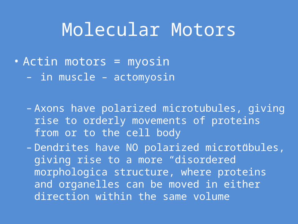
Molecular Motors
• Actin motors = myosin– in muscle – actomyosin
– Axons have polarized microtubules, giving rise to orderly movements of proteins from or to the cell body
– Dendrites have NO polarized microtubules, giving rise to a more “disordered” morphologica structure, where proteins and organelles can be moved in either direction within the same volume
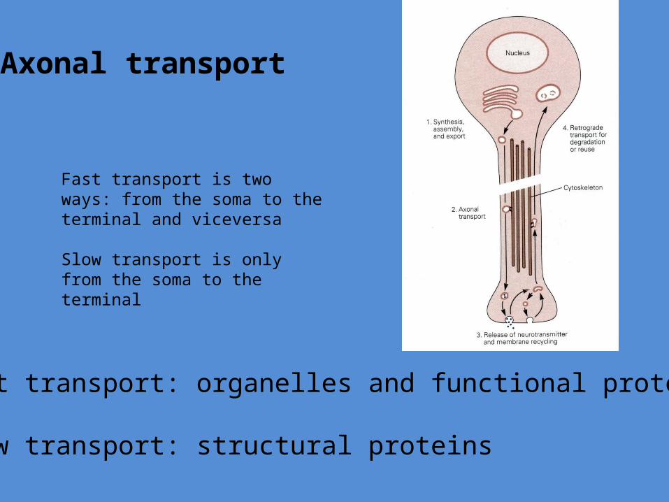
Axonal transport
Fast transport is two ways: from the soma to the terminal and viceversa
Slow transport is only from the soma to the terminal
Fast transport: organelles and functional proteins
Slow transport: structural proteins

Fast axonal transport 410mm/day


Tubulin
Clathrin
Neurofilament
Actin
Slow axonal transport = 0.2-2.5mm/day
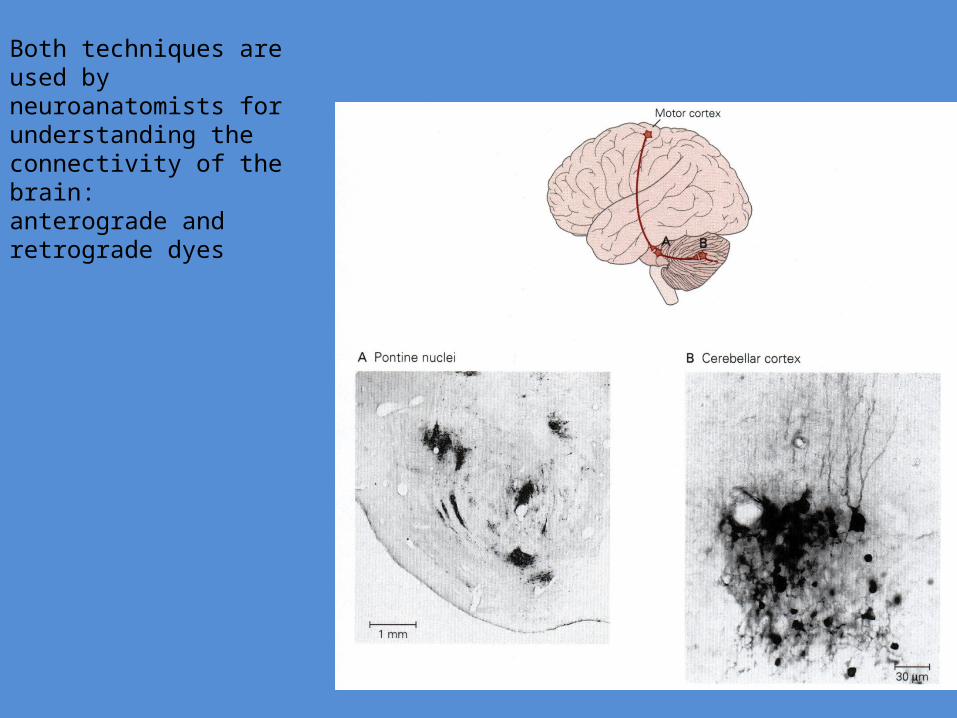
Both techniques are used by neuroanatomists for understanding the connectivity of the brain: anterograde and retrograde dyes

Inside the musclesimilar proteins organize muscle spindles where information is sent using an axon for conveying somatosensory information
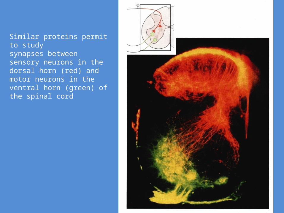
Similar proteins permit to study synapses between sensory neurons in the dorsal horn (red) and motor neurons in the ventral horn (green) of the spinal cord
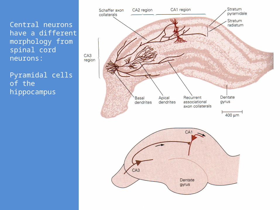
Central neurons have a different morphology from spinal cord neurons:
Pyramidal cells of the hippocampus
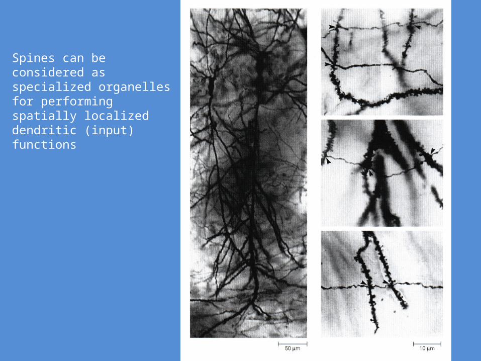
Spines can be considered as specialized organelles for performing spatially localized dendritic (input) functions
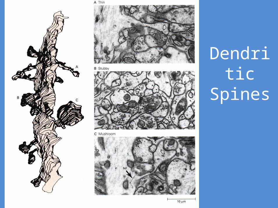
Dendritic Spines

http://multimedia.mcb.harvard.edu/anim_innerlife_hi.html