03 2020 university of cape town ventilation strategies in
TRANSCRIPT

Part Ii Anaesthesia Refresher Course – 2020 University of Cape Town 03
Ventilation Strategies in Paediatrics
Dr Lelanie Lambrechts
Dept of Anaesthesia & Perioperative Medicine Red Cross War Memorial Children’s Hospital
University of Cape Town
Introduction Paediatric ventilation is a very broad topic and would usually be covered in more than one chapter. To cover as much of the topic as possible I will focus on specific controversies and differences in the paediatric population with regards to ventilation.
Paediatric applied respiratory physiology
Infants are at risk of respiratory failure due to:
1. A decrease in respiratory reserve secondary to a smaller functional residual capacity (FRC). Their FRC is also closer to closing capacity (the lung volume at which alveoli and small airways begin to collapse)
2. The orientation of their ribs and diaphragm are more horizontal which limits the volume that can be displaced during inspiration
3. An increased oxygen consumption 4. The immature diaphragm muscle leads to quicker fatigue of inspiratory muscles 5. A decrease in airway radius causes an increase in resistance to the 4th power of change in
radius
The paediatric airway
1. Cuffed vs uncuffed endotracheal tubes (ETT)
Old airway beliefs:
• Funnel shaped airway
• The cricoid is the narrowest part of the airway These old airway beliefs lead to the usage of uncuffed endotracheal tubes in children younger than 8 years; the logic being to prevent pressure necrosis caused by an inflated cuff at the level of the cricoid. This historic belief stemmed from a manuscript by Dr Eckenoff in Anesthesiology in 1951 in which he referred to work done by Bayeux. Bayeux made plaster castings of 15 paediatric cadaveric larynxes ranging in age from 4 months to 14 years. In all 15 plaster cast cadaveric larynxes the internal circumference of the cricoid ring measured narrower than other parts of the airway. The concern with this experiment is that the distending pressure of the plaster may have altered the dimensions of the distensible parts of the airway when compared to the non-distensible regions like the cricoid. More recent airway imaging using newer modalities which includes bronchoscopic examination, CT and MRI have challenged these old beliefs. New airway beliefs:
• The airway is elliptical and not circular with a greater anterior-posterior(AP) diameter than transverse diameter
• The vocal cords and the subglottic area are the narrowest part of the airway Implications: From the above it becomes apparent that hearing a ‘leak’ with an uncuffed ETT doesn’t negate the risk of pressure necrosis on the airway. There may be a leak from the AP diameter, but still significant pressure on the lateral walls of the airway with a circular ETT. The benefit of a cuffed ETT is that the

Ventilation strategies in paediatrics Dr L Lambrechts
03 - 2
pliable cuff can seal off the airway with equal pressures at all points. To note is that although the cricoid area is not the narrowest area it may still be at risk for pressure necrosis, because it is rigid and not as displaceable as the vocal cords. Advantages of cuffed ETT:
1. Decrease need to exchange cuffed ETT 2. Better sealing of the airway with more reliable ventilation 3. Decrease incidence of sore throat 4. Prevention of oropharyngeal contamination with inhaled anaesthetic gases and high oxygen
concentrations 5. Decrease aspiration risk
The type of cuff further influences risk of aspiration. There is less risk of aspiration with the polyurethane cuff that is thinner and seals the trachea more efficiently than the polyvinylchloride cuff that may fold and thus lead to aspiration. Disadvantages of cuffed ETT:
1. Potential of cuff herniation through the glottis when the tip of the ETT is placed in the mid-trachea. This is secondary to the position of the cuff on the shaft and the more elliptical shape of the cuff.
2. Increased risk of pressure necrosis on the transverse walls when high pressure, low volume cuffs are inflated to a pressure of 20cmH2O, because the cuff doesn’t always cover the internal trachea at this pressure and still have a leak. Higher pressures are thus needed to seal the airway with an increased risk of pressure necrosis.
3. Potential to increase work of breathing when selecting a size smaller cuffed ETT due to the bulky cuff adding to the outer diameter
The new generation cuffed ETT e.g. the Microcuff ETT: The new generation cuffed ETT have all the advantages of the older generation cuffed ETT with the added benefit of nullifying most of the disadvantages the older generation cuffed ETT had. This is due to some new enhanced features:
• The cuff is spherical and more distal on the shaft and therefore when inflated there is less chance of cuff herniation through the glottis
• They have high volume, low pressure cuffs and therefore there is less chance for airway pressure necrosis
• They have an ultrathin cuff adding little to the outer diameter with less need to downgrade ETT sizes excessively
• The thin polyurethane cuff also seals the airway better with a resultant decreased aspiration risk
2. Oral vs nasal ETT
Oral intubations are technically easier to perform with less risk of pressure necrosis. Nasal ETTs are generally more secure with less risk of an unplanned extubation. When ventilation is anticipated post-op one should strongly consider using nasal endotracheal tubes especially in younger patients. Generally ventilated ICU patients are not deeply sedated or paralyzed and thus difficult to manage on a ventilator when they have an oral ETT in place. The disadvantages of nasal ETT are a technically more difficult intubation and the risk of pressure necrosis. It is therefore crucial that when strapping a nasal ETT in place one avoids causing any pressure on the nasal tip, nasal septum or lateral walls of the nose by strapping it in a downward fashion.
3. ETT size calculation
• Uncuffed ETT – Age/4 + 4
• Cuffed ETT – Age/4 + 3,5

Ventilation strategies in paediatrics Dr L Lambrechts
03 - 3
4. ETT length of insertion
Formula:
• Length (cm) at lip = 12 + age/2
• Length (cm) at nose = 15 + age/2 Unfortunately these formulas don’t always perform that accurately. An alternative method is to use the size of the ETT chosen as the corresponding length at which the ETT should be at the level of the vocal cords. For example if you use a size 4 ETT your ETT should be placed so that the 4cm marking on the ETT is at the level of the vocal cords. Another practical tip when placing an ETT is to make sure you confirm correct placement by auscultation in both the extended (when a roll is used under the shoulders for better airway visualization during laryngoscope) and the flexed airway position post-intubation.
Modes of ventilation Pressure vs volume control A pressure-controlled mode is usually preferred when there is a leak around the ETT that is commonly the case with an uncuffed ETT. In the pressure mode there is partial leak compensation, because this mode doesn’t target a set tidal volume, but rather gives a set pressure for a set time. In volume-controlled mode the ventilator targets the set tidal volume that is never attained, because of the leak. The machine will automatically increase the pressure to attain the set tidal volume that could potentially lead to barotrauma in parts of the lung that is more compliant than others. Cuffed ETT’s are used more commonly nowadays and therefore this is not as big a problem anymore. This led to the question being asked by many as to why pressure control is still so much preferred in paediatrics. The reason is probably because of its decelerating flow pattern. This flow pattern is more comfortable for the patient and has the potential to improve oxygenation. The improved oxygenation theory might be explained by the fact that there is minimal flow in the later part of the inspiratory phase with an inspiratory pause effectively being built into the breath that has the potential to improve V/Q matching. The advantages and disadvantages of pressure and volume control ventilation are summarised in the table below.
Pressure control Volume control
Advantages: Advantages:
• Partial leak compensation
• Guaranteed minimum minute ventilation, if no leak around tube
• Decelerating flow pattern prevents air hunger and thus more comfortable
• Set tidal volume
• Avoids high inspiratory pressures
• Potential to improve oxygenation
Disadvantages: Disadvantages:
• Change in lung compliance or resistance result in a change in tidal volume
• Fall in lung compliance will result in high alveolar pressure with risk of barotrauma
• Risk of volutrauma if an increase in lung compliance goes unnoticed
• Less suitable when uncuffed tube used
• Constant flow pattern might result in discomfort
Ventilatory modes and their main differences
1. Controlled mandatory ventilation • One of the first ventilator modes developed • Machine can’t sense patient’s own effort • Can lead to patient-ventilator asynchrony

Ventilation strategies in paediatrics Dr L Lambrechts
03 - 4
2. Intermittent mandatory ventilation • Set number of mandatory breaths • Allows for spontaneous ventilation, but doesn’t synchronize
3. Synchronized intermittent mandatory ventilation (SIMV) • Set number of mandatory breaths which are synchronized with spontaneous efforts • Patient can also take additional breaths that will be pressure supported if SIMV-PS selected
4. Assist control • On the newer ventilators this mode is sometimes confusingly referred to as e.g. pressure
control mode when in actual fact it is a pressure pre-set assist control mode • Breaths can be initiated by patient or ventilator (in contrast to pure controlled modes where all
breaths are initiated by the ventilator) • Characteristics of each breath is the same regardless if breath is initiated by patient or
ventilator (in contrast to SIMV) • Inspiratory time is set by clinician • Commonly used in the paediatric ICU in the form of pressure pre-set assist control mode
5. Pressure support • Breaths initiated by patient only • Inspiratory time controlled by patient (in contrast to assist control) • Clinician needs to set limit when machine should cycle from on (giving the set pressure and
facilitating inspiration) to off (stops delivering set pressure and allows for exhalation). Inspiratory to expiratory cycling thresholds:
▪ Default approximately 30% of peak inspiratory flow (PIF) ▪ In obstructive airways disease approximately 50% of PIF ▪ Patients with poorly compliant lungs approximately 5% of PIF ▪ In PICU mostly set to 5%, however in patients with a significant leak beware
that the flow may never fall to 5% of PIF
6. Pressure regulated volume control (PRVC) • Mode that combines features of both pressure and volume control • Ventilator automatically adjust inspiratory pressure to achieve a set tidal volume • Vital to set upper pressure limit appropriately to prevent excessive pressure being delivered.
Ventilator monitors each breath and compare the delivered tidal volume with the set tidal volume, if delivered volume is too low it increases the inspiratory pressure on the next breath, if too high it decreases the pressure.
• Great mode for asthmatic patient where airway resistance might change quickly with treatment or neurology/ neurosurgical patients where one needs to control CO2 tightly.
Setting the ventilator
1. Setting tidal volume (Vt) ➢ Target Vt 5-8 ml/kg predicted body weight (PBW) in normal lungs ➢ Target Vt 3-6 ml/kg PBW if poor compliance exists
There are no data to recommend optimal Vt and thus far only one observational study showed a lower mortality associated with Vt of 8 ml/kg actual body weight (ABW) compared with 10ml/kg
2. Setting the pressures ➢ Titrate pressure to attain above mentioned targeted tidal volumes
➢ Limit plateau pressure (Pplat) 28 cmH2O or 29-32 cmH2O if chest wall
elastance is increased or 30 cmH2O in obstructive airway disease
➢ Delta pressure 10 cmH2O if no lung pathology
The delta pressure/driving pressure (P=Vt/Crs) that is calculated as Pplat-PEEP, best stratified the risk for mortality in adults with ARDS. They have not been able to reproduce these observations in paediatrics except for one study where there was an independent association between the airway pressure gradient (PIP – PEEP) and mortality when measured under dynamic flow conditions.
Ventilation strategies in paediatrics Dr L Lambrechts
03 - 5
3. Setting PEEP PEEP help to prevent alveolar collapse. In children without lung injury the suggested PEEP level is 5cmH2O. In lung disease higher PEEP levels might be needed, but a balance needs to be found between alveolar recruitment and alveolar over-distenstion. There is no specific recommendation on how to best titrate PEEP. The following is a list of potential parameters to utilize when titrating PEEP:
• Intrinsic PEEP measured via an expiratory hold; keep the extrinsic PEEP less than the intrinsic PEEP
• Flow-time curve; ensure the expiratory flow returns to zero when increasing PEEP
• Alveolar dead space ({PaCO2-PetCO2} / PaCO2); take care not to increase alveolar dead space when increasing PEEP
• Oxygenation; titrate PEEP according to saturation and PaO2
• Compliance; in pressure mode if the tidal volume starts dropping with an increase in PEEP then you are probably over-distending the lung and need to bring the PEEP level back again
• Haemodynamics; if there is a decrease in pulse pressure with an increase in PEEP then stop the upward titration
4. Setting the I:E ratio/ inspiratory time
The table below can be used as a starting point when setting inspiratory times or the respiratory rate. From this point onwards one should interrogate the flow time curve of the patient to optimize flow dynamics.
Age Respiratory rate Inspiratory time (sec)
Newborn 40-60 bpm 0,35-0,5
Infant ( 1year) 25-35 bpm 0,5-0,7
Child (1-5years) 20-30 bpm 0,6-0,8
Child (6-12years) 15-20 bpm 0,7-1,2
Adolescent 12-15 bpm 1,0-1,5
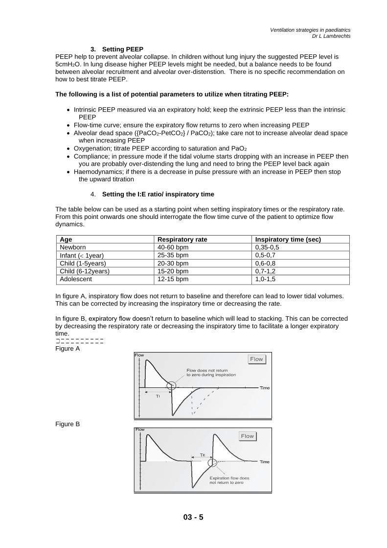
In figure A, inspiratory flow does not return to baseline and therefore can lead to lower tidal volumes. This can be corrected by increasing the inspiratory time or decreasing the rate. In figure B, expiratory flow doesn’t return to baseline which will lead to stacking. This can be corrected by decreasing the respiratory rate or decreasing the inspiratory time to facilitate a longer expiratory time.
Figure A
Figure B

Ventilation strategies in paediatrics Dr L Lambrechts
03 - 6
Goals of ventilation
1. Adequate oxygenation
Strategies to improve oxygenation Oxygenation is influenced by FiO2 and mean airway pressure. Mean airway pressure is determined by:
• Peak inspiratory pressure
• PEEP
• Inspiratory time
• Ventilator mode
When the mean airway pressure is increased oxygenation improves via several mechanisms: the recruitment of alveoli, increasing functional residual capacity, improvement of VQ matching and decreasing intrapulmonary shunting. Targets
• Sats > 95% in room air expected in children without lung injury or extra-pulmonary manifestations
• Permissive hypoxemia (i.e. Sats 92-97% when PEEP <10 cmH2O and 88-92% when
PEEP 10) in paediatric ARDS
• In children with congenital heart disease or chronic lung disease the clinician should be guided by trends in end organ perfusion when optimizing oxygenation. If end organ perfusion remains stable one may need to accept lower sats.
• In all situations the clinician needs to balance optimization of delivery of oxygen (DO2) with the harmful effects of ventilation
2. Adequate ventilation
Strategies to improve ventilation
• Increase minute ventilation This can be done by either increasing the tidal volume or the respiratory rate. However, if there is evidence of stacking, a decrease in the respiratory rate might be needed in order to increase minute ventilation
• Decrease dead space Targets
• Normal CO2 levels in normal lungs
• Normal pH and pCO2 levels in traumatic brain injury and pulmonary hypertension
• Permissive hypercapnia, targeting a pH of 7,15-7,3 in moderate/ severe paediatric ARDS except in conditions where permissive hypercapnia could be harmful e.g. increased intracranial pressure, severe pulmonary hypertension, selected congenital heart disease, haemodynamic instability and in patients with significant ventricular dysfunction.
• To note is that hypocarbia is as bad as hypercarbia and should be avoided.
3. Minimise adverse effects
Respiratory complications
• Ventilator induced lung injury (VILI): barotrauma, volutrauma, atelectrauma and biotrauma
• Oxygen toxicity
• Gas trapping
• Airway trauma
• Ventilator associated pneumonia(VAP)

Ventilation strategies in paediatrics Dr L Lambrechts
03 - 7
Cardiovascular complications Table – The effects of positive pressure ventilation
Physiological effects Clinical consequences
• Reduced left ventricle (LV) afterload
• Reduced preload
Good for:
• LV failure and pulmonary oedema
• Dilated cardiomyopathy
• Increased right ventricle (RV) afterload
• Reduced preload
Bad for:
• Glen/ Fontan circulation
• Hypovolaemia
• Pericardial effusion
• Pulmonary hypertension
Ventilation strategies in ARDS and Asthma
Asthma Ventilatory goals
• Acceptable (which doesn’t always equate to normal) level of oxygenation and ventilation
• Avoidance of lung hyperinflation due to incomplete exhalation Determining an appropriate respiratory rate and exhalation time
• Interrogate the flow time curve If expiratory flow has not returned to baseline, either decrease the respiratory rate or increase the exhalation time. However, to maintain adequate minute ventilation sufficient for CO2 clearance, a higher tidal volume and peak inspiratory pressure might need to be tolerated. Chasing a normal CO2
level might lead to VILI and thus permissive hypercapnia should be allowed. The rationale for using CPAP and PEEP in asthma CPAP in a spontaneously breathing patient with asthma:
• Decrease inspiratory work of breathing by decreasing the pressure gradient required to overcome intrinsic or auto PEEP
• Facilitates exhalation by moving the equal pressure point more proximal and towards the cartilaginous airways and thus preventing airway collapse
PEEP during positive pressure ventilation in a patient with asthma:
• By supplying extrinsic PEEP that is lower than the patient’s own intrinsic PEEP, one is able to move the equal pressure point more proximal and thus prevent airway collapse, as well as maintain a pressure gradient for air to move during exhalation.
Titrate extrinsic PEEP according to
1. Intrinsic PEEP measured via an expiratory hold 2. Flow-time curve; ensure the expiratory flow returns to zero when increasing PEEP 3. Measured CO2; alveolar dead space ({PaCO2-PetCO2} / PaCO2) should not increase when
PEEP is increased
Paediatric ARDS Limitations in AECC and Berlin ARDS definitions when describing paediatric ARDS:
1. Focused on adult lung injury 2. The use of PaO2 / FiO2 (P/F) ratio

Ventilation strategies in paediatrics Dr L Lambrechts
03 - 8
• Needs invasive measurement of PaO2
• Ratio influenced by ventilator pressures and, in PICU, greater variability in ventilatory management than adult ICU
3. Differences in risk factors/ etiology/ pathophysiology/ outcomes not considered in AECC/Berlin definitions
Differences in Paediatric ARDS (PARDS) definition compared to Berlin definition:
1. Use of oxygenation index (OI) or oxygen saturation index (OSI) rather than P/F ratio 2. Eliminated the requirement for bilateral pulmonary infiltrates 3. Excluded patients with perinatal related lung disease 4. Included special population groups
PARDS definition table taken from “PARDS: consensus recommendations from the pediatric acute lung injury consensus conference (PALICC)”
Summary of the PARDS management recommendations by the PALICC experts For each recommendation the agreement among the experts are displayed in brackets as either weak or strong Lung protective ventilation
1. Vt 5-8 ml/kg predicted body weight (PBW), 3-6 ml/kg PBW if poor compliance (weak)
2. Inspiratory Plat pressure <28 cmH2O, 29-32 if decreased chest wall compliance (weak)
3. Moderate PEEP 10-15 cmH2O titrated to oxygenation and hemodynamic response
• PEEP >15 cmH2O – severe PARDS, but limit Pplat (strong)
• Monitor DO2 (sats, PaO2)/ respiratory compliance/ haemodynamics as you increase PEEP (strong)
4. Careful recruitment manoeuvres (weak)
• Slow incremental and decremental PEEP steps

Ventilation strategies in paediatrics Dr L Lambrechts
03 - 9
• Sustained inflation maneuvers cant be recommended
5. After optimizing PEEP consider lower sats levels of 88-92% in PARDS with PEEP of at least 10cmH2O (strong)
6. When Sats <92% monitor SvO2 and markers of oxygen delivery (strong)
7. Practice permissive hypercapnia to minimize VILI (strong)
8. Maintain pH 7,15-7,30 within lung protective guidelines (weak)
• Exceptions – Increased intracranial pressure, severe pulmonary hypertension, selected congenital heart disease lesions, haemodynamic instability, significant ventricular dysfunction
9. Bicarbonate supplementation is not routinely recommended (strong) Mode of ventilation No recommendations HFOV
• Considered in – hypoxic respiratory failure, Pplat >28 in the absence of reduced chest wall compliance (weak)
• Paediatric evidence: Experience has proven its safety, but efficacy lack data. Small RCT and observational studies show improved oxygenation, but no difference in mortality, duration of mechanical ventilation or length of stay. PROSpect (paediatric RCT) ongoing
• 2 Adult studies: OSCILLATE – Use of HFOV in early mod-severe ARDS – increased mortality OSCAR – Use of HFOV in all patients with ARDS did not decrease mortality Cuffed ETT with conventional ventilation (strong) Nitric oxide (strong)
• Not recommended routine • Considered in: severe pulmonary hypertension, severe RV dysfunction, severe PARDS as
rescue from or bridge to ECLS
• Paediatric evidence: Studies all report improved oxygenation with no impact on mortality Prone position (weak)
• Not recommended routine • Considered in severe PARDS • In paeds dramatic initial improvement in oxygenation, but survival benefit difficult to prove
• Paediatric evidence:
RCT supports safety, but no difference in outcomes PROSpect ongoing
• Adult study: PROSEVA – Significantly decreased mortality in prone group. Complications did not differ much between prone and supine group except increase cardiac arrest in supine group
Ventilation strategies in paediatrics Dr L Lambrechts
03 - 10
Suctioning (strong)
• Insufficient data to recommend open/close suction system • Routine saline prior to suctioning not recommended, can use for lavage if thick secretions
High frequency oscillatory ventilation (HFOV)
The mechanism
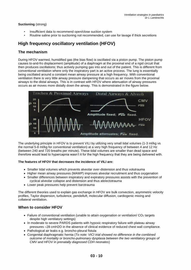
During HFOV warmed, humidified gas (the bias flow) is oscillated via a piston pump. The piston pump causes to-and-fro displacement (amplitude) of a diaphragm at the proximal end of a rigid circuit that then produces oscillations; thus actively pumping gas into and out of the patient. This is different from conventional ventilation where only the inspiratory part is an active process. The lung is essentially being oscillated around a constant mean airway pressure at a high frequency. With conventional ventilation there is very little airway pressure dampening that occurs as air moves from the proximal airways to the distal airways. This is in contrast with HFOV where attenuation of airway pressures occurs as air moves more distally down the airway. This is demonstrated in the figure below.
The underlying principle in HFOV is to prevent VILI by utilizing very small tidal volumes (1-3 ml/kg vs the normal 5-8 ml/kg for conventional ventilation) at a very high frequency of between 4 and 12 Hz (between 240 and 720 breaths per minute). These tidal volumes are smaller than dead space and therefore would lead to hypercapnia wasn’t it for the high frequency that they are being delivered with. The features of HFOV that decreases the incidence of VILI are:
• Smaller tidal volumes which prevents alveolar over-distension and thus volutrauma
• Higher mean airway pressures (MAWP) improves alveolar recruitment and thus oxygenation
• Smaller differences between inspiratory and expiratory pressures assists with the prevention of cyclical alveolar collapse and distension and thus atelectotrauma
• Lower peak pressures help prevent barotrauma
The different theories used to explain gas exchange in HFOV are bulk convection, asymmetric velocity profiles, Taylor dispersion, turbulence, pendelluft, molecular diffusion, cardiogenic mixing and collateral ventilation.
When to consider HFOV
• Failure of conventional ventilation (unable to attain oxygenation or ventilation/ CO2 targets despite high ventilatory settings)
• In moderate to severe PARDS patients with hypoxic respiratory failure with plateau airway
pressures 28 cmH2O in the absence of clinical evidence of reduced chest wall compliance.
• Pathological air leaks e.g. broncho-pleural fistula
• Congenital diaphragmatic hernia (To note: VICI trial showed no difference in the combined outcome of mortality or broncho-pulmonary dysplasia between the two ventilatory groups of CMV and HFOV in prenatally diagnosed CDH neonates)

Ventilation strategies in paediatrics Dr L Lambrechts
03 - 11
Managing a patient on HFOV Controlling oxygenation: MAP and FiO2 Oxygenation can be improved by adjusting your mean airway pressure (MAP) or FiO2. FiO2 should be adjusted according to Saturation targets set for that specific patient. A good starting point for MAP on the oscillator is 3-5 cmH2O above the MAP the patient had on conventional ventilation. After HFOV is initiated a CXR should be done to exclude under or over-distension of the lungs. The aim is to achieve lung inflation where the diaphragm lies between the 8th and 10th posterior ribs on an antero-posterior CXR. The Royal Children’s Hospital Melbourne gives the following guidelines with regards to setting MAP:
• Infant – MAP 18-25 (Be cautious in preterm infants however, where MAP’s as low as 8-10 could be sufficient)
• Child – MAP 20-30 Controlling ventilation: Amplitude and frequency Adequate ventilation and thus CO2 removal is dependant on the amplitude (delta P) of oscillations and frequency that is set. An increase in amplitude will lead to an increase in the tidal volume delivered and thus will aid in CO2 clearance. In conventional ventilation carefully increasing the respiratory rate, taking care not to introduce stacking, will lead to an increase in minute ventilation and thus an increase in CO2 removal. In HFOV increasing the frequency has the opposite effect on CO2 removal. By increasing the frequency you will decrease the area under the curve (i.e. tidal volume) and thus decrease CO2 removal. This concept is illustrated in the figure below where the amplitude is the same in the 2 pictures, but the frequency is higher in the picture on the right leading to a decrease in area under the curve and thus smaller tidal volumes.
The guideline below is a good starting point, but thereafter amplitude should be adjusted to achieve adequate chest wall vibrations that extend down to the level of the groin, referred to as the ‘chest wiggle factor’. Royal Children’s Hospital Melbourne guidelines for delta P and frequency:
• Infant - Delta P 30-40 cmH2O, Frequency 8-12 Hz
• Child – Delta P 40-60 cmH2O, Frequency 6-8 Hz Another method to promote CO2 elimination is to introduce a small cuff leak, but this might need periodic adjustment seeing that too big a leak makes it difficult to maintain a consistent MAP. Additional settings Bias flow The 3100A HFOV pocket guide recommends the following flows:
• Premature baby – 10-15 litres/min
• Term baby – 10-20 litres/min
• Small child – 15-25 litres/min
• Large child – 20-30 litres/min Inadequate bias flow will lead to an increase in dead space. Increased bias flow might be required to compensate for a leak in order to maintain the set MAP; e.g. during suctioning or in a patient with a broncho-pleural fistula.

Ventilation strategies in paediatrics Dr L Lambrechts
03 - 12
Inspiratory time Usually kept at 33% which translates to an I:E ratio of 1:2
Monitoring a patient on HFOV
• Chest wiggle factor (CWF)
Check for symmetrical, bilateral vibration that extends from the nipple line to the groin. A decrease in the CWF may indicate:
o Worsening compliance o The presence of secretions o ETT that has dislodged
Unilateral CWF may be a sign of:
o Endobronchial intubation o A pneumothorax o Unilateral lung collapse, consolidation or pleural effusion
The CWF may increase:
o With improvement in compliance
• Auscultation
Breath sounds wont be audible, but changes in the intensity of the piston sounds might be appreciated. To hear heart or gastro-intestinal sounds the piston can be stopped temporarily; lung inflation will be maintained.
• Transcutaneous PCO2 and PaCO2
End tidal CO2 monitoring is not possible during HFOV. Transcutaneous CO2 monitoring is used to monitor ventilation, but its drawback is its slow response time. Use regular blood gas analysis to correlate transcutaneous PCO2 to PaCO2. A rapid rise in CO2 may be a sign of:
o Acute airway obstruction (mucous plug) o Bronchospasm o A pneumothorax o Endobronchial intubation

Ventilation strategies in paediatrics Dr L Lambrechts
03 - 13
o Extubation o Worsening lung disease
Elevated CO2 refractory to increases in amplitude may indicate:
o A Mean airway pressure that is too low Rule of thumb – when the set amplitude approaches three times the set MAP it usually indicates too low lung volumes resulting in poor gas exchange
• Oxygen saturation and transcutaneous PO2 To monitor oxygenation A drop in oxygen saturation may be a sign of:
o Acute airway obstruction (mucous plug) o Disconnection o A pneumothorax o Changes in mean arterial pressure o Worsening lung disease
• Humidification
Monitoring of adequate, active humidification is essential for a patient on HFOV to prevent mucous plugging and a blocked ETT.
Complications of HFOV
• Irritability necessitating increased levels of sedation/ paralysis • Hypotension secondary to high mean airway pressures • Pneumothorax • ETT obstruction with secretions • Intraventricular haemorrhages in preterm infants • Bronchopulmonary dysplasia • Feeding intolerance
References
1. Bhalla A et al. Paediatric applied respiratory physiology – the essentials. Paediatrics and Child Health (2017) 2. Tobias JD. Pediatric airway anatomy may not be what we thought: implications for clinical practice and the use of cuffed
endotracheal tubes. Pediatric Anesthesia 25:9-19(2015) 3. Basic assessment and support in paediatric intensive care “Paediatric Basic course manual” November 2012 4. Lucking SE, Maffei FA, Tamburro RF, Thomas NJ. Pediatric critical care study guide. 2012 5. Gupta R, Rosen D. Paediatric mechanical ventilation in the intensive care unit. BJA Education, 16(12):422-426 (2016) 6. Kneyber MCJ, et al. Recommendations for mechanical ventilation of critically ill children from the Paediatric Mechanical
Ventilation Consensus Conference (PEMVECC). Intensive Care Med 43:1764-1780(2017) 7. Pediatric acute lung injury consensus conference group. Pediatric acute respiratory distress syndrome: consensus
recommendations from the pediatric acute lung injury consensus conference. Pediatric Critical Care Medicine 16(2015) 8. Orloff KE et al. The current state of pediatric acute respiratory distress syndrome. Pediatric allergy, immunology, and
pulmonology 32(2):35-44 (2019) 9. Pocket guide for the 3100A High frequency oscillatory ventilator by CareFusion