02_bm procedure and processing
DESCRIPTION
TMH proceedings 2010-2011,pdfTRANSCRIPT

Bone MarrowProcedure and Processing
Dr Khaliqur Rahman Senior Resident, Hematopathology LaboratoryDepartment of Pathology, TMH
History
• Initially entailed the drilling of cranial bones as a form of medical intervention for headaches and mental illnesses.
• However it was not until 1905, when the Italian physician Pianese reported bone marrow infiltration by the parasite Leishmania, that this procedure was applied toward clinical evaluation.
Parapia LA. Trepanning or trephines: a history of bone marrow biopsy. Br J Haematol. Oct 2007;139(1):14-9www.madametalbot.com, www.tuesday-johnson.tblr.com

Indications
• Pyrexia of unknown origin(Tuberculosis, leishmaniasis)• Pancytopenia• Thrombocytopenia • Refractory anaemia• Storage diseases• Leukaemia• Leukoerythroblastic picture in peripheral blood.• Paraproteinemias (rule out Myeloma)• Staging of neoplasm including lymphoma• For stem cell transplantations
Contraindication

Aspiration versus TrephineComplementary
Advantages
Bone marrow aspiration Fine cytological details, Wider range of cytochemical stains can be used, Ideal for microbiological culture, flow cytometry, cytogenetic and molecular studies.
Bone marrow Biopsy Complete assessment of cellularity and architecture.Detect focal lesions.Useful for assessment of aplastic anemia, metastasis etc.Archival material.
Bain BJ. Bone marrow trephine biopsy. J Clin Pathol. Oct 2001;54(10):737-42. Trewhitt KG. Bone marrow aspiration and biopsy: collection and interpretation. Oncol Nurs Forum. Oct 2001;28(9):1409-15;

Focal involvement in a case of neuroblastoma highlighted by synaptophysin

Focal Paratrabecular aggregate of lymphoma cellsin a case of Follicular Lymhpoma
Focal Paratrabecular aggregate of lymphoma cells, highlighted by cyclin D1 in a mantle cell lymphoma
Site for aspiration.
• Posterior superior iliac spine• Anterior superior iliac crest.• Spinous process of the
lumbar vertebrae.• The sternum. • The tibia is sampled only for
infants younger than 1 year

Needles used
Single use Needle
Reusable needle


Crushed Biopsy
Hemorrhagic Biopsy

Post OP care
• Firm pressure on the aspiration site.• If haemorrhage persists, place the patient in
the supine position• Analgesics to alleviate the pain.
Adverse events
Unilateral or Bilateral procedure?
Aspirate first or the biopsy first?

Adequacy of the specimen
• For a bone marrow biopsy, the accepted norm has been a length of 1.5cm, 5-6 inter-trabecular spaces and absence of handling or processing artifacts.

Processing of BM aspirate
Bone Marrow Aspirate
Anti-coagulated sampleSmear Preparation Clot preparation
Processed as biopsyFlow Cytometry (EDTA )
Cytogenetic studies (Heparin)
Molecular studies (EDTA)
Morphology
Cytochemistry
FISH (if required)
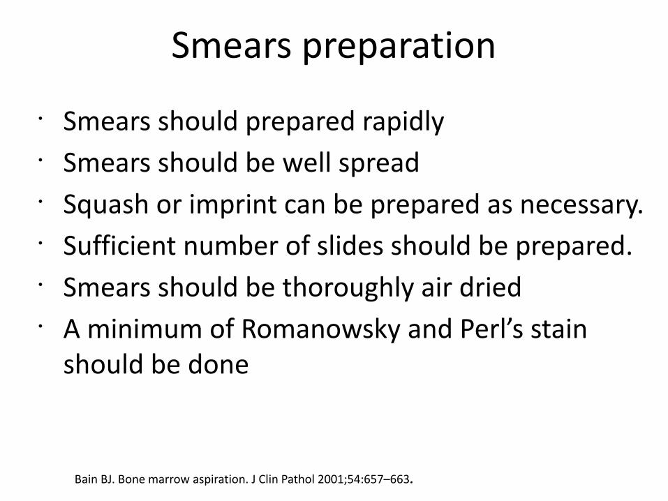
Smears preparation
• Smears should prepared rapidly• Smears should be well spread• Squash or imprint can be prepared as necessary.• Sufficient number of slides should be prepared.• Smears should be thoroughly air dried• A minimum of Romanowsky and Perl’s stain
should be done
Bain BJ. Bone marrow aspiration. J Clin Pathol 2001;54:657–663.


Lysed RBC due to improper drying
Properly dried smear

Wright’s Stain
Myeloperoxidase Perl’s Stain
Non Specific esterase

Processing of BM BiopsyBone Marrow biopsy
Fixation Imprint Smear
Morphological Correlation ( Better representation of marrow)
Decalcification&
Paraffin embedding
H&E, Special stains Immunohistochemistry
Sections

BM Biopsy Fixation
Bain BJ, Clark DM and Wilkins BS. Bone marrow Pathology. 4th edition. pp 601K N Naresh, I Lampert, R Hasserjian, D Lykidis. Optimal processing of bone marrow trephine biopsy: the Hammersmith Protocol. J Clin Pathol 2006;59:903–911.Bonds LA, Barnes P, Foucar K, et al. Acetic acid-zinc-formalin: a safe alternative to B-5 fixative. Am J Clin Pathol 2005;124:205–11.
Fixatives Minimum duration of fixation
10% neutral buffered saline 18 hrs (up to 48 hrs)
Bouins# 4-12 hrs
Zenkers$ 4hrs
B5$ 4 hrs, not more than 6 hrs
Aceto-zinc-Formalin(AZF)* Overnight
*Hammersmith protocol, $ Mercury based fixatives, #contains picric acid
Bouins and merrcury based fixatives are good for morphology but IHC is compromised
AZF is better over all fixatives in terms of preservation of morphology, IHC, DNA and RNA.
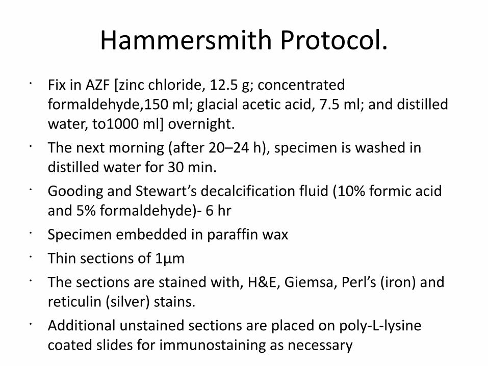
Hammersmith Protocol.• Fix in AZF [zinc chloride, 12.5 g; concentrated
formaldehyde,150 ml; glacial acetic acid, 7.5 ml; and distilled water, to1000 ml] overnight.
• The next morning (after 20–24 h), specimen is washed in distilled water for 30 min.
• Gooding and Stewart’s decalcification fluid (10% formic acid and 5% formaldehyde)- 6 hr
• Specimen embedded in paraffin wax• Thin sections of 1μm• The sections are stained with, H&E, Giemsa, Perl’s (iron) and
reticulin (silver) stains.• Additional unstained sections are placed on poly-L-lysine
coated slides for immunostaining as necessary
Decalcification
• 10%NITRIC ACID• HYDROCHLORIC ACID (HCL)• FORMIC ACID• EDTA
Embedding•Paraffin

Sections
• Thin sections, not more than 3μm.• Serial sections from multiple levels should be
examined.• A minimum of H&E and reticulin stain is
recommended.• Additional unstained slides should be cut in advance
for IHC stains.

LCA
H&E
H&E Retic

Take Home Message
• BM examination is an invaluable tool in work up of hematolymphoid neoplasm, PUO etc.
• It’s a painful procedure and sample obtained is precious.
• Aspiration and biopsy are the two facets of same coin.
• A good diagnosis rest on good processing.

Thanks