023.periodontal pocket
TRANSCRIPT
Dr Jaffar Raza Syed Page 1
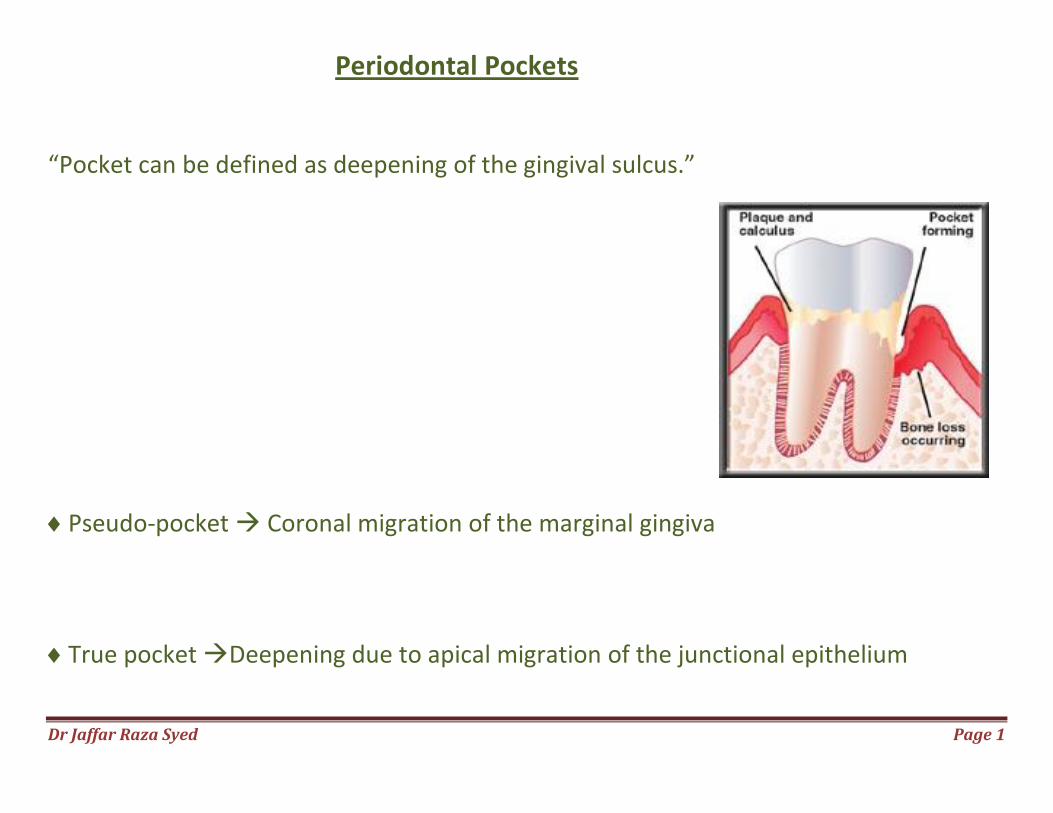
Periodontal Pockets “Pocket can be defined as deepening of the gingival sulcus.”
Pseudo-pocket Coronal migration of the marginal gingiva
True pocket Deepening due to apical migration of the junctional epithelium
Dr Jaffar Raza Syed
Classification Of Pockets
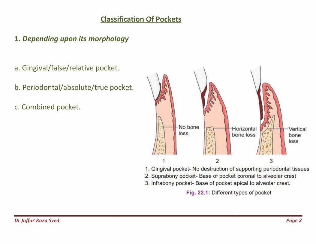
1. Depending upon its morphology a. Gingival/false/relative pocket. b. Periodontal/absolute/true pocket. c. Combined pocket.
Classification Of Pockets
Depending upon its morphology
.
b. Periodontal/absolute/true pocket.
Page 2
Dr Jaffar Raza Syed
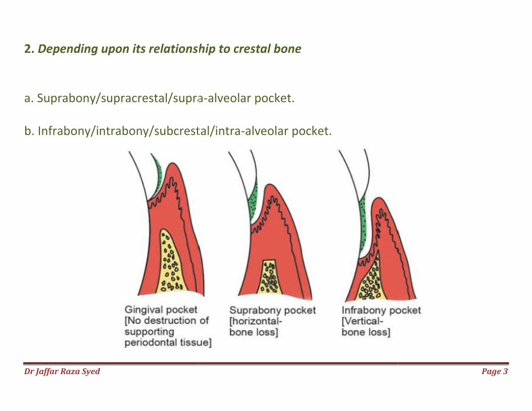
2. Depending upon its relationship to crestal bone a. Suprabony/supracrestal/supra b. Infrabony/intrabony/subcrestal/intra
ship to crestal bone
acrestal/supra-alveolar pocket.
abony/subcrestal/intra-alveolar pocket.
Page 3
Dr Jaffar Raza Syed
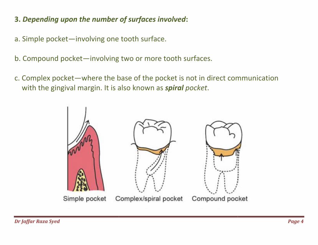
3. Depending upon the number of surfaces involved a. Simple pocket—involving one b. Compound pocket—involving two or more tooth c. Complex pocket—where the base of the pocket is with the gingival margin. It is also known as
Depending upon the number of surfaces involved:
involving one tooth surface.
involving two or more tooth surfaces.
where the base of the pocket is not in direct communication margin. It is also known as spiral pocket.
Page 4
not in direct communication
Dr Jaffar Raza Syed Page 5
4. Depending upon the nature of the soft tissue wall of the pocket a. Edematous pocket. b. Fibrotic pocket. 5. Depending upon the disease activity a. Active pocket. b. Inactive pocket.
Dr Jaffar Raza Syed Page 6
CLINICAL FEATURES
Enlarged, bluish-red marginal gingiva with a ‘rolled’ edge
A break in the faciolingual continuity of the interdental gingiva.
Shiny, discolored and puffy gingiva associated with exposed root surfaces.
Gingival bleeding, purulent exudate from the gingival margin.
Mobility, extrusion and migration of teeth.
The development of diastema where none had existed previously.
Dr Jaffar Raza Syed Page 7
Symptoms
Localized pain or a sensation of pressure in the gingival after eating, which
gradually diminishes.
A foul taste in localized areas.
A tendency to suck material from the interproximal spaces.
Radiating pain “deep in the bone”.
A “gnawing’ feeling or feeling of itching in the gums.
The urge to dig a pointed instrument into the gums and
relief is obtained from the resultant bleeding.
Patient complains that food “sticks between the teeth”
or that the teeth “feel loose” or a preference to “eat on the other side.”
Sensitivity to heat and cold; toothache in the absence of caries.
Dr Jaffar Raza Syed
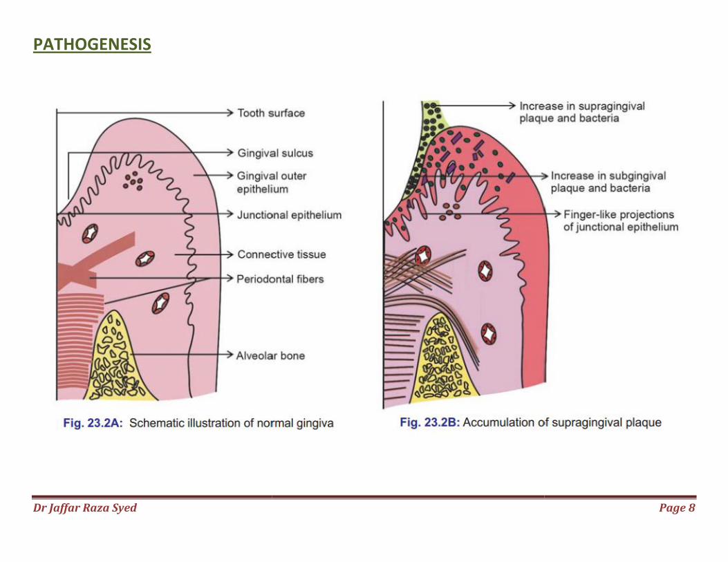
PATHOGENESIS
Page 8
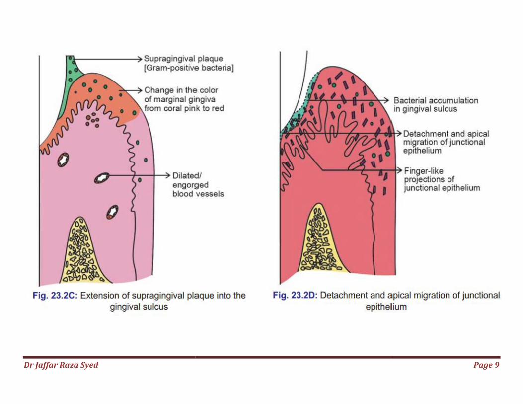
Dr Jaffar Raza Syed
Page 9
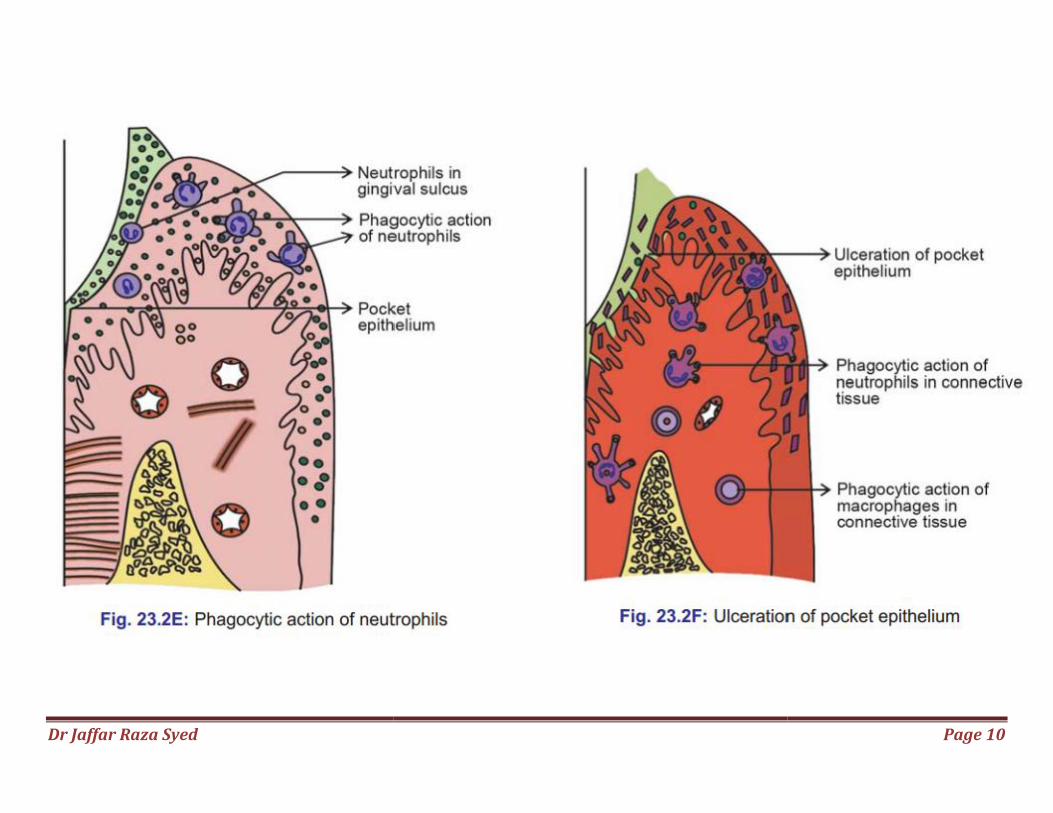
Dr Jaffar Raza Syed
Page 10
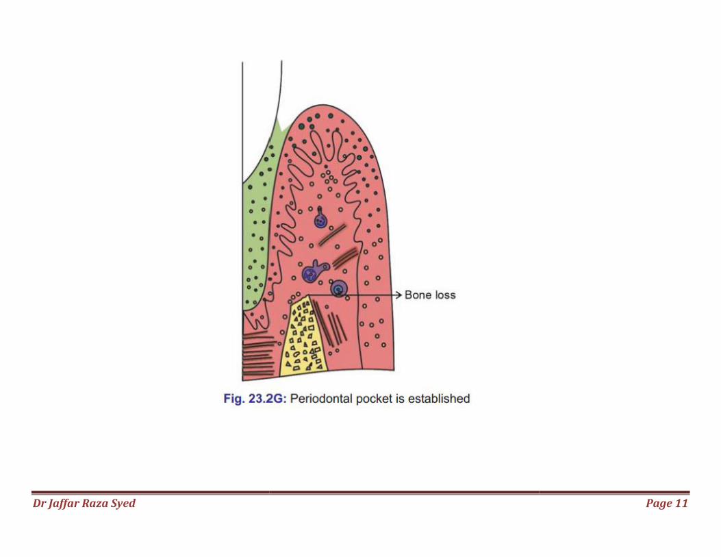
Dr Jaffar Raza Syed
Page 11
Dr Jaffar Raza Syed Page 12
Changes in the Soft Tissue Wall
blood vessels are engorged and dilated
connective tissue is edematous and densely infiltrated with plasma cells (80%), lymphocytes and PMNL
epithelium along the lateral wall of the pocket presents striking proliferative and degenerative changes
epithelial projection extends deep into the connective tissue and also extends further apically than the junctional epithelium.
Dr Jaffar Raza Syed Page 13
The epithelium is infiltrated with leukocytes and other inflammatory cells.
Degeneration and necrosis of the epithelium leading to ulceration of the epithelium and exposure of the underlying connective tissue.
Bacterial invasion along the lateral and apical areas of the pocket. Some bacteria traverse the basement lamina and invade the subepithelial connective tissue
Dr Jaffar Raza Syed Page 14
Pocket contents Mainly debris consisting of
microorganisms and their products
(enzymes, endotoxins, and other metabolic products),
gingival fluid,
food remnants,
salivary mucin,
desquamated epithelial cells,
leukocytes.
Plaque-covered calculus projecting from tooth surface.
Purulent exudate consists of
living, degenerated, and necrotic leukocytes;
living and dead bacteria;
serum
Dr Jaffar Raza Syed
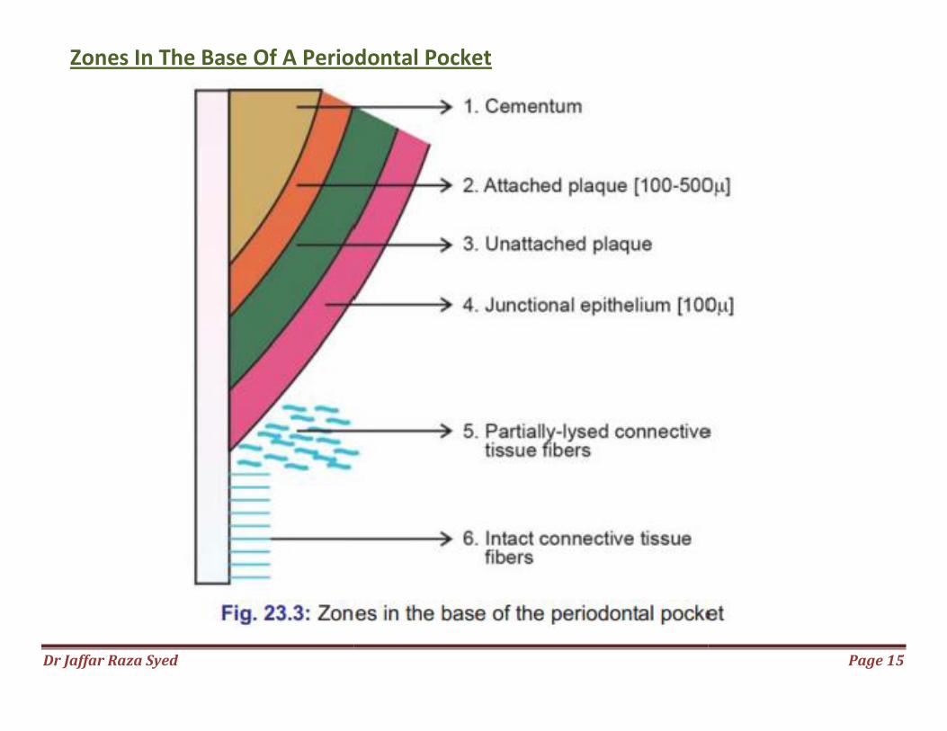
Zones In The Base Of A Periodontal Pocket
Periodontal Pocket
Page 15
Dr Jaffar Raza Syed Page 16
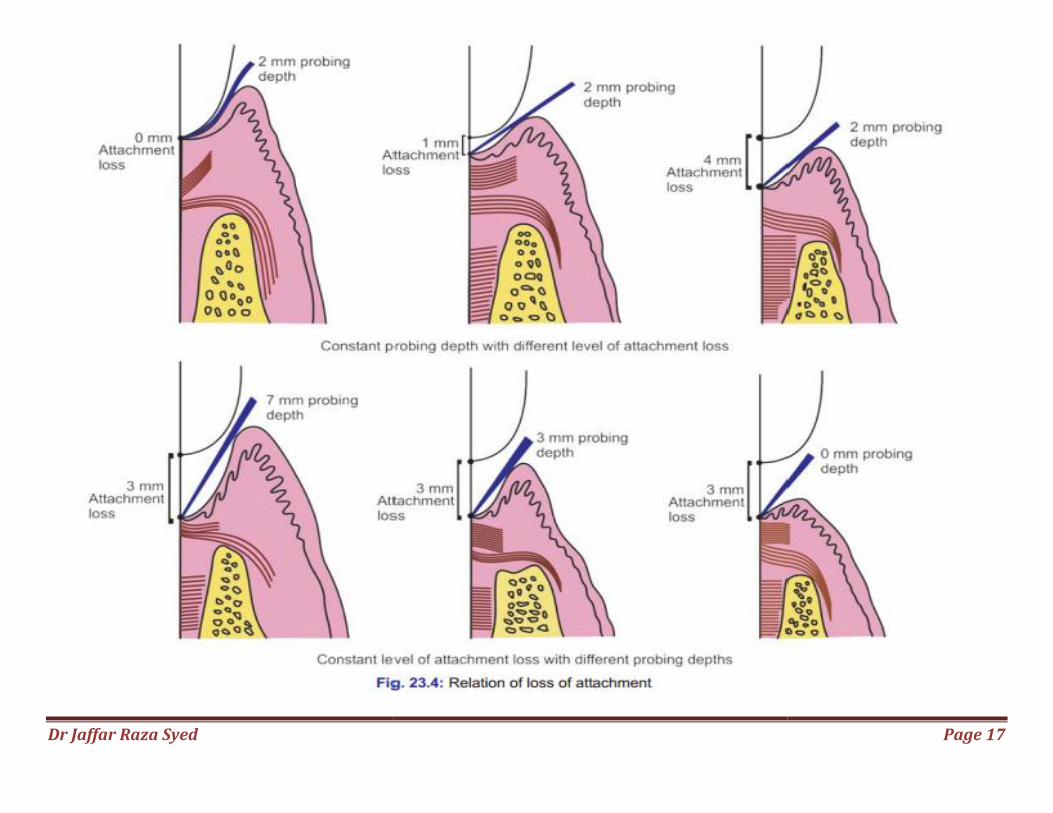
Relation of Loss of Attachment and Bone Loss to Pocket Depth
Pocket of same depth may be associated with different degree of attachment loss.
Pocket of different depth may be associated with same amount of attachment loss.
Area between the base of the pocket and the alveolar bone is always constant.
The radius of action of the plaque bacteria is 0.5 to 2.7 mm
Dr Jaffar Raza Syed
Page 17
Dr Jaffar Raza Syed
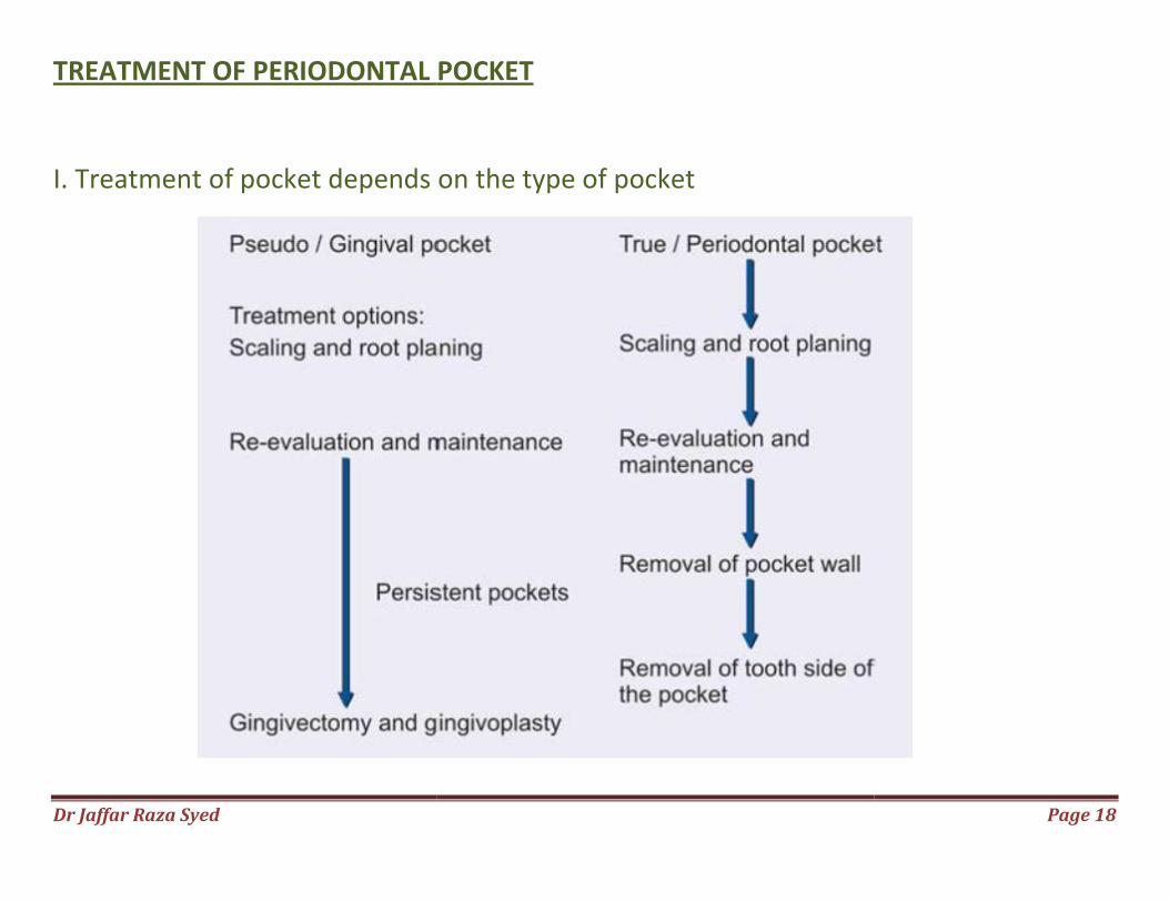
TREATMENT OF PERIODONTAL POCKE
I. Treatment of pocket depends on the type of pocket
REATMENT OF PERIODONTAL POCKET
Treatment of pocket depends on the type of pocket
Page 18
Dr Jaffar Raza Syed
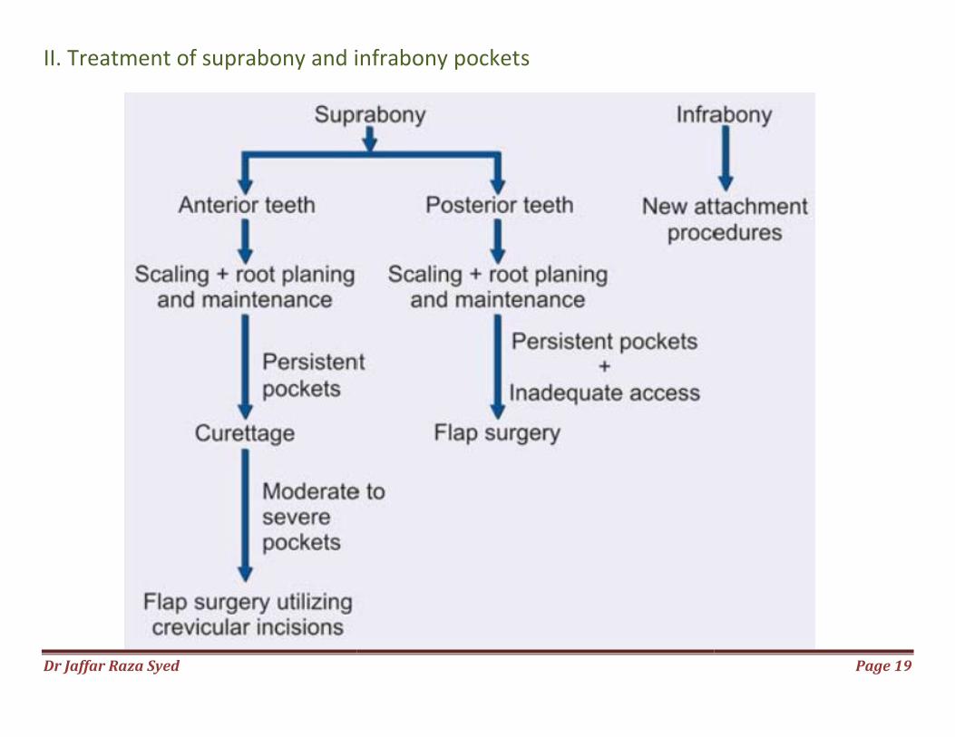
II. Treatment of suprabony and infrabony pocket
Treatment of suprabony and infrabony pockets
Page 19
Dr Jaffar Raza Syed Page 20
New attachment techniques:
It offers ideal result by reuniting the gingiva to the tooth at a position Coronal to the base of pre-existing pocket.
Here all the structures of lost periodontium are restored.
Following are the techniques for new attachment:
Non-graft associated new attachment procedures.
Graft associated new attachment procedures.
Combined techniques.
Dr Jaffar Raza Syed Page 21
Removal of pocket wall by, 1. Retraction or shrinkage, e.g. scaling and root planing. 2. Surgical removal by gingivectomy or by means of an undisplaced flap. 3. Apical displacement of pocket wall by apically displaced flap. Removal of the tooth side of the pocket, by tooth extraction or partial tooth extraction such as
hemisection or root resection.
Bicuspidization