0 med chem, chapter 10, 39 pages.ppt
TRANSCRIPT
5/15/2017
1
Chapter 10 - Drugs acting at miscellaneous targets
Transport Proteins
Structural Proteins
Biosynthetic Building Blocks
Protein synthesis - chain terminators
Protein-Protein Interactions
Cell Membrane Lipids
© Oxford University Press, 2013
Carbohydrates
Antigens and Antibodies
1
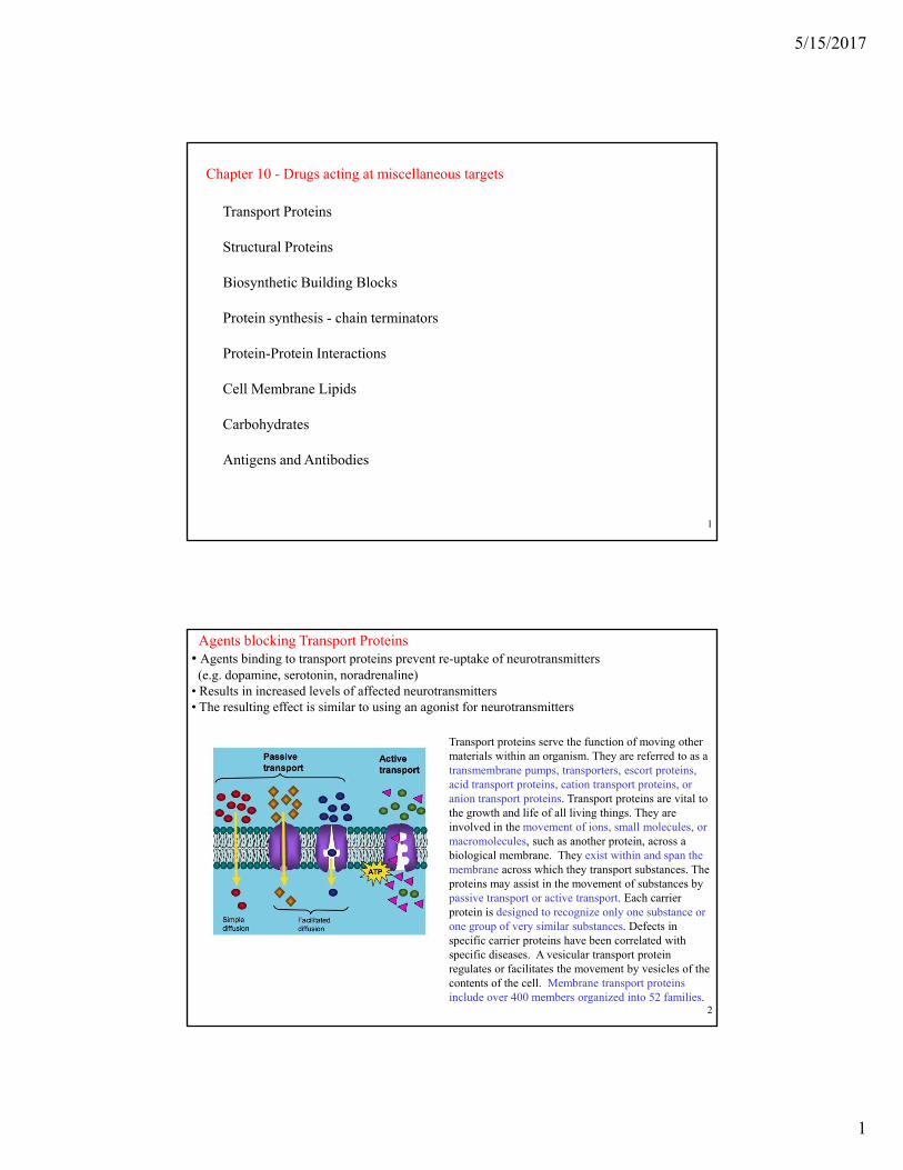
Agents blocking Transport Proteins• Agents binding to transport proteins prevent re-uptake of neurotransmitters (e.g. dopamine, serotonin, noradrenaline)
• Results in increased levels of affected neurotransmitters• The resulting effect is similar to using an agonist for neurotransmitters
Transport proteins serve the function of moving other materials within an organism. They are referred to as a g ytransmembrane pumps, transporters, escort proteins, acid transport proteins, cation transport proteins, or anion transport proteins. Transport proteins are vital to the growth and life of all living things. They are involved in the movement of ions, small molecules, or macromolecules, such as another protein, across a biological membrane. They exist within and span the membrane across which they transport substances. The proteins may assist in the movement of substances by
i i E h i
© Oxford University Press, 20132
passive transport or active transport. Each carrier protein is designed to recognize only one substance or one group of very similar substances. Defects in specific carrier proteins have been correlated with specific diseases. A vesicular transport protein regulates or facilitates the movement by vesicles of the contents of the cell. Membrane transport proteins include over 400 members organized into 52 families.

5/15/2017
2
CocaineN
O
CO2MeMe
C
O
H
Cocaine is a strong stimulant mostly used as a recreational drug. Mental effects include loss of contact with reality, an intense feeling of happiness, or agitation. Physical symptoms may include a fast heart rate, sweating, and large pupils.
HCl saltcrackcocaine
g pp , g y y p y , g, g p pHigh doses can result in very high blood pressure or body temperature. Effects begin within seconds to minutes of use and last between five and ninety minutes. Cocaine can be used medicinally for numbing and decreasing bleeding during nasal surgery (it is a vasoconstrictor).
Cocaine is very addictive due to its effect on the reward pathway in the brain. Its use increases the risk of stroke, myocardial infarction, lung problems (when smoked), blood infections, and sudden cardiac death (4300 in US in 2013).
Cocaine acts by inhibiting the reuptake of serotonin, norepinephrine, and dopamine. This results in greater concentrations of these three neurotransmitters in the brain. It can easily cross the blood–brain barrier and may lead to the breakdown of the barrier.
© Oxford University Press, 20133
The pharmacodynamics of cocaine involve the complex relationships of neurotransmitters (inhibiting monoamine uptake: serotonin, dopamine, norepinephrine). The most extensively studied is the blockade of the dopamine transporter protein. Dopamine transmitter released during neural signaling is normally recycled via the transporter; i.e., the transporter binds the transmitter and pumps it out of the synaptic cleft back into the presynaptic neuron, where it is taken up into storage vesicles. The dopamine transporter can no longer perform its reuptake function, and thus dopamine accumulates in the synaptic cleft. Cocaine also blocks sodium channels, thereby interfering with the propagation of action potentials giving it local anesthetic properties. Cocaine can often cause reduced food intake, many chronic users lose their appetite and can experience severe malnutrition and significant weight loss.
Agents blocking Transport Proteins Fluoxetine (Prozac) is a selective serotonin reuptake inhibitor antidepressant (SSRI). It is used to treat major depressive disorder, obsessive–compulsive disorder (OCD), bulimia nervosa and panic disorder. It is taken by mouth.
Common side effects include trouble sleeping, loss of appetite, dry mouth, rash, and abnormal dreams. Serious side effects include serotonin syndrome, mania, seizures, an increased risk of suicidal behavior in young people and an increased risk of bleeding. Its
h i f i i i l l b b li d b l d
O N
CH3
H
F3CFloxetine(Prozac)
mechanism of action is not entirely clear but believed to be related to increasing serotonin activity in the brain.
Prozac does not appreciably inhibit norepinephrine and dopamine reuptake at therapeutic doses, but it does delay the reuptake of serotonin, resulting in serotonin persisting longer when it is released.
The bioavailability of Prozac is high (72%), and peak plasma concentrations are reached in 6–8 hours. It is highly bound to
l i l lb i I i b li d i h li b
CYP2D6
O N
H
H
F3C norfloxetine
How?
© Oxford University Press, 20134
plasma proteins, mostly albumin. It is metabolized in the liver by cytochrome P450 enzymes, including CYP2D6. CYP2D6 has great genetic variability among people. CYP2D6 is responsible for converting Prozac to its only active metabolite, norfluoxetine. Both drugs are also potent inhibitors of CYP2D6. Floxetine and norfluoxetine have very long half lives (5 days and 16 days). Body concentrations build up over weeks. The drugs are still detectible after 7-8 weeks after discontinuing them.
5/15/2017
3
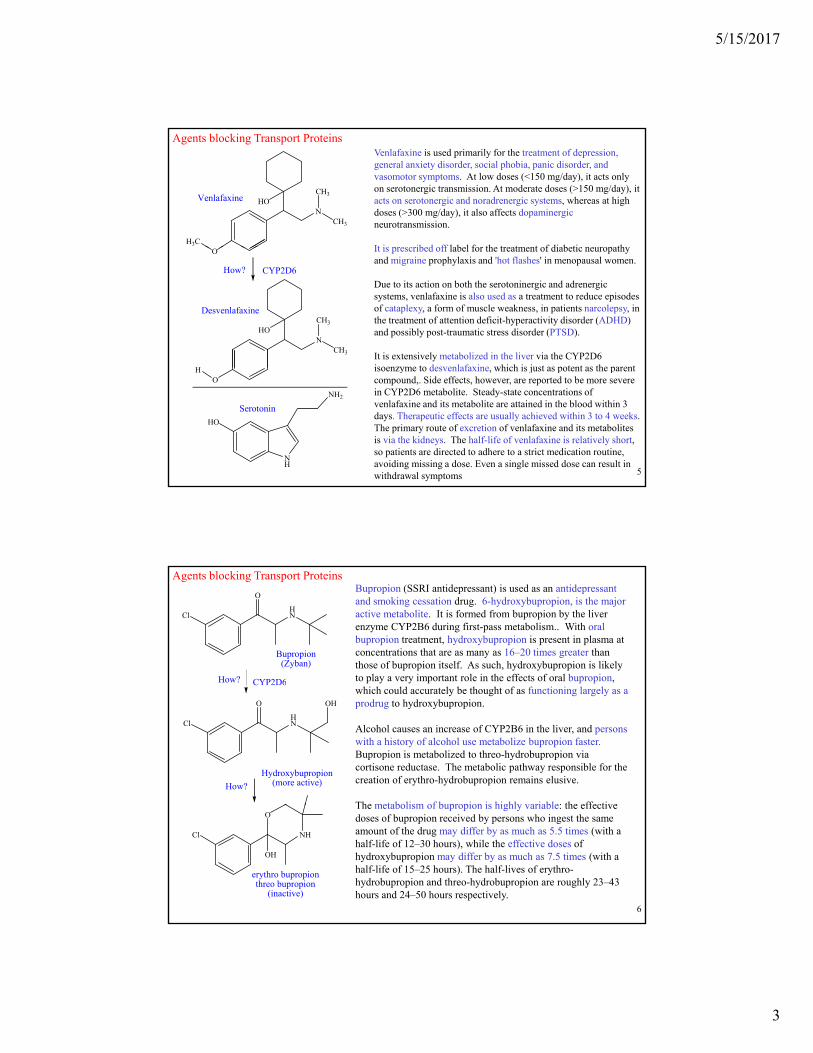
Agents blocking Transport ProteinsVenlafaxine is used primarily for the treatment of depression, general anxiety disorder, social phobia, panic disorder, and vasomotor symptoms. At low doses (<150 mg/day), it acts only on serotonergic transmission. At moderate doses (>150 mg/day), it acts on serotonergic and noradrenergic systems, whereas at high doses (>300 mg/day), it also affects dopaminergicneurotransmission.
It is prescribed off label for the treatment of diabetic neuropathy
N
CH3
CH3
HO
H3C
Venlafaxine
It is prescribed off label for the treatment of diabetic neuropathy and migraine prophylaxis and 'hot flashes' in menopausal women.
Due to its action on both the serotoninergic and adrenergic systems, venlafaxine is also used as a treatment to reduce episodes of cataplexy, a form of muscle weakness, in patients narcolepsy, in the treatment of attention deficit-hyperactivity disorder (ADHD) and possibly post-traumatic stress disorder (PTSD).
It is extensively metabolized in the liver via the CYP2D6 isoenzyme to desvenlafaxine which is just as potent as the parent
O
CYP2D6How?
N
CH3
CH3
HO
H
Desvenlafaxine
© Oxford University Press, 20135
isoenzyme to desvenlafaxine, which is just as potent as the parent compound,. Side effects, however, are reported to be more severe in CYP2D6 metabolite. Steady-state concentrations of venlafaxine and its metabolite are attained in the blood within 3 days. Therapeutic effects are usually achieved within 3 to 4 weeks. The primary route of excretion of venlafaxine and its metabolites is via the kidneys. The half-life of venlafaxine is relatively short, so patients are directed to adhere to a strict medication routine, avoiding missing a dose. Even a single missed dose can result in withdrawal symptoms
OH
NH
HO
NH2
Serotonin
Agents blocking Transport ProteinsBupropion (SSRI antidepressant) is used as an antidepressant and smoking cessation drug. 6-hydroxybupropion, is the major active metabolite. It is formed from bupropion by the liver enzyme CYP2B6 during first-pass metabolism.. With oral bupropion treatment, hydroxybupropion is present in plasma at concentrations that are as many as 16–20 times greater than those of bupropion itself. As such, hydroxybupropion is likely to play a very important role in the effects of oral bupropion,
Bupropion (Zyban)
CYP2D6How?
HN
O
Cl
which could accurately be thought of as functioning largely as a prodrug to hydroxybupropion.
Alcohol causes an increase of CYP2B6 in the liver, and persons with a history of alcohol use metabolize bupropion faster. Bupropion is metabolized to threo-hydrobupropion via cortisone reductase. The metabolic pathway responsible for the creation of erythro-hydrobupropion remains elusive.
The metabolism of bupropion is highly variable: the effective
C 6
Hydroxybupropion(more active)
HN
O
Cl
OH
How?
© Oxford University Press, 20136
The metabolism of bupropion is highly variable: the effective doses of bupropion received by persons who ingest the same amount of the drug may differ by as much as 5.5 times (with a half-life of 12–30 hours), while the effective doses of hydroxybupropion may differ by as much as 7.5 times (with a half-life of 15–25 hours). The half-lives of erythro-hydrobupropion and threo-hydrobupropion are roughly 23–43 hours and 24–50 hours respectively.
NH
O
Cl
OH
erythro bupropionthreo bupropion
(inactive)

5/15/2017
4
Agents blocking Transport ProteinsSibutramine (as HCl salt) is an oral anorexiant. Until 2010 it was marketed and prescribed as an adjunct in the treatment of obesity, along with diet and exercise. It has been associated with increased cardiovascular events and strokes and has been withdrawn from the market in several countries and regions, (small effect: 11.4% vs. 10.0% plecebo, ?). It is a derivative of amphetamine (Adderall, speed) which has been classified as a Schedule IV controlled substance in the US.
It is a monoamine reuptake inhibitor that, in humans, reduces the reuptake of norepinephrine (by 73%), serotonin (by 54%), and dopamine (by 16%), increasing the levels of these substances in synaptic clefts ( y %), p ( y %), g y pand helping enhance satiety, influencing the appetite. Older anorectic agents such as amphetamine and fenfluramine force the release of these neurotransmitters rather than affecting their reuptake. The simultaneous use of sibutramine and monoamine oxidase inhibitors (MAOIs, such as selegiline) is not indicated, as it
Sibutramine is well absorbed from the GI tract (77%), but undergoes considerable first-pass metabolism, reducing its bioavailability. The drug itself reaches its peak plasma level after 1 hour and has also a half-life of 1 hour. Sibutramine is metabolized by cytochrome P450 isozyme CYP3A4 into two pharmacologically-active primary and secondary amines (called active metabolites 1 and 2) with
© Oxford University Press, 20137
half-lives of 14 and 16 hours, respectively. Peak plasma concentrations of active metabolites 1 and 2 are reached after three to four hours. There are other inactive metabolites.
NHN NH2
Sibutraminet1/2 = 1 hour
metabolite 2t1/2 = 16 hours
metabolite 1t1/2 = 14 hours
cytochrome P450
cytochrome P450
Agents blocking Transport ProteinsAmphetamine (Adderall, speed) is a potent central nervous system (CNS) stimulant that is used in the treatment of attention deficit hyperactivity disorder (ADHD), narcolepsy, and obesity. It has been classified as a Schedule IV controlled substance in the US.
Methamphetamine is also a strong central nervous system (CNS) stimulant that is mainly used as a recreational drug. In low doses, methamphetamine can elevate mood, increase alertness, concentration and energy in fatigued individuals, reduce appetite and promote (initial) weight loss. At higher doses, it can induce psychosis, breakdown of skeletal muscle, seizures and bleeding in the brain. Chronic high-p y , , g C gdose use can cause unpredictable and rapid mood swings, prominent delusions and violent behavior. Methamphetamine is known to have a high addiction rates and withdrawal symptoms occur when discontinued. Heavy recreational use of methamphetamine may lead to a post-acute-withdrawal syndrome, which can persist for months. Unlike amphetamine, methamphetamine is neurotoxic and causes damage to human midbrain dopaminergic neurons. All of these factors can lead to a steep decline in the quality of life, or even life itself.
NH2
amphetamine
HN
© Oxford University Press, 20138
HO
HO
catecholcatecholamines
HO
HO
NH2
CO2H
L-DOPA
HO
HO
NH2
dopamine
HO
HO
HN
norepinephrine(noradrenoline)
OH
HO
HO
HN
epinephrine(adrenoline)
OH
pAdderall (speed)
methamphetamineMeth mouth

5/15/2017
5
Agents blocking Transport Proteins.Methylphenidate (Ritalin) is a central nervous system (CNS) stimulant of the phenethylamine and piperidine
classes that is used in the treatment of attention deficit hyperactivity disorder (ADHD) and narcolepsy(discovered in 1955).
Medical use began in 1960; the drug has become increasingly prescribed since the 1990s, when the diagnosis of ADHD became more widely accepted. Between 2007 and 2012, methylphenidate prescriptions increased by 50% and in 2013 global methylphenidate consumption increased to 2.4 billion doses, a 66% increase from the year before. The United States continues to account for more than 80% of global consumption.
ADHD and other similar conditions are believed to be linked to sub-performance of the dopamine and norepinephrine functions in the brain, primarily in the prefrontal cortex, responsible for executive function (e.g., reasoning, inhibiting behaviors, organizing, problem solving, planning, etc.). Methylphenidate's mechanism of action involves the inhibition of catecholamine reuptake, primarily as a dopamine reuptake inhibitor. It acts by blocking the dopamine transporter and norepinephrine transporter, leading to increased concentrations of dopamine and norepinephrine within the synaptic cleft. This effect in turn leads to increased neurotransmission of dopamine and norepinephrine.
© Oxford University Press, 20139
2. Agents acting on Structural Proteinsa. Agent binding to a viral structural protein
Host cellHost cell
Host cellHost cell
VirusVirusHelicalHelicalfoldingfolding
VirusVirus
gp41gp41 Fusion
Cell entry by HIV
• gp41 helices fold over on each other to form twice as many helices, which are half as long – draws virus and host cell together leading to fusion, gp41 is major target for HIV vaccines
•Enfuvirtide is a polypeptide containing 36 amino acids
EnfuvirtideEnfuvirtide
VirusVirus
Host cellHost cell
Fusion blocked
Host cell
VirusVirus
gp41gp41
Blocking fusion
© Oxford University Press, 2013
• Used vs HIV since 2003• Acts as a fusion inhibitor by binding to gp41 and preventing the folding of the helices.•Virus and host cell are not pulled together and fusion is blocked, gp41 cannot penetrate cell membrane
10
Enfuvirtide - cost is $25,000 / year

5/15/2017
6
b. Inhibitors of tubulin polymerisation
2. Agents acting on Structural Proteins
Colchicine inhibits microtubule polymerization by binding to tubulin (also maked up cell skeleton), one of the main constituents of microtubules. Availability of tubulin is essential to mitosis, so colchicine functions as a "mitotic poison" or spindle poison.
Colchicine
HN
O
O
O
The mitosis-inhibiting function of colchicine is of great use in the study of cellular genetics. To see the chromosomes of a cell under a light microscope, it is important that they be viewed near the point in the cell cycle in which they are most dense. This occurs near the middle of mitosis (specifically metaphase), so mitosis must be stopped before it completes. Adding
OCH3O
CH3O
CH3
H3C
microtubule
© Oxford University Press, 201311
pp p gcolchicine to a culture during mitosis is part of the standard procedure for doing karyotype studies.
Colchicine also inhibits neutrophil motility and activity, leading to a net anti-inflammatory effect. This has proven useful in the treatment of acute gout flares.
OH
CH2CH3N
NH
Vinblastine - anticancer agentb. Inhibitors of tubulin polymerisation
2. Agents acting on Structural Proteins
NH
MeO2C
MeO N
Me HOCO2Me
OCOMe
CH2CH3
H
Vinblastine is a chemotherapy medication, typically used with other medications, to treat a number of types of cancer. This includes Hodgkin's lymphoma, non-small cell lung cancer, bladder cancer, brain cancer, melanoma, and testicular cancer. It is given by injection into a vein. When used with bleomycin, and methotrexate it allows for lower doses and reduced overall toxicity with larger resting periods
© Oxford University Press, 201312
y g g pbetween chemotherapy cycles.
Common side effects include changes in sensation, constipation, weakness, loss of appetite, and headaches. Severe side effects include low blood cell counts and shortness of breath. It should not be given to people who have a current bacterial infection. Vinblastine works by blocking cell division.
At very low concentrations they suppress microtubule dynamics and at higher concentrations they reduce microtubule polymer mass. They also produce microtubule fragments.

5/15/2017
7
c. Inhibitors of tubulin depolymerisation2. Agents acting on Structural Proteins
Paclitaxel (Taxol)
oxetane
acetylbenzoyl
side chain
Paclitaxel is sold under the brand name Taxol and used to treat a number of types of cancer including
© Oxford University Press, 201313
Paclitaxel is sold under the brand name Taxol and used to treat a number of types of cancer, including ovarian cancer, breast cancer, lung cancer, Kaposi sarcoma, cervical cancer, and pancreatic cancer. It is given by injection into a vein.
Common side effects include hair loss, bone marrow suppression, numbness, allergic reactions, muscle pains, and diarrhea. Other serious side effects include heart problems, increased risk of infection, and lung inflammation. It works by interference with the normal function of microtubules during cell division using its side chain and the acetyl, benzoyl and oxetane groups. It used to require the bark from 3 slow growing "Yew" trees (600-700 years). It is now manufactured by cell culture.
3. Agents acting on Biosynthetic Building Blocks
O O
OH H3C
O
HO CH3
O
CH2OH
HO
HO
OOMe
HO
MeH3N
Cl
Vancomycin
• Important antibacterial agent
Cl
ON
O
OH H3C
N
NH
N CO2O
HN
HO
OH
N
O
OHOHHO
OH
CONH2
H H
CH3
NH2Me
H
H
H
H
H
© Oxford University Press, 2013
Important antibacterial agent• Caps the building block used in synthesis of bacterial cell wall• Contains a peptide chain which form H-bonds to the target• Vancomycin acts as a ‘receptor’ for the building block•Vancomycin is considered a last resort medication
14
Crystal structure of a short peptide L-Lys-D-Ala-D-Ala (bacterial cell wall precursor, in green) bound to vancomycin(blue) through hydrogen bonds

5/15/2017
8
vancomycin
Staphylococcus aureus (Staph aureus, S. aureus, or SA) is a common bacteria (a type of germ) in the nose and on the skin of people and animals ("staph"). MRSA means "methicillin-resistant Staphylococcus aureus." It is often resistant to (is not killed by) several types of antibiotic treatments. In general, healthy people with no cuts, abrasions, or breaks on their skin are at low risk for getting infected. About one out of every three people (33%) are estimated to carry staph in their nose, usually without any illness. About two in 100 (2%) carry MRSA. Both adults and children may have MRSA. MRSA can be harder to treat or become worse if the right treatment iscan be harder to treat or become worse if the right treatment is delayed. MRSA is one of the bacteria listed by the CDC as a "superbug" resistant to multiple antibiotics (about 18 "superbugs“ are listed). MRSA is transmitted from person to person by direct contact with the skin, inhaling droplets from coughing, or items touched by someone who has MRSA (for example, sink, bench, bed, and utensils). One way to keep visitors and health-care staff from carrying MRSA from one patient to others is to follow CDC-guided precautions by wearing disposable gloves, gowns and masks) when visiting hospitalized people who have MRSA. A deadly complication
peptide chain of bacterial cell wall
© Oxford University Press, 201315
of MRSA is a deep infection, necrotizing fasciitis, which causes rapid spread and destruction of human tissues. Some but not all strains of MRSA are more likely to behave like "flesh-eating bacteria." It is impossible to predict which MRSA infection will be "flesh-eating."
A simple substitution of a lactate for an alanine allows loss of a hydrogen bond and weaker binding and an increase in resistance to vancomycin.
MRSA infections can progress rapidly and needs to be treated aggressively.Initially the methoxy groups provided a steric
shield that made Meticillin more resistant (1960), but resistance soon developed. Meticillin is no longer used, but the term MRSA is still used in a symbollic way.
S
HN
O
O
The family of oxicillins is currently more commonly used today, as one of several types of antibiotics.
N
O
OOH
O
H
N
O
Meticillin
© Oxford University Press, 201316
N
S
O
OOH
HN
O
Oxacillin

5/15/2017
9
NAGGly......Transglycosidation
VancomycinGrowing cell wall
................................... ........................................
Cross-linking
3. Agents acting on Biosynthetic Building Blocks
N-acetylglucosamine (NAG)
OHO
HO
Cytoplasm
Cellmembrane
NAM
Carrierlipid
L-AlaD-GluL-Lys
L-Ala D-Ala D-Ala- D-Ala
O
OHOHO
HO
N-acetylmuramic acid (NAM)
OH
HN
HOHO
O
© Oxford University Press, 2013
• Building block partially constructed in cytoplasm• Constructed from a sugar (NAM) and a peptide chain • Transported across cell membrane and completed (NAM + glycines)• Linked to growing cell wall by enzyme (transglycosidation)
Amino acid
17
HNCHH3C
CO2H
O
Vancomycin
Buildingblock
3. Agents acting on Biosynthetic Building Blocks
Cell membrane
block
© Oxford University Press, 2013
• Vancomycin binds to peptide chain of building block• Caps the building block• Disguises building block from transglycosidation enzyme
18
5/15/2017
10
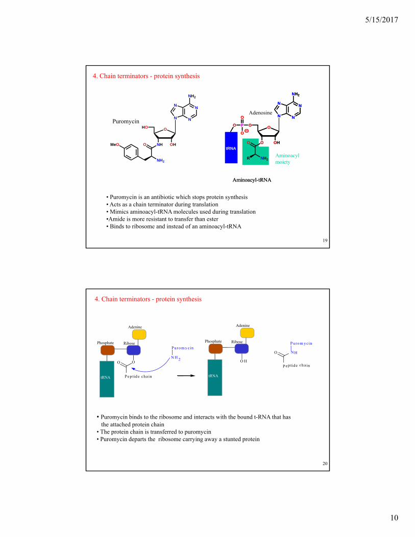
4. Chain terminators - protein synthesis
AdenosineN
NN
N
NH2
OOPO
O
N
NN
N
NH2
HOPuromycin
N
NN
N
NH2
OOPO
O
O
OHO
OPO
O
tRNA
O
R NH2
Aminoacyl-tRNA
O
OHNH
HO
O
NH2
MeO
Aminoacylmoiety
O
OHO
OPO
O
tRNA
O
R NH2
Aminoacyl-tRNA
© Oxford University Press, 2013
• Puromycin is an antibiotic which stops protein synthesis• Acts as a chain terminator during translation• Mimics aminoacyl-tRNA molecules used during translation•Amide is more resistant to transfer than ester• Binds to ribosome and instead of an aminoacyl-tRNA
19
4. Chain terminators - protein synthesis
Phosphate
Adenine
RibosePu ro m ycin
Phosphate
Adenine
Ribose P urom ycin
O
tRNA Pe p tid e cha in
O
y
N H 2 O H
tRNA
N H
P ep tide ch a in
O
© Oxford University Press, 2013
• Puromycin binds to the ribosome and interacts with the bound t-RNA that hasthe attached protein chain
• The protein chain is transferred to puromycin• Puromycin departs the ribosome carrying away a stunted protein
20

5/15/2017
11
O
O
PO O
N
NH
O
O
N
N N
N
O P O
O
OHH2N
O
O
P OO
O
PO O
O
NTP
N
HN
O P O
N
O
O
O
P OO
O
PO O
O
O
N
N
Chain terminating
group
Azidothymidine (AZT)Reminder
O
O
O
PO O
O
N
N
NH2
O
N
HN N
N
O
O
P OO
OO
H2N
O
H
Usual way of
B
O P O
O
O
P O
O
O
O
P OO
O
PO O
O
Chain terminating
group
O
Aciclovir
© Oxford University Press, 201321
O
P OO
Template
Growingchain
Usual way ofadding bases
ON
HN N
NO
H2N
R NN
N
NRH
H
NR R NN
N
H
BH
R NH
H B
Azides are very reactive and lose N2 easily (best leaving group in chemistry)
5. Protein-protein binding inhibitors
• Molecules capable of inhibiting protein-protein binding interactions
• Protein-protein interactions are important in many biochemical mechanisms
• Interactions sometimes involve a relatively few amino acids on each protein
• Feasible to design small molecules that mimic these groups on one of the proteins
• Small molecule should be capable of binding to the other protein and prevent protein-protein interactions
© Oxford University Press, 2013
• Examples already known - drugs interacting with tubulin
22

5/15/2017
12
5. Protein-protein binding inhibitors
5.2 Examples
Fibrinogen – important in blood clotting
The fibrinogen molecule is a soluble, large, and complex 340 kDa plasma glycoprotein, that is converted by thrombin into fibrin during blood clot formation. Fibrinogen is synthesized in the liver by the hepatocytes. The concentration of fibrinogen in the blood plasma is 200–400 mg/dL
Thrombin converts soluble fibrinogen into insoluble fibrin strands. These strands are then cross-linked by factor XIII to form a blood clot which binds to several adhesive proteins of various cells. Both the activation of factor XIII by thrombin and plasminogen activator are catalyzed by fibrin
© Oxford University Press, 2013
• Thought to mimic a tripeptide sequence (Arg-Gly-Asp) found in fibrinogen
• Binds to an integrin that normally binds fibrinogen• Blocks interaction between integrin and fibrinogen• Used as an anticoagulant 23
activator are catalyzed by fibrin. Fibrin also plays a key role in the inflammatory response and development of rheumatoid arthritis.
Integrins couple the extracellular matrix outside a cell to the cytoskeleton (the microfilaments) inside the cell.
5. Protein-protein binding inhibitors
• p53 acts to restrict cell growth or produce cell death in damaged cells or cells under stress (e.g. cancer cells)• p53 is normally suppressed by interaction with another protein (MDM2)•p53 gene is the most mutated gene in cancer cells (>50%)•The bromophenyl groups and the ethoxy group in nutlin-2 mimic three residues of p53 (L T Ph )
5.3 Nutlins
(Leu, Trp, Phe)•Murine double minute 2 homolog (mdm2) also known as E3 ubiquitin-protein ligase• Some cancer cells have excess MDM2 which prevents p53 acting• Nutlins have potential as anticancer agents by binding with the MDM2 protein. Nutlins have
been shown to affect the production of p53 within minutes.
ON
BrN
OHMeLeu-26
p53 ProteinO
N
BrN
OHO
N
BrN
OH
© Oxford University Press, 201324
N
N
Br
O
Me
O
Me
Nutlin-2
Me
HNPhe-19
Trp-23
Leu 26
Bromophenylgroups N
N
Br
O
Me
O
Me
Nutlin-2
Ethoxy group
Bromophenylgroups N
N
Br
O
Me
O
Me
Nutlin-2

5/15/2017
13
p53 becomes activated in response to various stressors, including but not limited to DNA damage (induced by either UV, IR, or chemical agents such as hydrogen peroxide), oxidative stress, osmotic shock, ribonucleotide depletion, and deregulated oncogene expression.
In unstressed cells, p53 levels are kept low through a continuous degradation of p53. A protein called Mdm2 (also called HDM2 in humans), binds to p53, preventing its action and transports it from the nucleus to the cytosol. Mdm2 also acts as ubiquitin ligase and covalently attaches ubiquitin to p53 and thus marks p53 for degradation by the proteasome. However, ubiquitylation of p53 is reversible.The novel molecule MI-63 binds to MDM2 making the action of p53 again possible in situations were p53's function has become inhibited. TP53 is a critical part of a signal transduction pathway that also helps cells respond to DNA damage.
© Oxford University Press, 201325
Peto's Paradox is the observation that, at the species level, the incidence of cancer does not appear to correlate with the number of cells in an organism. Humans have over 1000 times the number of cells as mice, yet live over 30 times longer. Also, the incidence of cancer in humans is much higher than the incidence of cancer in elephants (and whales), despite the fact that an elephant has many more cells than a human. If the probability of carcinogenesis were constant across cells, one would expect humans to have higher incidence of cancer than mice and elephants to have a higher incidence of cancer than humans. It has been found that elephants have 20 copies of the tumor suppressor gene TP53 in their genome, where humans and other mammals have only one, thus providing a possible solution to the paradox.
Wi hi b f h i i k d b d i b i i l l d A l i fWithin members of the same species, cancer risk and body size appear to be positively correlated. Analysis of the causes of death of 74,556 domesticated North American dogs found that cancer incidence was lowest in the smaller breeds, confirming results of other studies. In one experiment, laboratory mice were genetically altered to express active TP53 tumor antigens, similar to the ones found in elephants. The mutated mice exhibited increased tumor suppression ability, but also showed signs of premature aging.
© Oxford University Press, 201326

5/15/2017
14
5.4 Terphenyl-based structures
5. Protein-protein binding inhibitors
Scaffold
metasubstituent
OO
orthosubstituent
orthosubstituent
=
alpha-helix
R1
R4
R7
© Oxford University Press, 2013
CO2H
• Scaffold designed to mimic backbone of an alpha-helix• Substituents mimic amino acid residues at 1st, 4th and 7th residues of an alpha-
helix• Structure shown acts as an antagonist for calmodulin (calcium metabolism)
• Varying substituents varies the target protein
CO2Halpha helix
27
5. Protein-protein binding inhibitors
O
CO2H
5.4 Terphenyl-based structures B-cell lymphoma-extra large (Bcl-xL) is a transmembrane molecule in the mitochondria. It is a member of the Bcl-2 family of proteins, and acts as an anti-apoptotic protein by preventing the release of mitochondrial contents such as cytochrome c, which leads to caspase activation and ultimately, programmed cell death.
The relative amounts of pro- and anti-survival Bcl-2 family of proteins determine whether the cell will undergo cell death; if more Bcl-xL is present, then pores are non-permeable to pro-apoptotic molecules and the cell survives. However, if Bax and Bak become activated, and Bcl-xL is sequestered away by gatekeeper BH3-only factors causing a pore to form, cytochrome c is released leading to initiation of caspase cascade and apoptotic events.
© Oxford University Press, 2013
• Structure shown binds to the protein BCl-xL
• BCl-xLplays an important role in suppressing apoptosis
•Terphenyls may be useful in promoting apoptosis in tumour cells
CO2H
28
p p p
Bcl-xL has been implicated in the survival of cancer cells by inhibiting the function of p53, a tumor suppressor. In cancerous mouse cells, those which contained Bcl-xL were able to survive while those that only expressed p53 died in a small period of time.

5/15/2017
15
5. Protein-protein binding inhibitors
O2C
HOSer Glu
H3C
O2C
Glu
Target
5.5 Targeting transcription factor-coactivator interactions
-Helix of ESX transcription factor
CH3
3
CH3H3C
Ile
Leu
CH
H3C
HNTrp
© Oxford University Press, 2013
3H3C
CH3Ile
CH3Leu
Sur-2 protein
• Interaction between the ESX transcription factor and the co-activator protein Sur-2 involves an 8 amino acid -helix of ESX
• Trp forms a particularly important interaction 29
5. Protein-protein binding inhibitors
N N
O
Me
Me
Lead compound
N N
O
Me
Me isopropylgroup
N N
O
Me
MeUrea
isopropylgroup
N N
O
Me
MeUrea
isopropylgroup
N N
O
Me
Me
5.5 Targeting transcription factor-coactivator interactions
The interaction of the activation domain of ESX
ESX
Suv-2DRIP130
RAS
HER2
DNA
O N
N N Me
OH
O
NH
• Search for a lead compound containing an indole ring
Indole ring
O N
N N Me
OH
O
NH
Indole ring
O N
N N Me
OH
O
NH
Indole ring
O N
N N Me
OH
O
NHAdamanolol
Adamantanering
Indole ring
O N
N N Me
OH
O
NH
The interaction of the activation domain of ESX with the Ras linked coactivator Sur-2/DRIP130 is required for the overexpression of the Her2 oncogene (human epidermal growth factor receptor 2) and has been shown to play an important role in the development and progression of certain aggressive types of breast cancer. In recent years the protein has become an important biomarker and target of therapy for approximately 30% of breast cancer patients.
© Oxford University Press, 2013
• Search for a lead compound containing an indole ring to mimic Trp
• Lead compound = adamanolol (led to Wrenchnolol)• Adamantane thought to mimic Ile and Leu residues• Isopropyl group important for enforcing the shape of the molecule
• Eventhough 31 amino acids are involved with binding, only 8 aa’s account for 85% of binding.
30
app o a e y 30% o b eas ca ce pa e s.
Adamanolol was found to be an inhibitor of the interaction (and wrenchnolol on the next slide).
Remember, amides are flat.
R
O
NR
R
R
O
NR
R

5/15/2017
16
5. Protein-protein binding inhibitors
O
NH2
Drug optimisation 'Handle'
O
NH2
5.5 Targeting transcription factor-coactivator interactions
O N
N N
OH
O
N
S O
Wrenchnolol
'Jaws' 'Jaws'O N
N N
OH
O
N
S O
Wrenchnolol
© Oxford University Press, 2013
OMe
• More active and water soluble• Two hydrophobic jaws and a polar handle• Amphiphilic molecule mimicking amphiphilic alpha helix• Non-polar components clustered on one face of the molecule• The jaws mimic Try, Leu and Ile
OMe
31
6. Agents interacting with cell membrane lipids
Plasma membrane
© Oxford University Press, 2013
NucleusCytoplasm
Phospholipid bilayer
32

5/15/2017
17
Exterior High [Na+]
6. Agents interacting with cell membrane lipids
PhospholipidBilayer
© Oxford University Press, 2013
Interior
High [K+]
33
Drugs acting on cell membrane lipids - Anaesthetics and some antibiotics
Action of amphotericin B (antifungal agent)- builds tunnels through membrane and drains cell
6. Agents interacting with cell membrane lipids
OOH
HO
OHHOOC
OH
Me
OH
OHMe
OH O OH
Me
O
Me
H
HydrophilicHydrophilic
Hydrophilic
OOH
HO
OHHOOC
OH
Me
OH
OHMe
OH O OH
Me
O
Me
H
© Oxford University Press, 2013
Hydrophobic regionO
O
HONH2
HO
MeO
O
HONH2
HO
Me
34

5/15/2017
18
Polar tunnel formedEscape route for ions
TUNNEL
HO
HO
HO2C CO2H
Sugar
OH OH
OH
OH
Sugar
6. Agents interacting with cell membrane lipidsAmphotericin B is an antifungal medication used for serious fungal infections and leishmaniasis. For certain infections it is given with flucytosine. It is typically given by injection into a vein. Common side effects include a reaction with fever, chills, and h d h ft th di ti
CELLMEMBRANE
HO
HO
HO
HO
HO
HOOH
OH
OH
OH
OH
OH headaches soon after the medication is given, as well as kidney problems. Allergic symptoms including anaphylaxis may occur. Other serious side effects include low blood potassium and inflammation of the heart. There is a lipid formulation that has a lower risk of side effects. It works in part by interfering with the cell
© Oxford University Press, 2013Sugar
HO
HO
HO
HO
HO
HO
HO
OH OH
Sugar
HO2C CO2H
OH
OH
OH
OH
OH
OH
OH
35
by interfering with the cell membrane of the fungus.
6. Agents interacting with cell membrane lipids
• Peptide antibiotic thought to form a
© Oxford University Press, 2013
helix in the cell membrane• Two helices aligned end to end form an ‘escape tunnel’
• Hydrophobic exterior interacts with cell membrane lipids
• Hydrophilic interior allows uncontrolled passage of ionsIons
Cell
36

5/15/2017
19
Gramicidin is a heterogeneous mixture of three antibiotic compounds, gramicidins A, B and C, making up 80%, 6%, and 14%, respectively, all of which are obtained from the soil bacterial species Bacillus brevis and called collectively gramicidin D. Gramicidin D contains linear pentadecapeptides, that is chains made up of 15 amino acids.
Gramicidin is active against Gram-positive bacteria, except for the Gram-positive bacilli, and against select Gram-negative organisms. Its therapeutic use is limited to topical application, as it induces hemolysis in lower concentrations than bacteria cell death, so it cannot be administered internally. Since the exterior epidermis is composed of dead cells, applying it to the surface of the skin will not cause harm
© Oxford University Press, 201337
harm.
Magainin helix
6. Agents interacting with cell membrane lipids
Magainins• Polypeptide antibiotics containing 23 amino acids• Form helices in the cell membrane• Interact with polar head groups of cell membrane lipids• Create ‘wormholes’ that disrupt permeability
Cellmembrane
Cellmembrane
Cellmembrane
© Oxford University Press, 201338
5/15/2017
20
© Oxford University Press, 201339
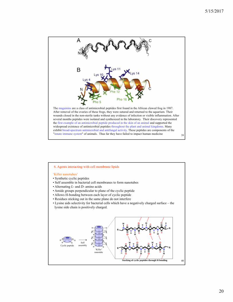
The magainins are a class of antimicrobial peptides first found in the African clawed frog in 1987. After removal of the ovaries of these frogs, they were sutured and returned to the aquarium. Their wounds closed in the non-sterile tanks without any evidence of infection or visible inflammation. After several months peptides were isolated and synthesized in the laboratory. Their discovery represented the first example of an antimicrobial peptide produced in the skin of an animal and supported the widespread existence of antimicrobial peptides throughout the plant and animal kingdoms. Many exhibit broad-spectrum antimicrobial and antifungal activity. These peptides are components of the "innate immune system" of animals. Thus far they have failed to impact human medicine
6. Agents interacting with cell membrane lipids
‘Killer nanotubes’• Synthetic cyclic peptides • Self assemble in bacterial cell membranes to form nanotubes• Alternating L- and D- amino acids• Amide groups perpendicular to plane of the cyclic peptide• Allows H-bonding between each layer of cyclic peptideg y y p p• Residues sticking out in the same plane do not interfere• Lysine aids selectivity for bacterial cells which have a negatively charged surface – the lysine side chain is positively charged.
RR
R R
HOH O
RN
N
N
N
NN
NN
O H OH
R
© Oxford University Press, 2013
Cyclic peptide
RRR R
R R
Selfassembly RR
R R
RRR
RRR R
RRR
RRR R
RRR
‘Killer’nanotube
OHO H
N N
H O H O
O
H O
H O
H
H
O
RN
N
N
N
NN
NN
H
O
O
HO
HO
H
R
Stacking of cyclic peptides through H-bonding 40

5/15/2017
21
6. Agents interacting with cell membrane lipids
L-LactateD-ValineL-Valine
D-Hyi
D-Hyi
NHO
NH
O
O OO
Me
Me
Me
Me
MeMe
MeMe
MeH
H H
H
ValinomycinValinomycin is a naturally occurring dodecadepsipeptide used in the transport of potassium and as an antibiotic. Valinomycin is obtained from the cells of several Streptomyces strains. It is a member of the group of natural neutral ionophores because it does not have a residual charge. Structures are alternately bound via amide
d t b id It h h d h bi
L-Valine
L-Lactate
D Valine
L-Lactate
D-Valine
O
NH
O
NHO
HN
O
HN
O
O O
O
OO
OOO
O
Me
Me
Me
Me
MMe
M
Me
Me
Me
Me
Me
H
H
H
H H
H
H
Hand ester bridges. It has hydrophobic groups on the exterior and polar carbonyl groups on the interior It functions as a potassium-specific transporter and facilitates the movement of potassium ions through lipid membranes "down" the electrochemical potential gradient. The stability constant K for the potassium-valinomycin complex is nearly 100,000 times larger than that of the sodium-valinomycin complex. This
© Oxford University Press, 2013
D-Valine
D-HyiL-Valine
MeMe Me Me
D-Hyi = D-Hydroxyisovaleric acid
41
sodium valinomycin complex. This difference is important for maintaining the selectivity of valinomycin for the transport of potassium ions (and not sodium ions) in biological systems. It is classified as an extremely hazardous substance in the United States, and is subject to strict reporting requirements by facilities which produce, store, or use it in significant quantities.
6. Agents interacting with cell membrane lipids
Mechanism of action• Acts as an ion carrier• Hydrophobic groups on exterior interact with
membrane lipids• Carbonyl groups interact with potassium ion• Allows uncontrolled escape of potassium ions from cell
Exterior
Cellmembrane
cell
K+
K+
K+
O
HN
O
NH
ONH
OO
O
OO
© Oxford University Press, 2013
Cytoplasm
membrane
Valinomycin
K+
K
O
HN
O NH
O
NH
O
O
OO
OOO
O
42

5/15/2017
22
An anesthetic is a drug to prevent pain during surgery. A wide variety of drugs are used in modern anesthetic practice. Anesthetics are categorized into two classes: general anesthetics, which cause a reversible loss of consciousness, and local anesthetics, which cause a reversible loss of sensation for a limited region of the body while maintaining consciousness. Combinations of anesthetics are sometimes used for their synergistic and additive therapeutic effects. Adverse effects, however, may also be increased. Local anesthetics are agents that prevent transmission of nerve impulses without causing unconsciousness. They act by binding to fast sodium channels from within (in an open state). Local anesthetics can be either ester- or amide-based.
Lidocaine (1946), also known as xylocaine, is a local anesthetic and used to numb tissue in a specific area. It can also be used for nerve blocks. Lidocaine mixed with a small amount of adrenaline (epinephrine) is available to allow larger doses for numbing, to decrease bleeding, and to make it last longer. When used as an injectable, it
© Oxford University Press, 201343
longer. When used as an injectable, it typically begins working within four minutes and lasts for half an hour to three hours. Lidocaine may also be applied directly to the skin for numbing.
HN
O
N
Lidocaine
Volatile agents are specially formulated organic liquids that evaporate readily into vapors, and are given by inhalation for general anesthesia. Nitrous oxide and xenon are gases at room temperature rather than liquids, so they are not considered volatile agents. The ideal anesthetic vapor or gas should be non-flammable, non-explosive, and lipid-soluble. It should possess low blood gas solubility, have no heart, liver or kidney toxicity or side-effects, should not be metabolized, and should not be an irritant to the respiratory
CH
F3C
CF3
OCH2
FSevoflurane
CH
H2C
CF3
OCH
FDesflurane
F
F
Cl O FIsoflurane
p ypathways of the patient. No anaesthetic agent currently in use meets all these requirements, nor can any anaesthetic agent be considered safe. There are inherent risks and drug interactions that are specific to each and every patient. Anesthetics in widespread current use are isoflurane, desflurane, sevoflurane, and nitrous oxide. Nitrous oxide is a common adjuvant gas, making it one of the most long-lived drugs still in current use. Because of its low potency, it cannot produce anesthesia on its own but is frequently combined with other agents.
CH
CF3
CH
F
© Oxford University Press, 201344
g
The most frequently used agent for inhalational induction is sevoflurane. Potency is directly related to lipid solubility. Also important is the blood/gas partition coefficient. This concept refers to the relative solubility of a given agent in blood. Those agents with a lower blood solubility make it easier for the anesthesia provider to titrate the depth of anesthesia, and permit a more rapid emergence upon discontinuing the anesthestic.

5/15/2017
23
Nitrous oxide is commonly known as laughing gas or nitrous. At room temperature, it is a colorless, odorless non-flammable gas. At elevated temperatures, nitrous oxide is a powerful oxidizer similar to molecular oxygen and can be used in motor racing and rocket fuel to increase power output. Nitrous oxide is used in surgery and dentistry, for its anaesthetic effects. It can provide an euphoric effect, a property that has led to its recreational use as a dissociative anaesthetic. Nitrous oxide occurs in small amounts in the atmosphere, but has been found recently to be a major
NN
ONitrous oxide
p y jscavenger of stratospheric ozone, with impact comparable to that of CFCs. It is estimated that 30% of the N2O in the atmosphere is the result of human agriculture activity.
Propofol is one of the most commonly used intravenous drugs employed to induce and maintain general anesthesia. It can also be used for sedation during procedures or in the ICU. Like the other agents mentioned above, it renders patients unconscious without producing pain relief. Propofol is used for induction and
OHsodium thiopental
S Na
© Oxford University Press, 201345
maintenance (in some cases) of anesthesia, having largely replaced sodium thiopental.because recovery from propofol is more rapid and "clear."
Sodium thiopental is a rapid-onset short-acting barbiturate general anesthetic. It has been supplanted by propofol. It was previously the first of three drugs administered during most lethal injections in the United States, but the U.S. manufacturer manufacturing the drug and the EU banned the export of the drug for this purpose.
Propofol
HN N
O O
Fentanyl is a potent, synthetic opioid pain medication with a rapid onset and short duration of action. It is also used as a recreational drug, leading to thousands of overdose deaths from 2000 to 2017. Deaths have also resulted from improper medical use. Fentanyl has a relatively wide therapeutic index (270) which makes it a safe surgical anesthetic when monitored carefully; however, its extreme potency requires careful measurements of highly diluted fentanyl in solution.
Fentanyl
N N
O
© Oxford University Press, 201346

5/15/2017
24
7. Agents acting on Carbohydrates
• Carbohydrates play important roles in cell recognition, regulation and growth• Potential targets for the treatment of bacterial and viral infection, cancer and
autoimmune disease• Carbohydrates act as antigens
Carbohydrate 'tag'
Cellmembrane
© Oxford University Press, 201347
Ceramide 'anchor'Carbohydrate 'tag'
7. Agents acting on Carbohydrates
HO
O
HO (CH2)16CH3
O
RO
HO
O
OHO
O
OHOH
(CH2)12CH3
HN (CH2)16CH3
O
Ceramide unit
SUGARS
Carbohydrates
© Oxford University Press, 2013
OH
(CH2)12CH3HO
NH2
Sphingosine
Fatty Acid (e.g. Stearic acid)O
OHHO
RO
OH Carbohydrate (R= various other carbohydrate structures)
48
5/15/2017
25
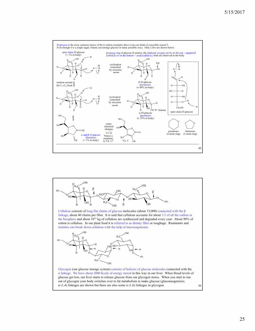
D-glucose is the most common choice of the 6 carbon examples above (can you think of a possible reason?). Even though it is a single sugar, Nature can arrange glucose in many possible ways. Only a few are shown below.
O
H
H H
H
H
O O
H2C
O
O
OH
O
H
H H
H
H
O OH
H2C
O
O
OHH
B
BH
O H
BH
open chain D-glucose(< 1% in body)
pyranose ring of glucose (6 atoms), the aldehyde oxygen can be on the top = equatorial (called ) or on the bottom = axial (called ), both are observed in the body
H
H
H
Hcyclization controlled
by enzymes
top
O
H
H OH
H
H
O H
H2C
OH
O
OH
O
H
H O
H
H
O H
H2C
O
O
OHH
rotation around the C1-C2 bond
B
BHcyclization controlled
by enzymes
-D-glucose(pyranose)
( 66% in body)
-D-glucose(pyranose)
CO H
H OH
HO H
H OH
H O
CH2OH
open chain D-glucose
H
B
HO
H
H
H
H
H
H
H
bottom
© Oxford University Press, 201349
(pyranose)( 33% in body)
O
OH
OH
HO
HO
HO
and D-glucose(furanose)
(< 1% in body)
OO
pyranoses(6 atom ring)
furanoses(5 atom ring)
O
O
OH
HO
HO
HO
Vit. C
Notice a similarityto Vit. C?
manychemicalchanges
O
H
H H
H
O O
H2C
OH
OH
HOO
OOHO
OH
H2C
O
OH
H2C
HO
OH
OH
etc.
etc.
Cellulose consists of long flat chains of glucose molecules (about 15,000) connected with the βlinkage about 40 chains per fiber It is said that cellulose accounts for about 1/2 of all the carbon in
O
H
H O
H
O H
H2C
OH
OH
HO OO
OH
H2COH
HO
OHO
etc.
linkage, about 40 chains per fiber. It is said that cellulose accounts for about 1/2 of all the carbon in the biosphere and about 1015 kg of cellulose are synthesized and degraded every year. About 90% of cotton is cellulose. In our plant food it is referred to as dietary fiber or roughage. Ruminants and termites can break down cellulose with the help of microorganisms.
© Oxford University Press, 201350
H OO
H2COH
Oetc.
Glycogen (our glucose storage system) consists of helicies of glucose molecules connected with the α linkage. We have about 2000 Kcals of energy stored in this way in our liver. When blood levels of glucose get low, our liver starts to release glucose from our glycogen stores. When you start to run out of glycogen your body switches over to fat metabolism to make glucose (gluconeogenisis). α (1,4) linkages are shown but there are also some α (1,6) linkages in glycogen.
5/15/2017
26
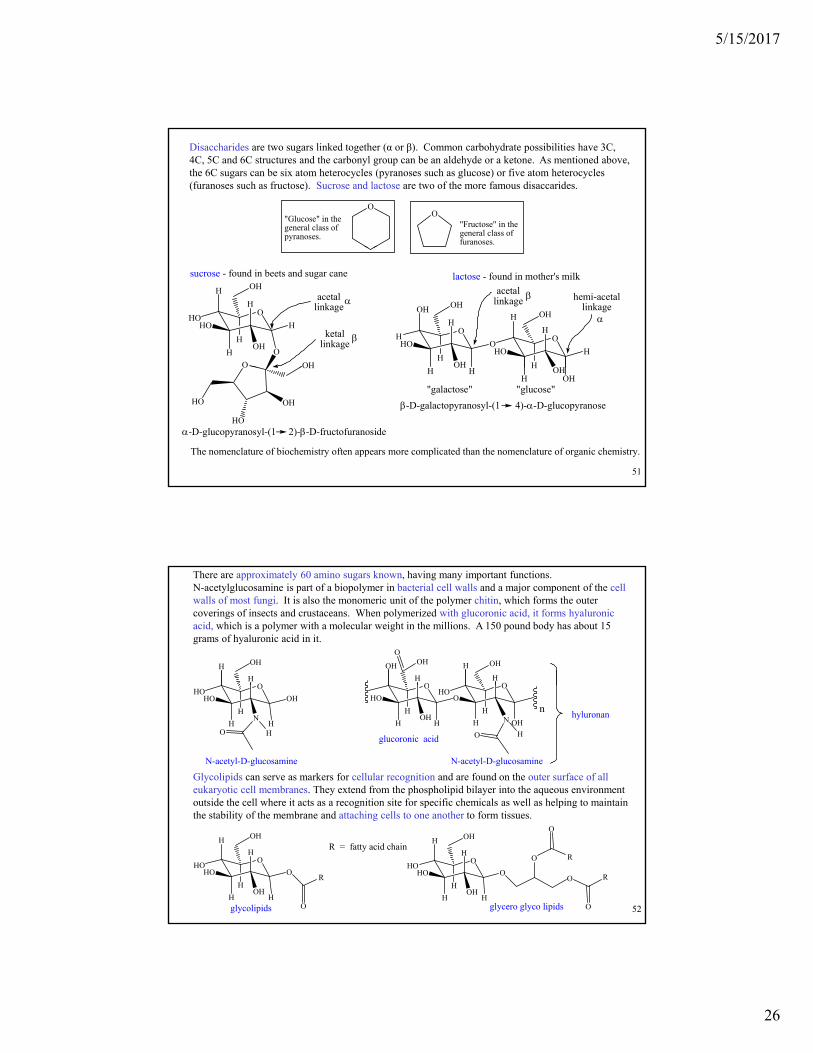
Disaccharides are two sugars linked together (α or β). Common carbohydrate possibilities have 3C, 4C, 5C and 6C structures and the carbonyl group can be an aldehyde or a ketone. As mentioned above, the 6C sugars can be six atom heterocycles (pyranoses such as glucose) or five atom heterocycles (furanoses such as fructose). Sucrose and lactose are two of the more famous disaccarides.
OO
"Glucose" in the general class of pyranoses.
"Fructose" in the general class of furanoses.
sucrose - found in beets and sugar cane lactose - found in mother's milk
acetallinkage
ketal linkage
O
O
O
H
H
H
HOHO
H
H
OH
OH
OH
O
O
H
OH
HHO
H
OH
OH
O
H
OH
HO H
H
H
OH
acetallinkage hemi-acetal
linkage
© Oxford University Press, 201351
-D-glucopyranosyl-(1 2)--D-fructofuranoside
The nomenclature of biochemistry often appears more complicated than the nomenclature of organic chemistry.
-D-galactopyranosyl-(1 4)--D-glucopyranose
O OH
HO
HO
OH
HOH
H OHH OH
H
"galactose" "glucose"
There are approximately 60 amino sugars known, having many important functions. N-acetylglucosamine is part of a biopolymer in bacterial cell walls and a major component of the cell walls of most fungi. It is also the monomeric unit of the polymer chitin, which forms the outer coverings of insects and crustaceans. When polymerized with glucoronic acid, it forms hyaluronic acid, which is a polymer with a molecular weight in the millions. A 150 pound body has about 15 grams of hyaluronic acid in it.
O
H
H
OH
O
OH
H
OH
O
H
H
OH
O
Glycolipids can serve as markers for cellular recognition and are found on the outer surface of all eukaryotic cell membranes. They extend from the phospholipid bilayer into the aqueous environment outside the cell where it acts as a recognition site for specific chemicals as well as helping to maintain
N-acetyl-D-glucosamine
hyluronan
O
OH
H
HOHO
HN
O
O
H
HO
HOH
H
O
H OH
H
HHO
HO
nN
HO
N-acetyl-D-glucosamine
glucoronic acid
© Oxford University Press, 201352
outside the cell where it acts as a recognition site for specific chemicals as well as helping to maintain the stability of the membrane and attaching cells to one another to form tissues.
glycolipids
O
O
H
H
HOHO
H
H
OH
OH
HO
R
R = fatty acid chain
O
O
H
H
HOHO
H
H
OH
OH
H
O
O
O
R
O
R
glycero glyco lipids

5/15/2017
27
Blood types are based, in part, on carbohydrate coatings linked to lipid molecules on the outer cell membranes. There is also a soluble form linked to proteins in the blood. About 75% of type A individuals have a repeat of the 3 terminal sugars. Type AB (4% in the US) have both A and B patterns on their cell surfaces and are universal acceptors. There is some variation in the position number of the connections and the α/β linkage is not indicated. Science News (vol 151, p 24, 1-11-97) discusses enzymes from chicken livers (type A) and coffee beans (type B) that can cleave the single sugar at the end of the cell coatings. This would convert type A and type B to type O, which would allow for universal donation. The article discusses the possible commercial production for blood donations. There are over 7,000,000,000 (billion) people on the earth. Every one of us has a unique biochemical profile that is uniquely recognized by our immune system. The blood types give you a hint at how this is possible.
type O (45% in US)universal donor
cell membrane
gluc galac N-acet galac
fucoseThis sequence is the same in all blood types (type O)
type A (40% in US)
gluc galac N-acet galac galac
O
OHHO
H3C
O
OH
OH
HO
OH
HO
OH OH
OH
fucose
galactose
The linkages are unspecified here.
© Oxford University Press, 201353
cell membranefucose
The extra galactosemakes this type A.
type B (11% in US)
cell membrane
gluc galac N-acet galac
fucose
N-acet
The extra N-acetylgalactosaminemakes this type B.
O
OH
OH
HO
O
OH
N
HO
OHOH
HO
glucose
H
ON-acetylgalactosamine
Blood types are based, in part, on carbohydrate coatings linked to lipid molecules on the outer cell membranes. There is also a soluble form linked to proteins in the blood. About 75% of type A individuals have a repeat of the 3 terminal sugars. Type AB (4% in the US) have both A and B patterns on their cell surfaces and are universal acceptors. There is some variation in the position number of the connections and the α/β linkage is not indicated. Scientists (2007) screened 2,500 fungi and bacteria and discovered two bacteria which contained potentially useful enzymes. They found that enzymes from both bacteria were able to remove both A and B antigens from red blood cells. Such a strategy has long been proposed, but has proved to be impractical. The new process cannot do anything about another antigen that can trigger an immune response. Blood which carries this antigen is known as rhesus positive. There are over 7,000,000,000 (billion) people on the earth. Every one of us has a unique biochemical profile that is uniquely recognized by our immune system. The blood types give you a hint at how this is possible.
type O (45% in US)universal donor
cell membrane
gluc galac N-acet galac
fucoseThis sequence is the same in all blood types (type O)
type A (40% in US)
gluc galac N-acet galac galac
O
OHHO
H3C
O
OH
OH
HO
OH
HO
OH OH
OH
fucose
galactose
The linkages are unspecified here.
© Oxford University Press, 201354
cell membranefucose
The extra galactosemakes this type A.
type B (11% in US)
cell membrane
gluc galac N-acet galac
fucose
N-acet
The extra N-acetylgalactosaminemakes this type B.
O
OH
OH
HO
O
OH
N
HO
OHOH
HO
glucose
H
ON-acetylgalactosamine

5/15/2017
28
7. Agents acting on Carbohydrates
Antibodies• Proteins produced by the immune system• Recognise and bind to foreign antigens• Y-shaped molecules consisting of 2 heavy and 2 light chains• Variable region at tips of the armsg p
H3NH3N
NH3NH3
Variable regions
Lightchain
© Oxford University Press, 2013
O2C CO2
chain
Heavychain
55
7. Agents acting on Carbohydrates
Antibodies
• Antibodies bind to foreign antigens on foreign cells• Mark out the cell for destruction• Immune system destroys the cell
Antibodies ha e been designed as anticancer agents• Antibodies have been designed as anticancer agents
Antibodies
Antibody
Invasion warning!
© Oxford University Press, 2013
Foreign cellAntigen
Antibodybinding
Cell destruction
56
5/15/2017
29
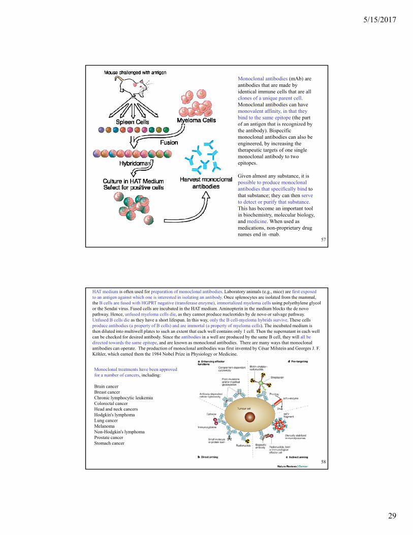
Monoclonal antibodies (mAb) are antibodies that are made by identical immune cells that are all clones of a unique parent cell. Monoclonal antibodies can have monovalent affinity, in that they bind to the same epitope (the part of an antigen that is recognized by g g ythe antibody). Bispecific monoclonal antibodies can also be engineered, by increasing the therapeutic targets of one single monoclonal antibody to two epitopes.
Given almost any substance, it is possible to produce monoclonal
© Oxford University Press, 201357
antibodies that specifically bind to that substance; they can then serve to detect or purify that substance. This has become an important tool in biochemistry, molecular biology, and medicine. When used as medications, non-proprietary drug names end in -mab.
HAT medium is often used for preparation of monoclonal antibodies. Laboratory animals (e.g., mice) are first exposed to an antigen against which one is interested in isolating an antibody. Once splenocytes are isolated from the mammal, the B cells are fused with HGPRT negative (transferase enzyme), immortalized myeloma cells using polyethylene glycol or the Sendai virus. Fused cells are incubated in the HAT medium. Aminopterin in the medium blocks the de novo pathway. Hence, unfused myeloma cells die, as they cannot produce nucleotides by de novo or salvage pathway. Unfused B cells die as they have a short lifespan. In this way, only the B cell-myeloma hybrids survive. These cells produce antibodies (a property of B cells) and are immortal (a property of myeloma cells). The incubated medium is then diluted into multiwell plates to such an extent that each well contains only 1 cell. Then the supernatant in each well can be checked for desired antibody. Since the antibodies in a well are produced by the same B cell, they will all be directed towards the same epitope, and are known as monoclonal antibodies. There are many ways that monoclonal antibodies can operate The production of monoclonal antibodies was first invented by César Milstein and Georges J Fantibodies can operate. The production of monoclonal antibodies was first invented by César Milstein and Georges J. F. Köhler, which earned them the 1984 Nobel Prize in Physiology or Medicine.
Monoclonal treatments have been approved for a number of cancers, including:
Brain cancerBreast cancerChronic lymphocytic leukemiaColorectal cancerHead and neck cancers
© Oxford University Press, 201358
Head and neck cancersHodgkin's lymphomaLung cancerMelanomaNon-Hodgkin's lymphomaProstate cancerStomach cancer

5/15/2017
30
A group of scientists demonstrates how CRISPR/Cas9 technology could be used to fight devastating diseases, by editing cells found in the human immune system called T cells (CD4 receptors recognize class II MHC sites, displays protein fragments from outside the cell, and CD8 receptors recognize class I MHC sites), displays protein fragments from inside the cell). CRISPR, which stands for clustered regularly interspaced short palindromic repeats, allows researchers to can go in and either delete or cut-and-paste pieces of genome within living cells with remarkable precision In
The CRISPR/Cas system is a prokaryotic immune system that confers resistance to foreign genetic elements such as those present within plasmids and phages that provides a form of acquired immunity. CRISPERs are sequences of genetic information from previous viral infections that allows the bacterium to recognize and destroy foreign genetic material to prevent infection. RNA harboring the spacer sequence helps Cas proteins recognize and cut exogenous foreign DNA CRISPRs are found in approximately 40% of sequenced bacterial genomes and 90% of sequenced
genome within living cells with remarkable precision. In a palindromic repeat, the sequence of nucleotides is the same in both directions. Each repetition is followed by short segments of spacer DNA from previous exposures to foreign DNA (e.g., a virus or plasmid). Small clusters of cas genes are located next to CRISPR sequences.Virus infected T cell
© Oxford University Press, 201359
foreign DNA. CRISPRs are found in approximately 40% of sequenced bacterial genomes and 90% of sequenced archaea.
By manipulating the genes found within these T cells, the researchers could make the cells less susceptible to HIV infection and even cancer. T cells are key coordinating cells in the immune system. They essentially set the stage for determining how the immune system will respond to infections, and have been found to be central to a lot of different disease processes.
CRISPR was used to eliminate a gene that encodes a particular protein on the surface of T cells (about 20% efficiency). The HIV virus uses this protein as a way to gain access to T cells and infect them, so the application may one day be used to fight off this deadly virus.