transportation- o 2 and co 2, nutrients, waste, hormones regulation- ph (buffers), heat, osmotic...
TRANSCRIPT
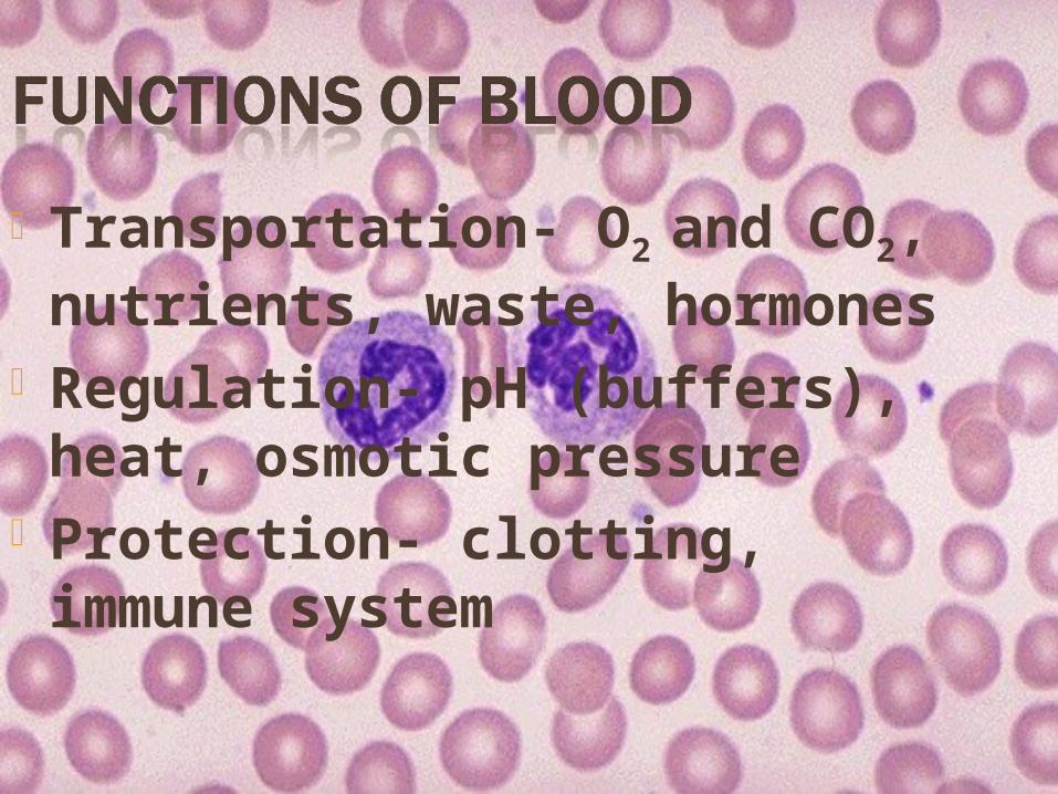
Transportation- O2 and CO2, nutrients, waste, hormones
Regulation- pH (buffers), heat, osmotic pressure
Protection- clotting, immune system
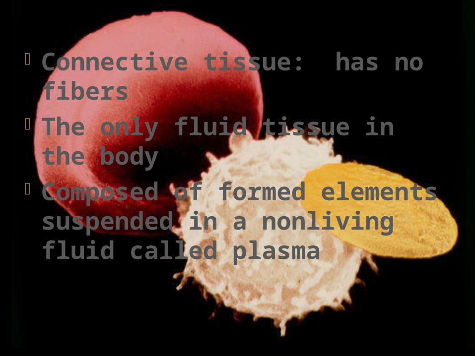
Connective tissue: has no fibers
The only fluid tissue in the body
Composed of formed elements suspended in a nonliving fluid called plasma

Can be separated into layers with centrifuge
Heavier, thicker, more viscous than H2O (5x)
Temperature of 100.4 F pH of 7.4 8% body weight 5-6 liters (1.5 gallons) in males 4-5 liters (1.2 gallons) in females
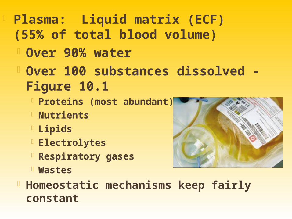
Plasma: Liquid matrix (ECF) (55% of total blood volume) Over 90% water Over 100 substances dissolved
- Figure 10.1 Proteins (most abundant) Nutrients Lipids Electrolytes Respiratory gases Wastes
Homeostatic mechanisms keep fairly constant
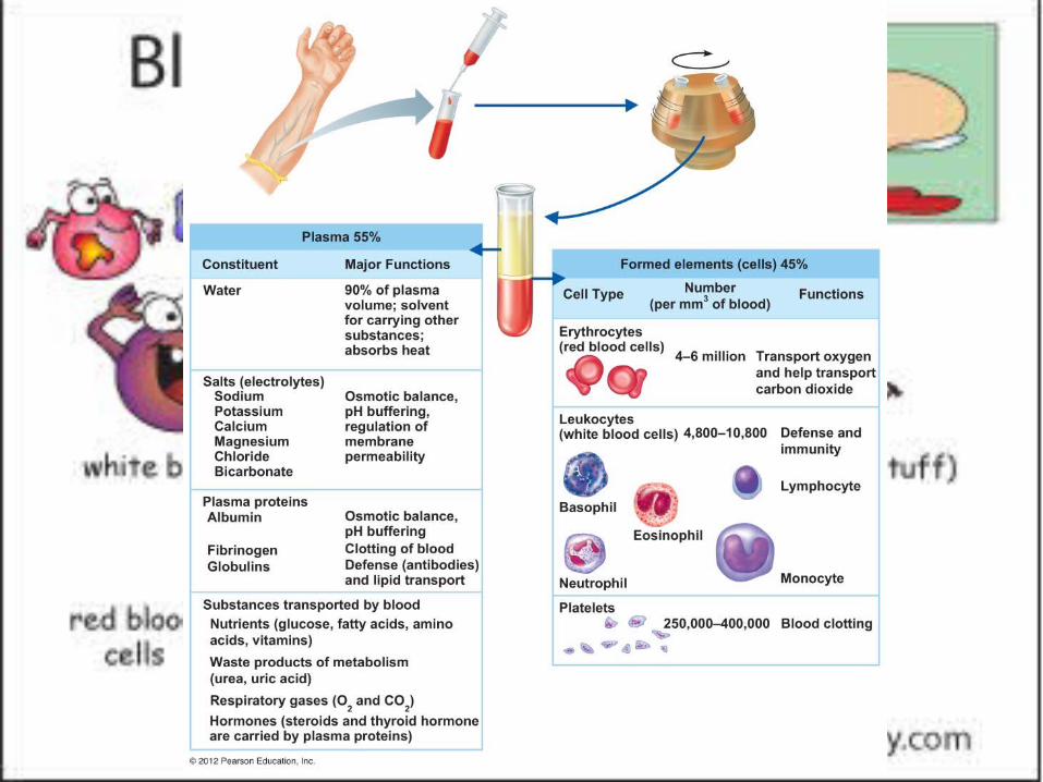
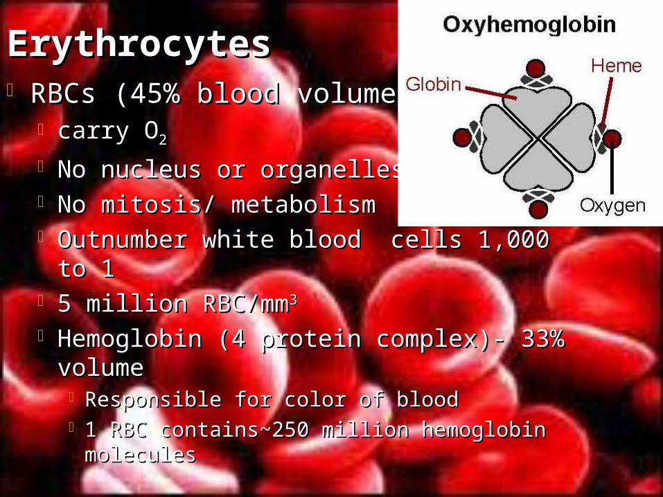
ErythrocytesErythrocytes RBCs (45% blood volume) RBCs (45% blood volume)
carry Ocarry O22
No nucleus or organellesNo nucleus or organelles No mitosis/ metabolismNo mitosis/ metabolism Outnumber white blood cells 1,000 to 1Outnumber white blood cells 1,000 to 1 5 million RBC/mm5 million RBC/mm3 3
Hemoglobin (4 protein complex)- 33% Hemoglobin (4 protein complex)- 33% volumevolume Responsible for color of bloodResponsible for color of blood 1 RBC contains~250 million hemoglobin 1 RBC contains~250 million hemoglobin
moleculesmolecules
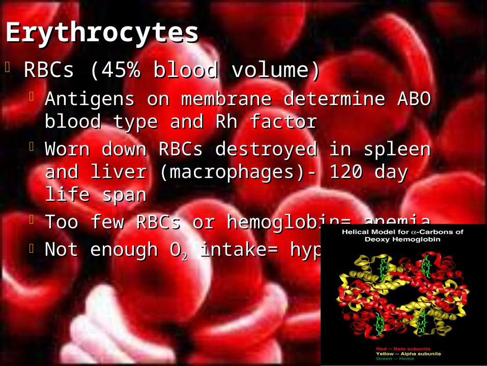
ErythrocytesErythrocytes RBCs (45% blood volume) RBCs (45% blood volume)
Antigens on membrane determine ABO Antigens on membrane determine ABO blood type and Rh factorblood type and Rh factor
Worn down RBCs destroyed in spleen Worn down RBCs destroyed in spleen and liver (macrophages)- 120 day life and liver (macrophages)- 120 day life spanspan
Too few RBCs or hemoglobin= anemiaToo few RBCs or hemoglobin= anemia Not enough ONot enough O22 intake= hypoxia intake= hypoxia
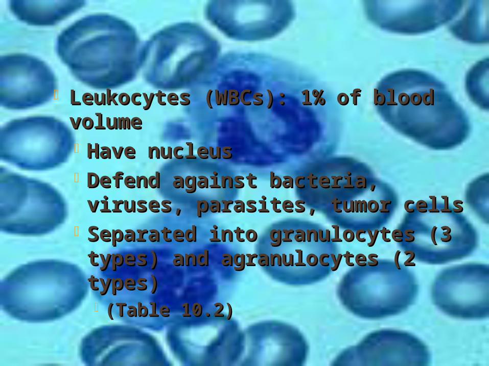
Leukocytes (WBCs): 1% of blood Leukocytes (WBCs): 1% of blood volumevolume Have nucleusHave nucleus Defend against bacteria, viruses, Defend against bacteria, viruses, parasites, tumor cellsparasites, tumor cells
Separated into granulocytes (3 Separated into granulocytes (3 types) and agranulocytes (2 types) and agranulocytes (2 types) types) (Table 10.2)(Table 10.2)
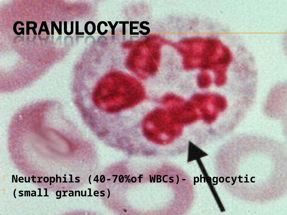
Neutrophils (40-70%of WBCs)- phagocytic (small granules)
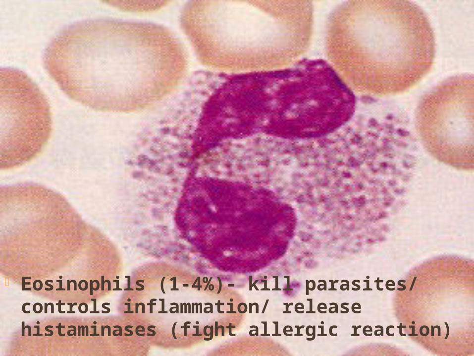
Eosinophils (1-4%)- kill parasites/ controls inflammation/ release histaminases (fight allergic reaction)
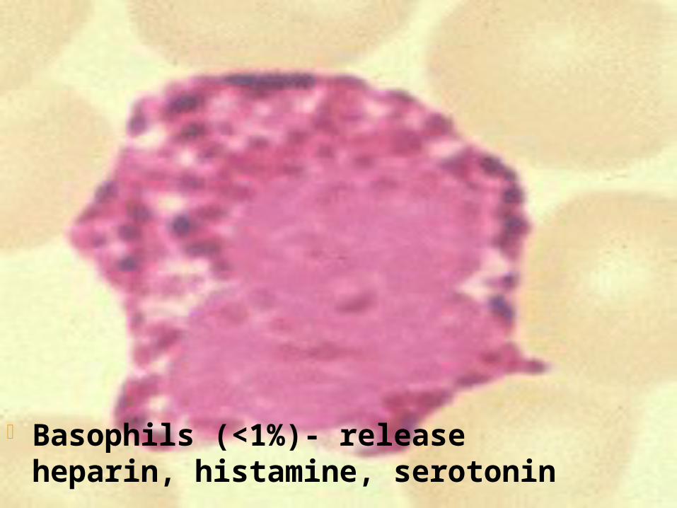
Basophils (<1%)- release heparin, histamine, serotonin

Monocytes (4-8%)- phagocytic (large)

Lymphocytes (20-45%)- provide immunity B and T cells produce antibodies
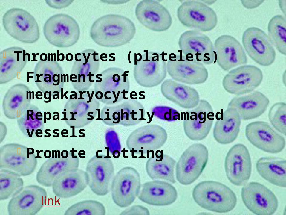
Thrombocytes (platelets) Fragments of megakaryocytes
Repair slightly damaged vessels
Promote clotting
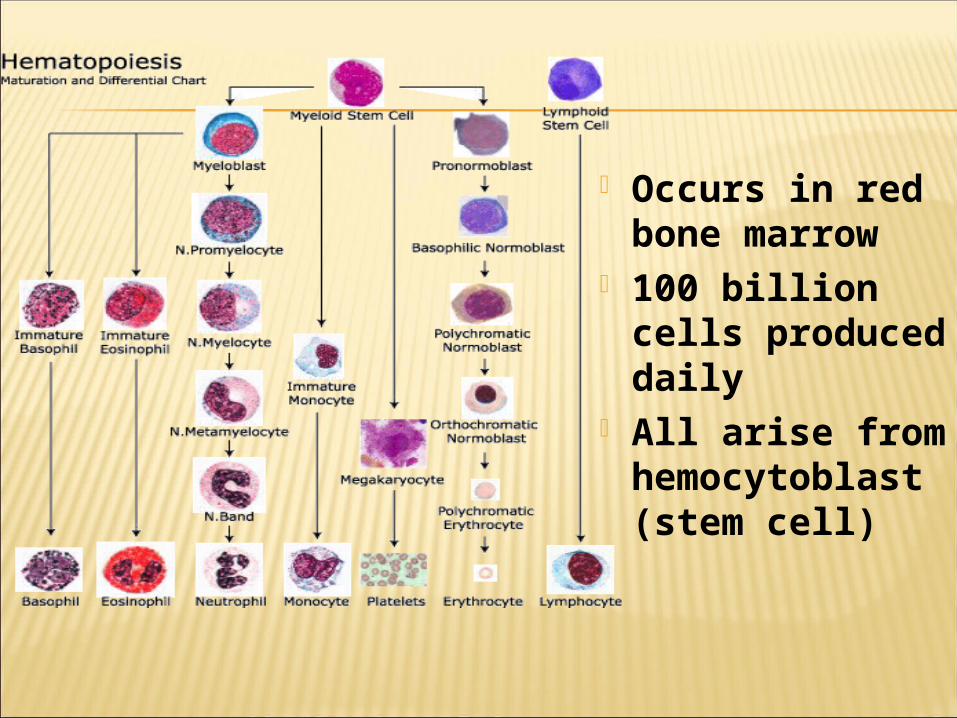
Occurs in red bone marrow
100 billion cells produced daily
All arise from hemocytoblast (stem cell)
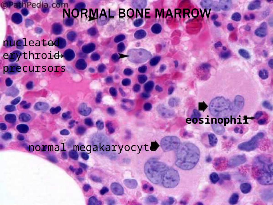
nucleated erythroid precursors
normal megakaryocyte
eosinophil

Process takes 3-5 days Rate of production controlled by
erythropoietin (hormone)
Stimulated by hormones Released by chemical signals
(inflammation, bacteria)

Three phasesa) Vascular spasmsb) Platelet plug formationc) Coagulation
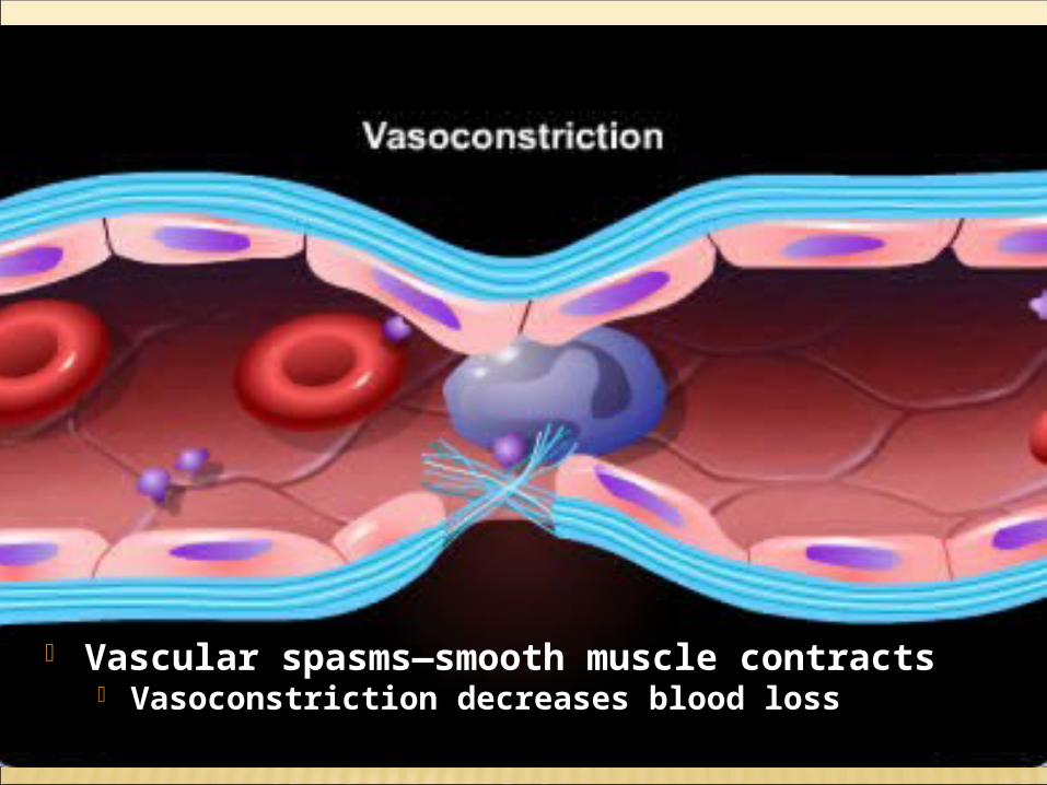
Vascular spasms—smooth muscle contracts Vasoconstriction decreases blood loss
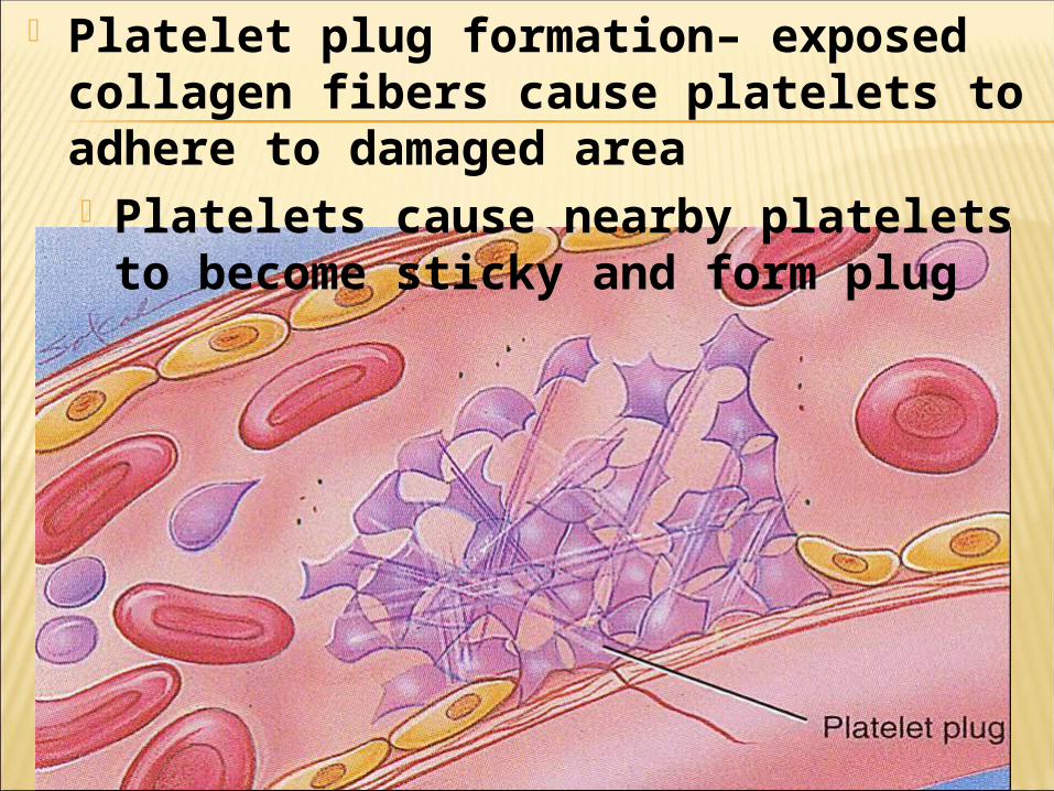
Platelet plug formation– exposed collagen fibers cause platelets to adhere to damaged area Platelets cause nearby platelets
to become sticky and form plug
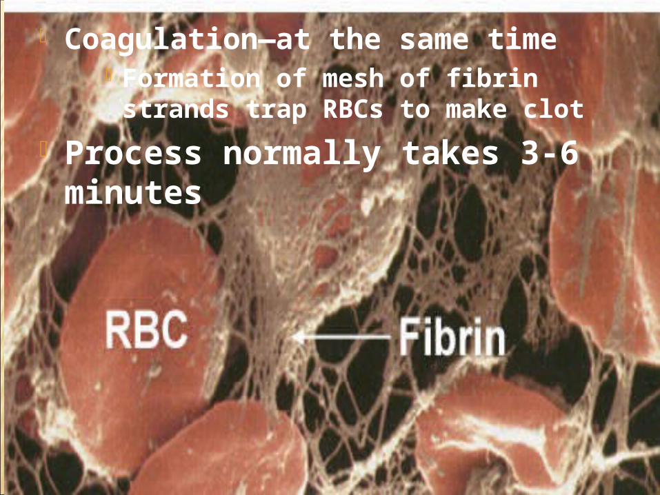
Coagulation—at the same time Formation of mesh of fibrin strands trap RBCs to make clot
Process normally takes 3-6 minutes
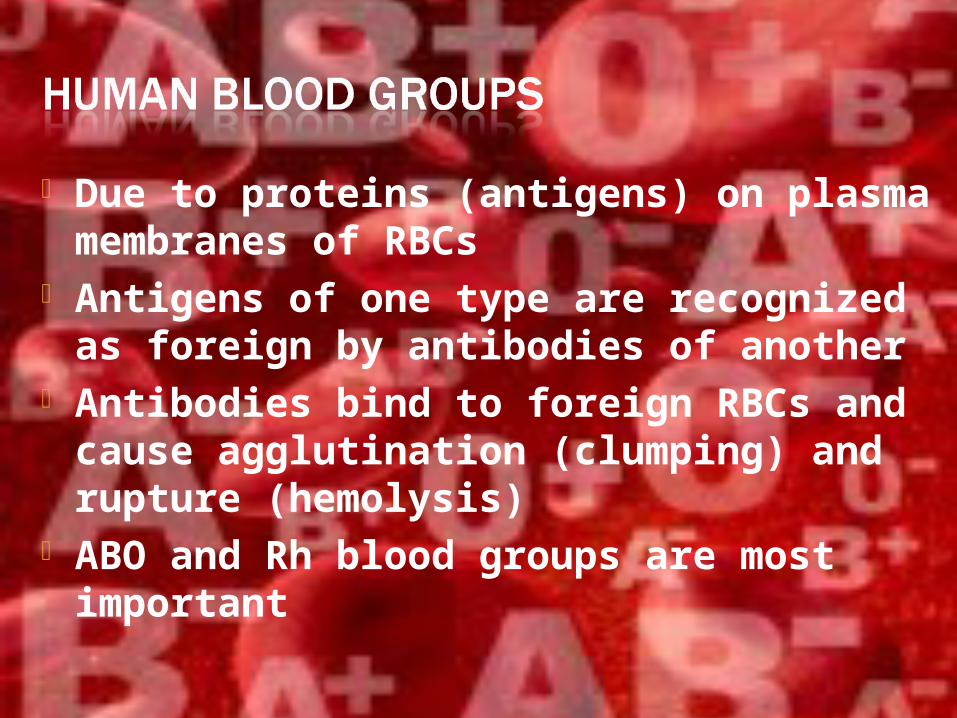
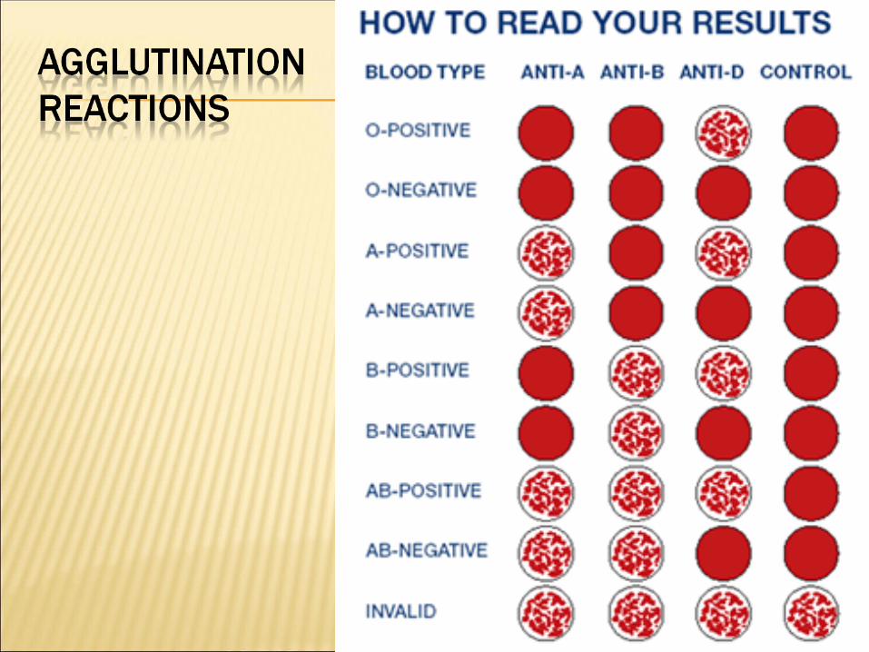
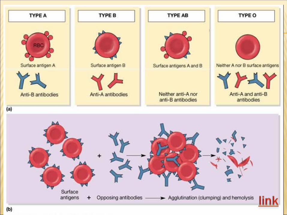
Due to proteins (antigens) on plasma membranes of RBCs
Antigens of one type are recognized as foreign by antibodies of another
Antibodies bind to foreign RBCs and cause agglutination (clumping) and rupture (hemolysis)
ABO and Rh blood groups are most important
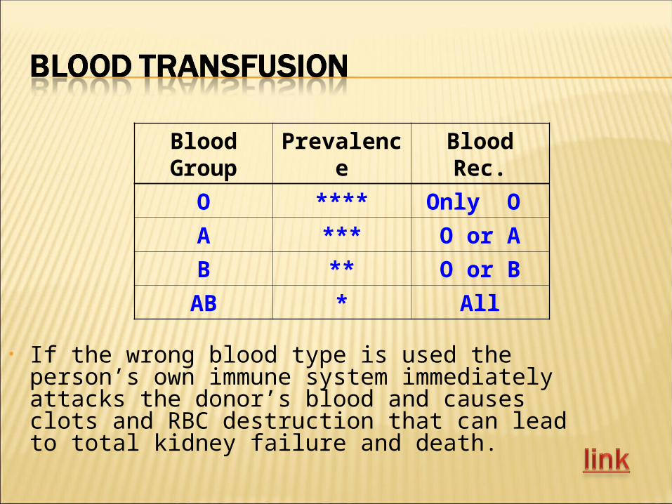
Blood Group
Prevalence Blood Rec.
O **** Only O
A *** O or A
B ** O or B
AB * All
• If the wrong blood type is used the person’s own immune system immediately attacks the donor’s blood and causes clots and RBC destruction that can lead to total kidney failure and death.
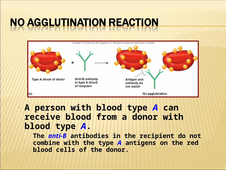
• A person with blood type A can receive blood from a donor with blood type A.– The anti-B antibodies in the recipient do not
combine with the type A antigens on the red blood cells of the donor.
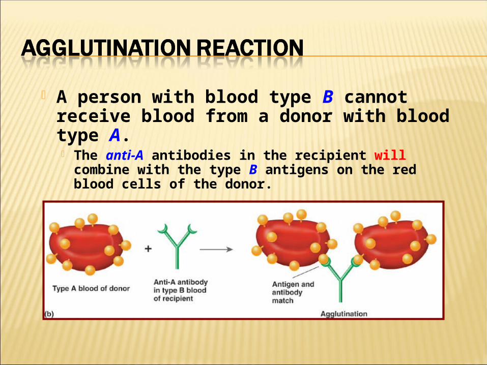
A person with blood type B cannot receive blood from a donor with blood type A. The anti-A antibodies in the recipient will
combine with the type B antigens on the red blood cells of the donor.
Rh+: have Rh antigen and no antibodies
Rh-: have no antigens and no antibodies
Rh-negative people will develop antibodies to the Rh antigen if they are exposed to the Rh-positive blood
If a Rh-negative woman becomes pregnant with a Rh-positive fetus she may make
antibodies to the fetus’ RBCs
This can be prevented with RhoGAM
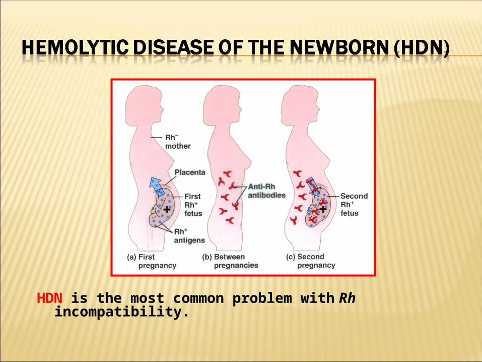
HDN is the most common problem with Rh incompatibility.