science news blog 20170522.d… · web vie
TRANSCRIPT

1 5/14/23 Name Student number
http://bit.ly/2rljvZrHeart attack risk increases 17-fold following respiratory
infectionsRespiratory infections can trigger a heart attack
The risk of having a heart attack is 17 times higher in the seven days following a respiratory infection, University of Sydney research has found.Published today in Internal Medicine Journal, this is the first study to report an association between respiratory infections such as pneumonia, influenza and bronchitis and increased risk of heart attack in patients confirmed by coronary angiography (a special X-Ray to detect heart artery blockages)."Our findings confirm what has been suggested in prior studies that a respiratory infection can act as a trigger for a heart attack," said senior author Professor Geoffrey Tofler, cardiologist from University of Sydney, Royal North Shore Hospital and Heart Research Australia."The data showed that the increased risk of a heart attack isn't necessarily just at the beginning of respiratory symptoms, it peaks in the first 7 days and gradually reduces but remains elevated for one month."The study was an investigation of 578 consecutive patients with heart attack due to a coronary artery blockage, who provided information on recent and usual occurrence of symptoms of respiratory infection.Seventeen per cent of patients reported symptoms of respiratory infection within 7 days of the heart attack, and 21 per cent within 31 days.Patients were interviewed about their activities before the onset of their heart attack, including if they experienced a recent "flu-like illness with fever and sore throat". They were considered affected if they reported sore throat, cough, fever, sinus pain, flu-like symptoms, or if they reported a diagnosis of pneumonia or bronchitis.
A second analysis was among those with symptoms restricted to the upper respiratory tract, which included the common cold, pharyngitis, rhinitis and sinusitis.Lead author Dr Lorcan Ruane, who conducted the work at University of Sydney said: "For those participants who reported milder upper respiratory tract infection symptoms the risk increase was less, but was still elevated by 13 fold.""Although upper respiratory infections are less severe, they are far more common than lower respiratory tract symptoms. Therefore it is important to understand their relationship to the risk of heart attacks, particularly as we are coming into winter in Australia," he said.Associate Professor Thomas Buckley, study investigator from Sydney Nursing School said: "The incidence of heart attacks is highest during winter in Australia.""This winter peak in seen not only in Australia but also in other countries around the world is likely due in part to the increased incidence of respiratory infections."People should take measures to reduce exposure to infection, including flu and pneumonia vaccines where appropriate."Professor Tofler added: "Possible reasons for why respiratory infection may trigger a heart attack include an increased tendency towards blood clotting, inflammation and toxins damaging blood vessels, and changes in blood flow.""Our message to people is while the absolute risk that any one episode will trigger a heart attack is low, they need to be aware that a respiratory infection could lead to a coronary event. So consider preventative strategies where possible, and don't ignore symptoms that could indicate a heart attack."The next step is to identify treatment strategies to decrease this risk of heart attack, particularly in individuals who may have increased susceptibility."The study was conducted at Royal North Shore Hospital.

2 5/14/23 Name Student number
http://bbc.in/2qZAUr3Plant chemicals hope for 'alternative contraceptives'
Could chemicals from wild plants be the key to a new generation of contraceptives?
By Philippa Roxby Health reporter, BBC NewsTwo compounds normally found in wild plants could make good alternatives to emergency contraceptives - if scientists only knew where to get enough of them. Chemicals from dandelion root and the "thunder god vine" plant have long been used in traditional medicines.Now, Californian researchers have found they can also block fertilisation.A UK sperm expert said the discovery could lead to a new and novel approach to male contraception. But the compounds existed at such low levels in plants that the cost of extraction was very high, the US team said.In tests, chemicals called pristimerin and lupeol stopped fertilisation by preventing human sperm from whipping its tail and propelling itself towards and into the woman's egg. The chemicals were acting like "molecular condoms", the study authors wrote in the journal Proceedings of the National Academy of Sciences.
The chemical lupeol is found in aloe vera leaves, in very small quantities Science Photo Library
In other words, they successfully blocked progesterone - which triggers the sperm's forceful swimming - but didn't damage the sperm."It doesn't kill sperm basal motility. It is not toxic to sperm cells; they still can move," said Polina Lishko, assistant professor of molecular and cell biology from the University of California, Berkeley."But they cannot develop this powerful stroke, because this whole activation pathway is shut down."
Lupeol is found in plants such as mango, dandelion root and aloe vera, while pristimerin is from the tripterygium wilfordii plant (also known as "thunder god vine") and is used in traditional Chinese medicine.The researchers found that the chemicals worked at very low doses and had no side-effects either, unlike hormone-based contraceptives.They concluded that the compounds could potentially be used as an emergency contraceptive, before or after intercourse, or as a permanent contraceptive via a skin patch or vaginal ring.'A good bet'Prof Lishko and her colleagues are now going to test how well these chemicals work in primates, whose sperm cells work in a similar way to humans. They also are searching for a cheap source of the chemicals, which are very expensive to extract from wild plants because they are present at very low levels.Allan Pacey, professor of andrology at the University of Sheffield said there was a real need for a non-hormone base male contraceptive."This is a very interesting study which shows that two natural compounds can knock out a key molecule on sperm that regulates how they swim in the final moments before fertilisation. "Moreover, because the molecule is specific to sperm, it seems a good bet that this could be a novel contraceptive target that might lead to a male contraceptive pill without any of the side-effects so far seen in trials with hormone-jab contraceptives."However, he said clinical trials were needed to show whether it worked in real people and this was likely to take a few years.
http://bit.ly/2qERjy3Why did hunter-gatherers first begin farming?
The beginnings of agriculture changed human history and has fascinated scientists for centuries.
Researchers from the Grantham Centre for Sustainable Futures at the University of Sheffield have shed light on how hunter-gatherers first began farming and how crops were domesticated to depend on humans.

3 5/14/23 Name Student number
Domesticated crops have been transformed almost beyond recognition in comparison with their wild relatives - a change that happened during the early stages of farming in the Stone Age.For grain crops like cereals, the hallmark of domestication is the loss of natural seed dispersal - seeds no longer fall off plants but have become dependent on humans or machines to spread them.Professor Colin Osborne, from the Grantham Centre for Sustainable Futures at the University of Sheffield, said: "We know very little about how agriculture began, because it happened 10,000 years ago - that's why a number of mysteries are unresolved. For example why hunter-gatherers first began farming, and how were crops domesticated to depend on people."One controversy in this area is about the extent to which ancient peoples knew they were domesticating crops. Did they know they were breeding domestication characteristics into crops, or did these characteristics just evolve as the first farmers sowed wild plants into cultivated soil, and tended and harvested them?"The new research, published in the journal Evolution Letters, shows the impact of domestication on vegetable seed size.Any selective breeding of vegetables by early farmers would have acted on the leaves, stems or roots that were eaten as food, but should not have directly affected seed size.Instead, any changes in vegetable seed size must have arisen from natural selection acting on these crops in cultivated fields, or from genetic links to changes in another characteristic like plant or organ size. In the last instance, people might have bred crops to become bigger, and larger seeds would have come along unintentionally.The University of Sheffield researchers gathered seed size data from a range of crops and found strong evidence for a general enlargement of seeds due to domestication.They discovered domesticated maize seeds are 15 times bigger than the wild form, soybean seeds are seven times bigger. Wheat, barley and other grain crops had more modest increases in size (60 per cent
for barley and 15 per cent for emmer wheat) but these changes are important if they translate into yield."We found strong evidence for a general enlargement of seeds due to domestication across seven vegetable species," said Professor Osborne."This is especially stunning in a crop like a sweet potato, where people don't even plant seeds, let alone harvest them. The size of this domestication effect falls completely within the range seen in cereals and pulse grains like lentils and beans, raising the possibility that at least part of the seed enlargement in these crops also evolved during domestication without deliberate foresight from early farmers."Professor Osborne added: "Our findings have important implications for understanding how crops evolved, because they mean that major changes in our staple crops could have arisen without deliberate foresight by early farmers."This means that unconscious selection was probably more important in the genesis of our food plants than previously realised. Early increases in the yields of crops might well have evolved in farmers' fields rather than being bred artificially."
http://bit.ly/2r1rZp3An immunity gene evolved in Southeast Asia to protect
against leprosyProliferation of an immune system gene mutation in SE Asia 50,000
years ago because it likely conferred protection against leprosyA mutation in an immune system gene rapidly rose in frequency in Southeast Asia approximately 50,000 years ago because it likely conferred protection against leprosy, which spread to the region from Africa around the same time.The findings, published May 16th in Cell Reports, show that the gene variant, called HLA-B*46:01, encodes a protein that binds to molecules derived from the bacterium that causes leprosy--a chronic infection of the skin and peripheral nerves.

4 5/14/23 Name Student number
This HLS protein then presents these foreign molecules to the immune system, which destroys the infected cells."Our study suggests that HLA-B*46:01 may provide protection against severe leprosy because it is better adapted to present pathogen-derived peptide antigens for immunosurveillance by the immune system," says lead author Hugo Hilton (@Hilton_HG) of Stanford University School of Medicine. "The findings may explain why HLA-B*46:01 evolved 50,000 years ago and spread to become one of the most prevalent immunity gene variants in Southeast Asia."Population expansion, cultural changes, and migration during the last 100,000 years exposed humans to pathogens against which they had not evolved effective resistance. Due to strong selective pressure, human leukocyte antigen (HLA) genes have evolved to provide immunity against diverse and rapidly evolving pathogens. "New HLA gene variants, or alleles, are thought to arise in human populations during episodes of Darwinian selection, but there is little direct evidence for the nature of this process," says senior study author Peter Parham of Stanford University School of Medicine.One compelling example of such an episode is the HLA-B*46:01 allele, which is now carried by approximately 110 million individuals of Southeast Asian descent. This HLA-B gene variant formed through genetic recombination between its two parent alleles: HLA-B*15:01 and HLA-C*01:02. "HLA-B*46:01 has since become the most common HLA-B allele in Southeast Asia, suggesting that it fills an immunological niche not afforded by either parent or any other HLA variant found in the region," Hilton says.In the new study, Hilton and Parham set out to determine why HLA-B*46:01 rapidly rose in frequency in Southeast Asia over a relatively short period. To do so, the researchers used high-resolution mass spectrometry to compare the peptide sequences presented by the HLA-B*46:01 protein with those presented by its parent alleles. They found that
HLA-B*46:01 binds a small, distinct, and less diverse set of peptides compared with its most closely related parent, suggesting that the HLA molecule is specialized to protect against one or a small number of closely related pathogens. Moreover, 21% of HLA-B*46:01 peptides strongly bind to a natural killer cell receptor called KIR2DL3, allowing the HLA molecule to trigger an effective immune response.Using an algorithm that predicts binding affinities of HLA molecules to peptides, the researchers found that HLA-B*46:01 is predicted to bind a significantly higher number of peptides derived from Mycobacterium leprae--the pathogen that causes leprosy--compared with its most closely related parent. But surprisingly, HLA-B*46:01 is predicted to bind equal or lower numbers of peptides derived from Salmonella Enteritidis, HIV-1, or H1N1-influenza as compared to its parents.The new findings are consistent with epidemiological studies showing that HLA-B*46:01 carriers are protected against a severe, life-threatening form of leprosy but are more susceptible to other infectious diseases, such as malaria, HIV, and SARS coronavirus. Moreover, this gene variant predisposes individuals to autoimmune disorders such as myasthenia gravis and Grave's disease, in addition to a rare type of head and neck cancer."Taken together, these observations support the notion that HLA-B*46:01 poses an immunological trade-off between protection against leprosy and protection against other diseases," Hilton says. "This suggests that the selective pressure exerted by leprosy in Southeast Asia must have been a stronger force over the past tens of thousands of years compared with the collective fitness detriment imposed by many other serious diseases in the region."Major funding for this work was provided by the National Institutes of Health.Cell Reports, Hilton et al.: "The Intergenic Recombinant HLA-B*46:01 Has a Distinctive Peptidome that Includes KIR2DL3 Ligands" http://www.cell.com/cell-reports/fulltext/S2211-1247(17)30570-3

5 5/14/23 Name Student number
http://bit.ly/2rncmI4Findings do not support steroid injections for knee
osteoarthritisPeriodic injection of a corticosteroid resulted in significantly greater
cartilage loss and no significant difference in knee painAmong patients with knee osteoarthritis, an injection of a corticosteroid every three months over two years resulted in significantly greater cartilage volume loss and no significant difference in knee pain compared to patients who received a placebo injection, according to a study published by JAMA.Symptomatic knee osteoarthritis was estimated to affect more than 9 million individuals in the United States in 2005 and is a leading cause of disability and medical costs.Treatments for osteoarthritis are primarily prescribed to reduce symptoms, with no interventions known to influence structural progression. Synovitis (inflammation of a membrane that lines the joints) is common and is associated with progression of structural characteristics of knee osteoarthritis. Intra-articular corticosteroids (an injection in the joint) could reduce cartilage damage associated with synovitis but might have adverse effects on cartilage and bone.Timothy E. McAlindon, D.M., M.P.H., of Tufts Medical Center, Boston, and colleagues randomly assigned 140 patients with symptomatic knee osteoarthritis with features of synovitis to injections in the joint with the corticosteroid triamcinolone (n = 70) or saline (n = 70) every 12 weeks for two years. The researchers found that injections with triamcinolone resulted in significantly greater cartilage volume loss than did saline (average change in cartilage thickness of -0.21 mm vs -0.10 mm) and no significant difference on measures of pain. The saline group had three treatment-related adverse events compared with five in the triamcinolone group.Several limitations of the study are noted in the article, including that any transient benefit on pain ending within the 3-month period between each injection could have been missed by methods used in
the study. "These findings do not support this treatment for patients with symptomatic knee osteoarthritis," the authors write.
http://bit.ly/2q2blRSUK researchers identify macrophages as key factor for
regeneration in mammalsResearchers found depleted macrophages were required to initiate a
regenerative response to injuryLEXINGTON, Ky. - A team of University of Kentucky researchers has discovered that macrophages, a type of immune cell that clears debris at injury sites during normal wound healing and helps produce scar tissue, are required for complex tissue regeneration in mammals. Their findings, published today in eLife, shed light on how immune cells might be harnessed to someday help stimulate tissue regeneration in humans."With few examples to study, we know very little about how regeneration works in mammals; most of what we know about organ regeneration comes from studying invertebrates or from research in amphibians and fish," said Ashley Seifert, senior author of the study and assistant professor of biology in the UK College of Arts and Sciences. "If we want to apply what we learn from basic regenerative biology to humans, it would be helpful to understand what cell types and molecules regulate regeneration in a mammal where it occurs naturally."Scientists have been trying to learn for years why some animals, like salamanders and zebrafish, are able to regrow body parts following injury, while others -- like humans -- can only produce scar tissue in response. Seifert's lab learned nearly eight years ago that African spiny mice are one of the few mammalian models capable of complex tissue regeneration, making them particularly fascinating subjects. But what remained unclear was exactly how an identical injury in spiny mice and non-regenerating lab mice could produce dramatically different healing responses.

6 5/14/23 Name Student number
Seifert and his colleagues decided to investigate how the inflammatory environment might differ between the regenerative response observed in spiny mice compared to the typical scarring response observed in lab mice. Although white blood cell profiles were the same in uninjured animals from both species, injury elicited different local responses."We asked whether inflammatory cells positively or negatively regulate tissue regeneration in spiny mice," said Jennifer Simkin, lead author of the paper and postdoctoral scholar in biology. "Comparing spiny mice to common house mice, we discovered that subtypes of macrophages active during regeneration are different than those active during scarring."Using the African spiny mice, the researchers depleted macrophages in the ear pinna and found these cells were required to initiate a regenerative response to injury. When they allowed macrophages to re-invade the wound site, regeneration occurred. When the team looked at different types of macrophages in healing tissue they found that a pro-inflammatory type of macrophage was highly abundant during scarring, but very rare during regeneration."There is growing appreciation that macrophages can adopt both regenerative and pathological functions," said John Gensel, assistant professor of physiology in the UK College of Medicine and the Spinal Cord and Brain Injury Research Center."Our findings imply that macrophage activation in our model favors regeneration. The next step is to identify the components of macrophage activation that are necessary for regeneration. Since we are actively developing clinically feasible therapies that selectively activate macrophages, identifying targetable components of macrophage activation opens new areas of discovery with real potential for improving tissue regeneration in humans."The team's results demonstrate an essential role for inflammatory cells to regulate a regenerative response. The next step is to explore these different types of macrophages and how the local tissue environment
alters which types are present in response to injury. The hope is that studying these cellular mechanisms will lead to novel clinical approaches to restore damaged tissue in humans.Gensel, Seifert and Simkin, along with postdoctoral scholar Thomas Gawriluk, are co-authors on the study. The paper can be found at http://dx.doi.org/10.7554/eLife.24623.
http://bit.ly/2rCrC0fCan Plants Hear?
Flora may be able to detect the sounds of flowing water or munching insects
By Marta Zaraska on May 17, 2017Pseudoscientific claims that music helps plants grow have been made for decades, despite evidence that is shaky at best. Yet new research suggests some flora may be capable of sensing sounds, such as the gurgle of water through a pipe or the buzzing of insects.In a recent study, Monica Gagliano, an evolutionary biologist at the University of Western Australia, and her colleagues placed pea seedlings in pots shaped like an upside-down Y. One arm of each pot was placed in either a tray of water or a coiled plastic tube through which water flowed; the other arm had only soil. The roots grew toward the arm of the pipe with the fluid, regardless of whether it was easily accessible or hidden inside the tubing. “They just knew the water was there, even if the only thing to detect was the sound of it flowing inside the pipe,” Gagliano says. Yet when the seedlings were given a choice between the water tube and some moistened soil, their roots favored the latter. Gagliano hypothesizes that these plants use sound waves to detect water at a distance but follow moisture gradients to home in on their target when it is closer.The research, reported earlier this year in Oecologia, is not the first to suggest flora can detect and interpret such information. A 2014 study showed the rock cress Arabidopsis, a relative of cabbage, can distinguish between caterpillar chewing sounds and wind vibrations—the plant produced more chemical toxins after “hearing” a recording of feeding insects. “We tend to underestimate plants because their responses are usually less visible to us. But leaves turn out to be

7 5/14/23 Name Student number
extremely sensitive vibration detectors,” says lead study author Heidi Appel, an environmental scientist now at the University of Toledo.Another hint that plants can hear comes from the phenomenon of “buzz pollination,” in which a bee buzzing at a particular frequency has been shown to stimulate pollen release. Other experiments have found that sounds can lead to hormonal changes in plants, influence their oxygen uptake and change their growth rates. A study published earlier this year revealed that sound waves can even influence gene expression in Arabidopsis.Michael Schöner, a biologist at University of Greifswald in Germany, who was not involved in the new research, believes that plants may have organs that can perceive noises. “Sound vibrations could trigger a response of the plant via mechanoreceptors—these could be very fine, hairy structures, anything that could work like a membrane,” he says.This research raises questions about whether acoustic pollution affects plants as well as animals, Gagliano observes: “Noise could block information channels between plants, for example, when they need to warn each other of insects.” So next time you turn on a noisy leaf blower or a hedge trimmer in your garden, consider the lilies.
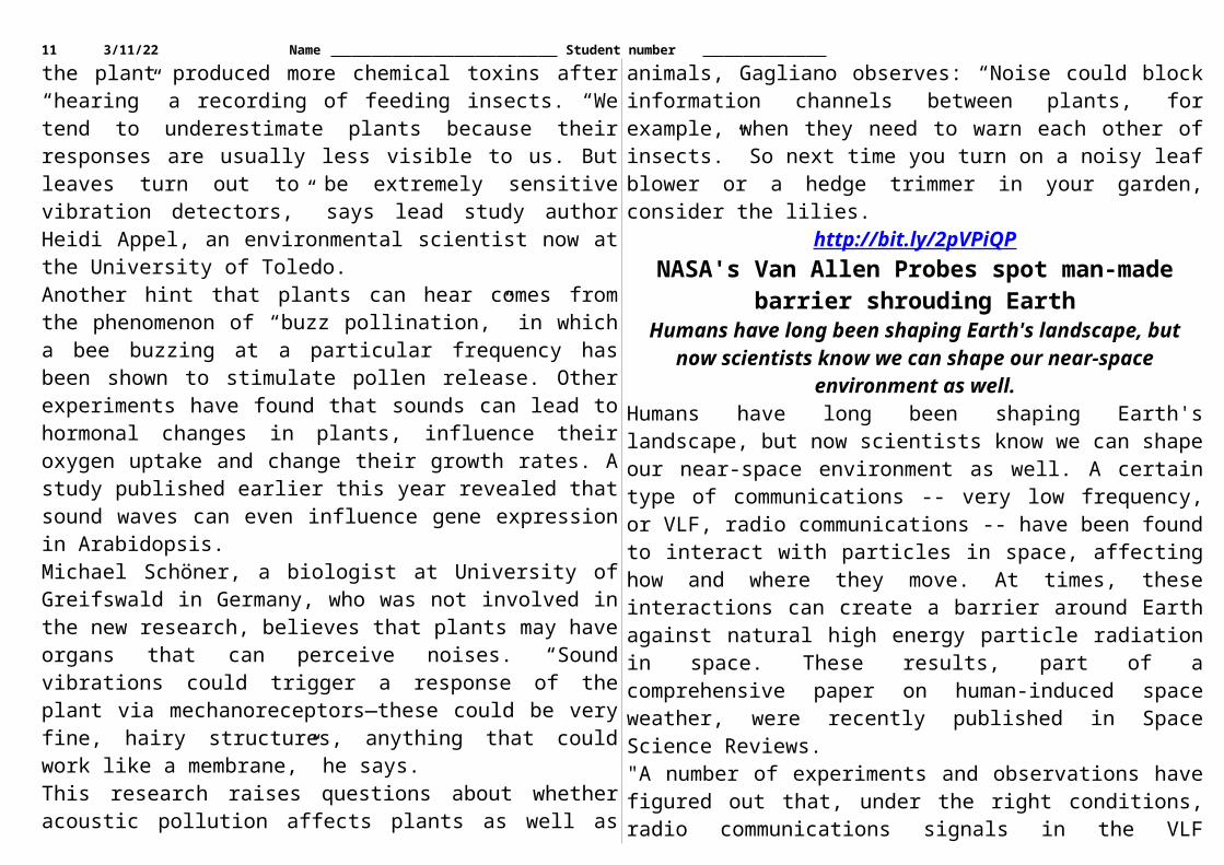
http://bit.ly/2pVPiQPNASA's Van Allen Probes spot man-made barrier
shrouding EarthHumans have long been shaping Earth's landscape, but now
scientists know we can shape our near-space environment as well.Humans have long been shaping Earth's landscape, but now scientists know we can shape our near-space environment as well. A certain type of communications -- very low frequency, or VLF, radio communications -- have been found to interact with particles in space, affecting how and where they move. At times, these interactions can create a barrier around Earth against natural high energy particle radiation in space. These results, part of a comprehensive paper on
human-induced space weather, were recently published in Space Science Reviews."A number of experiments and observations have figured out that, under the right conditions, radio communications signals in the VLF frequency range can in fact affect the properties of the high-energy radiation environment around the Earth," said Phil Erickson, assistant director at the MIT Haystack Observatory, Westford, Massachusetts.VLF signals are transmitted from ground stations at huge powers to communicate with submarines deep in the ocean. While these waves are intended for communications below the surface, they also extend out beyond our atmosphere, shrouding Earth in a VLF bubble. This bubble is even seen by spacecraft high above Earth's surface, such as NASA's Van Allen Probes, which study electrons and ions in the near-Earth environment.The probes have noticed an interesting coincidence -- the outward extent of the VLF bubble corresponds almost exactly to the inner edge of the Van Allen radiation belts, a layer of charged particles held in place by Earth's magnetic fields. Dan Baker, director of the University of Colorado's Laboratory for Atmospheric and Space Physics in Boulder, coined this lower limit the "impenetrable barrier" and speculates that if there were no human VLF transmissions, the boundary would likely stretch closer to Earth. Indeed, comparisons of the modern extent of the radiation belts from Van Allen Probe data show the inner boundary to be much farther away than its recorded position in satellite data from the 1960s, when VLF transmissions were more limited.With further study, VLF transmissions may serve as a way to remove excess radiation from the near-Earth environment. Plans are already underway to test VLF transmissions in the upper atmosphere to see if they could remove excess charged particles -- which can appear during periods of intense space weather, such as when the sun erupts with giant clouds of particles and energy.
8 5/14/23 Name Student number
http://bit.ly/2ro9x9TLarge volcanic eruption may have caused the first mass
extinctionResearchers in the U.S. and Japan say they may have found the
cause of the first mass extinction of life on Earth.There have been five mass extinctions since the divergent evolution of early animals 600 to 450 million years ago (Figure 1). Volcanic activity was the cause of both the third and fourth, while an asteroid impact led to the fifth. But triggers of the first and second mass extinctions had, until now, been unknown. The new study strongly suggests volcanic activity caused the first mass extinction.It occurred at the end of the Ordovician. This age is between the divergence of the Ordovician and land invasion of vascular land plants and animals. Animals in the Ordovician-Silurian comprised marine animals like corals, trilobites, sea scorpions, orthoceras, brachiopods, graptolite, crinoid and jawless fish. Approximately 80 percent of species disappeared at the end of the Ordovician.The researchers found Hg enrichments in sedimentary rocks deposited in North
America and southern China 445-443 million years ago. Hg enrichments are products of multiple phases of a large igneous province volcanism. This, they
say, could have led to the environmental changes that caused the disappearance of many marine animal species.
A team led by Dr. David S. Jones of Amherst College and Professor Kunio Kaiho of Tohoku University looked into possible triggers of the first mass extinction. They took sedimentary rock samples from two places—North America and southern China—and analyzed their
mercury (Hg) content. They found Hg enrichments coinciding with the mass extinction in both areas. This, they believe, is the product of large volcanic eruptions, because the Hg anomaly was also observed in other large igneous province volcanisms.Huge volcanic eruptions can produce sulfate aerosols in the stratosphere. Sulfate aerosols are strong, light-reflecting aerosols, and cause global cooling. This rapid climate change is believed to be behind the loss of marine creatures.Kaiho's team is now studying the second mass extinction in the hopes of further understanding the cause and processes behind it.
http://go.nature.com/2pVS1K2Rescue old data before it’s too late
If we don’t act soon to preserve past records, invaluable knowledge will be lost, warns Elizabeth Griffin.
In the late nineteenth century, astronomers began to photograph stars using prisms and gratings. They recorded stellar spectra — the dispersal of starlight into colours — to learn what the stars are made of. Since then, those photographic plates have become useful for another purpose: they let scientists map past concentrations of ozone in Earth’s stratosphere, and help to reveal whether some changes to the ozone hole are natural. The hardest part is getting hold of these glass plates. I know, because I spent many weeks going through collections at observatories across the world, from Germany to Australia, to search for them.What other historic data could be useful? Tales abound. The thousands of logs recorded during ship voyages in past centuries are a bonanza for studying weather patterns today. Photos of glaciers from the past and the present have startled the world, and yielded incontrovertible evidence of climate change. Medical records on dusty punch cards, abandoned in the late 1950s and decoded decades later, have helped to show how varying levels of cholesterol predict later disease.

9 5/14/23 Name Student number
To model the future, we need to be able to examine the past. But our chances to do so are fading fast, sped by misunderstanding and negligence. Few forms of ‘heritage data’ — whether stored on glass plates, paper, old tapes or floppy disks — are easily available for today’s research, so the information on them is effectively lost.Scientists used to complain that they could never obtain enough data. Today, we speak of Big Data as if it were an untameable beast. Measurements collected now are increasingly sophisticated, but they tell us only about the present. Measurements recorded long ago can show us how Earth’s weather, ecosystems and more are changing, and data taken from individuals in decades past can inform modern medical and policy guidelines. If we want those data, we need to start recovering them now.Why aren’t scientists from all domains scrambling to preserve old records, the better to study long-term trends? Part of the answer is human psychology. At one talk I gave on the need to bring astronomy’s near-lost data into lasting, easily shared formats, an audience member challenged the effort. “Modern data are so much better,” he said.He missed the point. Few want to poke around musty archives for heritage data captured using yesterday’s technology, but these provide information not available in any other form. Hydrologists in Cape Town, South Africa, have converted 70-year-old, handwritten stream data to deduce how non-native tree species affect water distribution across a landscape. High-resolution, full-colour photographs of extant birds cannot replace images of extinct passenger pigeons and laughing owls. “Treasure troves of data, and the knowledge they could offer, are left mouldering on shelves.”The time is ripe to rescue heritage data. In many cases, the original scientists are still alive to provide context. Technologies for digitizing many sorts of records are cheap and convenient.Digitization will not preserve everything. At least one epidemiologist has tracked the spread of cholera in the Iberian Peninsula by sniffing
envelopes. How? For centuries, post offices used vinegar to disinfect outgoing mail from afflicted towns, and the smell has persisted.So, what can be done? The Data Rescue Interest Group, part of the international Research Data Alliance, offers guidelines to steer a researcher through the initial stages of rescuing data, determining the equipment needed and deciding how best to tackle the rescue. The most important data capture conditions from before large-scale human changes were felt. Fields such as biodiversity (http://rebind.bgbm.org), volcanology and oceanography have made strides in preserving old data, but more needs to be done — soon, and with better coordination.We will not be able to save all data. Prioritizing means looking for the potential to illuminate questions that could not be answered otherwise. Too often, researchers dismiss heritage materials without considering what uses they might have. Treasure troves of data, and the knowledge they could offer, are left mouldering on shelves.Everyone can help. The first challenge is to locate records, photographs or other items, or simply to recognize their value. Most have not been used for yonks, and are stored in some almost-forgotten location where damp, spiders and mice are probably doing their best to destroy them.The second is to ascertain that the necessary metadata (such as date, location and limitations) are available, so that when data are converted into modern formats, they can be assigned accurately to time and place.Finding the resources for preservation is often difficult. Funding is sparse and erratic, but enthusiasts have secured grants from agencies ranging from NASA to the US Agency for International Development and the German Research Foundation. It is worth casting a wide net. University archivists can supply expertise. Citizen-science groups have also been mobilized.One overlooked resource is success stories. When researchers learn of once-neglected data that have been revived and transformed into

10 5/14/23 Name Student number
modern insight, they themselves are more likely to recognize hidden opportunities. The next heroic rescue tale could be your undertaking.But hurry. Some data are decaying as I write, some will have gone past retrieval by tomorrow, and the ageing memories needed to make them meaningful might not be available much longer.
http://bit.ly/2qBw0z8Researchers discover first human antibodies that work
against all ebolavirusesFirst natural human antibodies that can neutralize and protect animals against all three major disease-causing ebolaviruses
After analyzing the blood of a survivor of the 2013-16 Ebola outbreak, a team of scientists from academia, industry and the government has discovered the first natural human antibodies that can neutralize and protect animals against all three major disease-causing ebolaviruses. The findings, published online today in the journal Cell, could lead to the first broadly effective ebolavirus therapies and vaccines.Ebolaviruses infections are usually severe, and often fatal. There are no vaccines or treatments approved by the Food and Drug Administration for treating these viruses. Some two dozen ebolavirus outbreaks have occurred since 1976, when the first outbreak was documented in villages along the Ebola River in the Democratic Republic of Congo (formerly Zaire). The largest outbreak in history -- the 2013-16 Western African epidemic -- caused more than 11,000 deaths and infected more than 29,000 people.Monoclonal antibodies, which bind to and neutralize specific pathogens and toxins, have emerged as one of the most promising treatments for Ebola patients. A critical problem, however, is that most antibody therapies target just one specific ebolavirus. For example, the most advanced therapy -- ZMappTM, a cocktail of three monoclonal antibodies -- is specific for Ebola virus (formerly known as "Ebola Zaire"), but doesn't work against two related ebolaviruses (Sudan virus and Bundibugyo virus) that have also caused major outbreaks.
"Since it's impossible to predict which of these agents will cause the next epidemic, it would be ideal to develop a single therapy that could treat or prevent infection caused by any known ebolavirus," says study co-leader Zachary A. Bornholdt, Ph.D., director of antibody discovery at Mapp Biopharmaceutical, Inc. "Our discovery and characterization of broadly neutralizing human antibodies is an important step toward that goal," adds study co-leader, Kartik Chandran, Ph.D. , professor of microbiology & immunology at Albert Einstein College of Medicine.The study was also co-led by John M. Dye, Ph.D., chief of viral immunology at the U.S. Army Medical Research Institute of Infectious Diseases (USAMRIID).In earlier research, Dr. Bornholdt and Laura M. Walker, Ph.D., a senior scientist at Adimab, LLC, isolated 349 distinct monoclonal antibodies from a survivor of the 2013-16 Ebola epidemic. In the current study, the multi-institutional research team found that two of those 349 antibodies, known as ADI-15878 and ADI-15742, potently neutralized infection by all five known ebolaviruses in tissue culture. Both antibodies were able to protect animals (mice and ferrets) that had been exposed to a lethal dose of the three major agents: Ebola virus, Bundibugyo virus and Sudan virus.Follow-up studies showed that the two antibodies isolated from the Ebola patient work by interfering with a critical step in the process by which ebolaviruses infect cells and then multiply inside them. The two antibodies encounter the virus while it's still in the bloodstream, and bind to glycoproteins (proteins to which carbohydrate chains are attached) that project from its surface. The virus, with its hitchhiking antibodies still bound to it, then attaches to a cell and enters the lysosome -- a membrane-bound structure within the cell that is filled with enzymes for digesting foreign and cellular components. The virus must then fuse with the lysosome membrane to escape into the host cell's cytoplasm, where it can multiply. However, the antibodies prevent the virus from breaking out of its lysosomal "prison," thus stopping infection in its tracks.
11 5/14/23 Name Student number
"Knowing precisely where the antibodies attach to the glycoprotein molecules and when and how they act to neutralize ebolaviruses, we can begin to craft broadly effective immunotherapies," says Dr. Dye. "That knowledge has already allowed us to create a cocktail of monoclonal antibodies that we are testing in larger animal models for possible use in treating infected patients," adds Dr. Bornholdt.The researchers also pinpointed the human genes that are the likely source of the immune cells that produce the two antibodies. These and other findings could help speed the development of vaccines to prevent ebolavirus infection. "We'd like to synthesize vaccine immunogens [proteins that trigger antibody production] that can elicit the same types of broadly protective antibodies in people," says Dr. Chandran.The study is titled "Antibodies from a human survivor define sites of vulnerability for broad protection against ebolaviruses." Other Einstein researchers include co-first author Anna Z. Wec, M.S., Elisabeth K. Nyakatura, Ph.D., Jens Maximilian Fels, Rohit K. Jangra, Ph.D., M.V.Sc., B.V.Sc. & A.H., and Jonathan R. Lai, Ph.D. Additional contributors are co-first author Andrew S. Herbert, Ph.D., Rebekah M. James, and Russell R. Bakken, of USAMRIID, Fort Detrick, MD; Shihua He, Ph.D., Marc-Antoine de La Vega, Wenjun Zhu, Ph.D., and Xiangguo Qiu, M.D., of National Microbiology Laboratory, Public Health Agency of Canada and University of Manitoba, Manitoba, Canada; Charles D. Murin, Ph.D., Hannah L. Turner, and Andrew B. Ward, Ph.D. of The Scripps Research Institute, La Jolla, CA; Eileen Goodwin of Adimab, LLC, Lebanon, NH; and Dafna M. Abelson and Larry Zeitlin, Ph.D. of Mapp Biopharmaceutical Inc., San Diego, CA.The study was funded by grants from the National Institutes of Health (U19 AI109762), JSTO- Defense Threat Reduction Agency (CB4088 and HDTRA1-13-C-0018), and the Public Health Agency of Canada. The authors declare no financial conflicts of interest.
http://bit.ly/2pW13Xt100-year-old fertility technique reduces need for IVF
Infertile couples have a major opportunity to achieve a successful pregnancy without the need for IVF, thanks to new research into a
100-year-old medical technique.The now lesser known technique -- which involves flushing the woman's fallopian tubes with an iodised poppy seed oil -- has been proven to have significant benefits for fertility, according to the
largest study undertaken by a team involving researchers in the Netherlands and Australia.The results of the study will today be published in The New England Journal of Medicine. They will also be presented at the 13th World Congress on Endometriosis in Vancouver, Canada, by project leader Professor Ben Mol, from the University of Adelaide's Robinson Research Institute, and a member of the South Australian Health and Medical Research Institute's Healthy Mothers, Babies and Children theme.Known as the H2Oil study, the project compared the benefits of flushing the fallopian tubes with either an oil-based or water-based solution in 1119 women. With Professor Mol, this work was conducted by Dr Kim Dreyer and Dr Velja Mijatovic from the Department of Reproductive Medicine, VU University Medical Centre, Amsterdam, and a research team from 27 medical centres in the Netherlands.100-year-old techniqueThe procedure, known as hysterosalpingography (HSG), is a dye test of the fallopian tubes conducted under X-ray. The procedure was first carried out in 1917, and since the 1950s both water-based and oil-based solutions have been used."Over the past century, pregnancy rates among infertile women reportedly increased after their tubes had been flushed with either water or oil during this X-ray procedure. Until now, it has been unclear whether the type of solution used in the procedure was influencing the change in fertility," says Professor Mol, who himself was conceived after his mother underwent such a procedure."Our results have been even more exciting than we could have predicted, helping to confirm that an age-old medical technique still has an important place in modern medicine," he says.Results show clear benefits of oil-based solution

12 5/14/23 Name Student number
Almost 40% of infertile women in the oil group and 29% of infertile women in the water group achieved successful pregnancies within six months of the technique being performed.The oil-based product used in the study was Lipiodol® Ultra-Fluid, an iodised solution of fatty acids from poppy seeds. This product is currently available in 47 countries around the world."The rates of successful pregnancy were significantly higher in the oil-based group, and after only one treatment. This is an important outcome for women who would have had no other course of action other than to seek IVF treatment. It offers new hope to infertile couples," Professor Mol says.The big question: why?"It was long believed that testing a woman's fallopian tubes could have fertility benefits through 'flushing out' the kind of debris that hinders fertility. The reality is, we still don't really understand why there is a benefit, only that there is a benefit from this technique, in particular for women who don't present with any other treatable fertility symptoms," Professor Mol says."Further research would need to be conducted into the mechanisms behind what we're seeing. For now, and considering the technique has been used for 100 years without any known side-effects, we believe it is a viable treatment for infertility prior to couples seeking IVF."Not only is there a known benefit, but this flushing procedure is also a fraction of the cost of one cycle of IVF. Considering that 40% of women in the oil-based group achieved a successful pregnancy, that's 40% of couples who could avoid having to go through the huge costs and emotions associated with IVF treatment," he says.Turning around infertility - a family historyUntil he embarked on this study, Professor Mol had no idea that he himself was the result of a successful pregnancy following such a procedure.In the 1960s, after being considered infertile for nine years, Professor Mol's mother underwent an HSG which, coincidentally, also used
Lipiodol®. "It was only after I started researching this technique that my family told me what had happened," Professor Mol says."My mother went from being infertile for many years to becoming pregnant, and I was born in 1965. I also have a younger brother. So it's entirely possible - in fact, based on our team's research, it's highly likely - that my brother and I are both the result of this technique helping my mother to achieve fertility."What can infertile couples do?"The use of used Lipiodol® itself is not currently practiced widely, so the first thing couples need to do is to speak with their doctor about it," Professor Mol says."Professional bodies responsible for guidelines, funders of health care, and fertility clinics all have a role to play in assisting infertile couples to make this intervention available to couples before IVF is started," he says.This study received no financial assistance from the makers of Lipiodol®. Professor Mol's research is supported by the National Health and Medical Research Council (NHMRC).
http://dailym.ai/2q6D9nwMeningitis W: Students urged to get vaccine
Young people starting university or college this autumn are being urged to get a vaccine against meningitis.
By Smitha Mundasad Health reporterPublic Health England says the jab will help protect against meningitis W in particular - a sometimes deadly strain that is on the rise. Officials say new students are at risk as they often mix closely with groups of unfamiliar people - some who may unknowingly carry the bug.Wales has also renewed calls for school-leavers to take up the jab. The injection - known as the Men ACWY vaccine - was first introduced for new university students in the UK last year. 'Highly aggressive'It protects against the A, C, Y and W strains of the disease - all forms that can cause death or disability. But health experts say they are

13 5/14/23 Name Student number
particularly concerned about "a highly aggressive strain" of meningitis W bacteria.Some 22 people got meningitis W in 2009 in England, compared with almost 200 people in the last 12 months.Meningitis W infection is fatal in one in 10 cases and can lead to long-term health problems including deafness, epilepsy and amputations. 'Horrific side-effects'She said: "At first I thought I had the flu and felt very tired. But by the next day, I was covered in a rash, felt extremely unwell and was rushed to hospital. "I spent three weeks in intensive care on life-support. My organs failed, and my family was told I was the most unwell person in the hospital." The infection spread to her bloodstream and bones and damaged her feet. She had toes on both feet amputated, and later her left leg was also amputated. She added: "The jab was not available when I was 18. I would encourage everyone to get the vaccine who can. "It takes just five minutes, and is just one injection that can save your life or save you from getting horrific side-effects."GPs in England are inviting 17 and 18-year-olds to come for a vaccine. First-time students under the age of 25 are eligible too. People who missed out on the jab last year should also see their doctor, experts say. 'Save lives'And though students are the focus of the campaign, other young people are strongly advised to get the jab - whether they are planning on attending university or college or not. Dr Mary Ramsay, at Public Health England, said students needed to remain vigilant to signs of the disease. She added: "Protecting young people from this potentially deadly disease as they embark upon one of the most important periods of their lives is vitally important. "The vaccination will save lives and prevent lifelong devastating disability."Meningitis
Meningitis is an infection of the meninges - the membranes that surround the brain and spinal cord Meningococcal bacteria are common and carried harmlessly in the nose or throat by about one in 10 people They are passed on through close contact Symptoms can include a fever, tiredness, and general aches at first. These can get rapidly worse, with agitation, confusion, vomiting and headaches People should seek help as soon as possible and should not wait for a rash to appear before getting advice Vinny Smith, of the Meningitis Research Foundation, said: "By getting this free meningitis vaccine students are not only protecting themselves from a potentially deadly disease, but also protecting others by stopping the spread."It is also vital to watch out for friends if they are unwell. If they have meningitis it can be like a very bad hangover that quickly gets worse. It can be deadly so it is important to act fast and get medical help."Meanwhile Liz Brown, at the charity Meningitis Now, said people must not get complacent about the threat of meningitis. She added: "Up to a quarter of students carry the bacteria that can cause meningitis compared to one in 10 of the general population. "In the UK, every university could experience at least one case of meningitis amongst its students within the first term."Since 2015 the vaccine has also been rolled out for younger teenagers at schools across the UK. The ultimate aim is to ensure teenagers are offered the vaccine before they leave school. In the meantime officials in Scotland and Wales say any school-leavers who have not had the vaccine should speak to their doctor.
http://bit.ly/2qIawPjStudies link healthy brain aging to omega-3 and omega-6
fatty acids in the bloodTwo new studies link patterns of polyunsaturated fatty acids in the blood to the integrity of brain structures and cognitive abilities that
are known to decline early in aging.

14 5/14/23 Name Student number
CHAMPAIGN, Ill. -- The studies add to the evidence that dietary intake of omega-3 and omega-6 fatty acids can promote healthy aging, the researchers said. Further research is needed to test this hypothesis, they said.The brain is a collection of interconnected parts, each of which ages at its own pace. Some brain structures, and the abilities they promote, start to deteriorate before others, said University of Illinois M.D./Ph.D student Marta Zamroziewicz, who led the new research with psychology professor Aron Barbey.
New studies link specific nutrients to the structure and function of brain regions that are particularly sensitive to aging and neurodegenerative disease.
Julie McMahon"We studied a primary network of the brain -- the frontoparietal network - that plays an important role in fluid intelligence and also declines early, even in healthy aging," Zamroziewicz said. Fluid intelligence describes the ability to solve problems one has never encountered before."In a separate study, we examined the white matter structure of the fornix, a group of nerve fibers at the center of the brain that is important for memory," she said.Previous research has shown that the fornix is one of the first brain regions to be compromised in Alzheimer's disease.In both studies, the researchers looked for patterns of polyunsaturated fatty acids in the blood of adults ages 65 to 75. They analyzed the relationship between these nutrient patterns and subjects' brain structure and performance on cognitive tests. This research differs
from other such studies, which tend to focus on only one or two polyunsaturated fatty acids, Zamroziewicz said."Most of the research that looks at these fats in health and healthy aging focuses on the omega-3 fatty acids DHA and EPA, but those come from fish and fish oil, and most people in the Western Hemisphere don't eat enough of those to really see the benefits," she said. Other fatty acids, like alpha-linolenic acid and stearidonic acid, are precursors of EPA and DHA in the body. Those fats can be derived from land-based foods such as nuts, seeds and oils."A central goal of research in nutritional cognitive neuroscience is to understand how these nutrients affect brain health," Zamroziewicz said. "Some of these nutrients are thought to be more beneficial than others."In a study reported in the journal Nutritional Neuroscience, the researchers looked for relationships between several omega-3 fatty acids in the blood, the relative size of structures in the frontal and parietal cortices of the brain, and performance on tests of fluid intelligence in healthy elderly adults.The team found correlations between blood levels of three omega-3 fatty acids -- ALA, stearidonic acid and ecosatrienoic acid -- and fluid intelligence in these adults. Further analyses revealed that the size of the left frontoparietal cortex played a mediating role in this relationship. People with higher blood levels of these three nutrients tended to have larger left frontoparietal cortices, and the size of the frontoparietal cortex predicted the subjects' performance on tests of fluid intelligence."A lot of research tells us that people need to be eating fish and fish oil to get neuroprotective effects from these particular fats, but this new finding suggests that even the fats that we get from nuts, seeds and oils can also make a difference in the brain," Zamroziewicz said.In the second study, the team found that the size of the fornix was associated with a balance of omega-3 and omega-6 fatty acids in the blood, and that a more robust fornix coincided with memory

15 5/14/23 Name Student number
preservation in older adults. Again, the researchers saw that brain structure played a mediating role between the abundance and balance of nutrients in the blood and cognition (in this case, memory). The findings are reported in the journal Aging & Disease."These findings have important implications for the Western diet, which tends to be misbalanced with high amounts of omega-6 fatty acids and low amounts of omega-3 fatty acids," Zamroziewicz said."These two studies highlight the importance of investigating the effects of groups of nutrients together, rather than focusing on one at a time," Barbey said. "They suggest that different patterns of polyunsaturated fats promote specific aspects of cognition by strengthening the underlying neural circuits that are vulnerable to disease and age-related decline."Abbott Nutrition funded this work through the Center for Nutrition, Learning and Memory at the U. of I.The paper "Determinants of fluid intelligence in healthy aging: Omega-3 Polyunsaturated fatty acid status and frontoparietal cortex structure" is available online and from the U. of I. News Bureau.The paper "Predictors of memory in healthy aging: Polyunsaturated fatty acid balance and fornix white matter integrity" is available online " and from the U. of I. News Bureau.
http://bit.ly/2q3MEEMBrain stent to let five paralysed people control
exoskeletonMIND CONTROL without the side effects. That’s the aim of a
device that could help people control robotic limbs using thought alone – without the need for brain surgery.
By Alice KleinThe device will be trialled in people with paralysis next year.Several groups are developing brain-machine interfaces that allow people who are paralysed to operate a bionic exoskeleton just by thinking about it. These devices decode electrical brain signals and translate them into movement of robotic limbs.Usually, brain signals are detected via electrodes attached to the scalp or implanted directly in the brain. Placing them on the scalp avoids surgery, but the signals are muffled by the skull. Direct implantation
allows precise recordings but the electrodes can stop working because the brain treats them as foreign bodies and wraps them in scar tissue.Now, a research team led by Thomas Oxley at the University of Melbourne has developed a way of implanting electrodes in the brain without opening up the skull. Their electrodes are attached to a metallic mesh tube that is guided through a small incision in the jugular vein in the neck and up into a blood vessel in the brain. There, the electrode can measure signals from nearby brain cells on the other side of the vessel wall.The technique is borrowed from cardiologists, who slide similar tubes called stents into arteries to keep them open.The electrode-studded stent – or “stentrode” – was tested in the brains of live sheep in 2016 (Nature Biotechnology, doi.org/f8dczh). Like a cardiac stent, it sat in the blood vessel without causing any adverse effects. Because the metallic mesh does not directly touch brain tissue, no inflammation or scarring occurred over the six-month trial. “The brain doesn’t even know it’s there,” says David Grayden at the University of Melbourne, who oversaw the engineering of the device.The matchstick-sized stentrode was able to clearly detect electrical brain signals. “The recordings are not quite as detailed as those from directly implanted electrodes, but they’re close,” says Grayden.The team is now planning a clinical trial at the Royal Melbourne Hospital that will start next year. Up to five patients with no use of their arms or legs due to spinal cord injury, stroke, motor neurone disease or muscular dystrophy will be involved.
The “invisible” stentrode University of MelbourneThe stentrode will be inserted into a blood vessel that runs along the motor cortex, the part of the brain that controls movement. Fine wires

16 5/14/23 Name Student number
will run from the electrodes down through the blood system into a recording device implanted in the chest. This device will then wirelessly transmit the information to an external computer.“The end goal is that the person will be able to think about moving and an exoskeleton will obey”By asking participants to think about a particular action, like “move right fist”, the computer will learn to recognise the exact pattern of brain signals corresponding to each thought.“The end goal is that the person will be able to think about moving and an exoskeleton will obey,” says Grayden.The research is exciting, but it’s still unclear whether the stentrode will be able to pick up meaningful signals in the brains of humans, says Nick Ramsey at University Medical Center Utrecht in the Netherlands. “The stentrode research is worth doing, but I would not dismiss the technologies that are already much further ahead.”
http://bit.ly/2roz4jnLab-Grown Blood Stem Cells Produced at Last
Two research teams cook up recipe to make long-sought cells in mice and people
By Amy Maxmen, Nature magazine on May 18, 2017After 20 years of trying, scientists have transformed mature cells into primordial blood cells that regenerate themselves and the components of blood. The work, described today in Nature, offers hope to people with leukaemia and other blood disorders who need bone-marrow transplants but can’t find a compatible donor. If the findings translate into the clinic, these patients could receive lab-grown versions of their own healthy cells.One team, led by stem-cell biologist George Daley of Boston Children’s Hospital in Massachusetts, created human cells that act like blood stem cells, although they are not identical to those found in nature. A second team, led by stem-cell biologist Shahin Rafii of Weill Cornell Medical College in New York City, turned mature cells from mice into fully fledged blood stem cells.
“For many years, people have figured out parts of this recipe, but they’ve never quite gotten there,” says Mick Bhatia, a stem-cell researcher at McMaster University in Hamilton, Canada, who was not involved with either study. “This is the first time researchers have checked all the boxes and made blood stem cells.”Daley’s team chose skin cells and other cells taken from adults as their starting material. Using a standard method, they reprogrammed the cells into induced pluripotent stem (iPS) cells, which are capable of producing many other cell types. Until now, however, iPS cells have not been morphed into cells that create blood.The next step was the novel one: Daley and his colleagues inserted seven transcription factors—genes that control other genes—into the genomes of the iPS cells. Then they injected these modified human cells into mice to develop. Twelve weeks later, the iPS cells had transformed into progenitor cells capable of making the range of cells found in human blood, including immune cells. The progenitor cells are “tantalizingly close” to naturally occurring ‘haemopoetic’ blood stem cells, says Daley. Bhatia agrees. “It’s pretty convincing that George has figured out how to cook up human haemopoetic stem cells,” he says. “That is the holy grail.”Bloody goodBy contrast, Rafii’s team generated true blood stem cells from mice without the intermediate step of creating iPS cells. The researchers began by extracting cells from the lining of blood vessels in mature mice. They then inserted four transcription factors into the genomes of these cells, and kept them in Petri dishes designed to mimic the environment inside human blood vessels. There, the cells morphed into blood stem cells and multiplied.When the researchers injected these stem cells into mice that had been treated with radiation to kill most of their blood and immune cells, the animals recovered. The stem cells regenerated the blood, including immune cells, and the mice went on to live a full life—more than 1.5 years in the lab.

17 5/14/23 Name Student number
Because he bypassed the iPS-cell stage, Rafii compares his approach to a direct aeroplane flight, and Daley’s procedure to a flight that takes a detour to the Moon before reaching its final destination. Using the most efficient method to generate stem cells matters, he adds, because every time a gene is added to a batch of cells, a large portion of the batch fails to incorporate it and must be thrown out. There is also a risk that some cells will mutate after they are modified in the lab, and could form tumours if they are implanted into people.But Daley and other researchers are confident that the method he used can be made more efficient, and less likely to spur tumour growth and other abnormalities in modified cells. One possibility is to temporarily alter gene expression in iPS cells, rather than permanently insert genes that encode transcription factors, says Jeanne Loring, a stem-cell researcher at the Scripps Research Institute in La Jolla, California. She notes that iPS cells can be generated from skin and other tissue that is easy to access, whereas Rafii’s method begins with cells that line blood vessels, which are more difficult to gather and to keep alive in the lab.Time will determine which approach succeeds. But the latest advances have buoyed the spirits of researchers who have been frustrated by their inability to generate blood stem cells from iPS cells. “A lot of people have become jaded, saying that these cells don’t exist in nature and you can’t just push them into becoming anything else,” Bhatia says. “I hoped the critics were wrong, and now I know they were.”
http://bit.ly/2rCT9icWarm-bloodedness possibly much older than previously
thoughtCharacteristic may have developed 20 million years earlier, a study
by the Universities of Cape Town and Bonn showsWarm-bloodedness in land animals could have developed in evolution much earlier than previously thought. This is shown by a recent study at the University of Bonn, which has now been published in the journal Comptes Rendus Palevol.
People who like watching lizards often get the best opportunity to do so in the morning, as they can usually be found sunbathing at this time of day. This is because they rely on an external energy supply to reach their operating temperature. However, mice and other mammals make themselves nice and cozy in a different way: they burn calories and can even keep themselves warm during a bitterly cold winter's night.Mammals are thus referred to as warm-blooded. Until now, it was thought that the "body heater" was invented in four-legged land animals around 270 million years ago. "However, our results indicate that warm-bloodedness could have been created 20 to 30 million years earlier," explains Prof. Martin Sander from the Steinmann Institute for Geology, Mineralogy and Paleontology at the University of Bonn.
Ophiacodon mirus Wikimedia CommonsBones as a thermometerFor long-extinct animals, it is naturally not possible to simply determine body temperature using a thermometer. However, warm-bloodedness leaves behind tell-tale signs in fossils. It not only means that the animal is not reliant on the ambient temperature, but also enables faster growth. "And this is shown in the structure of the bones," explains Sander.Bones are composites of protein fibers, collagen, and a biomaterial, hydroxyapatite. The more orderly the arrangement of the collagen fibers, the more stable the bone, but the more slowly it normally grows as well. The bones of mammals thus have a special structure. This allows them to grow quickly and yet remain stable. "We call this bone form fibrolamellar," says the paleontologist.Together with his PhD student Christen D. Shelton (now at the University of Cape Town), the scientist looked at humerus bones and femurs from a long-extinct land animal: the mammal predecessor Ophiacodon. This lived 300 million years ago. "Even in Ophiacodon, the bones grew as fibrolamellar bones," says Sander to summarize the analysis results. "This indicates that the animal could already have been warm-blooded".
18 5/14/23 Name Student number
Ophiacodon was up to two meters long, but otherwise resembled today's lizards -- and not without good reason: mammals and reptiles are related; they thus share a predecessor. In the family tree, Ophiacodon is very close to the place where these two branches separate.Were the first reptiles warm-blooded?However, lizards, turtles and other reptiles living today are cold-blooded. Until now, it has been assumed that this was the original form of the metabolism -- i.e. that the shared ancestor of both animal groups was cold-blooded. Warm-bloodedness would thus be a further development, which arose over the course of mammalian evolution.However, Ophiacodon appears a very short time after the division between mammals and reptiles. "This raises the question of whether its warm-bloodedness was actually a completely new development or whether even the very first land animals before the separation of both branches were warm-blooded," says Sander. That is just speculation. However, if this theory is correct, we would have to drastically correct our image: the first reptiles would then also have been warm-blooded -- and would have only discarded this type of metabolism later.Publication: Christen D. Shelton, P. Martin Sander: Long bone histology of Ophiacodon reveals the geologically earliest occurrence of fibrolamellar bone in the mammalian stem lineage; Comptes Rendus Palevol; DOI: 10.1016/j.crpv.2017.02.002
http://bit.ly/2rEPMIxA single mutation may explain why Zika exploded in the
AmericasBetween 2007 and 2016, Zika mutated to more easily board biting
mosquitoes.Beth Mole - 5/19/2017, 5:35 AM
A single mutation may explain why Zika suddenly erupted from obscurity to become the alarming re-emerging infectious disease it is today, researchers report in Nature.According to researchers from Texas and China, the mutation boosts Zika’s ability to hop into feasting mosquitoes that can then shuttle the virus to more victims. Based on archived viral strains, the mutation
popped up sometime between the virus’ low-profile outbreaks in Southeastern Asia (which took place in 2007 and 2012) and Zika’s explosive emergence in the Americas beginning in 2015.“Our data offer a potential explanation for the recent re-emergence of ZIKV [Zika virus],” the authors conclude. And, they go on, the findings suggest that co-evolution between a virus and its vector—mosquitoes, in this case—is just as important for outbreak risk as co-evolution with its hosts—us.Since Zika burst onto the scene in Brazil, researchers have been sifting through its genetics to figure out how it went from a relatively benign African virus, attracting little notice for decades, to a sudden international crisis, causing devastating birth defects. While researchers are working out all the ways the virus ravages the brains and bodies of developing babies, the authors of the Nature study wanted to figure out why the virus took off in Brazil when it did. After all, Zika was first discovered in 1947 (in Uganda’s Zika forest) and caused few noticeable outbreaks in the decades following.Itching for dataThe researchers, led by Pei-Yong Shi of the University of Texas and Gong Cheng of Beijing’s Tsinghua University, started off by comparing a Zika strain collected in 2010 with one from 2016. The 2010 strain was linked to the 2007 to 2012 outbreaks in Southeastern Asia, while the 2016 virus was linked to the strain circulating in the Americas at the time.In experiments with mosquito-bitten, Zika-infected mice, the researchers quickly noticed that the 2016 virus was far better at infecting Aedes aegypti mosquitoes (a main Zika carrier) than the 2010 virus. The 2016 virus also produced much higher levels of a protein called “nonstructural protein 1,” or NS1.NS1 is known to be critical to the virus’ spread. During an infection, virus-ridden cells secrete NS1, which then tours the body, fighting off immune responses. The researchers hypothesized that extra doses of NS1 in the blood of infected hosts helps overcome defenses in a

19 5/14/23 Name Student number
feasting mosquito. This then allows the virus to settle in for a ride to a new host. The idea held up in experiments. When the researchers knocked back NS1 levels in blood using a special antibody, the virus wasn’t as good at hitching a ride. When they added NS1 to blood, the milder 2010 virus became a more frequent flier in the biting insects.The researchers traced the boosted NS1 levels to a specific mutation in the gene that codes for the protein. The mutation—an alanine-to-valine amino acid substitution at residue 188 of the gene—was present in the explosive 2016 Zika virus but absent from its tamer 2010 relative. In cell experiments, the researchers found that this substitution mutation alone could switch NS1 levels from low to high. But they don’t know why, exactly.Nevertheless, the researchers say the mutation may explain why Zika blazed through much of the Americas in the last few years and is now threatening to storm farther north. It's not definitive, of course, and the study doesn’t rule out the possibility that other factors—genetic or otherwise—sparked the devastating re-emergence. Researchers need more data to say for sure. But certainly, the authors note, “increases in the infectivity of mosquito-borne viruses within their vectors results in high epidemic potential.” Nature, 2017. DOI: 10.1038/nature22365 (About DOIs).
http://bit.ly/2rGgRu1Study on how rats process smell may address larger issue
of experiment reproducibilitySuggests an explanation for the much written-about "replication
crisis" in some fields of science and points to better ways of designing experiments
May 18, 2017 by Carla ReiterUniversity of Chicago psychology professor Leslie Kay and her research group set out to resolve a 15-year-old scientific dispute about how rats process odors. What they found not only settles that argument, it suggests an explanation for the much written-about
"replication crisis" in some fields of science and points to better ways of designing experiments. Reproducible experimental results are part of the bedrock of scientific method. But a concern is that researchers, particularly in psychology and medicine, are too often unable to replicate the findings of colleagues in other labs.This has certainly been true of understanding how rats—and by extension, possibly humans—process smell. "There was simply a disagreement in the literature," Kay said. "Different labs tried to get the same result, and they were unsuccessful."The diverging results came from two camps, doing similar but slightly different experiments. What Kay and her group found was that while both were correct, they were asking different questions without realizing it. Their experiments were not, in fact, comparable.Kay's group's study, published this spring in Journal of Neuroscience, shows that the disparate conclusions arise from small but crucial differences in the way the two sets of experiments were set up. By eliminating those differences, and then doing both experiments rather than only one, the group was able to tease out similarities underlying the varying results and discover a general truth about how rats smell.In both kinds of experiments, rats were trained to recognize pairs of odors by sniffing them and then discriminating between the two after being asked. In the first type of experiment, if a rat smelled odor A (banana, for example), it poked its nose into a hole and got a reward. If it smelled odor B (sweaty socks), it did nothing and received no treat. In the second type of experiment there were two holes; the rat poked its nose into one if it smelled odor A, and into the other if it smelled odor B. Both earned a reward.The labs that did the one-hole experiments concluded that rats sniff deliberatively, gathering information over time. The two-hole experimenters concluded that rats just do a quick sniff and leave, getting by with whatever information they gather in that short time.

20 5/14/23 Name Student number
Researchers discussed whether the tasks made the rats respond differently in the two experiments. Did reacting to smell B in the one-hole studies make rats slower overall, or did the different number of rewards affect rats in other ways?Controlling the variablesDonald Frederick, a graduate student in Kay's lab, decided to explore those questions by conducting both kinds of experiments in an extremely controlled way, testing the rats on many different pairs of odors. The experiments were designed to be identical to the point that the rats learned to recognize the second odor and discriminate between the two odors by choosing holes."For each type of task, we got results that were comparable to what had been found before for that type of task," Kay said. When faced with the two-hole task, the rats sniffed quickly and acted quickly. When faced with the one-hole task, they took an additional sniff before acting.Previous researchers had concluded, mistakenly, that their results for a single task held true for the way rats smell in all situations. Because Kay's group looked at both tasks in experiments that were set up identically, they were able to see that the differences in experimental design between the two types of studies had an enormous effect on the outcomes. The type of rewards used, the precise way the rats were trained and how hungry they were when they did the tasks, among many other factors, all affected the results."It's a little bit overwhelming when you start to realize that everything is going to affect how the animals behave," Kay said. "But we really have to pay attention to that. Many non-replications may be due to experimental details that people think are unimportant," she said. "They aren't necessarily non-replications at all; they're doing a different experiment."Once they eliminated the "noise" created by the differences in the experimental set-ups, Kay and her colleagues were able to discern an underlying similarity in the rats' approach to the two situations.
"By doing both experiments, we found that the rats are doing the same thing, they're just doing it in a more compressed fashion for one task than the other," Kay said. "Because we employed so many different odors and did such a carefully balanced study, we were able to show that, in fact, in both tasks, they're accumulating information over time. And they extend their sampling times in both tasks when it's hard to discriminate between the odors."The important message, Kay said, is that, "it's crucial to use multiple tasks in trying to come to general conclusions. We're all searching for general truths, and we forget that we've found a specific truth. When we forget that, we stop looking for what's really the general truth. Only by using multiple ways of addressing a question within the same lab can we get at the underlying truths about cognition. More information: Donald E. Frederick et al. Task-Dependent Behavioral Dynamics Make the Case for Temporal Integration in Multiple Strategies during Odor Processing, The Journal of Neuroscience (2017). DOI: 10.1523/JNEUROSCI.1797-16.2017
http://bit.ly/2rF1QcHHow RNA formed at the origins of life
A single process for how a group of molecules called nucleotides were made on the early Earth, before life began, has been suggested
by a UCL-led team of researchers.Nucleotides are essential to all life on Earth as they form the building blocks of DNA or RNA, and understanding how they were first made is a long-standing challenge that must be resolved to elucidate the origins of life.In a study, published today in Nature Communications and funded by the Engineering and Physical Sciences Research Council, the Simons Foundation and the Origins of Life Challenge, researchers from UCL, Harvard University and Massachusetts General Hospital suggest a single chemical mechanism by which both classes of nucleotides—purines and pyrimidines—could have formed together.
21 5/14/23 Name Student number
Before now, scientists thought that the two classes of nucleotide must have been made separately and under mutually incompatible conditions. This study is the first to show that both purines and pyrimidines can be formed from a common precursor molecule that existed before life began."We provide a new perspective on how the original RNA molecules were made and suggest a simple chemical solution for delivering both purine and pyrimidine nucleotides at the origins of life," explained corresponding author, Dr Matthew Powner (UCL Chemistry).
This is a computer graphic of an RNA molecule. Richard Feldmann/Wikipedia"RNA is the corner stone of all life on Earth and probably carried the first information at the outset of life, but making RNA requires both purine and pyrimidine nucleotides to be simultaneously available. A solution to this problem has remained elusive for more than 50 years."The team demonstrated how purines and pyrimidine nucleotides can both be assembled on the same sugar scaffold to form molecules called ribonucleotides which are used to construct RNA.Purine and pyrimidine nucleotides are used to create the DNA and RNA. The purine and pyrimidine nucleotides bind to one another through specific molecular interactions that provide a mechanism to copy and transfer information at the molecular level, which is essential for genetics, replication and evolution. Therefore understanding the origins of nucleotides is thought to be key to understanding the origins of life itself.The team discovered that molecules, called 8-oxo-adenosine and 8-oxo-inosine, which are purine ribonucleotides, can be formed under the same chemical conditions as the natural pyrimidine
ribonucleotides. They also found that one chemical precursor can divergently yield both purine and pyrimidine ribonucleotides."The mechanism we've reported gives both classes of molecule the same stereochemistry that is universally found in the sugar scaffold of biological nucleic acids, suggesting that 8-oxo-purine ribonucleotides may have played a key role in primordial nucleic acids," said Dr Shaun Stairs (UCL Chemistry), first author of the study.The team now plans to further investigate mechanisms that use 8-oxo-purines to transfer information, which could help scientists better understand life's first informational transfer systems. More information: 'Divergent prebiotic synthesis of pyrimidine and 8-oxo-purine ribonucleotides', Nature Communications (2017). DOI: 10.1038/NCOMMS15270
http://bit.ly/2q62uyFTo curb medical errors, physicians must be better trained
to admit mistakesPenn researchers call for use of social psychology methods to
transform culture of medical error disclosurePHILADELPHIA -- Medical errors are a leading cause of death in the United States, with some research suggesting that errors can cause as many 250,000 fatalities each year. The medical community has made strides to normalize and encourage error disclosure for physicians and medical trainees in order to improve patient safety and health care outcomes, but these guidelines fall short when it comes to addressing the social psychology that influences how and when physicians and medical trainees disclose errors and how they manage the consequences of those errors. In a paper published this month in Medical Education authors Neha Vapiwala, MD, an associate professor of Radiation Oncology and vice chair of Education in the Perelman School of Medicine at the University of Pennsylvania and Jason Han, a fourth-year student in the Perelman School of Medicine, call for better education and training focused on the psychological challenges that coincide with errors and

22 5/14/23 Name Student number
error disclosure in order to improve outcomes and reduce the number and severity of medical errors."We must transform the culture of error disclosure in the medical community from one that is often punitive to one that is restorative and supportive," Vapiwala said. "And to do that, we must tend to the psychological challenges that medical professionals wrestle with when they face the possibility of disclosing an error." Initiatives such as the Disclosure, Apology, and Offer model have helped make moderate gains in creating a culture of transparency in health systems, but these efforts primarily focus on the legal and financial aspects of error disclosure and do not address other barriers, such as the fear, shame, and guilt that come with error disclosure."Arguably, these psychological factors are harder to overcome, especially in this modern age of social media where health care providers can be reviewed and scrutinized in very public forums," Vapiwala said. "There is real concern that any little slip-up can live on the internet for the rest of someone's career." The authors identified two main cognitive biases that often hinder error disclosure: Fundamental Attribution Error (FAE), which is the tendency overestimate one's own role in a situation, and Forecasting Error (FE), the tendency to overestimate impact and duration of negative consequences while underestimating the ability to recover from those circumstances.For example, if an error led to a patient injury, the physician might initially overstate his own role in that error rather than examine any systematic reasons for why that error occurred. Secondly, he may then also overestimate the long-term consequences or recovery time for that patient, leading to feelings of both self-blame and exaggerated doom, both of which damage the physician-patient relationship and may impede a care provider from reporting the error. "Overcoming these biases is akin to suppressing a reflex. It requires self-awareness, practice, and most importantly, education and training," Vapiwala said.
Looking at other fields that have high-stake consequences when an error occurs, such as the airline industry, the authors offer several strategies to overcome these patterns of thought, utilizing elements of social psychology to transform the current culture of error disclosure. Recommendations include incorporating standardized patients (SPs), actors who simulate patients, not only to "practice" difficult patient encounters, but also to help model interactions with family members, peers, and administrators in order to teach various behavior and coping mechanisms. SPs have been proven to effectively mimic the psychosocial elements of error disclosure, including profound guilt, feelings of ineptitude, and fear of repercussions. Virtual reality (VR) is another tool that can offer immersive and realistic technology to supplement traditional curricula, while also offering tremendous scalability at a lower cost than SPs. The authors cite an example of a recent VR exercise which allowed viewers to experience the perspective of a 12-year Syrian refugee to incite more compassion and understanding. While VR medical content doesn't currently exist, it is on the horizon for many medical trainees and professions. However, both SP and VR do have limitations, as the users ultimately know that the scenario is simulated. "Standardized patients and other simulated scenarios provide an excellent foundation, but until you are put into a real-world situation and forced to confront your mistake and its potential consequences, you can't truly understand the psychosocial challenges," Han said. Finally, the authors recommend implementing a professional standard for trainees, including a formal evaluation of the skills needed to disclose and cope with medical errors. This standard would further normalize error disclosure and make it a common practice among physicians and trainees. The authors conclude that the primary change will need to be cultural, not just among trainees, but at every level of medical practice, in order
23 5/14/23 Name Student number
to successfully pivot away from the current stigma related to error disclosure. "Administrators must make a shift from asking 'who is at fault' to asking 'why' and 'how' did a situation occur, creating a culture that embraces error disclosure and seeks to solve the many systematic factors that lead to an error in the first place. This approach will not only normalize error disclosures but also help us better understand why they happen so we can prevent more of them in the future," Vapiwala said.
http://bit.ly/2rGflblUnderstanding the architecture of our 'second brain'Important step in understanding the organisation of nerve cells
embedded within the gutScientists have made an important step in understanding the organisation of nerve cells embedded within the gut that control its function -- a discovery that could give insight into the origin of common gastrointestinal diseases, including irritable bowel syndrome and chronic constipation.The findings, published in Science, reveal how the enteric nervous system -- a chaotic network of half a billion nerve cells and many more supporting cells inside the gut wall -- is formed during mouse development.
Nerve cells (yellow) from the same parent cell are organized in 3-D columns that extend through the layers of the gut wall. Reena Lasrado
The research was led by the Francis Crick Institute, in collaboration with the University of Leuven, Stanford University, the Hubrecht Institute and the Quadram Institute Bioscience. The work was funded by the Francis Crick Institute, the Medical Research Council and the UK Biotechnology and Biological Sciences Research Council.
Often known as the 'second brain' for its vast number of neurons and complex connectivity, the enteric nervous system has a crucial role in maintaining a healthy gut. Therefore, understanding how this neural mosaic is organised could help scientists find treatments for common gastrointestinal disorders."The gut wall is home to many types of nerve cells which appear to be distributed randomly," says Vassilis Pachnis, Group Leader at the Francis Crick Institute. "But despite this chaos, the neural networks of the gut are responsible for well organised and stereotypic functions such as production of stomach acid, movement of food along the gut, communication with immune cells and bacteria, and relay of information to the brain. We wanted to find out how organised activity emerges from such a chaotic system."During development, a unique and dynamic population of cells known as progenitor cells divide to produce copies of themselves, which can then generate many other types of cells. Using genetic tools, the team labelled individual progenitor cells of the enteric nervous system with unique colours and followed their descendants -- also marked with the same colour -- through development and into the adult animal. By examining the type of cells produced by single progenitors, they could understand their properties.They found that some progenitors only produced nerve cells, others only produced nerve-supporting cells called glia, and some produced both. Neurons and glia originating from the same parent stayed close to each other, forming relatively tight groups of cells. Cell groups that descended from different but neighbouring parent cells overlapped like a Venn diagram that could be viewed on the gut surface. Interestingly, this close relationship was maintained by the descendants of single progenitors down through all layers of the gut wall thereby forming overlapping columns of cells."We uncovered a set of rules that control the organisation of the 'second brain' not just along a single gut layer but across the 3D space

24 5/14/23 Name Student number
of the gut wall," says Reena Lasrado, first author of the paper and researcher in Vassilis's lab at the Crick.The team explored whether this intricate structure of the enteric nervous system also contributes to nerve cell activity in the gut."A subtle electrical stimulation to the enteric nervous system showed that nerve cells generated by the same parent cell responded in synchrony," says Vassilis. "This suggests that developmental relationships between cells of the enteric nervous system of mammals are fundamental for the neural regulation of gut function.""Now that we have a better understanding of how the enteric nervous system is built and works, we can start to look at what happens when things go wrong particularly during the critical stages of embryo development or early life. Perhaps mistakes in the blueprint used to build the neural networks of the gut are the basis of common gastrointestinal problems."
http://bit.ly/2q99z0PMussel gloop can be used to make wounds knit without
any scarsThe humble mussel could soon help us prevent scarring.
By Alice KleinA sticky substance naturally secreted by the marine animal is one element of a glue that closes skin wounds seamlessly in rats. The glue could be used to prevent unsightly scars after accidental cuts or surgical operations.“If this can be replicated in humans, it might be the next big thing for scar therapy,” says Allison Cowin at the University of South Australia, who wasn’t involved in the study.Scars form when the collagen scaffolding in skin is broken apart. Instead of re-forming in their original and neat basket-weave arrangement, the collagen fibres grow back in parallel bundles that create the characteristic lumpy appearance of scars.One way to reduce scarring is to apply decorin, a skin protein involved in collagen organisation. But because decorin has a highly
complex physical structure it is hard to synthesise and therefore not used in the clinic.Sticky substanceTo get round this problem, Hyung Joon Cha at Pohang University of Science and Technology in South Korea and his colleagues have created a simplified version of decorin. They combined a small section of the decorin protein with a collagen-binding molecule and a sticky substance secreted by mussels.The resulting glue was tested on rats with deep, 8-millimetre-wide wounds. The glue was spread over each wound and covered with clear plastic film. Rats in a control group had their wounds covered in plastic without any glue.By day 11, 99 per cent of the wound was closed in the treated rats compared with 78 per cent in the control group. By day 28, treated rats had fully recovered and had virtually no visible scarring. In comparison, control rats had thick, purple scars (see images below).
The wounds that received the new treatment (bottom) showed less scarring than those that didn’t (top)Jeon EY, Choi B-H, Jung D, Hwang BH, Cha HJ.
Closer inspection under the microscope confirmed that collagen fibres in the treated wounds had returned to their original basket-weave

25 5/14/23 Name Student number
arrangement. The new skin had also developed hair follicles, blood vessels, oil glands and other structures that aren’t regenerated in scars.The glue is able to promote normal collagen growth because negative charges on the decorin fragments hold the fibres apart, says Cha. In doing so, the fibres are more easily able to weave in and out between each other instead of sticking together randomly.Cowin says the results are impressive but there is still a way to go before the results can be translated to humans. “Rats have loose skin, whereas we have tight skin, and they tend to heal better and have less scarring than we do,” she says. As a result, the glue may not be as effective in people as in rats.Cha says that the glue will now be tested in pigs, whose skin better resembles our own.Adding to the toolboxNew scar treatments are greatly needed because the existing ones don’t work very well, says Cowin. Silicone gels, steroids, pressure bandages, cryotherapy and laser treatments are often used to reduce the appearance of scars, but they cannot erase them completely.Cowin is developing a scar treatment that uses monoclonal antibodies to block a type of protein that impairs wound healing. Other groups are applying embryonic stem cells to wounds, based on the observation that skin abrasions in embryos and early fetuses don’t scar. These approaches are still being tested in animals.Journal reference: Biomaterials, DOI: 10.1016/j.biomaterials.2017.04.041
http://bit.ly/2qE8BNqItaly law obliges parents to vaccinate
Italy's cabinet has approved a law obliging parents to vaccinate their children against infectious diseases as politicians spar over a spike
in measles cases.Isla Binnie, Reuters
Children up to six years old will now need to be immunised to be eligible for nursery school, and parents who send their children to
school after that age without vaccinating them first will be liable for fines.Vaccines against measles, mumps, rubella, chicken pox and meningitis, which were previously only recommended, will now become mandatory, Prime Minister Paolo Gentiloni said."The lack of appropriate measures over the years and the spread of anti-scientific theories, especially in recent months, has brought about a reduction in protection," Gentiloni told a news conference in Rome.The law will also oblige inoculation against polio, diphtheria, tetanus, hepatitis B, whooping cough, and haemophilus influenzae type B.Italy's Higher Health Institute warned in April that a fall-off in vaccinations had led to a measles epidemic. The United States warned visitors to Italy about exposure to the potentially fatal disease.The institute has recorded some 2395 measles cases so far this year compared with some 840 in all of 2016 and 250 in 2015.The website of the European Commission, the European Union's executive, says it encourages all member states to "ensure that as many children as possible receive the main childhood vaccines".Gentiloni's centre-left government has accused the anti-establishment 5-Star Movement of sowing fear among parents by questioning the safety of some vaccines and the scruples of multinational pharmaceutical firms.5-Star members, who run Rome's city hall, abstained on Thursday from a vote on obliging schoolchildren in the capital to be vaccinated, stoking fresh controversy over their stance."5-Star is riding the wave of disinformation, nourishing fear and favouring a position that is anti-science and dangerous for the whole community," said Federico Gelli, a deputy from the ruling Democratic Party.Paola Ferrara, 5-Star's leader in city hall, said the party had abstained because of the pending vote in parliament, and considered vaccinations "essential".

26 5/14/23 Name Student number
http://bit.ly/2rFYqpdNew hope for patients with severe lung disease
'Game-changing' clinical trial could lead to transformation of lives of patients suffering from severe lung disease
Patients suffering from severe lung disease could see their lives transformed thanks to a 'game-changing' clinical trial carried out by UK experts and led by the team from the Lane Fox Respiratory Service based at Guy's and St Thomas' in London.The HOT-HMV trial (Home Oxygen Therapy-Home Mechanical Ventilation), which involved giving selected patients a breathing machine to be used in their home in addition to oxygen therapy, was found to reduce readmissions to hospital following an acute infection.The results of the trial, which have been published in the Journal of the American Medical Association (JAMA), were announced today at the American Thoracic Society's prestigious annual conference in Washington, DC.In the UK alone, approximately 30,000 people die from Chronic Obstructive Pulmonary Disease (COPD) every year. The World Health Organisation (WHO) estimates that COPD will be the world's third largest killer disease by 2020. Chronic Obstructive Pulmonary Disease (COPD) is an umbrella term used to describe progressive lung diseases including emphysema, chronic bronchitis, refractory (non-reversible) asthma, and some forms of bronchiectasis. This disease is characterized by increasing breathlessness.Respiratory experts Professor Nicholas Hart and Dr Patrick Murphy, who co-ordinated the UK-wide trial from St Thomas' Hospital, said the trial results could pave the way for a complete change in the way that the most severely affected COPD patients across the world are treated.Professor Hart explains: "The only current treatment we have to give these patients is oxygen therapy, but now we can give them oxygen as well as a ventilator in their home. We have managed to reduce the likelihood of readmission to hospital by almost 50%.
"In the trial we used a home ventilator that co-ordinates itself with the individual patient's breathing. The mask ventilator machine works by blowing in air and oxygen to keep oxygen levels high and carbon dioxide, the waste gas, low." The trial was carried out thanks to funding and equipment from manufacturers Philips Respironics and ResMed, and Guy's and St Thomas' Charity.COPD, or Chronic Obstructive Pulmonary Disease, is one of the world's biggest killers, but the addition of a home ventilator to oxygen treatment reduces admissions to hospital as well as maintaining quality of life.Ronnie Ward, 74, from Brighton, has suffered from COPD for five years and uses his home ventilator every night, to support his breathing. Since being recruited for the trial, he and his wife Julie, 55, have had to make far fewer trips to hospital."Ronnie was in and out of hospital, sometimes spending weeks and months on the wards. Coming back and forth and spending so much time in hospital was stressful and very demoralising," she says."We were finding that just weeks after he'd been discharged from hospital, Ronnie would need to be readmitted because he was struggling to breathe again. Using the breathing machine every night has taken a lot of pressure off us."Meanwhile, the trial follow up will continue, as patients are monitored for survival rates over the next three and five years."These results are extremely promising but the work will continue. So far we have found that patients using home oxygen with a home ventilator device are two-thirds less likely to be readmitted within 28 days," says Professor Hart."This is very important because not only does it maintain a patient's quality of life but also it has the potential to significantly increase our ability to care for these patients without the need for a hospital stay. At Guy's and St Thomas' around 1,000 patients are admitted each year with COPD. If we can keep them comfortable at home for longer, this will have a big impact."

27 5/14/23 Name Student number
http://bit.ly/2qGE95vFirst-line antibiotics can fail almost a quarter of
pneumonia patientsNew research on the effectiveness of antibiotics for pneumonia
patients suggests doctors may need to rethink their prescriptions, writes Andrew Masterson.
Nearly a quarter of Americans with community-acquired pneumonia – as opposed to hospital-acquired pneumonia – do not respond to first-line antibiotics, a disturbing new study has found.The finding – uncovered by a team led by James McKinnell of LA BioMed in California – is cause for concern because pneumonia is the leading cause of death arising from infectious disease in the US.McKinnell and colleagues examined the case histories of 251,947 adult patients who received outpatient treatment for pneumonia between 2011 and 2015.Combing the records, they looked for cases that required a repeat antibiotic prescription, a change of antibiotic, a visit to an emergency room, or hospitalisation, within 30 days of diagnosis.They found that a total of 22.1% of patients met the criteria for failing to respond to initial treatment. In patients treated with certain types of antibiotics – notably a class called beta-lactams – the failure rate actually topped 25%. Patients over the age of 65 were heavily represented in the failure figures.“The additional antibiotic therapy noted in the study increases the risk of antibiotic resistance and complications which are difficult to treat and may be life-threatening, especially for older adults,” comments McKinnell. The study, presented at a recent conference and published in the American Journal of Respiratory and Critical Care Medicine, concludes that doctors should change their approach to treating at-risk pneumonia patients in the community.“Our findings suggest that the community-acquired pneumonia treatment guidelines should be updated with more robust data on risk factors for clinical failure,” says McKinnell.