, nam young kim , jung-shik bae , hyun sook lee , eun ji kimsu-min lim1, jae in jung1, nam young...
TRANSCRIPT

Food and Nutrition Sciences, 2019, 10, 128-147 http://www.scirp.org/journal/fns
ISSN Online: 2157-9458 ISSN Print: 2157-944X
DOI: 10.4236/fns.2019.102011 Feb. 2, 2019 128 Food and Nutrition Sciences
Cyanidine-3-O-Galactoside Enriched Aronia melanocarpa Extract Inhibits Adipogenesis and Lipogenesis via Down-Regulation of Adipogenic Transcription Factors and Their Target Genes in 3T3-L1 Cells
Su-Min Lim1, Jae In Jung1, Nam Young Kim2, Jung-Shik Bae2, Hyun Sook Lee3, Eun Ji Kim1*
1Center for Efficacy Assessment and Development of Functional Foods and Drugs, Hallym University, Chuncheon, Republic of Korea 2R&D Center, Wellfine Co., Ltd., Chuncheon, Republic of Korea 3Department of Food Science & Nutrition, Dongseo University, Busan, Republic of Korea
Abstract Aronia melamocarpa (AM) is a rich source of anthocyanins, which are known to help prevent obesity. The cyanidine-3-O-galactoside enriched AM extract (AM-Ex) containing more cyanidine-3-O-galactoside than conven-tional AM extract was recently developed. The objective of this study was to examine the effect of AM-Ex on adipogenesis and its action mechanisms in vitro using 3T3-L1 adipocytes. To examine the anti-obesity effect of AM-Ex, 3T3-L1 cells were induced adipocyte differentiation and incubated with vari-ous concentration of AM-Ex. Lipid accumulation, cellular triglyceride con-tent, mRNA expression of transcription factors and adipogenic genes were analyzed. Treatment with 100 - 400 μg/mL of AM-Ex resulted in a dose-de- pendent decrease in adipocyte differentiation and triglyceride accumulation. mRNA expression of adipogenic transcription factors, such as peroxisome proliferator-activated receptor gamma, CCAAT/enhancer binding protein α, sterol regulatory element-binding protein 1 were decreased. The level of gene expression of adipogenesis and lipogenesis-related genes, such as adipocyte protein 2, lipoprotein lipase, acetyl-CoA carboxylase, ATP-citrate lyase and fatty acid synthase were decreased. These results suggest that AM-Ex alle-viated risk factors related to obesity by modulating multiple pathways asso-ciated with adipogenesis.
How to cite this paper: Lim, S.-M., Jung, J.I., Kim, N.Y., Bae, J.-S., Lee, H.S. and Kim, E.J. (2019) Cyanidine-3-O-Galactoside En- riched Aronia melanocarpa Extract Inhibits Adipogenesis and Lipogenesis via Down- Regulation of Adipogenic Transcription Factors and Their Target Genes in 3T3-L1 Cells. Food and Nutrition Sciences, 10, 128-147. https://doi.org/10.4236/fns.2019.102011 Received: January 3, 2019 Accepted: January 30, 2019 Published: February 2, 2019 Copyright © 2019 by author(s) and Scientific Research Publishing Inc. This work is licensed under the Creative Commons Attribution International License (CC BY 4.0). http://creativecommons.org/licenses/by/4.0/
Open Access

S.-M. Lim et al.
DOI: 10.4236/fns.2019.102011 129 Food and Nutrition Sciences
Keywords Obesity, Adipocyte, Adipogenesis, Lipogenesis, Transcription Factor, Adipocyte Protein 2
1. Introduction
The prevalence of obesity has increased dramatically worldwide and has become a major global health problem. Obesity is related to increased mortality and morbidity in a number of chronic diseases, such as metabolic syndrome and vascular disease [1] [2]. Therefore, it is important to control obesity to prevent various chronic diseases and improve health.
Various drugs have been developed and applied to treat obesity through regu-lating appetite and controlling fat absorption and oxidation. However, these an-ti-obesity drugs have been reported to cause negative side effects and rebound weight gain when the medication was ceased [3] [4]. Thus, the need exists for functional foods or drugs with high efficacy and no negative side effects. Re-cently explored substances which may potentially cure and prevent obesity in-clude numerous foods and plant-derived bioactive compounds, such as catechin [5] [6] and hydroxycitric acid [7] [8].
Aronia melamocarpa (AM), known as aronia or chokeberry, belongs to the Rosaceae family, and originated in North America. Its red-purple fruits are fre-quently made into juice or jams. Also, it has traditionally been used in Russia and some Eastern European countries to treat chronic diseases [9]. Currently, AM has attracted a lot of research attention because of its high phytochemical content, compounds known to be beneficial to health. AM contains various phenolic compounds, including anthocyanins, phenolic acids, and flavonoids [10]. In vitro and in vivo studies suggested that the phenolic compounds con-tained in AM possess a wide range of beneficial health functions, including an-tioxidant [11] [12], anti-hyperlipidemic [13] [14], anti-diabetic [15] [16], hepa-toprotective [17], cardioprotective [18] [19] [20], and gastroprotective [21] ac-tivities. Recent animal studies demonstrated that AM reduced diet-induced ob-esity [22] [23]. Kim et al. [24] reported that the content of cyanidin-3-O-galacto- side (C-3-Gal), a major anthocycnin in AM, is present in different amounts in AM extracts depending on the extraction method, and that the greater the C-3-Gal content in the extract, the greater the anti-obesity effect. However, in-formation regarding the anti-obesity effect of AM is limited, and the molecular mechanism underlying these effects has yet to be elucidated.
Adipogenesis is constituted by a set of processes, which include preadipocyte proliferation, differentiation, and fatty acid synthesis, and is regulated by various molecular factors. Adipogenesis is related to both the occurrence and develop-ment of obesity [25]. To understand whether and how AM exhibits anti-obesity effect, we investigated the anti-adipogenic effect of AM.

S.-M. Lim et al.
DOI: 10.4236/fns.2019.102011 130 Food and Nutrition Sciences
Recently, we developed a new kind of C-3-Gal enriched AM extract (AM-Ex), containing more C-3-Gals than conventional AM extract. In the present study, we investigated the effect of AM-Ex on adipogenesis and its action mechanisms in vitro using 3T3-L1 adipocytes in order to elucidte the anti-obesity of AM-Ex.
2. Materials and Methods 2.1. Materials
The materials used in this study were purchased from the indicated suppliers: Dulbecco’s Modified Eagle’s Medium (DMEM) and other miscellaneous cell culture reagents from Welgene (Daegu, Korea); fetal bovine serum (FBS) and bovine calf serum (BCS) Gibco-Thermo Fisher Scientific Inc. (Waltham, MA, USA); 3-(4,5-dimethylthiazol-2-yl)-2,5-diphenyl tetrazolium bromide (MTT), 3-isobutyl-1-methylxanthine, dexamethasone, insulin, and Oil red O from Sig-ma-Aldrich Co. (St. Louis, MO, USA); and cyanidine-3-O-galactoside (C-3-Gal) from Polyphenols AS (Sandnes, Norway). Unless noted otherwise, all other ma-terials were purchased from Sigma-Aldrich Co.
2.2. Preparation of Cyanidine-3-O-Galactoside Enriched Aronia Melanocarpa Nero Extract (AM-Ex)
The freeze-dried powder of Aronia melanocarpa Nero fruits harvested from Go-seong (Korea) was purchased from Goseong Happy Aronia Farm (Goseong, Korea). The dried fruit powder was extracted with 70% ethanol by adding 100 g of the dried powder to 2 L of 70% ethanol using a high pressure homogenizer (Micronox, Seongnam, Korea) at room temperature. The intermediate extract was additionally extracted under reduced pressure using a rotary evaporator at 40 Mpa pressure at 30˚C for 2 h. The extract was concentrated with a rotary evaporator, and lyophilized for 72 h in a lyophilizer (IlshinBioBase, Dong-ducheon, Korea). The resulting powder was used as Aronia melanocarpa Nero extract (AM-Ex) and stored at −20˚C until further use. The control 70% ethanol extract (C-AM-Ex) was extracted for 24 h at 80˚C from 100 g of the dried fruit powder placed into 2 L of 70% ethanol. This C-AM-Ex was concentrated with a rotary evaporator, and lyophilized for 72 h in a lyophilizer (IlshinBioBase).
2.3. High-Performance Liquid Chromatography (HPLC) Analysis
Both AM-Ex and C-AM-Ex were analyzed using HPLC (Ultimate 3000, Thermo Fisher Scientific, Waltham, MA, USA) with Jupiter 5 μ C18 columns (150 × 4.6 mm, 3000 A, Phenomenex, Torrance, CA, USA) at 520 nm, and the operating conditions were as follows. The mobile phase solvents used were (A) HCOOH- H2O (1:9) and (B) HCOOH-MeOH-H2O (1:5:4), with (A) 100% in the 0 - 2 min period, with (A) 30% and (B) 70% in the 2 - 20 min period, with (B) 100% in the 20 - 22 min period, and with (A) 100% in the 22 - 24 min period. Before per-forming HPLC, the samples were filter using 0.45 μm filter, and the flow velocity was set to 0.8 mL/min.

S.-M. Lim et al.
DOI: 10.4236/fns.2019.102011 131 Food and Nutrition Sciences
2.4. Cell Culture and Adipocyte Differentiation Induction
Mouse 3T3-L1 preadipocytes were purchased from the Korean Cell Line Bank (Seoul, Korea). 3T3-L1 cells were cultured in growth medium (GM; DMEM supplemented with 100 mL/L BCS, 100,000 U/L penicillin, and 100 mg/L strep-tomycin) at 37˚C in a humidified atmosphere of 5% CO2 and 95% air. To induce adipocyte differentiation, at 2 days post-confluence (referred to as day 0), 3T3-L1 cells were exposed to differentiation medium (DM; DMEM with 100 mL/L FBS, containing 0.5 mM 3-isobutyl-1-methylxanthine, 1 μM dexametha-sone, and 5 μg/mL insulin) for 2 days. On day 2, the medium was replaced with DM (DMEM with 100 mL/L FBS, containing 5 μg/mL insulin only). On day 4, the medium was replaced with DMEM supplemented with 100 mL/L FBS and cells were incubated for the next 4 - 8 days to fully differentiate. To examine the effects of AM-Ex on adipocyte differentiation, the cells were incubated in D-M in the presence or absence of various concentration of AM-Ex at 2 day intervals when the medium was replenished.
2.5. Cell Viability Assay
Cell viability was determined by the MTT assay, as described previously [26]. In brief, 3T3-L1 cells were plated in 24-well plates at a density of 3 × 10⁴ cells/well. After incubation for 24 h, the cells were treated with AM-Ex at concentrations ranging from 0 to 2000 μg/mL and incubated for 72 h. At the end of the treat-ment period, the media were removed and 1 mg/mL MTT solution was added, and the cells were incubated at 37˚C for 2 h. After incubation, MTT solution was removed, 0.5 mL of isopropanol was added to dissolve formazan crystals, and the absorbance was measured at 570 nm with a microplate reader (Molecular Devices, Sunnyvale, CA, USA).
2.6. Oil Red O Staining and Lipid Accumulation Quantification
3T3-L1 cells were induced differentiation and treated with various concentration of AM-Ex as described above. Eight days after induction of differentiation and treatment, the fully differentiated 3T3-L1 cells were fixed using 4% paraformal-dehyde in phosphate buffered saline (PBS) for 1 h, washed with PBS, then stained with Oil red O solution for 1 h. After removing excess staining solution, the stained cells were rinsed with water and dried. The stained cells were visua-lized by light microscopy (AxioImager, Carl Zeiss, Jena, Germany). Intracellular lipid accumulation was analyzed by dissolving the stained lipid droplets in 100% isopropanol and measuring the absorbance at 520 nm.
2.7. Measurement of Cellular Triglyceride Contents
Following differentiation and treatment with AM-Ex, total lipids in cells were extracted, according to the conventional extraction method [27], with minor modification. In brief, the cells were collected, homogenized immediately and chloroform/methanol (2:1) solution was added. The cell lysate was mixed, incu-

S.-M. Lim et al.
DOI: 10.4236/fns.2019.102011 132 Food and Nutrition Sciences
bated at 37˚C for 30 min, and centrifuged at 5000 rpm for 10 min. The bottom organic layer was collected and dried using a high-efficiency concentrated cen-trifuge. The lipid pellet was reconstituted in 20 μL chloroform. Triglyceride con-tents were assayed by the TG-S kit (Asan Pharmaceutical, Hwaseong, Korea), according to the manufacturer’s instruction.
2.8. Quantitative Real-Time RT-PCR
Eight days after induction of differentiation and treatment with AM-Ex, total RNA was isolated with the RNeasy kit (Qiagen, Valencia, CA, USA) according to the manufacturer’s instruction. The content and purity of total RNA were esti-mated using a micro-volume spectrophotometer (BioSpec-nano, Shimadzu, Kyoto, Japan). Complementary DNA was synthesized from 1 μg of total RNA and 1 μM of Oligo-dT primer using HyperScriptTM RT master mix (GeneAll Biotechnology, Seoul, Korea) according to the manufacturer’s instruction. Quantitative real-time polymerase chain reaction (PCR) was conducted using a Rotor-gene 3000 PCR (Corbett Research, Mortlake, Australia) and Rotor- GeneTM SYBR Green kit (Qiagen) according to the manufacturer’s instruction. The sequences of the primers used in this study are shown in Table 1. PCR am-plification of cDNA was carried out at 94˚C for 3 min, followed by 40 cycles as follows: 95˚C for 10 s, 60˚C for 15 s, and 72˚C for 20 s. The results were analyzed with Rotor-Gene 6000 Series System Software program, version 6 (Corbett Re-search), and normalized to those of glyceraldehyde 3-phosphate dehydrogenase (GAPDH). Table 1. Gene-specific primers used for real-time PCR analysis.
Primer Sequence (5’ - 3’)
ACC1 Forward Reverse
GGAGATGTACGCTGACCGAGAA ACCCGACGCATGGTTTTCA
ACL Forward Reverse
TGGATGCCACAGCTGACTAC GGTTCAGCAAGGTCAGCTTC
aP2 Forward Reverse
GGATTTGGTCACCATCCGGT TTCACCTTCCTGTCGTCTGC
C/EBP-α Forward Reverse
TGGACAAGAACAGCAACGAGTAC GCAGTTGCCCATGGCCTTGAC
FAS Forward Reverse
AGGGGTCGACCTGGTCCTCA GCCATGCCCAGAGGGTGGTT
LPL Forward Reverse
CCAATGGAGGCACTTTCCA CACGTCTCCGAGTCCTCTCTCT
PPAR-γ Forward Reverse
CAAAACACCAGTGTGAATTA ACCATGGTAATTTCTTGTGA
SREBP-1c Forward Reverse
CACTTCTGGAGACATCGCAAAC ATGGTAGACAACAGCCGCATC
GAPDH Forward Reverse
CATCAAGAAGGTGGTGAAGCAGG CCACCACCCTGTTGCTGTAGCCA

S.-M. Lim et al.
DOI: 10.4236/fns.2019.102011 133 Food and Nutrition Sciences
2.9. Statistical Analysis
Results are presented as the mean ± SEM. Statistical analyses were performed using the Student’s t-test to test the differences between the undifferentiated group (GM—treated group) and the differentiated group (DM—treated group). Analysis of variance (ANOVA) test, followed by Duncan’s multiple comparison test was performed to determine whether AM-Ex has significant effects on the differentiated group. P < 0.05 was considered to be statistically significant.
3. Results 3.1. Quantification of C-3-Gal in AM-Ex
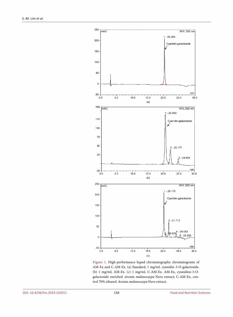
HPLC chromatograms of AM-Ex and C-AM-Ex are shown in Figure 1. The standard peak of C-3-Gal was compared with the peaks of C-3-Gal in AM-Ex and C-AM-Ex. Higher amoumts of C-3-Gal were estimated in AM-Ex than in C-AM-Ex, 2408 mg/100g versus 1049 mg/100g for AM-Ex and C-AM-Ex, re-spectively. These HPLC measurements of C-3-Gal content in AM-Ex indicate that AM-Ex produced by the high pressure homogenizing method contain a large amount of C-3-Gal.
3.2. Effect of AM-Ex on Cell Viability of 3T3-L1 Preadipocyte
To investigate the concentration of AM-Ex that was not cytotoxic, we deter-mined the effect of AM-Ex on 3T3-L1 cell viability by conducting the MTT as-say. Concentrations of AM-Ex between 500 to 2000 μg/mL showed significant reductions in the viability of 3T3-L1 cells. However, the viability of cells treated with lower doses of AM-Ex (1 to 400 μg/mL) were not significantly different compared to non-treated cells (Figure 2). Therefore, we used it at concentra-tions of 0 - 400 μg/mL in subsequent experiments to exclude the possibility that the inhibitory effect of AM-Ex on adipogenesis is due to its cytotoxic effect on 3T3-L1 cells.
3.3. AM-Ex Inhibits Adipocyte Differentiation in 3T3-L1 Preadipocytes
3T3-L1 preadipocytes undergo morphologic changes from a spindle-like to round shape and accumulate intracellular lipids after addition of differentiation inducing reagents [28]. To investigate the effect of AM-Ex on adipocyte diffe-rentiation and adipogenesis, post-confluent 3T3-L1 cells were induced by DM with or without the various concentration of AM-Ex. Accumulated lipid droplets in the differentiated 3T3-L1 adipocyte were visualized and quantified by Oil Red O staining. As shown in Figure 3(a), on day 8 after differentiation, 3T3-L1 adi-pocytes accumulated intracellular lipid droplets. AM-Ex significantly decreased the accumulation of lipid droplets. Cells treated with 400 μg/mL AM-Ex showed markedly reduced accumulation of lipid droplets (37.2% reduction) compared to non-AM-Ex-treated control cells (Figure 3(b)). These results demonstrated that AM-Ex inhibited adipocyte differentiation and adipogenesis
S.-M. Lim et al.
DOI: 10.4236/fns.2019.102011 134 Food and Nutrition Sciences
Figure 1. High-performance liquid chromatography chromatograms of AM-Ex and C-AM-Ex. (a) Standard, 1 mg/mL cyanidin-3-O-galactoside. (b) 1 mg/mL AM-Ex. (c) 1 mg/mL C-AM-Ex. AM-Ex, cyanidine-3-O- galactoside enriched Aronia melanocarpa Nero extract; C-AM-Ex, con-trol 70% ethanol Aronia melanocarpa Nero extract.

S.-M. Lim et al.
DOI: 10.4236/fns.2019.102011 135 Food and Nutrition Sciences
Figure 2. Effect of AM-Ex on cell viability in 3T3-L1 preadipocyte. 3T3-L1 cells were plated at 3 × 10⁴ cells/well and incubated for 24 h. After incubation for 24 h, the cells were incubated for 48 h in medium con-taining concentrations of AM-Ex ranging from 0 to 2000 μg/mL. Cell viability of 3T3-L1 cells was measured by the MTT assay. Values are ex-pressed as mean ± SEM. *P < 0.05, **P < 0.01, ***P < 0.01 significantly different from that of non-AM-Ex treated cells. AM-Ex, cyani-dine-3-O-galactoside enriched Aronia melanocarpa Nero extract.
in 3T3-L1 preadipocytes.
3.4. AM-Ex Inhibits Triglyceride Accumulation in 3T3-L1 Adipocytes
To investigate the effect of AM-Ex on lipogenesis, we determined intracellular triglyceride accumulation in 3T3-L1 adipocytes. AM-Ex at concentrations of 100, 200 or 400 μg/mL reduced the triglyceride levels in 3T3-L1 adipocytes by 24.4%, 28.6%, and 36.2%, respectively, compared to the non-AM-Ex-treated control (Figure 4). These results indicate that AM-Ex suppressed lipogenesis in 3T3-L1 adipocyte.
3.5. AM-Ex Inhibits the Expression of Adipogenic Transcription Factors
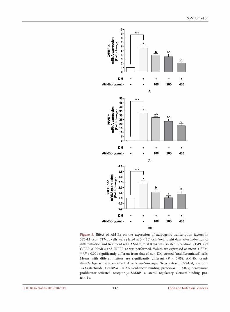
Adipogenesis is the process by which the differentiation from preadipocytes to mature adipocytes occurs. Several transcription factors such as CCAAT/enhancer binding proteins (C/EBPs), peroxisome proliferator-activated receptorγ (PPARγ) and sterol regulatory element-binding protein 1c (SREBP-1c) are in-volved in adipogenesis [29]. Thus, we next examined whether AM-Ex sup-pressed the expression of adipogenic transcription factors. Compared to the non-DM-treated (undifferentiated) cells, the DM-treated (differentiated) cells exhibited dramatically increased C/EBP-α, PPAR-γ, and SREBP-1c mRNA ex-pression. AM-Ex significantly decreased C/EBP-α, PPAR-γ, and SREBP-1c mRNA expression (Figure 5). These results suggest that AM-Ex inhibited

S.-M. Lim et al.
DOI: 10.4236/fns.2019.102011 136 Food and Nutrition Sciences
Figure 3. Effect of AM-Ex on adipocyte differentiation in 3T3-L1 cells. (a) 3T3-L1 cells were plated at 3 × 10⁴ cells/well and induced differentiation of adipocyte in medium containing various concentrations of AM-Ex. The differentiated 3T3-L1 cell were stained with Oil red O solution and observed by light microscopy. (b) The accumulated lipid droplets in stained 3T3-L1 adipocyte with Oil red O solution were quantified. Values are expressed as mean ± SEM. ***P < 0.001 significantly different from that of non-DM-treated (undifferentiated) cells. Means with different letters are significantly different (P < 0.05). AM-Ex, cyanidine-3-O-galac- toside enriched Aronia melanocarpa Nero extract.
Figure 4. Effect of AM-Ex on intracellular triglyceride accumulation in 3T3-L1 cells. 3T3-L1 cells were plated at 3 × 10⁴ cells/well. 3T3-L1 cells were differentiated and treatment with AM-Ex. The intracellular triglyce-ride accumulation in differentiated 3T3-L1 adipocyte was measured. Values are expressed as mean ± SEM. ***P < 0.001 significantly different from that of non-DM-treated (undifferentiated) cells. Means with differ-ent letters are significantly different (P < 0.05). AM-Ex, cyanidine-3-O- galactoside enriched Aronia melanocarpa Nero extract.
S.-M. Lim et al.
DOI: 10.4236/fns.2019.102011 137 Food and Nutrition Sciences
Figure 5. Effect of AM-Ex on the expression of adipogenic transcription factors in 3T3-L1 cells. 3T3-L1 cells were plated at 3 × 10⁴ cells/well. Eight days after induction of differentiation and treatment with AM-Ex, total RNA was isolated. Real-time RT-PCR of C/EBP-α, PPARγ, and SREBP-1c was performed. Values are expressed as mean ± SEM. ***P < 0.001 significantly different from that of non-DM-treated (undifferentiated) cells. Means with different letters are significantly different (P < 0.05). AM-Ex, cyani-dine-3-O-galactoside enriched Aronia melanocarpa Nero extract; C-3-Gal, cyanidin 3-O-galactoside; C/EBP-α, CCAAT/enhancer binding protein-α; PPAR-γ, peroxisome proliferator-activated receptor-γ; SREBP-1c, sterol regulatory element-binding pro-tein-1c.

S.-M. Lim et al.
DOI: 10.4236/fns.2019.102011 138 Food and Nutrition Sciences
adipocyte differentiation and adipogenesis through down-regulation of tran-scription factor, including C/EBP-α, PPAR-γ, and SREBP-1c.
3.6. AM-Ex Attenuates the Expression of Adipogenesis and Lipogenesis-Related Genes in 3T3-L 1 Cells
Adipogenic transcription factors, including C/EBP-α, PPAR-γ, and SREBP-1c, cooperatively induce the expression of specific genes involved in adipogenesis and lipogenesis [30]. Since C/EBP-α, PPAR-γ, and SREBP-1c were down-regulated by AM-Ex, we examined the gene regulation for adipogenesis and lipogenesis in AM-Ex-treated 3T3-L1 adipocytes using quantitative real-time RT-PCR. The mRNA expression of adipocyte protein 2 (aP2) and lipoprotein lipase (LPL), specific adipogenesis-related genes, were decreased by AM-Ex treatment. At 400 μg/mL, AM-Ex considerably reduced mRNA expression of aP2 and LPL by 76.2% and 34.8%, respectively, compared to the non-AM-Ex-treated control (Figure 6). As shown in Figure 7, AM-Ex significantly down-regulated the
Figure 6. Effect of AM-Ex on the expression of adipogenesis-related genes in 3T3-L1 cells. 3T3-L1 cells were plated at 3 × 10⁴ cells/well. Eight days after induction of differentiation and treatment with AM-Ex, total RNA was isolated. Real-time RT-PCR of aP2 and LPL was performed. Values are expressed as mean ± SEM. ***P < 0.001 significantly different from that of non-DM-treated (undifferentiated) cells. Means with different letters are significantly different (P < 0.05). AM-Ex, cyanidine-3-O-galactoside enriched Aronia melanocarpa Nero extract; aP2, adipocyte protein 2; LPL, lipoprotein lipase.

S.-M. Lim et al.
DOI: 10.4236/fns.2019.102011 139 Food and Nutrition Sciences
Figure 7. Effect of AM-Ex on the expression of lipogenesis-related genes in 3T3-L1 cells. 3T3-L1 cells were plated at 3 × 10⁴ cells/well. Eight days after induction of differentiation and treatment with AM-Ex, total RNA was isolated. Real-time RT-PCR of ACC1, ACL and FAS was performed. Values are expressed as mean ± SEM. ***P < 0.001 significantly different from that of non-DM-treated (undifferentiated) cells. Means with different let-ters are significantly different (P < 0.05). AM-Ex, cyanidine-3-O-galactoside enriched Aronia melanocarpa Nero extract; ACC1, acetyl-CoA carboxylase 1; ACL, ATP-citrate lyase; FAS, fatty acid synthase.

S.-M. Lim et al.
DOI: 10.4236/fns.2019.102011 140 Food and Nutrition Sciences
mRNA expression of lipogenesis-related genes, including acetyl-CoA carbox-ylase 1 (ACC1), ATP-citrate lyase (ACL), and fatty acid synthase (FAS). How-ever, there was no significant difference in the expression of ACC1, ACL, and FAS at AM-Ex concentrations of 100, 200, or 400 µg/mL (Figure 7). These results suggest that AM-Ex effectively suppressed adipogenesis and lipogene-sis by down-regulating expression of major adipogenic and lipogenic target genes.
4. Discussion
Aronia melanocarpa (Chokeberry, AM) is a rich source of polyphenols, espe-cially anthocyanins present as different forms of cyaniding-glycosides, procyani-dins, and flavonoids. Numerous health promoting effects of AM, related to an-tioxidant activity have been reported [31]. Cyanidine glycosides are known to be effective in preventing obesity associated with metabolic diseases [32]. Antho-cyanins also have some beneficial effects similar to other polyphenols including anti-inflammatory, anti-obesity, anti-diabetic, and anti-hypertensive effects. However, the mechanisms of its anti-obesity effect are still unclear compared to other AM functions. In the present study, we examined the effect of AM-Ex, an AM extract with a high content of C-3-Gal, on adipogenesis and lipogenesis in vitro using 3T3-L1 adipocytes.
Increased adipose tissue and adipocyte dysfunction related to obesity have been shown to be associated with abnormal adipogenesis regulation [33]. Adi-pocyte hypertrophy and hyperplasia both increase in adipose tissue, leading to obesity. Gene expression is tightly controlled and regulated by multiple compo-nents of various molecular circuits. In this process, transcription factors play an important role in different biological processes, such as differentiation, deve-lopmental process, and response to external and internal stimuli [30]. The process by which preadipocytes differentiate into mature adipocytes is regulated by hundreds of downstream protein-coding genes responsible for adipogenesis and by long noncoding RNAs (lncRNAs). This means that a large network of transcription factors acting together directly or indirectly, control the differen-tiation of adipocytes and the phenotypic characteristics of mature adipocytes [30] [34] [35] [36].
PPARγ and C/EBPα are crucial transcription factors in adipogenesis. PPAR is considered a critical factor in glucose and energy metabolism [37] [38]. C/EBPα plays an important role in adipogenesis, which directly induces diverse adipo-cyte genes. These two transcription factors play essential roles in determining the fate of differentiating preadipocytes [34] [39]. However, C/EBPα alone cannot induce adipogenesis without PPARγ. [37]. When PPARγ was overexpressed in mature 3T3-L1 adipocytes, both the adipocyte size and intracellular triglyceride content were increased [40]. Therefore, finding a functional food ingredient that can regulate PPARγ activity may be a complementary treatment for obesi-ty-related diseases [41]. SREBP1c is also a critical factor that mediates induction

S.-M. Lim et al.
DOI: 10.4236/fns.2019.102011 141 Food and Nutrition Sciences
of lipid biosynthesis in adipocytes by increasing gene expression of major lipo-genesis genes [42]. Many other transcription factors have been shown to exert a positive or negative effect on adipogenesis. For example, EBF1, KLF4, KLF5, KLF6, KLF15, EGR2, CEBPB, CEBPG and ARNTL promote adipogenesis. In contrast, GATA2, GATA3, KLF2, KLF3, IRF3, and IRF4 inhibit adipogenesis [34].
In the present study, we observed that AM-Ex treatment decreased PPARγ, C/EBPα, and SREBP1c mRNA expression in 3T3-L1 cells (Figure 5). These transcription factors regulate aP2, LPL, ACC, ACL, and FAS genes that control adipogenesis in the adipose tissue. For example, aP2, also called fatty acid bind-ing protein 4 (FABP4) is postulated to be an early marker of the metabolic syn-drome. Blocking this protein may represent a treatment for heart disease [43], diabetes [44], asthma [45], obesity [46], and fatty liver disease [47]. LPL acts as a key enzyme in the catabolic pathway of triglyceride-rich lipoproteins. LPL is synthesized and secreted by adipocytes and muscles, and is transported to capil-lary endothelial cells to hydrolyze the triglyceride core of circulating very low density lipoprotein and chylomicrons into fatty acids and monoglyceride. The hydrolysis products are taken up by the tissue. Depending on the energy state, LPL causes fatty acids to be transported to fat tissue for storage, and in the fast-ing state, fatty acids are broken down in muscles for use as fuel.
When adipose LPL is increased, free fatty acid produced by hydrolysis of li-poprotein stimulates PPAR transcription factor. In adipose tissue, LPL stimu-lates PPARγ and the LPL gene exhibits a mutually positive feedback loop that is stimulated by PPARγ [48]. Cinnamon and green tea polyphenols also have been reported to inhibit LPL and mRNA expression of other lipogenic genes [49] [50]. ACC regulates the metabolism of fatty acids. When ACC is activated, ma-lonyl-CoA is formed to synthesize new fatty acids and inhibit the β-oxidation of fatty acid in mitochondria by inhibiting the transfer of fatty acyl groups from acyl CoA to carnitine with carnitine acyltransferase. In mammals, two main iso-forms of ACC are expressed, ACC1 and ACC2, which differ in both tissue dis-tribution and function. Although ACC1 is found in the cytoplasm of all cells, it is abundant in lipogenic tissue, such as adipose tissue and lactating mammary glands, while ACC2 is found in many oxidative tissues such as skeletal muscle and heart [51]. ACC1 and ACC2 are highly expressed in the liver where both fatty acid oxidation and synthesis are important [52]. This difference in tissue distribution means that ACC1 is involved in the regulation of fatty acid synthesis and ACC2 is involved in the regulation of fatty acid oxidation. ACL is an en-zyme that catalyzes the hydrolysis of ATP and the conversion of citrate and CoA to acetyl-CoA and oxaloacetate [53]. Acetyl-CoA is used as a precursor in several important biosynthetic pathways, including triglyceride and cholesterol produc-tion [54]. FAS is a key enzyme in de novo lipogenesis. Metabolism and homeos-tasis of FAS are transcriptionally regulated by Upstream Stimulatory Factors (USF1 and USF2) and SREBP-1c in response to feeding/insulin in living animals
S.-M. Lim et al.
DOI: 10.4236/fns.2019.102011 142 Food and Nutrition Sciences
[55]. FAS mRNA and protein levels were increased in obese Zucker rats [56], and polyphenols of cinnamon [49] and dietary green tea [50] were reported to inhibit FAS mRNA expression in diet-induced insulin-resistant animals. Re-cently, aronia juice or extract intake in animal models and humans was reported to prevent or treat obesity. Qin and Anderson [22] reported that chokeberry ex-tract intake lowered blood glucose, triglyceride, cholesterol, LDL-cholesterol, and epididymal fat pads in Wister rats fed fructose-rich diet. And also the ex-pression of PPARγ and adiponectin mRNA were up-regulated and aP2, FAS, and LPL mRNA levels were inhibited by chokeberry extract consumption. Ta-kahashi et al. [23] reported that aronia fruit consumption inhibited hyperglyce-mia as well as visceral fat accumulation in high-fat diet-induced diatomic obese rats. Yamane et al. [16] reported that aronia juice had a beneficial effect on di-abetes and obesity by decreasing dipeptidyl peptidase and a-glucosidase activity. Kardum et al. [57] reported that aronia juice was effective in improving obesity in abdominally obese women aged 45 - 65 years.
Studies on the mechanism of the molecular regulation associated with the an-ti-obesity effects of AM have recently begun. Kowalska et al. [58] reported that various combinations of berries, including AM, in 3T3-L1 adipose cells reduced adipogenesis and oxidative stress. In this study, cells treated with 100 μg/mL of mixed berry extract down-regulated PPARγ (67%), C/EBPα (72%), SREBP1 (62%), aP2 (24%), HSL (39%), and PLIN1 (32%). In the present study, the data showed that AM-Ex treatment decreased aP2, LPL, ACC1, ACL, and FAS. In particular, aP2 expression was decreased by AM-Ex treatment in a dose-depen- dent manner (Figure 6 and Figure 7).
5. Conclusion
Our results showed that the anti-obesity effect of AM-Ex occured through down-regulation of the transcription factors PPARγ, C/EBPα, SREBP-1c, and adipogenesis and lipogenesis-related genes, aP2, LPL, ACC1, ACL, and FAS. These results demonstrate that AM-Ex can be used as a preventive or therapeutic agent for obesity. Future, animal and human studies are needed to further inves-tigate the mechanism and proper concentration of AM to be used as anti-obesity agents.
Acknowledgements
This research was supported by the Ministry of Trade, Industry & Energy (MOTIE), Korea Institute for Advancement of Technology (KIAT) through the Encouragement Program for the Industries of Economic Cooperation Region (P0000824).
Conflicts of Interest
The authors declare no conflicts of interest regarding the publication of this pa-per.

S.-M. Lim et al.
DOI: 10.4236/fns.2019.102011 143 Food and Nutrition Sciences
References [1] Elagizi, A., Kachur, S., Lavie, C.J., Carbone, S., Pandey, A., Ortega, F. and Milani,
R.V. (2018) An Overview and Update on Obesity and the Obesity Paradox in Car-diovascular Diseases. Progress in Cardiovascular Diseases, 61, 142-150. https://doi.org/10.1016/j.pcad.2018.07.003
[2] Kopelman, P.G. (2000) Obesity as a Medical Problem. Nature, 404, 635-643. https://doi.org/10.1038/35007508
[3] Baretić, M. (2013) Obesity Drug Therapy. Minerva Endocrinologica, 38, 245-254.
[4] Halford, J.C.G. (20016) Obesity Drugs in Clinical Development. Current Opinion Investigational Drugs, 7, 312-318.
[5] Klaus, S., Pültz, S., Thöne-Reineke, C. and Wolfram, S. (2005) Epigallocatechin Gallate Attenuates Diet-Induced Obesity in Mice by Decreasing Energy Absorption and Increasing Fat Oxidation. International Journal of Obesity, 29, 615-623. https://doi.org/10.1038/sj.ijo.0802926
[6] Wang, H., Wen, Y., Du, Y., Yan, X., Guo, H., Rycroft, J.A., Boon, N., Kovacs, E.M.R. and Mela, D.J. (2010) Effects of Catechin Enriched Green Tea on Body Composition. Obesity (Silver Spring, Md.), 18, 773-779. https://doi.org/10.1038/oby.2009.256
[7] Hayamizu, K., Ishii, Y., Kaneko, I., Shen, M., Okuhara, Y., Shigematsu, N., Tomi, H., Furuse, M., Yoshino, G. and Shimasaki, H. (2003) Effects of Garcinia Cambogia (Hydroxycitric Acid) on Visceral Fat Accumulation: A Double-Blind, Randomized, Placebo-Controlled Trial. Current Therapeutic Research, Clinical and Experimen-tal, 64, 551-567. https://doi.org/10.1016/j.curtheres.2003.08.006
[8] Semwal, R.B., Semwal, D.K., Vermaak, I. and Viljoen, A. (2015) A Comprehensive Scientific Overview of Garcinia Cambogia. Fitoterapia, 102, 134-148. https://doi.org/10.1016/j.fitote.2015.02.012
[9] Kokotkiewicz, A., Jaremicz, Z. and Luczkiewicz, M. (2010) Aronia Plants: A Review of Traditional Use, Biological Activities, and Perspectives for Modern Medicine. Journal of Medicinal Food, 13, 255-269. https://doi.org/10.1089/jmf.2009.0062
[10] Slimestad, R., Torskangerpoll, K., Nateland, H.S., Johannessen, T. and Giske, N.H. (2005) Flavonoids from Black Chokeberries, Aronia melanocarpa. Journal of Food Composition and Analysis, 18, 61-68. https://doi.org/10.1016/j.jfca.2003.12.003
[11] Moyer, R.A., Hummer, K.E., Finn, C.E., Frei, B. and Wrolstad, R.E. (2002) Antho-cyanins, Phenolics, and Antioxidant Capacity in Diverse Small Fruits: Vaccinium, Rubus, and Ribes. Journal of Agricultural and Food Chemistry, 50, 519-525. https://doi.org/10.1021/jf011062r
[12] Valcheva-Kuzmanova, S., Gadjeva, V., Ivanova, D. and Belcheva, A. (2007) Anti-oxidant Activity of Aronia melanocarpa Fruit Juice in Vitro. Acta Alimentaria, 36, 425-428. https://doi.org/10.1556/AAlim.36.2007.4.5
[13] Park, C.H., Kim, J.H., Lee, E.B., Hur, W., Kwon, O.J., Park, H.J. and Yoon, S.K. (2017) Aronia melanocarpa Extract Ameliorates Hepatic Lipid Metabolism through PPARγ2 Downregulation. PLoS ONE, 12, e0169685. https://doi.org/10.1371/journal.pone.0169685
[14] Valcheva-Kuzmanova, S., Kuzmanov, K., Mihova, V., Krasnaliev, I., Borisova, P. and Belcheva, A. (2007) Antihyperlipidemic Effect of Aronia melanocarpa Fruit Juice in Rats Fed a High-Cholesterol Diet. Plant Foods for Human Nutrition, 62, 19-24. https://doi.org/10.1007/s11130-006-0036-2
[15] Jeon, Y.D., Kang, S.H., Moon, K.H., Lee, J.H., Kim, D.G., Kim, W., Kim, J.S., Ahn,

S.-M. Lim et al.
DOI: 10.4236/fns.2019.102011 144 Food and Nutrition Sciences
B.Y. and Jin, J.S. (2018) The Effect of Aronia berry on Type 1 Diabetes in Vivo and in Vitro. Journal of Medicinal Food, 21, 244-253. https://doi.org/10.1089/jmf.2017.3939
[16] Yamane, T., Kozuk, M., Konda, D., Nakano, Y., Nakagaki, T., Ohkubo, I. and Ariga, H. (2016) Improvement of Blood Glucose Levels and Obesity in Mice Given Aronia Juice by Inhibition of Dipeptidyl Peptidase IV and α-Glucosidase. The Journal of Nutritional Biochemistry, 31, 106-112. https://doi.org/10.1016/j.jnutbio.2016.02.004
[17] Valcheva-Kuzmanova, S., Borisova, P., Galunska, B., Krasnaliev, I. and Belcheva, A. (2004) Hepatoprotective Effect of the Natural Fruit Juice from Aronia Melanocarpa on Carbon Tetrachloride-Induced Acute Liver Damage in Rats. Experimental and Toxicologic Pathology, 56, 195-201. https://doi.org/10.1016/j.etp.2004.04.012
[18] Bell, D.R. and Gochenaur, K. (2006) Direct Vasoactive and Vasoprotective Proper-ties of Anthocyanin-Rich Extracts. Journal of Applied Physiology, 100, 1164-1170. https://doi.org/10.1152/japplphysiol.00626.2005
[19] Cebova, M., Klimentova, J., Janega, P. and Pechanova, O. (2017) Effect of Bioactive Compound of Aronia Melanocarpa on Cardiovascular System in Experimental Hypertension. Oxidative Medicine and Cellular Longevity, 2017, 1-8. https://doi.org/10.1155/2017/8156594
[20] Ryszawa, N., Kawczyńska-Drózdz, A., Pryjma, J., Czesnikiewicz-Guzik, M., Ada-mek-Guzik, T., Naruszewicz, M., Korbut, R. and Guzik, T.J. (2006) Effects of Novel Plant Antioxidants on Platelet Superoxide Production and Aggregation in Atheros-clerosis. Journal of Physiology and Pharmacology, 57, 611-626.
[21] Matsumoto, M., Hara, H., Chiji, H. and Kasai, T. (2004) Gastroprotective Effect of Red Pigments in Black Chokeberry Fruit (Aronia Melanocarpa Elliot) on Acute Gastric Hemorrhagic Lesions in Rats. Journal of Agricultural and Food Chemistry, 52, 2226-2229. https://doi.org/10.1021/jf034818q
[22] Qin, B. and Anderson, R.A. (2012) An Extract of Chokeberry Attenuates Weight Gain and Modulates Insulin, Adipogenic and Inflammatory Signalling Pathways in Epididymal Adipose Tissue of Rats Fed a Fructose-Rich Diet. British Journal of Nu-trition, 108, 581-587. https://doi.org/10.1017/S000711451100599X
[23] Takahashi, A., Shimizu, H., Okazaki, Y., Sakaguchi, H., Taira, T., Suzuki, T. and Chiji, H. (2015) Anthocyanin-Rich Phytochemicals from Aronia Fruits Inhibit Vis-ceral Fat Accumulation and Hyperglycemia in High-Fat Diet-Induced Dietary Ob-ese Eats. Journal of Oleo Science, 64, 1243-1250. https://doi.org/10.5650/jos.ess15181
[24] Kim, M.Y., Lee, J.M., Lee, J.Y. and Lee, H.Y. (2016) Enhancement of Anti-Obesity Activities of Aronia Melanocarpa Elliot Extracts from Low Temperature Ultrasoni-fication Process. Korean Journal of Medicinal Crop Science, 24, 309-316. https://doi.org/10.7783/KJMCS.2016.24.4.309
[25] Gregoire, F.M., Smas, C.M. and Sul, H.S. (1998) Understanding Adipocyte Diffe-rentiation. Physiological Reviews, 78, 783-809. https://doi.org/10.1152/physrev.1998.78.3.783
[26] Denizot, F. and Lang, R. (1986) Rapid Colorimetric Assay for Cell Growth and Sur-vival. Modifications to the Tetrazolium Dye Procedure Giving Improved Sensitivity and Reliability. Journal of Immunological Methods, 8, 271-277. https://doi.org/10.1016/0022-1759(86)90368-6
[27] Folch, J., Lees, M. and Sloane Stanley, G.H. (1957) A Simple Method for the Isola-tion and Purification of Total Lipides from Animal Tissues. The Journal of Biologi-cal Chemistry, 226, 497-509.
S.-M. Lim et al.
DOI: 10.4236/fns.2019.102011 145 Food and Nutrition Sciences
[28] He, Y., Li, Y., Zhao, T., Wang, Y. and Sun, C. (2013) Ursolic Acid Inhibits Adipo-genesis in 3T3-L1 Adipocytes through LKB1/AMPK Pathway. PLoS ONE, 8, e70135. https://doi.org/10.1371/journal.pone.0070135
[29] Rosen, E.D. and MacDougald, O.A. (2006) Adipocyte Differentiation from the In-side Out. Nature Reviews Molecular Cell Biology, 7, 885-896. https://doi.org/10.1038/nrm2066
[30] Farmer, S.R. (2006) Transcriptional Control of Adipocyte Formation. Cell Metabol-ism, 4, 263-273. https://doi.org/10.1016/j.cmet.2006.07.001
[31] Crubasik, C., Li, G. and Crubasik, S. (2010) The Clinical Effectiveness of Chokeber-ry: A Systematic Review. Phytotherapy Research, 24, 1107-1114. https://doi.org/10.1002/ptr.3226
[32] Sikora, J., Broncel, M., Markowicz, M., Chatubinski, M., Wojdan, K. and Miki-ciuk-Olasik, E. (2012) Short-Term Supplementation with Aronia Melanocarpa Ex-tract Improves Platelet Aggregation, Clotting, and Fibrinolysis in Patients with Me-tabolic Syndrome. European Journal of Nutrition, 51, 549-556. https://doi.org/10.1007/s00394-011-0238-8
[33] Rodríguez-Acebes, S., Palacios, N., Botella-Carretero, J.I., Olea, N., Crespo, L., Peromingo, R., Gómez-Coronado, D., Lasunción, M.A., Vázquez, C. and Martínez- Botas, J. (2010) Gene Expression Profiling of Subcutaneous Adipose Tissue in Mor-bid Obesity Using a Focused Microarray: Distinct Expression of Cell-Cycle- and Differentiation-Related Genes. BMC Medical Genomics, 3, 61. https://doi.org/10.1186/1755-8794-3-61
[34] Lefterova, M.I. and Lazar, M.A. (2009) New Developments in Adipogenesis. Trends in Endocrinology and Metabolism, 20, 107-114. https://doi.org/10.1016/j.tem.2008.11.005
[35] Stephens, J.M. (2012) The Fat Controller: Adipocyte Development. PLoS Biology, 10, e1001436. https://doi.org/10.1371/journal.pbio.1001436
[36] Sun, L., Goff, L.A., Trapnell, C., Alexander, R., Lo, K.A., Hacisuleyman, E., Sauva-geau, M., Tazon-Vega, B., Kelley, D.R., Hendrickson, D.G., Yuan, B., Kellis, M., Lo-dish, H.F. and Rinn, J.L. (2013) Long Noncoding RNAs Regulate Adipogenesis. Proceeding of the National Academy of Sciences of the United States of America, 110, 3387-3392. https://doi.org/10.1073/pnas.1222643110
[37] Tontonoz, P. and Spiegelman, B.M. (2008) Fat and Beyond: The Diverse Biology of PPARgamma. Annual Review of Biochemistry, 77, 289-312. https://doi.org/10.1146/annurev.biochem.77.061307.091829
[38] Rosen, E.D., Hsu, C.H., Wang, X., Sakai, S., Freeman, M.W., Gonzalez, F.J. and Spiegelman, B.M. (2002) C/EBP Alpha Induces Adipogenesis through PPARgam-ma: A Unified Pathway. Genes & Development, 16, 22-26. https://doi.org/10.1101/gad.948702
[39] Tontonoz, P., Hu, E., Graves, R.A., Budavari, A.I. and Spiegelman, B.M. (1994) mPPAR Gamma 2: Tissue-Specific Regulator of an Adipocyte Enhancer. Genes, 15, 1224-1234. https://doi.org/10.1101/gad.8.10.1224
[40] Tamori, Y., Masugi, J., Nishino, N. and Kasuga, M. (2002) Role of Peroxisome Pro-liferator-Activated Receptor-Gamma in Maintenance of the Characteristics of Ma-ture 3T3-L1 Adipocytes. Diabetes, 51, 2045-2055. https://doi.org/10.2337/diabetes.51.7.2045
[41] Ortuño Sahagún, D., Márquez-Aguirre, A.L., Quintero-Fabián, S., López-Roa, R.I. and Rojas-Mayorquín, A.E. (2012) Modulation of PPAR-γ by Nutraceutics as Com-plementary Treatment for Obesity-Related Disorders and Inflammatory Diseases.

S.-M. Lim et al.
DOI: 10.4236/fns.2019.102011 146 Food and Nutrition Sciences
PPAR Research, 2012, Article ID: 318613.
[42] Kim, J.B., Sarraf, P., Wright, M., Yao, K.M., Mueller, E., Solanes, G., Lowell, B.B. and Spiegelman, B.M. (1998) Nutritional and Insulin Regulation of Fatty Acid Syn-thetase and Leptin Gene Expression through ADD1/SREBP1. Journal of Clinical Investigation, 101, 1-9. https://doi.org/10.1172/JCI1411
[43] Makowski, L., Boord, J.B., Maeda, K., Babaev, V.R., Uysal, K.T., Morgan, M.A., Parker, R.A., Suttles, J., Fazio, S., Hotamisligil, G.S. and Linton, M.F. (2001) Lack of Macrophage Fatty-Acid-Binding Protein aP2 Protects Mice Deficient in Apolipo-protein E against Atherosclerosis. Nature Medicine, 7, 699-705. https://doi.org/10.1038/89076
[44] Furuhashi, M., Tuncman, G., Görgün, C.Z., Makowski, L., Atsumi, G., Vaillancourt, E., Kono, K., Babaev, V.R., Fazio, S., Linton, M.F., Sulsky, R., Robl, J.A., Parker, R.A. and Hotamisligil, G.S. (2007) Treatment of Diabetes and Atherosclerosis by Inhibiting Fatty-Acid-Binding Protein aP2. Nature, 447, 959-965. https://doi.org/10.1038/nature05844
[45] hum, B.O., Mackay, C.R., Gorgun, C.Z., Frost, M.J., Kumar, R.K., Hotamisligil, G.S. and Rolph, M.S. (2006) The Adipocyte Fatty Acid-Binding Protein aP2 Is Required in Allergic Airway Inflammation. Journal of Clinical Investigation, 116, 2183-2192. https://doi.org/10.1172/JCI24767
[46] Maeda, K., Cao, H., Kono, K., Gorgun, C.Z., Furuhashi, M., Uysal, K.T., Cao, Q., Atsumi, G., Malone, H., Krishnan, B., Minokoshi, Y., Kahn, B.B., Parker, R.A. and Hotamisligil, G.S. (2005) Adipocyte/Macrophage Fatty Acid Binding Proteins Con-trol Integrated Metabolic Responses in Obesity and Diabetes. Cell Metabolism, 1, 107-119. https://doi.org/10.1016/j.cmet.2004.12.008
[47] Makowski, L. and Hotamisligil, G.S. (2005) The Role of Fatty Acid Binding Proteins in Metabolic Syndrome and Atherosclerosis. Current Opinion in Lipidology, 16, 543-548. https://doi.org/10.1097/01.mol.0000180166.08196.07
[48] Schoonjans, K., Peinado-Onsurbe, J., Lefebvre, A.M., Heyman, R.A., Briggs, M., Deeb, S., Staels, B. and Auwerx, J. (1995) PPARα and PPARγ Activators Direct a Distinct Tissue-Specific Transcriptional Response via a PPRE in the Lipoprotein Lipase Gene. The EMBO Journal, 15, 5336-5348. https://doi.org/10.1002/j.1460-2075.1996.tb00918.x
[49] Qin, B., Polansky, M.M. and Anderson, R.A. (2010) Cinnamon Extract Regulates Plasma Levels of Adipose-Derived Factors and Expression of Multiple Genes Re-lated to Carbohydrate Metabolism and Lipogenesis in Adipose Tissue of Fruc-tose-Fed Rats. Hormone and Metabolic Research, 42, 187-193. https://doi.org/10.1055/s-0029-1242746
[50] Lee, M.S., Kim, C.T. and Kim, Y. (2009) Green Rea (-)-epigallocatechin-3-fallate Reduces Body Weight with Regulation of Multiple Genes Expression in Adipose Tissue of Diet-Induced Obese Mice. Annals of Nutrition and Metabolism, 54, 151-157. https://doi.org/10.1159/000214834
[51] Kim, T.S., Leahy, P. and Freake, H.C. (1996) Promoter Usage Determines Tissue Specific Responsiveness of the Rat Acetyl-CoA Carboxylase Gene. Biochemical and Biophysical Research Communications, 225, 647-653. https://doi.org/10.1006/bbrc.1996.1224
[52] Barber, M.C., Price, N.T. and Travers, M.T. (2005) Structure and Regulation of Acetyl-CoA Carboxylase Genes of Metazoa. Biochimica et Biophysica Acta, 1733, 1-28. https://doi.org/10.1016/j.bbalip.2004.12.001
[53] Sun, T., Hayakawa, K., Bateman, K.S. and Fraser, M.E. (2010) Identification of the

S.-M. Lim et al.
DOI: 10.4236/fns.2019.102011 147 Food and Nutrition Sciences
Citrate-Binding Site of Human ATP-Citrate Lyase Using X-Ray Crystallography. The Journal of Biology Chemistry, 285, 27418-27428. https://doi.org/10.1074/jbc.M109.078667
[54] Guay, C., Madiraju, S.R., Aumais, A., Joly, E. and Prentki, M. (2007) A Role for ATP-Citrate Lyase, Malic Enzyme, and Pyruvate/Citrate Cycling in Glucose-In- duced Insulin Secretion. The Journal of Biology Chemistry, 282, 35657-35665. https://doi.org/10.1074/jbc.M707294200
[55] Latasa, M.J., Griffin, M.J., Moon, Y.S., Kang, C. and Sul, H.S. (2003) Occupancy and Function of the -150 Sterol Regulatory Element and -65 E-Box in Nutritional Regu-lation of the Fatty Acid Synthase Gene in Living Animals. Molecular and Cellular Biology, 23, 5896-5907. https://doi.org/10.1128/MCB.23.16.5896-5907.2003
[56] Guichard, C., Dugail, I., Le, L.X. and Lavau, M. (1992) Genetic Regulation of Fatty Acid Synthetase Expression in Adipose Tissue: Over-Transcription of the Gene in Genetically Obese Rats. The Journal of Lipid Research, 33, 679-687.
[57] Kardum, N., Petrović-Oggiano, G., Takic, M., Glibetić, N., Zec, M., Debeljak-Mar- tacic, J. and Konić-Ristić, A. (2014) Effects of Glucomannan-Enriched, Aronia Juice-Based Supplement on Cellular Antioxidant Enzymes and Membrane Lipid Status in Subjects with Abdominal Obesity. Scientific World Journal, 2014, Article ID: 869250. https://doi.org/10.1155/2014/869250
[58] Kowalska, K., Olejnik, A., Szwajgier, D. and Olkowicz, M. (2017) Inhibitory Activity of Chokeberry, Bilberry, Raspberry and Cranberry Polyphenol-Rich Extract towards Adipogenesis and Oxidative Stress in Differentiated 3T3-L1 Adipose Cells. PLoS ONE, 12, e0188583. https://doi.org/10.1371/journal.pone.0188583