epithelial tissue connective tissue muscle tissue nervous tissue endo derm mesoderm ectoderm ...
TRANSCRIPT

Vocabulary Chapter 5 Tissues Sept 26 Due
Epithelial tissue Connective tissue Muscle tissue Nervous tissue Endo derm Mesoderm Ectoderm Gastrulation Histogenesis Membranous epithelium Glandular epithelium Squamous Cuboidal Columnar
Pseudosratified Exocrine glands Endocrine glands Apocrine glands Holocrine glands Merocrine glands Adipose tissue Reticular tissue Hyaline cartilage Elastic cartilage Fibrocartilage Smooth muscle Cardiac muscle Skeletal muscle Hematopoiesis

Chapter 5 TissuesTest 10/4/2012
Epithelial tissue: membranous tissue covering internal organs and other internal surfaces of the body.
The function of epithelial tissue include: transportation of materials, protection of underlying tissues, absorption of water and nutrients and secretion of waste products.

Connective Tissue
In our body, Connective Tissues bind structures together, form a framework and support for organs and the body as a whole, store fat, transport substances, protect against disease, and help repair tissue damage. Connective Tissues occur throughout the body and are characterized by an abundance of intercellular matrix with relatively few cells. Connective tissue cells are able to reproduce but not as rapidly as epithelial cells. Most but not all connective tissues have a good blood supply.In our body, numerous cell types are found in Connective Tissue. Three of the most common cell types in Connective Tissues are the fibroblast, macrophage, and mast cell. The types of connective tissue include Loose Connective Tissue, Adipose Tissue, Dense Fibrous Connective Tissue, Elastic Connective Tissue, Cartilage, Osseous Tissue (bone), and Blood.

Connective tissue

Muscle Tissue
A body tissue composed of sheets or bundles of cells that contract to produce movement or increase tension. Muscle cells contain filaments made of the proteins actin and myosin, which lie parallel to each other. When a muscle is signaled to contract, the actin and myosin filaments slide past each other in an overlapping pattern. Skeletal muscle effects voluntary movement and is made up of bundles of elongated cells (muscle fibers), each of which contains many nuclei. Smooth muscle provides the contractile force for the internal organs and is controlled by the autonomic nervous system. Smooth muscle cells are spindle-shaped and each contains a single nucleus. Cardiac muscle makes up the muscle of the heart and consists of a meshwork of striated cells.

Muscle Tissue

Nervous Tissue
Nervous tissue is specialised to react to stimuli and to conduct impulses to various organs in the body which bring about a response to the stimulus. Nerve tissue (as in the brain, spinal cord and peripheral nerves that branch throughout the body) are all made up of specialised nerve cells called neurons. Neurons are easily stimulated and transmit impulses very rapidly. A nerve is made up of many nerve cell fibres (neurons) bound together by connective tissue.
5 functions of epithelial tissue
Since epithelial tissue marks the border between our bodies and some kind of space, the epithelial tissue must be able to handle whatever it encounters within that space. If the space is our external world (that the skin has to come into contact with), the epithelial tissue must be pretty hearty. If the space is a tiny little air passage deep within your lung, not much besides air would ever get in there and the epithelial tissue does not have to offer protection.
Also, as a bordering tissue, it may also have to allow materials to pass through it. Think again about epithelial tissue in the deep air passageways within the lungs-- oxygen has to be able to freely cross this epithelial tissue to enter our bodies.
protection-- as a barrier between the outer world (or inner spaces) and our bodies.
secretion-- when our bodies need to release material, like hormones into the blood, this tissue has to allow for such material to pass through. Often, it is the cells within the epithelial tissue that make the material for secretion.
absorption-- epithelial tissue facing our digestive tract has to be very good at absorbing nutrients from the digestive tract lumen in order for us to get what we need from what we eat.
excretion-- epithelial tissue even lines the excretory lumina, like the tracts from the kidneys through to the urethra.are:
Sensory – specialized structures found in the eyes, ears, nose, and skin

Histogenesis
The formation of different tissues from undifferentiated cells. These cells are constituents of three primary germ layers, the endoderm, mesoderm, and ectoderm. The science of the microscopic structures of the tissues formed within histogenesis is termed histology.
Ectoderm: The outermost germ layer of cells derived from the inner cell mass of the blastocyst; gives rise to the nervous system, sensory organs, skin, and related structures.
Mesoderm: The middle layer of cells in embryonic development; gives rise to muscles, bones, and structures associated with reproduction. The middle embryonic tissue layer. Cells and structures arising from the mesoderm include the bone, blood, muscle, skin, and reproductive organs.
Endoderm:
Apocrine gland
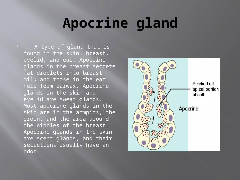
A type of gland that is found in the skin, breast, eyelid, and ear. Apocrine glands in the breast secrete fat droplets into breast milk and those in the ear help form earwax. Apocrine glands in the skin and eyelid are sweat glands. Most apocrine glands in the skin are in the armpits, the groin, and the area around the nipples of the breast. Apocrine glands in the skin are scent glands, and their secretions usually have an odor.

Holocrine Gland Holocrine glands are glands that
secrete whole cells that have completely broken down for elimination from the body. This is unique because other forms of secretion do not decimate entire cells.
Sebaceous glands are the only holocrine glands that exist in the body. They are a type of exocrine gland, which means they use ducts to transport secretions to a specific location outside the body; the prefix “exo” means “outside” or “on top of.” Since sebaceous glands are located on the epidermal layer of the skin, they transport secretions directly to the skin’s surface.

Merocrine Gland Sometimes referred to as eccrine
sweat glands, merocrine sweat glands are glands that extend from the outer layer of the skin to the inner layers. The merocrine sweat gland helps to regulate the body’s overall temperature, keeping it within a range that is considered to be healthy.
Merocrine sweat glands are found on the entire surface of the human body. There are several areas where the concentration of sweat glands is particularly plentiful. Three examples of areas that are highly concentrated with merocrine sweat glands are the soles of the feet, the palms of both hands, and the forehead.

Functions of Connective tissue
This is the most widespread and abundant type of tissue in the human body. Its function is primarily to support, anchor and connect various parts of the body. Although connective tissue exists in a number of forms, all types have three basic structural elements -- cells, fibres and intercellular substance (ground substance).
The most common cell types are fibroblasts, which produce fibres and other intercellular materials. The two most common types of fibres are: collagen (collagenous) and elastic. Collagen fibres are for strength while the elastic ones are for elasticity of the tissue. Both the cells and the fibres are embedded in the intercellular substance. The consistency of this substance is highly variable from gelatin-like to a much more rigid material.
The proportions of the cells, fibres, and intercellular substance vary, depending on a particular nature and function of the connective tissue. For example, a strong connective tissue needs a greater proportion of the collagen fibres and fewer cells. An example would be a dense regular connective tissue, which is found in tendons and ligaments. On the other hand, a connective tissue composed of mostly cells would not be very strong. An example would be an adipose (fat) connective tissue.

Cartilage It is a stiff yet flexible connective
tissue found in many areas in the bodies of humans and other animals, including the joints between bones, the rib cage, the ear, the nose, the elbow, the knee, the ankle, the bronchial tubes and the intervertebral discs. It is not as hard and rigid as bone but is stiffer and less flexible than muscle.
Cartilage is composed of specialized cells called chondrocytes that produce a large amount of extracellular matrix composed of collagen fibers, abundant ground substance rich in proteoglycan, and elastin fibers. Cartilage is classified in three types, elastic cartilage, hyaline cartilage and fibrocartilage, which differ in the relative amounts of these three main components.
Bone
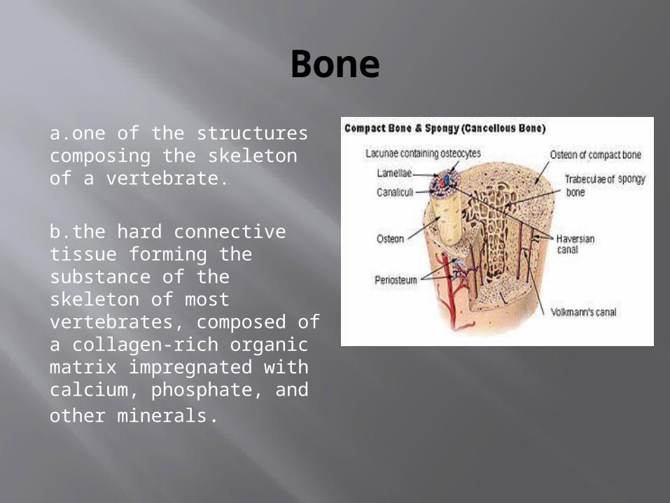
a.one of the structures composing the skeleton of a vertebrate.
b.the hard connective tissue forming the substance of the skeleton of most vertebrates, composed of a collagen-rich organic matrix impregnated with calcium, phosphate, and other minerals.

Blood The average adult has about five liters of blood
living inside of their body, coursing through their vessels, delivering essential elements, and removing harmful wastes. Without blood, the human body would stop working.
Blood is the fluid of life, transporting oxygen from the lungs to body tissue and carbon dioxide from body tissue to the lungs. Blood is the fluid of growth, transporting nourishment from digestion and hormones from glands throughout the body. Blood is the fluid of health, transporting disease fighting substances to the tissue and waste to the kidneys
Because it contains living cells, blood is alive. Red blood cells and white blood cells are responsible for nourishing and cleansing the body. Since the cells are alive, they too need nourishment.
Vitamins and Minerals keep the blood healthy. The blood cells have a definite life cycle, just as all living organisms do. Approximately 55 percent of blood is plasma, a straw-colored clear liquid. The liquid plasma carries the solid cells and the platelets which help blood clot. Without blood platelets, you would bleed to death.

Smooth Muscle
Muscle tissue that contracts without conscious control, having the form of thin layers or sheets made up of spindle-shaped, unstriated cells with single nuclei and found in the walls of the internal organs, such as the stomach, intestine, bladder, and blood vessels, excluding the heart.

Skeletal muscle Skeletal muscles move and
support the skeleton. They make up fifty percent of your body weight. There are 640 individually named skeletal muscles. A skeletal muscle links two bones across its connecting joint. When these muscles contract or shorten, your bone moves. Muscles are arranged in layers over the bones. Those nearest to the skin are called superficial muscles. Those closest to the inside of the body are called deep muscles. Skeletal muscles are voluntary muscles. These are muscles that we can consciously control.

Cardiac Muscle Cardiac muscles are the muscles of the
heart. They are self-contracting, autonomically regulated and must continue to contract in rythmic fashion for the whole life of the organism. Hence they have special features.
The cells are Y shaped and are shorter and wider than skeletal muscle cells. They are predominatly mononucleated. The arrangement of actin and myosin is similar to skeletal striated muscle.
Some of the cardiac muscle cells are auto-rhythmic, i.e they contract even in the absence of neuronal innervation (known as pacemaker cells). Intercalated disks are located between cardiac muscles cells. These contain gap junctions which provide communicating channels between cells.The intercalated disks allows waves of depolarisations to sweep across the cells thus synchronising muscle contraction.