a closed system of the heart and blood vessels the heart pumps blood blood vessels allow blood to...
TRANSCRIPT
A closed system of the heart and blood vesselsThe heart pumps bloodBlood vessels allow blood to circulate to all
parts of the body The function of the cardiovascular
system is to deliver oxygen and nutrients and to remove carbon dioxide and other waste products
LocationThorax between the lungs in the inferior
mediastinum Orientation
Pointed apex directed toward left hipBase points toward right shoulder
About the size of your fist
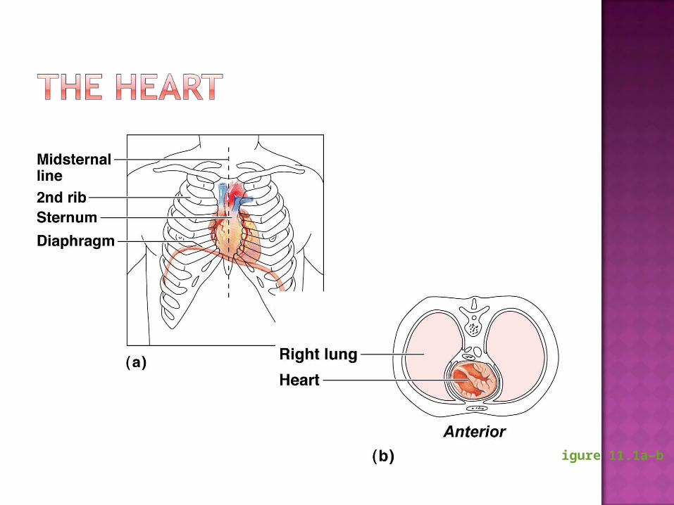
Figure 11.1a–b

Figure 11.1c

Figure 11.2a

Pericardium—a double-walled sacFibrous pericardium is loose and superficialSerous membrane is deep to the fibrous
pericardium and composed of two layers Visceral pericardium
Next to heart; also known as the epicardium Parietal pericardium
Outside layer that lines the inner surface of the fibrous pericardium
Serous fluid fills the space between the layers of pericardium

Figure 11.2b
Three layersEpicardium
Outside layerThis layer is the visceral pericardiumConnective tissue layer
MyocardiumMiddle layerMostly cardiac muscle
EndocardiumInner layerEndothelium
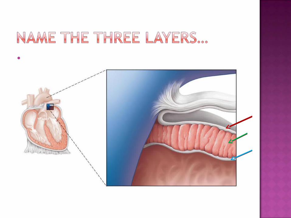
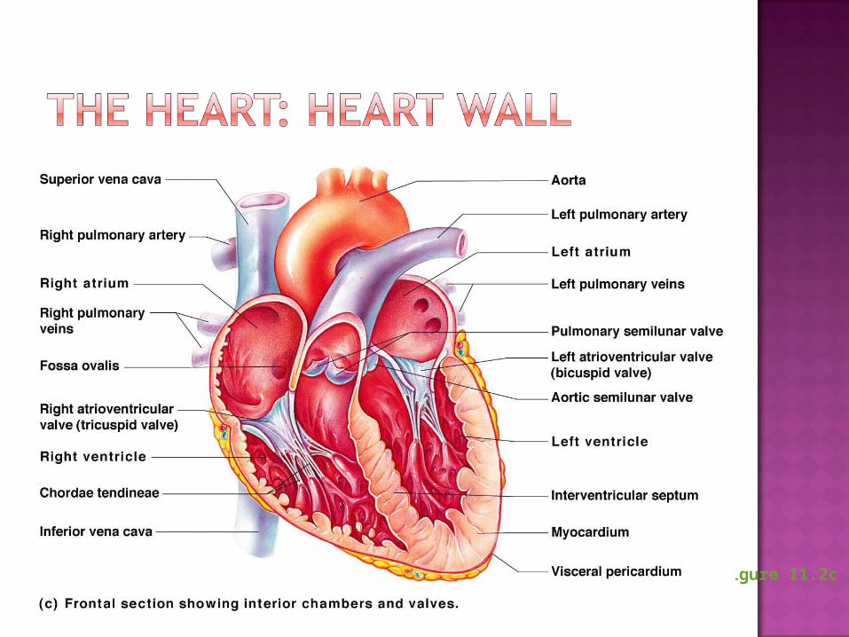
Figure 11.2c

Right and left side act as separate pumps
Four chambersAtria
Receiving chambersRight atriumLeft atrium
Ventricles Discharging chambers
Right ventricleLeft ventricle

Figure 11.2c

Figure 11.4
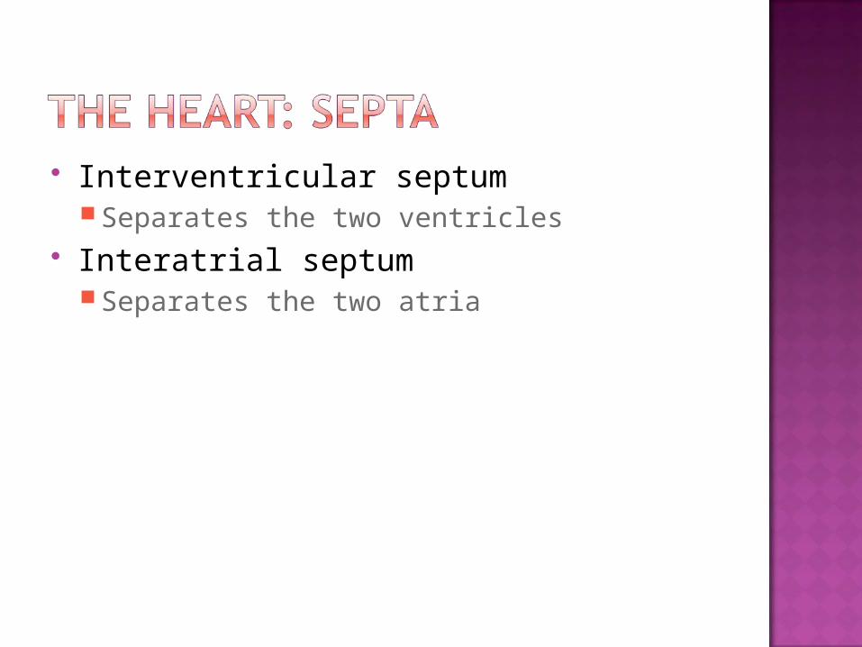
Interventricular septum Separates the two ventricles
Interatrial septum Separates the two atria
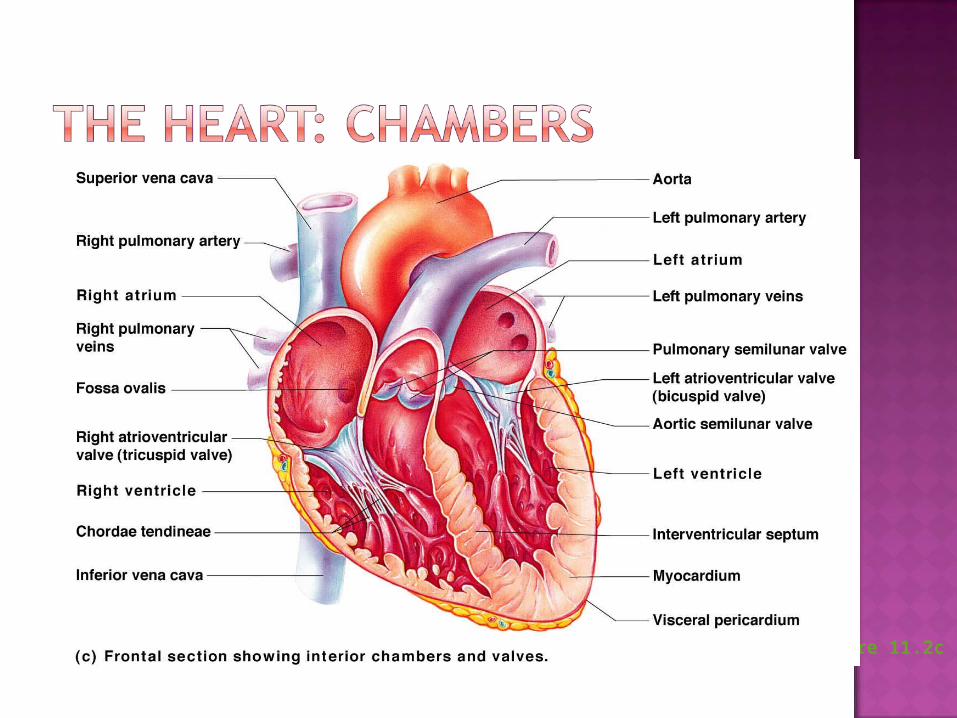
Figure 11.2c

Allow blood to flow in only one direction to prevent backflow
Four valvesAtrioventricular (AV) valves—between atria
and ventricles Bicuspid (mitral) valve (left side of heart) Tricuspid valve (right side of heart)
Semilunar valves—between ventricle and artery Pulmonary semilunar valve Aortic semilunar valve
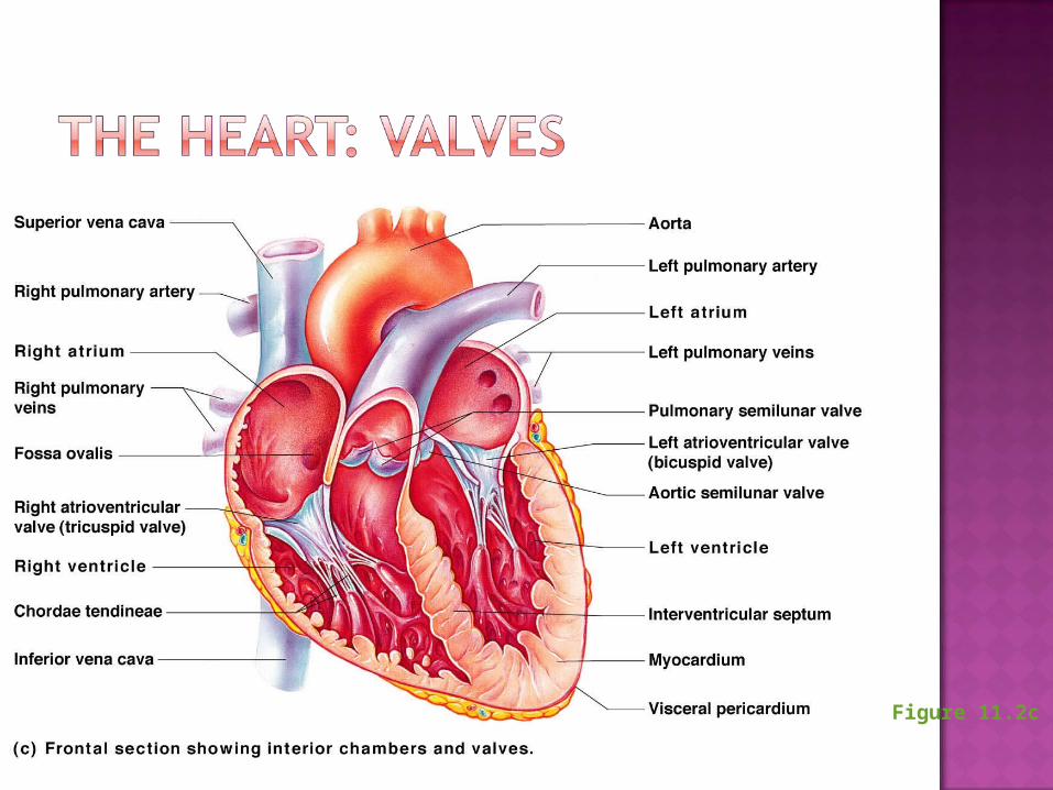
Figure 11.2c

AV valves Anchored in place by chordae tendineae
(“heart strings”)Open during heart relaxation and closed
during ventricular contraction Semilunar valves
Closed during heart relaxation but open during ventricular contraction
Notice these valves operate opposite of one another to force a one-way path of blood through the heart
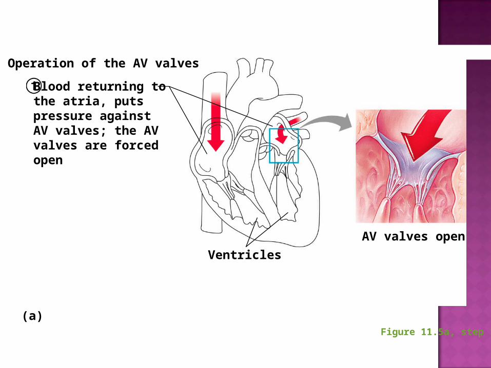
Figure 11.5a, step 1
Blood returning tothe atria, putspressure againstAV valves; the AVvalves are forcedopen
Ventricles
Operation of the AV valves
AV valves open
(a)
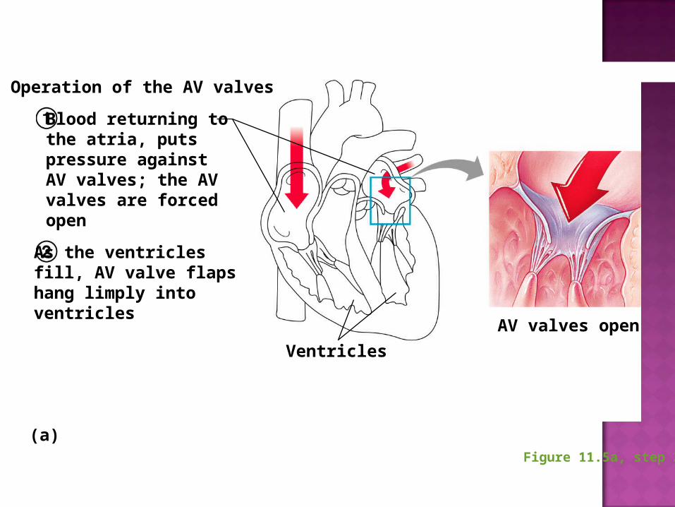
Figure 11.5a, step 2
Blood returning tothe atria, putspressure againstAV valves; the AVvalves are forcedopen
As the ventriclesfill, AV valve flapshang limply intoventricles
Ventricles
Operation of the AV valves
AV valves open
(a)
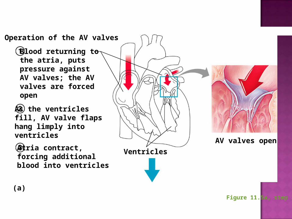
Figure 11.5a, step 3
Blood returning tothe atria, putspressure againstAV valves; the AVvalves are forcedopen
As the ventriclesfill, AV valve flapshang limply intoventricles
Atria contract,forcing additionalblood into ventricles
Ventricles
Operation of the AV valves
AV valves open
(a)
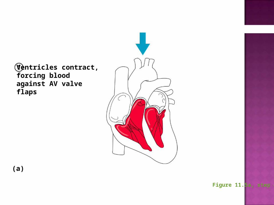
Figure 11.5a, step 4
Ventricles contract,forcing bloodagainst AV valveflaps
(a)
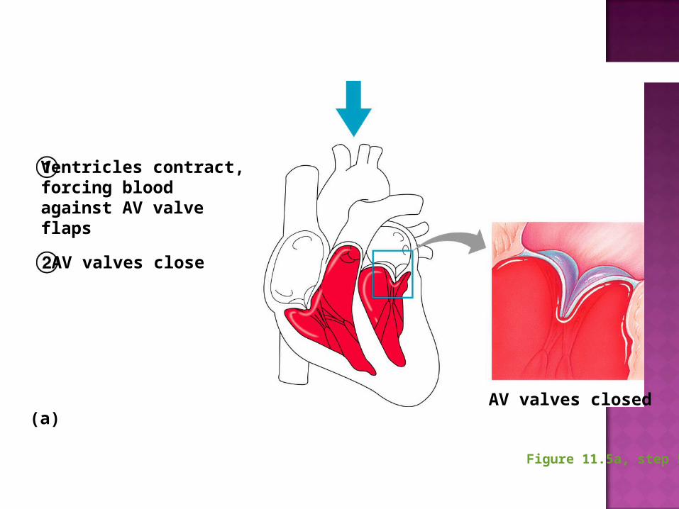
Figure 11.5a, step 5
Ventricles contract,forcing bloodagainst AV valveflaps
AV valves close
AV valves closed(a)

Figure 11.5a, step 6
Ventricles contract,forcing bloodagainst AV valveflaps
AV valves close
Chordae tendineaetighten, preventingvalve flaps fromeverting into atria
AV valves closed(a)
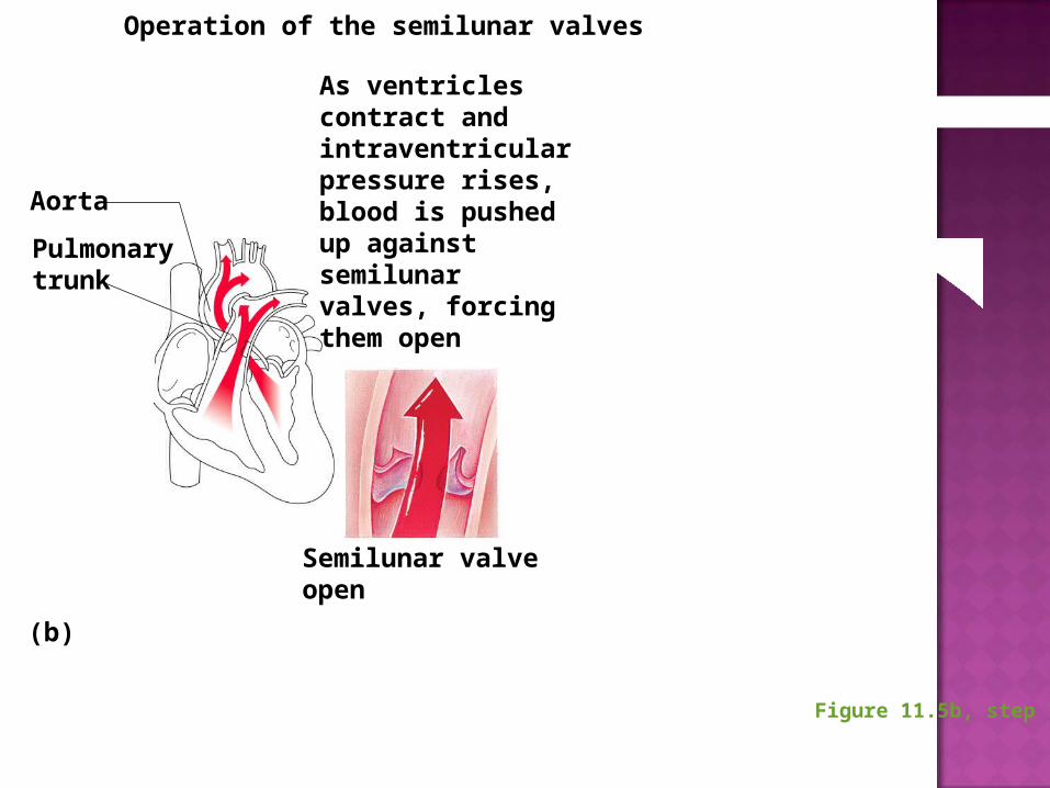
Figure 11.5b, step 1
As ventriclescontract andintraventricularpressure rises,blood is pushedup againstsemilunarvalves, forcingthem open
Aorta
Pulmonarytrunk
Semilunar valveopen
Operation of the semilunar valves
(b)

Figure 11.5b, step 2
As ventriclescontract andintraventricularpressure rises,blood is pushedup againstsemilunarvalves, forcingthem open
Aorta
Pulmonarytrunk
Semilunar valveopen Semilunar valve
closed
As ventriclesrelax, andintraventricularpressure falls,blood flowsback fromarteries, fillingthe leaflets of semilunarvalves andforcing themto close
Operation of the semilunar valves
(b)
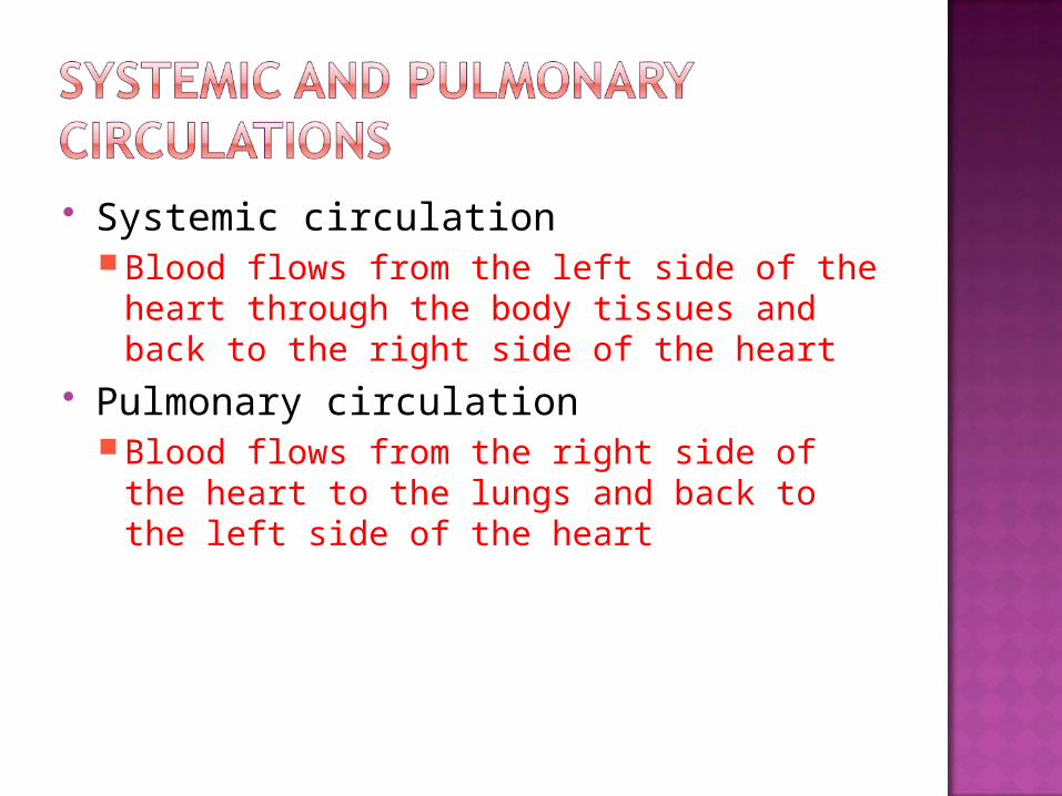
Systemic circulationBlood flows from the left side of the heart
through the body tissues and back to the right side of the heart
Pulmonary circulationBlood flows from the right side of the heart
to the lungs and back to the left side of the heart

Figure 11.3
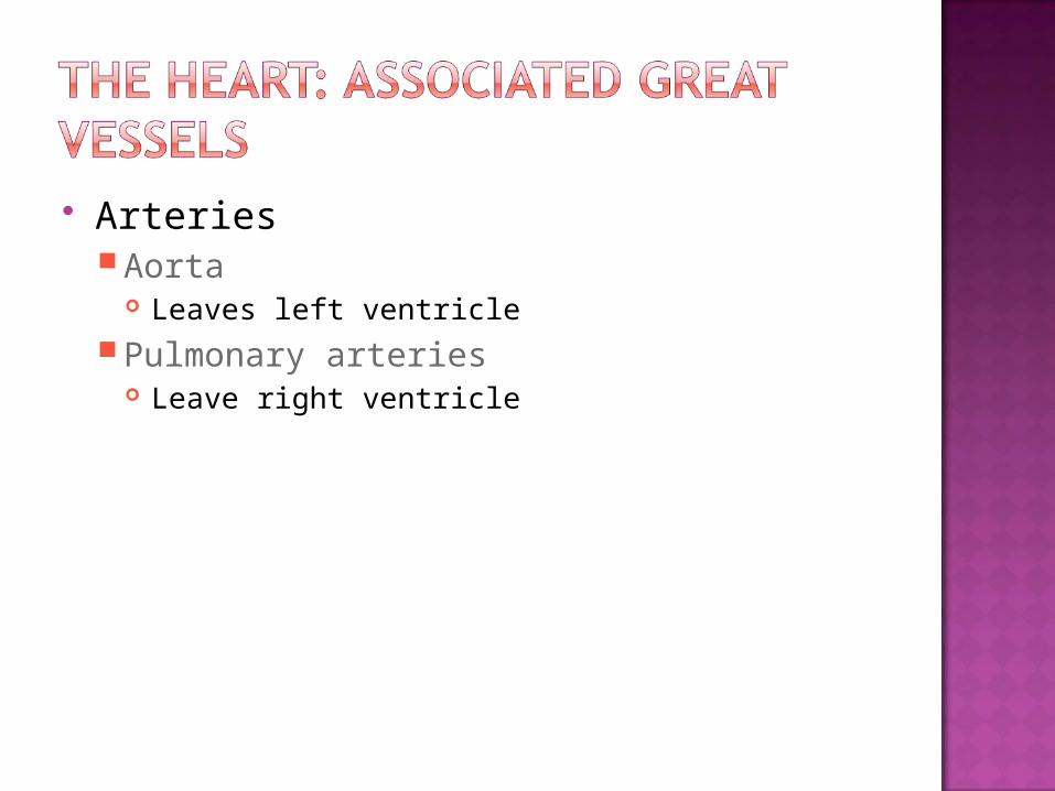
ArteriesAorta
Leaves left ventriclePulmonary arteries
Leave right ventricle
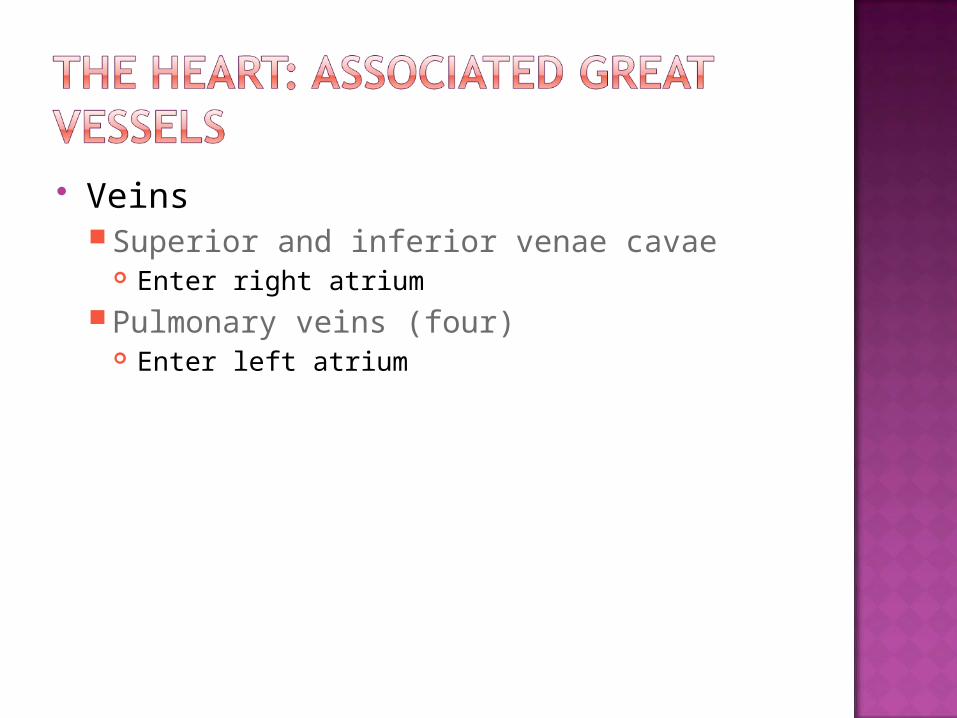
VeinsSuperior and inferior venae cavae
Enter right atriumPulmonary veins (four)
Enter left atrium
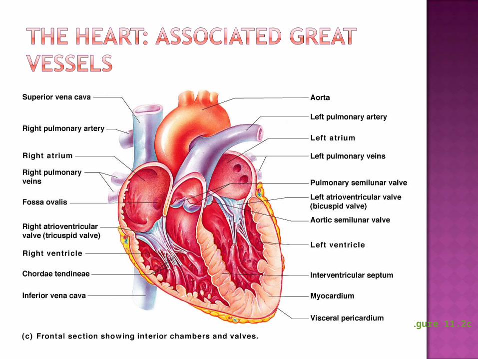
Figure 11.2c

Superior and inferior venae cavae dump blood into the right atrium
From right atrium, through the tricuspid valve, blood travels to the right ventricle
From the right ventricle, blood leaves the heart as it passes through the pulmonary semilunar valve into the pulmonary trunk
Pulmonary trunk splits into right and left pulmonary arteries that carry blood to the lungs

Oxygen is picked up and carbon dioxide is dropped off by blood in the lungs
Oxygen-rich blood returns to the heart through the four pulmonary veins
Blood enters the left atrium and travels through the bicuspid valve into the left ventricle
From the left ventricle, blood leaves the heart via the aortic semilunar valve and aorta
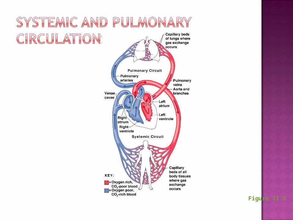
Figure 11.3

Blood in the heart chambers does not nourish the myocardium
The heart has its own nourishing circulatory system consisting of Coronary arteries—branch from the aorta to
supply the heart muscle with oxygenated blood
Cardiac veins—drain the myocardium of blood
Coronary sinus—a large vein on the posterior of the heart, receives blood from cardiac veins
Blood empties into the right atrium via the coronary sinus

1. What is the location of the heart in the thorax?
2. How does the function of the systemic circulation differ from that of the pulmonary circulation?
3. Why are the heart valves important?4. Why might a thrombus in a corarnary
artery cause a sudden death?

5. Draw a diagram of the heart showing the three layers composing its wall and its four chambers. Label each. Show where the AV and semilunar valves are. Show and label all blood vessels entering and leaving the heart chambers.
6. Trace a drop of blood from the time it enters the right atrium of the heart until it enters the left atrium. What is this circuit called?
7. What is the function of the fluid that fills the pericardial sac?
8. Define systole, diastole, stroke volume, and cardiac cycle.
9. How does the hearts ability to contract differ from that of other muscles of the body.