arteriosclerosissta.uwi.edu/fms/mdsc1001/athero.pdf · 9/13/2010 1 arteriosclerosis • hardening...
TRANSCRIPT

9/13/2010
1
ARTERIOSCLEROSIS
• Hardening of arteries due to thickening and loss of elasticityelasticity.
• The three types of arteriosclerosis are‐
• 1) Atherosclerosis (AS): #1 Killer, CVS disease
• 2) Monckeberg’s arteriosclerosis – Medial calcification, >50 yrs of age
3) Arteriolosclerosis: associated with hypertension.
Atherosclerosis (AS):
• slow progressive disease of large and mediumi d l d l l ti t isized muscular and large elastic arteries
• characterized by an elevated fibro fatty intimalplaques formed by lipid deposition, smoothmuscle proliferation and synthesis of extracellular matrix.

9/13/2010
2
• It begins in childhood and manifests in middle and later life
• Risk Factors: Major – Non Modifiable: age, male gender, family historyage, male gender, family history
• Potentially controllable: Hyperlipidemia, hypertension, cig.smoking & D.Mellitus(deadly quartet)
• Minor – obesity, HDL, Physical inactivity, stress post menopause High carbohydratestress, post menopause, High carbohydrate diet, alcohol, homocysteine, lipoprotein (a), chlamydial infection etc.
Etio‐Pathogenesis
• Hypotheses:
• 1) Insudation hypothesis (Virchow)
• 2) Encrustation hypothesis (Rokitansky)
• 3) Monoclonal hypothesis (Benditt)
• 4) Reaction to injury hypothesis (Ross).

9/13/2010
3
Reaction to Injury
Chronic endothelial injury Hyperlipid,HTN,smoking, DM,Hemodyn,Homocyst,Toxins etc
Endothelial Dysfunction
SMC emigration & Macrophage activation Chronic inflammatory disease
IL, TNF,GF’s,Metaloprot, Adh mol,
SMC proliferation, Lipid, collagen &ECM deposition – Well formed plaque
HTN
Toxins cig smoke
Hemodynamic stress
LDL
D dDamage to endo
PTL adhDiff.Plasma Prot
Mig Mono
OxidationPDGF
Oxidation LDL
UptakeFoam cells
Cytokine releaseCollagen Syn
Prolif.SMC
9/13/2010
4
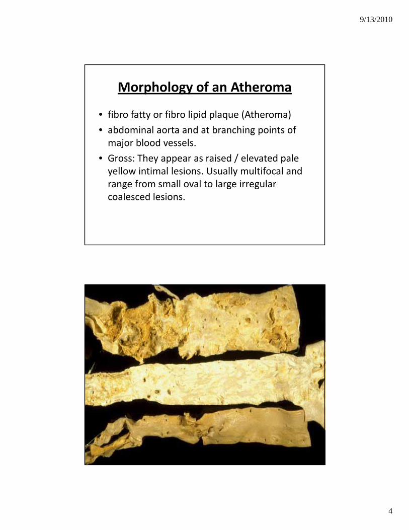
Morphology of an Atheroma
• fibro fatty or fibro lipid plaque (Atheroma)
• abdominal aorta and at branching points of major blood vessels.
• Gross: They appear as raised / elevated pale yellow intimal lesions. Usually multifocal and range from small oval to large irregularrange from small oval to large irregular coalesced lesions.

9/13/2010
5
• Microscopic findings: 1.Fibrous cap‐Located between the lumen and tunicamedia. It is composed of proliferatingsmooth muscle cells Leukocytes & Densesmooth muscle cells, Leukocytes & Denseconnective tissue matrix
• 2. Central necrotic core: Contains deadcells, lipid, cholesterol clefts, foamymacrophages, plasma proteins and smoothmuscle cellsmuscle cells.
3.Periphery consists of proliferatingcapillaries.

9/13/2010
6

9/13/2010
7
Two common variants of plaque
• Fatty streaks
• Complicated plaques

9/13/2010
8
• Clinical significance: Asymptomatic fordecades. Progression of disease orcomplication of AS leads to:
a)Insidious progressive narrowing of vasculara)Insidious, progressive narrowing of vascularlumen resulting in ischemicatrophy/gangrene of tissue/ organ affected.
b) Plaque rupture & superimposedthrombosis results in sudden occlusion andischemic necrosis/ infarction as in MI &stroke.

9/13/2010
9
• c) Atheroembolism: Rupture and release of core of the plaque.
• d) Aneurysm: weakening of the wall.
• Organs often affected by AS are heart, brain, kidney, lower extremities and intestines
• Is Atherosclerosis reversible?
• Vaccine for AS?
Aneurysms
• A true aneurysm is a localized, permanent, abnormal dilatation of a blood vessel causedabnormal dilatation of a blood vessel caused by weakness of the vessel wall
• (> 50% of normal diameter)• False (pseudo) aneurysm or pulsating hematoma is an extra vascular hematoma that
i t ith th i t lcommunicates with the intravascular space. This usually follows an injury.

9/13/2010
10
• Aneurysms can be classified by their location/ name of the vessel, configuration / morphology & etiology
• Morphologic classification:Morphologic classification:
• 1. Fusiform:
• 2. Saccular:
• 3. Cylindrical:
• 4 Berry:4. Berry:
• 5. Dissecting hematoma
• 6. A‐V aneurysm
Aneurysms classified by etiology
• Atherosclerotic Aneurysm: They are l l t d i bd i l tcommonly located in abdominal aorta
between renal and common iliac vessels. They may also be seen in descending thoracic aorta.

9/13/2010
11
• Etiology: Atherosclerotic weakening of the wall
• Morphology – Gross: They are fusiform or sacc lar Wall sho s complicated plaq essaccular. Wall shows complicated plaques with mural thrombus.
• Micro: Features of a complicated plaque.• Clinical Features: >50yrs of age, Males, Associated with Hypertension. Presents with abdominal pain, pulsatile abdominal mass. Lesions >5 cms diameter is at risk of rupture and fatal hemorrhage.

9/13/2010
12
Syphilitic (Luetic) aneurysm
• tertiary stage of syphilis.
• Aneurysm is seen in the ascending and arch of the aorta. It can extend proximally resulting in aortic valve ring dilatation, rolling of leaflets and valvular insufficiency. This condition is associated with marked LVH (Cor bovinum). ( )

9/13/2010
13
Fundamental lesion: Syphilitic aortitis due to obliterative endarteritis of the vasa vasorum.
Morphology: Gross‐ Fusiform aneurysm and p gy yintima shows “ Tree barking “ appearance
Micro: Loss of elastic fibres, smooth muscle cells due to aortic medial ischemia and weakening of the wall Blood vessels (vasaweakening of the wall. Blood vessels (vasa vasorum) show perivascular lympho‐plasmacytic cuffing.

9/13/2010
14
Dissecting Aneurysm (Aortic dissection)
• characterized by dissection of blood along the laminar planes of the aortic media with formation of intramural blood filled channel
• men between 40 to 60yrs• strongly associated with hypertension• Other predisposing factors are Copper deficiency, Marfan’s Syndrome in young men, y y y gAortopathy, pregnancy, Bicuspid valve, Iatrogenic
• cystic medial degeneration of the aorta.
• Morphology: Gross‐
1. Intimal tear within 10cms of aortic valve ring is the portal of entry of blood. Some times it could result from bleeding vasa vasorumresult from bleeding vasa vasorum.
• 2. Dissection occurs between middle and outer thirds in the media and it extends into major vessels of the neck, coronaries etc
• 3. Outer wall is thin and can rupture into extra vascular space pericardium mediastinum andvascular space‐ pericardium, mediastinum, and pleural and abdominal cavity.

9/13/2010
15
9/13/2010
16
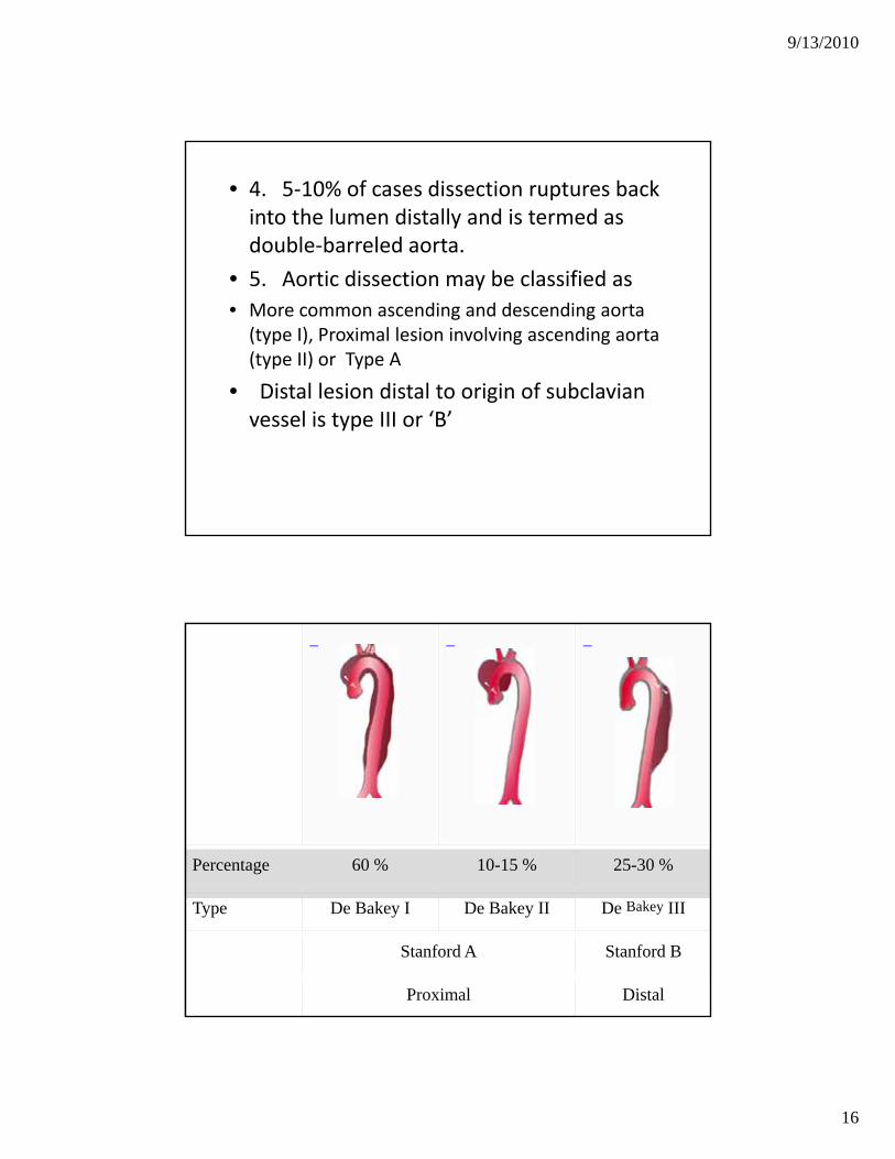
• 4. 5‐10% of cases dissection ruptures back into the lumen distally and is termed as double‐barreled aorta.
5 A ti di ti b l ifi d• 5. Aortic dissection may be classified as• More common ascending and descending aorta (type I), Proximal lesion involving ascending aorta (type II) or Type A
• Distal lesion distal to origin of subclavian vessel is type III or ‘B’
Percentage 60 % 10-15 % 25-30 %
Type De Bakey I De Bakey II De Bakey III
Stanford A Stanford B
Proximal Distal

9/13/2010
17
• Micro: Aortic dissection associated with hypertension shows non –specific degenerative changes, elastic fragmentation and collection of excessfragmentation and collection of excess amorphous interstitial material. In Marfans syndrome cystic medial necrosis is typical of the lesion.
C/F: Acute onset of severe tearing chest pain mimicking MI Loss of peripheral pulsesmimicking MI. Loss of peripheral pulses, Aortic insufficiency, fatal hemorrhage and cardiac tamponade.

9/13/2010
18
• Berry Aneurysm: Small aneurysms seen at the branching points in Circle of Willis. These are due to defect in muscular wall replaced by thin fibrous tissue Rupturereplaced by thin fibrous tissue. Rupture leads to subarachnoid hemorrhage.
• Mycotic aneurysm (Infective): Seen in association with bacterial endocarditis. Bacterial/fungal infection of arterial wall with tendency to rupture and hemorrhage.

9/13/2010
19