© 2010 shilpa naidu beesabathuni - ideals
TRANSCRIPT

© 2010 Shilpa Naidu Beesabathuni

EVALUATION OF IMMOBILIZING HORSERADISH PEROXIDASE AND
ALCOHOL OXIDASE IN PVA-AWP POLYMER
BY
SHILPA NAIDU BEESABATHUNI
THESIS
Submitted in partial fulfillment of the requirements
for the degree of Master of Science in Agricultural and Biological Engineering
in the Graduate College of the
University of Illinois at Urbana-Champaign, 2010
Urbana, Illinois
Adviser:
Assistant Professor Mary-Grace Danao

ii
ABSTRACT
Biosensor technology is a powerful alternative to conventional analytical techniques,
harnessing the specificity and sensitivity of biological systems in small, low cost devices. One of
the factors that affect the performance of a biosensor is the immobilization of the biological
sensing element, or bioreceptor, on the transducer surface. The objectives of this study were to
determine the effects of polymer concentration, ultraviolet (UV) light exposure, and film thickness
on the activity and stability of horseradish peroxidase (HRP) and alcohol oxidase (AOX) when
immobilized in a photo-crosslinkable and water-soluble polymer, poly (vinyl alcohol) azide-unit
water pendant (PVA-AWP). The immobilized enzyme films were to be used in biosensing
applications and their stability and activity were determined colorimetrically and
electrochemically. Compared to other gel immobilization techniques, PVA hydrogels offer several
advantages, such as better elasticity, low-toxicity, biocompatibility with enzymes and yeast cells,
mechanical and long-term stability, and biodegradability.
To determine the activity and stability of immobilized HRP, UV–Vis spectroscopy was
used to analyze changes in HRP structure since the position of the Soret absorption band at 402-
403 nm of the enzyme’s heme prosthetic group can provide information on protein conformation.
Position of Soret band peaks occurred between 402-403 nm for HRP immobilized in agarose and
PVA-AWP (3.8 and 5% w/v). Peak absorbance of the Soret band in AWP 5% (w/v) was found to
be 22%, 30%, 25% higher than those using AWP 3.8% (w/v) for UV exposure times 5, 10 and
15 minutes, respectively. From the above results, it can be concluded that UV exposure did not
affect the conformation of HRP immobilized in PVA-AWP and the activity was higher in AWP
(5% w/v). There was no difference (p < 0.05) observed in the enzyme activity of HRP
immobilized in PVA-AWP, for UV exposure times of 5, 10 and 15 minutes, and PVA-AWP
concentrations of 3.8% (w/v) and 5% (w/v). However, enzyme activity was lower (p < 0.05)
when HRP was immobilized in glutaraldehyde. There was also no difference (p < 0.05) in
enzyme leaching for UV 5, 10 and 15 minute exposure when immobilized in PVA-AWP.
However, enzyme leaching was higher (p < 0.05) for HRP immobilized in glutaraldehyde. It can

iii
be concluded that HRP immobilized in PVA-AWP had higher activity and mechanical stability
when compared to HRP immobilized in glutaraldehyde.
Results from the preceding tests were applied to immobilizing AOX in PVA-AWP. An
AOX-PVA-AWP mixture containing 0.5 units of AOX activity was drop-coated on a screen
printed carbon electrode. The immobilized AOX was tested with varying concentrations of ethanol
solution ranging from 17.1 µM to 1.71 mM. The electrical current produced during the oxidation
of hydrogen peroxide by cobalt phthalocyanine was measured chronoamperometrically. The
magnitude of the current output was dependent on thickness of the film on the electrode. There was
no difference (p < 0.05) observed in the rate of current output with change in ethanol concentration
between AOX immobilized in PVA-AWP and non-immobilized or free AOX, but it was higher (p
< 0.05) for AOX immobilized in glutaraldehyde. Enzyme activity decreased by 40%, 36% and 7%
when stored for 24 hours at 23°C, 3°C and -17°C respectively. When AOX was immobilized in
approximately 0.09 mm thick PVA-AWP polymer film, results showed that the lower limit of
ethanol detection was 171.3 µM.

iv
ACKOWLEDGMETS
I would like to express my sincere gratitude to my advisor, Dr. Mary-Grace Danao, who
not only guided me for the past two years but was instrumental in molding a naive student to
adopt a greater understanding of science. She is a mentor, friend and a source of inspiration and I
will cherish the lessons learnt from her. I am grateful to my committee members, Dr. Steven
Eckhoff and Dr. Vijay Singh, for their invaluable discussions and meticulous reviews of this
thesis. I am thankful to Dr. Eckhoff for making me gain a different perspective in my approach
to research and life.
I would like to thank Julio Soares for his technical assistance. I am thankful to every
member of the Department of Agricultural and Biological Engineering for providing wisdom,
meaningful breaks, coffee sessions and constant humor. Thanks to my colleague, Shih-Fang,
who was always there to discuss my experiments and bounce ideas around. Along with Anna
Oldani and Sidney Knight, she has made our lab cheerful and a fun place. Hard work would not
have been so much fun without my friends at Champaign who have converted this place into
another home. I owe most of the completion of my work to Joy Deep Nath, who not only was a
constant source of encouragement but always lent a patient ear. He has redefined the word
optimism for me.
My journey so far would have been impossible without the support of my loving family,
particularly my nephew Aadit. A smile on his face was enough to keep me going through trying
times. My father, mother, sister, brother-in-law and nephew have shaped my personality. They
have taught me to dream big and more importantly, that I can achieve those dreams.
v
TABLE OF COTETS
Chapter 1 INTRODUCTION ........................................................................................................ 1
Chapter 2 LITERATURE REVIEW ............................................................................................. 5
2.1 Biosensor ............................................................................................................................ 5
2.2 Immobilization of the Bioreceptor ..................................................................................... 6
2.3 Polyvinyl (alcohol) Azide-Unit Pendant Water-Soluble Photopolymer (PVA-AWP) .... 14
2.4 Horseradish Peroxidase .................................................................................................... 15
2.5 Alcohol Oxidase ............................................................................................................... 15
Chapter 3 EVALUATION OF IMMOBILIZING HORSERADISH PEROXIDASE IN PVA-
AWP POLYMER ....................................................................................................... 17
3.1 Introduction ...................................................................................................................... 17
3.2 Materials and Methods ..................................................................................................... 19
3.3 Results and Discussion ..................................................................................................... 26
3.4 Conclusions ...................................................................................................................... 34
Chapter 4 EVALUATION OF IMMOBILIZING ALCOHOL OXIDASE IN PVA-AWP
POLYMER ................................................................................................................. 36
4.1 Introduction ...................................................................................................................... 36
4.2 Materials and Methods ..................................................................................................... 38
4.3 Results and Discussion ..................................................................................................... 45
4.4 Conclusions ...................................................................................................................... 53
Chapter 5 CONCLUSIONS AND FUTURE WORK ................................................................. 54
REFERENCES .............................................................................................................................. 55
APPENDIX A ............................................................................................................................ 64
A.1. Soret band ........................................................................................................................ 64
A.2. Determination of Activity of HRP Immobilized in Glutaraldehyde and PVA-AWP ..... 67
A.3. Determination of Activity of HRP Leached from HRP-Glutaraldehyde and HRP-AWP
films ................................................................................................................................. 68
APPENDIX B ............................................................................................................................... 69
B.1 Effect of Film Thickness and UV Exposure on Sensitivity ............................................. 69
B.2 Effect of Immobilization Medium on Sensitivity of Biosensor ....................................... 73
B.3 Response times ................................................................................................................ 75

1
CHAPTER 1
ITRODUCTIO
Biosensors are widely applied to many fields such as biocatalytic process, medical care,
food, environment, industries, security and defense (Alcock and Turner, 1994; Dennison and
Turner, 1995; Kress-Rogers, 1996; White and Turner, 1997). Enzyme-based biosensors are
analytical devices that utilize immobilized enzymes as biological recognition components and a
transducer to generate a measurable response. The immobilized enzyme acts as a biocatalyst in a
heterogeneous phase reaction in which the products are captured at the interface of the
immobilized enzyme and transducer.
Since the second half of the last century, numerous efforts have been devoted to the
development of insoluble immobilized enzymes for a variety of applications (Silman and
Katchalski, 1966). These applications can clearly benefit from use of the immobilized enzymes
rather than the soluble counterparts as reusable heterogenous biocatalysts. For example, the use
of immobilized enzymes can reduce production costs by efficient recycling and control of the
process (Vandamme, 1983; Schulze and Wubbolts, 1999); form the basis for stable and reusable
devices for analytical and medical applications (Stetter, 1951; Clark and Lyons, 1962; Campbell
et al., 1951; Watanabe et al., 1988; Chang, 1977; Klein and Langer, 1986; Kircka and Thorpe,
1986); and serve as selective adsorbents for purification of proteins and enzymes (Dunlap, 1974),
tools for solid-phase protein chemistry (Bickerstaff, 1984; Martinek and Mozhaev, 1985), and
microdevices for controlled release of protein drugs (Cristallini et al., 1997). It is recognized that
the availability of a robust immobilized enzyme will enable early insight into process
development and save costs not only in process development but also in production. However,
the lack of guidelines for performance to be expected of an immobilized enzyme seriously
hampers application of a rational approach to the design of such robust immobilized enzymes
(Van Roon et al., 2002).
Enzymes belong to the category of natural catalyst which includes DNA, RNA and
catalytic antibodies. A unique function of enzymes is that all the reactions they catalyze can be

2
performed sequentially, selectively and precisely under mild physiological reaction conditions.
There is, however, no doubt that many are not ideal catalysts for industrial applications. For
example in the manufacture of fine chemicals (Liese and Filho, 1999; Schulze and Wubbolts,
1999) and pharmaceuticals and their intermediates (Bommarius et al., 1998), enzymes are
usually exposed to unnatural conditions such as elevated substrate concentrations, high pH, and
temperature, and the presence of deleterious organic solvents. They must be used in the
immobilized forms to reduce production cost by facilitating downstream processing such as
recycling and separation (Tischer and Kasche, 1999).
The problem of enzyme immobilization is not how to immobilize the enzyme but how to
achieve the desired performance for a given application by selecting an appropriate means of
immobilization. Although enzyme immobilization and improvement of enzyme performance by
immobilization share the same principles, the emphasis is different. The former is mainly
associated with efforts to find suitable immobilization methods for enzymes that must be
immobilized for certain applications. The immobilization technique developed is mainly
intended to retain the major catalytic functions of the native enzymes. In contrast, improvement-
by-immobilization is focused mainly on utilization of available immobilization techniques to
alter (or improve) enzyme performance, to suit the desired application. The native enzyme might
be not suitable for a desired process, because of its poor performance such as lower activity, or
stability or selectivity. The technique to be developed should improve the performance of the
enzyme besides immobilizing it.
Stabilization by immobilization has been studied since the 1970s, when immobilized
enzymes became increasingly used in industrial processes, in which the cost-contribution of the
immobilized enzyme is often the indicator of process viability (Clark, 1994). The stability of a
native enzyme (i.e. a non-immobilized or modified enzyme) is principally determined by its
intrinsic structure whereas the stability of an immobilized enzyme is highly dependent on many
factors, including:
• the properties of its interaction with the carrier;
• the binding position and the number of the bonds;
• the freedom of the conformation change in the matrix;
• the microenvironment in which the enzyme molecule is located;

3
• the chemical and physical structure of the carrier;
• the properties of the spacer (for example, charged or neutral, hydrophilic or hydrophobic,
size, length) linking the enzyme molecules to the carrier; and
• the conditions under which the enzyme molecules were immobilized.
Activity by immobilization is often regarded as an extra benefit rather than the primary
goal of enzyme immobilization. Activity retention by carrier-bound immobilized enzymes is
usually approximately 50%. At high enzyme loading, especially, diffusion limitation might occur
as a result of the unequal distribution of the enzyme within a porous carrier, leading to a
reduction of apparent activity (Janssen et al., 2002). The conditions for high activity retention are
often marginal, often requiring laborious screening of immobilization conditions such as enzyme
loading, pH, carrier and binding chemistry (Taylor, 1985). Next to the microenvironment effect
mentioned above, it has been demonstrated that immobilized enzymes can be more active than
the native enzymes, when the inhibiting effect of the substrate was reduced (Taylor, 1985).
The criteria for assessing the robustness of the immobilized enzymes remain the same –
industrial immobilized enzymes must be highly active (high activity in a unit of volume, U/ml),
highly selective (to reduce side reactions), highly stable (to reduce cost by effective reuse), cost-
effective (low cost contribution thus economically attractive), safe to use (to meet safety
regulations) and innovative (for recognition as intellectual property). As with the volume activity
(U enzyme per gram of carrier used), most enzymes bound to carriers with particle sizes above
100 µm (minimum size requirements for a carrier-bound immobilized enzyme have a loading (or
payload) ranging from 0.001 to 0.1 (Tischer and Kasche, 1999). The volume ratio of catalyst to
reactor is usually in the range 10-20%. The productivity of most immobilized enzymes is still
lower than in chemical processes, mainly because of the small number of active sites per mass of
biocatalyst (low volume activity) (Straathof et al., 2002).
As a result, it is to be expected that the focus should be the development of a new method
of enzyme immobilization that combines the advantages of carrier-free and carrier-bound
methods. The new method should be able to provide high enzyme loading (close to that of
carrier-free enzymes), high retention of activity, and broad reactor configurations. There is no
currently available method that meets these criteria. A vast number of methods of immobilization

4
are currently available but how to have control over the performance of the immobilized enzyme
remains elusive.
In this study, photolinkable polyvinyl (alcohol) azide-unit pendant water-soluble
photopolymer (PVA-AWP) was used for immobilizing enzymes. PVA hydrogels have been
widely used because of their rubbery elasticity, high degree of swelling in aqueous solutions,
inherent non-toxicity and biocompatibility with enzymes, mechanical and long-term stability,
and biodegradability. The photocrosslinking or degree of immobilization can be spatially and
temporally controlled by the irradiation of light (Stammen et al., 2001; Li et al., 1998; Hyon et
al., 1994; Boyd and Yamazaki, 1994). Horseradish peroxidase (HRP) and alcohol oxidase
(AOX) are widely used enzymes in biosensing for the detection of hydrogen peroxide and
ethanol respectively.
The objectives of this study were to:
1. Determine the effects of polymer concentration and ultraviolet (UV) exposure on the
conformation, activity and stability of HRP immobilized in PVA-AWP polymer;
2. Determine the effect of film thickness of PVA-AWP and UV exposure on sensitivity of
detection; and the effect of immobilizing AOX in PVA-AWP on biosensor sensitivity.

5
CHAPTER 2
LITERATURE REVIEW
2.1 Biosensor
A biosensor is described as a compact analytical device, incorporating a biological or
biomimetic sensing element, either closely connected to, or integrated within, a transducer
system (Figure 2.1). It uses specific biochemical reactions mediated by isolated enzymes,
antibodies, tissues, organelles or whole cells to detect chemical compounds usually by electrical,
thermal or optical signals (Velasco-Garcia and Mottram, 2003). Biosensors are a powerful
alternative to conventional analytical techniques, harnessing the specificity and sensitivity of
biological systems in small, low cost devices. Its applications are varied, ranging from
agriculture - detecting of food borne pathogens and identification of infectious diseases in crops
and livestock, measuring food quality parameters during processing, monitoring animal fertility
and screening therapeutic drugs in veterinary testing (Velasco-Garcia and Mottram, 2003; Amine
et al., 2006); health care – measurement of gases, blood, ions and metabolites required in the
human body; to environmental monitoring. One of the factors that affect the performance of a
biosensor is the immobilization of the biological sensing element, or bioreceptor, on the
transducer surface.
Figure 2.1. The sample comes into contact with a biosensor which recognizes the analyte
and converts into sensible information.

6
2.2 Immobilization of the Bioreceptor
Immobilization attaches the bioreceptors to an inert support material, thereby rendering
them insoluble and enhancing their stability and activity. The flexibility and porosity of the
immobilized bioreceptor layer must still allow for free diffusion of substrates and products into
and out of the layer. Enzyme-based biosensors offer many advantages over conventional
chemical methods, which mainly stem for their intrinsic specificity, sensitivity, and ability to
operate under mild operational conditions (Vojinovic et al., 2006). The nature and specificity of
enzymatic catalytic activity makes them excellent tools for chemical analysis and the reactions
can be followed by simple, widely available spectroscopic or electrochemical methods (Azavedo
et al., 2005). Since the development of the first enzymatic sensor comprising glucose oxidase, a
multitude of enzyme-based sensors have been constructed. Nevertheless, their implementation in
commercially successful instruments has been hampered, mainly because of the limited stability
of the biological component (Gibson, 1999). The biological component should retain a high
degree of stability not only during storage but also during operation (Gibson and Woodward,
1992). For enzyme-based biosensors, the enzymes can be immobilized by physical adsorption,
covalent attachment, encapsulation, entrapment into various polymers and crosslinking
(Kandimalla et al., 2006). Adsorption techniques are easy to perform, but the bonding of the
enzymes to the transducer surface is often weak. Covalent linkage methods tend to be tedious
and the compounds involved usually inactivate or reduce the activity of the enzymes.
Entrapment in sol-gel systems allows for retention of water in the bioreceptor layer and promotes
long-term stability of the enzyme, but the resulting beads or films are susceptible to cracking.
There is a need, therefore, for an immobilization procedure that enhances the activity and
stability of the enzymes without compromising the mass transfer of analytes through the
bioreceptor layer. Additionally, the procedure would need to be practical and cost-effective to
enable the mass production of portable enzyme-based biosensors for in situ environmental,
bioprocess, food, and biomedical analysis.
The efficiency of an immobilization process can be measured by the following criteria:
• a high percentage of the enzymes must be initially retained in after immobilization;
• the enzymes must be mechanically stable and physically restrained from diffusing
back into the substrate solution at a later time;

7
• the enzymes must be biocompatible with the immobilization medium; and
• the mass transfer of the analytes through the immobilized layer must not be limited.
Because enzymes are biological catalysts that promote the rate of reactions but are not
consumed in the reactions in which they participate, they may be used repeatedly for as long as
they remain active. However, in most of the industrial, analytical, and clinical processes,
enzymes are mixed in a solution with substrates and cannot be economically recovered after the
exhaustion of the substrates. This single use is obviously quite wasteful when the cost of
enzymes is considered. Thus, there is an incentive to use enzymes in an immobilized or
insolubilized form so that they may be retained in a biochemical reactor to catalyze further the
subsequent feed. The use of an immobilized enzyme makes it economically feasible to operate
an enzymatic process in a continuous mode.
The majority of enzyme immobilization methods can be classified into five main
categories: adsorption, microencapsulation, matrix entrapment, crosslinking and covalent
bonding (Figure 2.2).
2.2.1 Adsorption
Immobilization by adsorption is the simplest method and involves reversible surface
interactions between the enzyme and the support material. The forces involved are mostly
electrostatic, such as van der Waals forces, ionic and hydrogen bond interactions. These forces
are very weak, but sufficiently large in number to enable reasonable binding. Existing surface
chemistry between the enzyme and support is utilized so no chemical activation of modification
is required and the enzyme structure is not altered (Messing, 1976; Woodward, 1985; Eggins
2002).
The procedure consists of mixing together the biological components and a support with
adsorption properties for a period of incubation, followed by collecting the immobilized material
and extensive washing to remove unbound biological components. This procedure offers several
advantages such as little or no damage to enzymes; simple, economic and quick; no chemical
changes to support the enzyme; and reversible to allow regeneration with fresh enzymes.

8
Adsorption Covalent Bonding
Microencapsulation
Entrapment Crosslinking
Enzyme
Support
material
Figure 2.2. The majority of enzyme immobilization methods can be classified into five main
categories: adsorption, covalent bonding, microencapsulation, entrapment and
crosslinking.
However, the enzymes can leak from the immobilization medium; the binding is
nonspecific; overloading on and steric hindrance by the matrix are commonly observed.
Nonspecific binding occurs if substrate, product, residual contaminants are charged and interact
with the support. This can lead to diffusion limitations and reaction kinetics problems (Goldstein,
1976; Rudge and Bickerstaff, 1984; Toher et al., 1990). Overloading the support can lead to low
catalytic activity, and the absence of a suitable spacer between the enzyme molecule and the
support can produce problems related to steric hindrance.

9
2.2.2 Covalent Bonding
Covalent bonding involves a carefully designed bond between a functional group in the
biomaterial and the support matrix (Woodward, 1985; Porath and Axén, 1976; Cabral and
Kennedy, 1991). The bond is normally formed between functional groups present on the surface
of the support and functional groups belonging to amino acid residues on the surface of the
enzyme. A number of amino acid functional groups are suitable for participation in covalent
bond formation like the amino group (NH2) of lysine or arginine, the carboxyl group (CO2H) of
aspartic acid or glutamic acid, the hydroxyl (OH) group of serine or threonine, and the sulfydryl
group (SH) of cysteine (Srere and Uyeda, 1976).
For covalent bonding of enzymes to support materials, functional groups on the support
material are activated by a specific reagent and the enzyme is added in a coupling reaction to
form a covalent bond with the support material. The activation reaction is designed to make the
functional groups on the support strongly electrophilic or electron deficient. In the coupling
reaction, these groups will react with strong nucleophiles (electron donating), such as the amino
functional group of certain amino acids on the surface of the enzyme, to form a covalent bond
(Bickerstaff, 1995).
2.2.3 Microencapsulation
In microencapsulation, the biomaterial is held in place behind a membrane, giving close
contact between the biomaterial and the transducer. This does not interfere with the reliability of
the enzyme, and limits contamination and biodegradation. Many materials, such as alginate,
nylon, and cellulose nitrate, have been used to construct microcapsules varying from 10-100 µm
in diameter. The problems associated with diffusion of analytes are more acute and may result in
rupture of the membrane if products from a reaction accumulate rapidly. The immobilized
enzyme particle also may have a density similar to that of the bulk solution with consequent
problems in reactor configuration and flow dynamics (Kierstan and Coughlan, 1991; Nilsson,
1987; Groboillot et al., 1994).

10
2.2.4 Entrapment
Enzymes immobilized by entrapment are free in solution, but restricted in movement by
the lattice structure of a gel (Bickerstaff, 1995; O’Driscoll, 1976). Matrix entrapment has a fine
wire-mesh structure and can more effectively hold smaller enzymes in its cages. The degree of
crosslinking depends on the polymerization of the polymer and there is a degree of control over
the matrix formation. Because there is a statistical variation in the mesh size, some of the enzyme
molecules gradually diffuse toward the outer shell of the gel and eventually leak in to the
surrounding medium. Thus, even in the absence of loss in the intrinsic enzyme activity, there is a
need to replenish continually the lost enzymes to compensate for the loss of apparent activity. In
addition, because an immobilized enzyme preparation is used for a prolonged period of
operation, there is also a gradual, but noticeable, decline in the intrinsic enzyme activity even for
the best method. Eventually, the entire immobilized enzyme packing must be replaced. Besides
the leakage of enzymes, another problem associated with the entrapment method of
immobilization is the mass transfer resistance to substrates, products, and inhibitors. Product
inhibition may occur for some immobilized enzymes. Thus, ideally the network of crosslinking
should be coarse enough so that the passage of substrate and product molecules in and out of a
gel bead is as unhindered as possible. It can also have advantages since harmful cells, proteins,
and enzymes are prevented from interaction with the immobilized biocatalyst (Brodelius, 1985;
Bucke, 1983). Most polymerization reactions that cause crosslinking and gel formation in
entrapment methods do not directly involve the formation of bonds between the support material
and the enzyme molecules.
Entrapment can be achieved by temperature-induced gelation (e.g., agarose or gelatin),
organic polymerization by chemical or photochemical reaction (e.g., polyacrylamide), or
precipitation from an immiscible solvent (e.g., polystyrene). The pore size of the gel and its
mechanical properties are determined by the relative amounts of monomer and the lattice
structure can be influenced. The formed polymer may be broken up into particles of a desired
size, or polymerization can be arranged to form beads of defined size (Bickerstaff, 1997).

11
2.2.5 Crosslinking
In crosslinking, the biomaterials are joined to each other to form a large, three-
dimensional complex structure. Covalent bond formation occurs between the biomaterial by
means of a crosslinking reagent, such as glutaraldehyde (Broun, 1976). However, the toxicity of
such reagents is a limiting factor in applying this method to living cells and many enzymes
which might be damaged. In addition, the mechanical strength of the system is poor (Eggins,
2002).

12
Tab
le 2
.1. A
com
pari
son
of
dif
fere
nt
typ
es o
f im
mob
iliz
ati
on
med
ia u
sed
for
imm
ob
iliz
ati
on
.
Tec
hn
iqu
e A
dvan
tages
L
imit
ati
on
s R
efer
ence
Poly
acry
lam
ide
(Entr
apm
ent)
1.
Non-i
onic
2.
Pro
per
ties
of
enzym
es a
re
only
min
imal
ly m
odif
ied
3.
Dif
fusi
on o
f ch
arged
sub
stra
te
and p
roduct
s not
affe
cted
1.
Dim
eth
yla
min
opro
pio
nit
rile
,
the
poly
mer
izat
ion i
nit
iato
r, i
s
hig
hly
tox
ic a
nd m
ust
be
han
dle
d w
ith g
reat
car
e.
2.
Monom
er s
olu
tion s
hould
be
purg
ed w
ith n
itro
gen
Tre
van
and G
rover
, 1979
Cal
cium
Alg
inat
e
(mic
roen
capsu
lati
on)
1.
Hig
her
enzym
e ac
tivit
y y
ield
s
2.
Cal
cium
ions
can b
e ea
sily
dis
pla
ced b
y o
ther
ions
and
hen
ce,
easy
rec
over
y o
f
enzym
e
Cal
cium
ions
can b
e ea
sily
dis
pla
ced b
y o
ther
ions.
Ohls
on e
t al
., 1
979;
Vai
ja e
t al
., 1
982;
Lee
and W
oodw
ard, 198
3
Gel
atin
(mic
roen
capsu
lati
on)
1.
Req
uir
es o
nly
sim
ple
equip
men
t an
d t
he
reag
ents
are
rela
tivel
y i
nex
pen
sive
and
nonto
xic
2.
Ret
enti
on r
ates
of
25-5
0%
of
free
enzym
e
3.
Mas
s tr
ansf
er r
esis
tance
rela
tivel
y l
ow
Rat
e of
enzym
e lo
ss d
ue
to
leak
age
is h
igh
de
Alt
erii
s et
al.
, 1985

13
Tab
le 2
.1 (
con
t.)
Avid
in-B
ioti
n
(coval
ent
bondin
g)
1.
Str
ong n
onco
val
ent
inte
ract
ions
whic
h m
inim
ize
leac
hin
g o
f en
zym
e
2.
Spec
ific
bin
din
g
3.
Gre
ater
oper
atio
nal
sta
bil
ity
1.
Len
gth
y p
roce
ss
2.
Ex
pen
sive
Goodno e
t al
., 1
981;
Janoli
no a
nd S
wai
sgood,
1982;
Wal
sh a
nd S
wai
sgood, 1
994;
Huan
g, et
al.
, 1995;
Janoli
no e
t al
., 1
996
Glu
tera
ldeh
yde
(cro
ssli
nkin
g)
Thin
lay
er c
ross
linkin
g
1.
Tox
ic a
nd n
eeds
to b
e han
dle
d
wit
h c
are
2.
Poor
mec
han
ical
sta
bil
ity
Mas
safe
ra e
t al
., 2
009
Poly
vin
yl
(alc
ohol)
hav
ing
stil
baz
oli
um
gro
ups
(PV
A-S
bq)
(mat
rix
entr
apm
ent)
1.
Rap
id d
iffu
sion o
f su
bst
rate
into
the
acti
ve
mem
bra
ne
to
max
imiz
e th
e ca
taly
tic
rate
2.
Bio
com
pat
ible
poly
mer
3.
Bet
ter
stora
ge
stab
ilit
y
4.
Good m
echan
ical
res
ista
nce
5.
Good r
esponse
tim
e
Oper
atio
nal
sta
bil
ity o
f th
e
imm
obil
ized
enzym
e is
not
ver
y h
igh
fro
m a
com
mer
cial
poin
t of
vie
w.
Ichim
ura
, 1980;
Mar
ty e
t al
., 1
992;
Jean
ty e
t al
., 1
998;
Wan
et
al., 1
999;
Cam
pas
et
al., 2
005;
Chan
g e
t al
., 2
007;
Kudo e
t al
., 2
008

14
2.3 Polyvinyl (alcohol) Azide-Unit Pendant Water - Soluble Photopolymer
(PVA-AWP)
PVA hydrogels have been widely used as an immobilization medium by entrapment
because of their rubbery elasticity, high degree of swelling in aqueous solutions, inherent non-
toxicity and biocompatibility with enzymes and yeast cells, mechanical and long-term stability,
and biodegradability (Stammen et al., 2001; Li et al., 1998; Hyon et al., 1994; Boyd and
Yamazaki, 1994). The photocrosslinking process is spatially and temporally controllable by the
irradiation of initiation light. Azide derivatives are typically used as photolabeling reagents for
biomolecules in aqueous solutions (Bayley and Knowles, 1977; Staros, 1980; Fink et al., 1980).
Pyridine moiety (pKa = 5.25) in the side chain of AWP (Figure 2.3) provided both water-
solubility and adhesiveness to anionic surface, e.g., a glass substrate (Ishizuka et al., 2006). The
thickness of the film layer depended on the concentration of AWP (Ito et al., 2005).
Figure 2.3. Chemical structure of AWP.
Oxidase enzymes and DNA immobilized in PVA-AWP polymer have been found to have
better diagnostic sensitivity compared to sol-gel matrix or glutaraldehyde entrapment and the
degree of immobilization in PVA-AWP can be controlled by spatially and temporally controlling
the irradiation of ultraviolet light (Ishizuka et al., 2006; Iguchi et al., 2007; Gurban et al., 2008).
PVA-AWP immobilizes enzymes, horseradish peroxidase and alcohol oxidase, by entrapment.
Horseradish peroxidase and alcohol oxidase are widely used in biosensing.

15
2.4 Horseradish Peroxidase
Horseradish peroxidase (HRP) is an important heme-containing enzyme and is widely
used in biosensing. It functions as an indicator in oxidase-based coupled enzyme assays, in
enzyme immunoassay (Tijssen, 1985), cytochemistry (Oliver et al., 1984) and DNA probes
(Renz and Kurz, 1984). It has great potential for use in biosensor configurations (Sanchez et al.,
1990; Cowell et al., 1992). It has good stability at 37°C, high activity at a neutral pH, is
nontoxic, and is used in conjugates to determine the presence of a target analyte in coupled
enzyme assays, chemiluminescent assays, and immunoassays (Veitch, 2004). Its activity in
organic solvents enhances its usefulness in these applications (Dordick, 1992; Kazandjian et al.,
1986; Takahashi et al., 1984). Although it acts on a narrow range of peroxides, HRP can be used
in a wide range of hydrogen donors, including a range of chromogenic and luminescent
compounds (Childs and Bardsley, 1975; Bos et al., 1981; Whitehead et al., 1983; Thorpe et al.,
1985; Tijssen, 1985). Its ability to form easily detectable compounds like these has contributed to
its widespread use as a detection or probe system. HRP is used for the electrocatalytic reduction
of hydrogen peroxide (H2O2).
HRP is a moderately stable protein. This is likely due to its tightly bound calcium ions,
extensive glycosylation at up to eight different sites, and its four disulfide bridges (Welinder,
1979). Thermal inactivation of the commercial preparation deviates from a first-order decay at
75°C. HRP has good stability characteristics (Dunford, 1991), contributing to its widespread
use. Stability of HRP can be improved by modifying the enzyme (Ryan et al., 1994). In oxidase-
based sensing, the product hydrogen peroxide can be detected colorimetrically or
electrochemically using HRP.
2.5 Alcohol Oxidase
Alcohol oxidase (AOX) is an oligomeric enzyme containing a strongly bound cofactor,
flavin adenine dinucleotide (FAD), molecule per sub-unit (Vonck and Bruggen, 1990). Its
quarternary structure ranges from four to eight identical subunits arranged in a quasi-cubic
spatial distribution. It is produced by methylotropic yeasts (e.g., Hansenula, Pichia, Candida)
located and assembled in peroxisomes. AOX has been explored in the development of biosensors
for the detection of alcohols (Yildiz and Toppare, 2006). It uses molecular oxygen (O2) as the

16
electron acceptor and oxidizes alcohols to their corresponding aldehydes. Enzyme activity may
be followed by the decrease in the O2 concentration or the production of hydrogen peroxide.
Applications of alcohol oxidase-based biosensors range from the quantification and
detection of ethanol in liquid fermentation samples and to monitoring trace levels of ethanol and
methanol in mammalian breath as a result of high gut flora (Patel et al., 2001; Mitsubayashi et
al., 2005).

17
Chapter 3
EVALUATIO OF IMMOBILIZIG HORSERADISH
PEROXIDASE I PVA-AWP POLYMER
3.1 Introduction
One of the factors that affect the performance of a biosensor is the immobilization of the
biological sensing element, or bioreceptor, on the transducer surface. Immobilization attaches
the bioreceptors to an inert support material, thereby rendering them insoluble and enhancing
their stability and activity. The flexibility and porosity of the immobilized bioreceptor layer must
still allow for free diffusion of substrates and products into and out of the layer. For enzyme-
based biosensors, the enzymes can be immobilized by physical adsorption, covalent attachment,
entrapment into various polymers, microencapsulation or crosslinking (Kandimalla et al., 2006).
There is a need for an immobilization procedure that enhances the activity and stability of the
enzymes without compromising the mass transfer of analytes through the bioreceptor layer.
Polyvinyl (alcohol) hydrogels have been widely used as an immobilization medium by
entrapment because of their rubbery elasticity, high degree of swelling in aqueous solutions,
inherent nontoxicity and biocompatibility with enzymes and yeast cells, mechanical and long-
term stability, and biodegradability (Stammen et al., 2001; Li et al., 1998; Schemdlen et al.,
2002; Hyon et al., 1994; Boyd and Yamazaki, 1994). One example of polyvinyl (alcohols) is
polyvinyl (alcohol) azide unit water pendant polymer (PVA-AWP), which polymerizes upon
exposure to ultraviolet (UV) light. Enzymes, such as horseradish peroxidase, can be entrapped
and immobilized within the polymerized matrix and be used multiple times in biosensing and
bioprocessing. Horseradish peroxidase (HRP), a heme-containing enzyme produced from
horseradish roots, is an important and widely used enzyme in biosensing. It is used in conjugates
to determine the presence of a target analyte in coupled enzyme assays, chemiluminescent
assays, and immunoassays (Veitch, 2004). Optical absorption spectra of heme proteins and
heme complexes exhibit an intense absorption band called Soret band at approximately 400 nm,
attributed to a π→ π*
electronic transition (Eaton et al., 1978; Eaton and Hofrichter, 1981;
Makinen and Churg, 1983). The maximum Soret absorption band is at 403 nm for the heme

18
group of native HRP (Veitch and Williams, 1990; Kamiya et al., 2000). The position of the Soret
absorption band of heme prosthetic group for heme proteins and intensity changes can provide
information on protein conformation (George and Hanania, 1953; Herskovits and Jaillet, 1969;
Uno et al., 1984). When HRP is denatured, the Soret band will shift or disappear. A shift of 1 to
2 nm in the position of the peak absorbance of the Soret band compared to the native state of the
protein does not affect the biological activity of the heme protein but a shift of 5 nm or greater
indicates a change in the structure of the enzyme (Liu and Hu, 2003; Xu et al., 2005).
In this study, horseradish peroxidase was immobilized in three types of immobilization
media – agarose (HRP-agarose), glutaraldehyde (HRP-glutaraldehyde) and PVA-AWP (HRP-
AWP). Agarose is a polysaccharide, whose monomer unit is a disaccharide of D-galactose and
3,6-anhydro-L-galactopyranose. In aqueous solutions below 35°C, the polymer strands of
agarose are held together in a porous gel structure by non-covalent interactions like hydrogen
bonds and electrostatic interactions which are broken when heated and are re-established on
cooling. Agarose gels are formed by gelation though hydrogen bonding and electrostatic
interactions thereby entrapping and immobilizing HRP. Since agarose neither absorbs nor
polymerizes when exposed to UV light, the effects of UV exposure time on the conformation and
activity of free HRP can be determined by measuring the position and magnitude of the Soret
band of HRP immobilized in agarose (Wang et al., 2004).
Glutaraldehyde is used for immobilizing enzymes by crosslinking where the enzymes are
joined to each other to form a three-dimensional complex structure (Massafera et al., 2009).
Immobilization of enzymes in glutaraldehyde by crosslinking is a standard and widely used
method in biosensing. However, crosslinked enzymes exhibit low activity retention, poor
reproducibility and low mechanical stability (Sheldon, 2007). In entrapment method of
immobilization, the enzyme is entrapped while the material is being formed, producing low-
leaching bioactive films (Turner et al., 2004). Leaching is the loss of enzyme from the
immobilized films and is a measure of the mechanical stability of the films (Novick and Rozzell,
2005).
Colorimetric, chemiluminescent and fluorescent measurements are typically used to
detect the activity of immobilized HRP. HRP is used to reduce hydrogen peroxide (H2O2) to
water (H2O) at the expense of hydrogen donor molecules (Figure 3.1). The most popular

19
substrate used is ABTS (2, 2’-azino-bis(3-ethylbenzthiazoline-6-sulfonic acid)) (Pappa and Cass,
1993). The oxidized form of ABTS has a bluish green color and is detected at 415 nm (Azavedo
et al., 2005).
Oxidized ABTS
(bluish green)
Immobilized enzyme layer
Oxidized ABTS
(bluish green)Hydrogen Peroxide + ABTS O2 +
(HRP)
Horseradish
peroxidase
Hydrogen
Peroxide
O2
ABTS
Figure 3.1. The oxidation of ABTS can be used to measure the activity of HRP during the
reduction of hydrogen peroxide.
The objectives of this study were to determine
1. the effect of ultraviolet (UV) exposure on the conformation of HRP immobilized by
entrapment method in PVA-AWP and agarose, as measured by the position and absorbance
of its Soret band; and
2. the effect of immobilizing medium on the relative activity and leaching of HRP
immobilized by entrapment method in PVA-AWP and crosslinking method in
glutaraldehyde.
3.2 Materials and Methods
3.2.1 Chemicals and Other Reagents
Horseradish peroxidase (E.C. 232-668-6, 1550 units/mg solid) from Pichia pastoris was
purchased from Sigma-Aldrich (St. Louis, MO). PVA-AWP was received from Toyo Gosei Co.
(Chiba, Japan) in the form of 6% (w/v) aqueous solution. ABTS (2, 2’-azino-bis(3-
ethylbenzthiazoline-6-sulfonic acid), agarose, bovine serum albumin (BSA, 96%, w/v),
20
potassium phosphate (≥ 99.0%), dimethylformamide (DMF), hydrogen peroxide (30% w/w)
were of analytical reagent grade and purchased from Sigma-Aldrich (St. Louis, MO). Hydrogen
peroxide standard solution (0.3% w/w) was prepared immediately before use from a stock
solution of 30% (w/w) by diluting in deionized water. Potassium phosphate buffer (100 mM, pH
7) solution was prepared with deionized water and stored at 4oC until use. HRP stock solution of
18 mg/ml was prepared in phosphate buffer to give the required activity per milliliter and was
stored at -17°C.
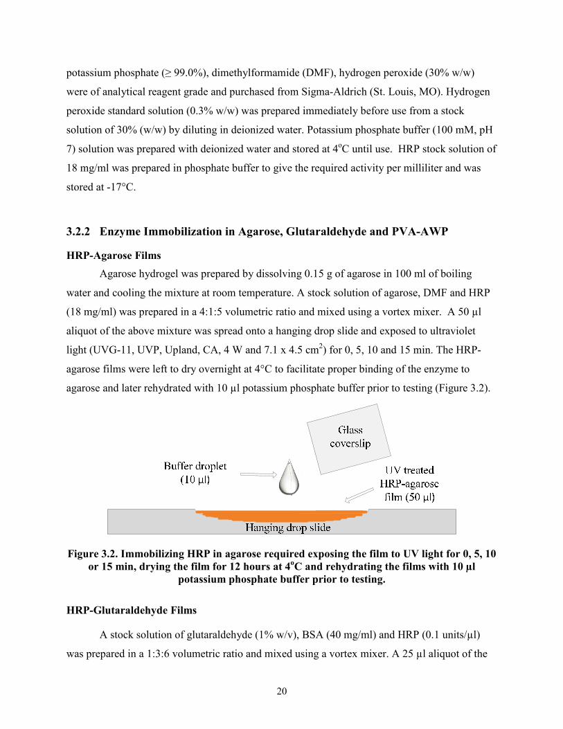
3.2.2 Enzyme Immobilization in Agarose, Glutaraldehyde and PVA-AWP
HRP-Agarose Films
Agarose hydrogel was prepared by dissolving 0.15 g of agarose in 100 ml of boiling
water and cooling the mixture at room temperature. A stock solution of agarose, DMF and HRP
(18 mg/ml) was prepared in a 4:1:5 volumetric ratio and mixed using a vortex mixer. A 50 µl
aliquot of the above mixture was spread onto a hanging drop slide and exposed to ultraviolet
light (UVG-11, UVP, Upland, CA, 4 W and 7.1 x 4.5 cm2) for 0, 5, 10 and 15 min. The HRP-
agarose films were left to dry overnight at 4°C to facilitate proper binding of the enzyme to
agarose and later rehydrated with 10 µl potassium phosphate buffer prior to testing (Figure 3.2).
Figure 3.2. Immobilizing HRP in agarose required exposing the film to UV light for 0, 5, 10
or 15 min, drying the film for 12 hours at 4oC and rehydrating the films with 10 µl
potassium phosphate buffer prior to testing.
HRP-Glutaraldehyde Films
A stock solution of glutaraldehyde (1% w/v), BSA (40 mg/ml) and HRP (0.1 units/µl)
was prepared in a 1:3:6 volumetric ratio and mixed using a vortex mixer. A 25 µl aliquot of the

21
mixture was spread on the hanging drop slide and left to dry for 3 h at room temperature to allow
for crosslinking between the enzyme and glutaraldehyde. The films were washed with 100 µl of
buffer to remove any unbound enzyme prior to testing.
HRP-AWP Films
HRP-AWP films were prepared by mixing PVA-AWP at 5.0% or 3.8% (w/v) in
potassium phosphate buffer with HRP (18 mg/ml) in a 1:1 volumetric ratio. The HRP-AWP
mixture was kept warm at 40°C to reduce the viscosity of the solution. A 50 µl aliquot of the
mixture was spread onto a hanging drop slide and exposed to UV light for 5, 10 or 15 min to
promote photo-polymerization of the matrix (Figure 3.3). After polymerization, the films were
rehydrated with 10 µl potassium phosphate buffer prior to testing.
For determining the activity of HRP, HRP-AWP films were prepared in the same way but
using HRP (0.1 units/ µl) and 25 µl aliquot of the HRP-AWP mixture. Activity of free HRP was
maintained at 1.25 units in each film. Instead of rehydrating the films with buffer, they were
washed with 100 µl buffer solution to remove any unbound enzyme prior to testing.
BSA Films
For control experiments, BSA (18 mg/ml), a non-catalytic protein was used in place of
HRP and BSA-agarose, BSA-glutaradehyde and BSA-AWP films were prepared using the same
procedures above. Absorbance of the substrate on the films in the absence of enzyme
demonstrates the ability of the porous membrane to facilitate substrate transport necessary for
enzymatic activity and indicates that the immobilized enzyme is solely responsible for
subsequent oxidation (Turner et al., 2004).

22
25µl AWP in potassium
phosphate (100mM, pH 7)
25µl HRP (18 mg/ml)
Buffer droplet
(10 µl)HRP – AWP film
Hanging drop slide
Hanging drop slide
Photocrosslinking (UV light)
HRP – AWP film (50 µl)
Hanging drop slide
0.13 W/cm2
5.8 cm
Figure 3.3. HRP-AWP mixtures were exposed to UV light for 5, 10, and 15 min and
rehydrated with 10 µl potassium phosphate buffer prior to testing.
3.2.3 Experimental Design
Effect of UV Light on Conformation of Immobilized Enzyme
UV–Vis spectroscopy was used to analyze changes in HRP structure since the position of
the maximum absorbance in the Soret absorption band occurs at 403 nm for native HRP. HRP
immobilized in agarose was used as a conventional and equivalent method for comparison. The
absorbances of the immobilized HRP and BSA films in agarose and PVA-AWP, between 300-
500 nm, were measured using a Cary 5G spectrophotometer (Varian Inc., Palo Alto, CA) and
recorded using Cary WinUV software.

23
Effect of Immobilization Medium on HRP Activity
A 20 µl aliquot of ABTS and 5 µl of hydrogen peroxide were added to the washed HRP-
glutaraldehyde, HRP-AWP, BSA-glutaraldehyde and BSA-AWP films to determine the activity
of HRP. The diffusion of ABTS and hydrogen peroxide through the films were monitored over a
period of two min and the absorbance at 415 was measured using a Cary 5G spectrophotometer.
Effect of Immobilization Medium on Leaching of HRP
Leaching of HRP from the films was tested by washing the HRP films with 100 µl of
buffer and measuring the activity, via ABTS assays, of the leached HRP in the buffer solution
(Figure 3.4).
Figure 3.4. Immobilized HRP films were washed with buffer and the activity of leached
HRP in the buffer was measured at 415 nm.
Data Analysis
Three replications were conducted for each type of film for determining the Soret band.
Data were corrected by subtracting the values of absorbance (absorbance units) of BSA from that
of HRP in the immobilized films, thereby removing the effect of the film on the enzyme (Figure
3.5). The first derivative of the absorbance values (AU) over wavelength was approximated

24
using a 10-point central difference numerical method. The position of the peak of the Soret band
coincided with the wavelength at which the first derivate was zero or changed from a positive
value to a negative value. This was repeated for the HRP-AWP films.
Regression analysis (R, Version 2.9.0, St. Louis, MO) was used to compare the linearity
of the absorbance units (AU) over time for HRP-AWP, HRP-glutaraldehyde, BSA-AWP and
BSA-glutaraldehyde films. The slope of the regression curve (AU/min) is the enzyme activity of
the films. Data were corrected by subtracting the values of activity (AU/min) of immobilized
BSA from that of immobilized HRP films. The experimental setup was arranged as a 2 x 3
factorial in a complete randomized design. Analysis of variance (ANOVA) and Fisher’s least
significant difference (LSD) test (R, Version 2.9.0, St. Louis, MO) were used to compare the
means of final enzyme activity values (AU/min). The level selected to show statistical
significance was 5% (p < 0.05).

25
Abso
rban
ce [
AU
]
0.0
0.2
0.4
0.6
0.8
1.0
(a) AHRP
ABSA
Abso
rban
ce [
AU
]
0.0
0.2
0.4
0.6
0.8
1.0
(b) AHRP - ABSA
Wavelength [nm]
Abso
rban
ce [
AU
]
Fir
st d
eriv
ativ
e ap
pro
xim
atio
n
300 350 400 450 500
0.0 -0.2
0.2 0.0
0.4 0.2
0.6 0.4
0.8 0.6
1.0 0.8
(c) AHRP - ABSA
First derivative approximation
Figure 3.5. Data were corrected by taking (a) original immobilized HRP sample and
immobilized BSA control and (b) subtracting the absorbance values of immobilized BSA
from those of HRP. (c) The first derivative of the absorbance over wavelength was
approximated using a 10-point central difference numerical method to determine the
position of the peak of the Soret band which occurred at the point where the first derivative
curve crossed the zero line.

26
3.3 Results and Discussion
3.3.1 Effect of UV Light on Conformation of Immobilized HRP
The position of Soret band peaks occurred approximately between 402-404 nm for HRP
immobilized in agarose, PVA-AWP (3.8% and 5% w/v) (Figure 3.6). The position of the Soret
band peak occurred at 404 nm for HPR immobilized in agarose and exposed to UV light for 15
min (Figure 3.6). This is due to longer exposure to UV light as compared to UV 5 and 10 min.
The position of Soret band peak in native HRP occurs at 403 nm. A shift of 1 nm is not
significant and the biological activity of the heme protein was not affected. Protein structure of
the enzyme was not affected by entrapment with UV exposure times of 5, 10 and 15 min.
An increase in the Soret absorption indicates that some change at the active site might be
responsible for the high enzyme activity. If there is no significant shift in the Soret band but
enzyme activity is high, it signifies that the active site maintains a conformation very similar to
that of the native enzyme although there might be a change in the bulk structure with a preserved
tertiary structure but a few changes in its secondary structure (Zhou et al., 2002). Prolonged UV
irradiation leads to protein denaturation (Neves-Petersen et al., 2007).The peak absorbance
(absorbance units) of the Soret band decreased as the UV exposure time increased in HRP-
agarose films. Peak absorbances of HRP-AWP films were higher than HRP-agarose films
(Figure 3.7).

27
Posi
tion o
f S
ore
t B
and [
nm
]
400
401
402
403
404
405
HRP-Agarose
Posi
tion o
f S
ore
t B
and [
nm
]
400
401
402
403
404
405
HRP-AWP(3.8% w/v)
UV Exposure Time [min]
Posi
tion o
f S
ore
t B
and [
nm
]
-5 0 5 10 15 20
400
401
402
403
404
405
HRP-AWP(5% w/v)
Figure 3.6. The average of three replications is reported for the position of the Soret band
in HRP-agarose films at 0, 5, 10, 15 min, HRP-AWP (3.8 and 5% w/v) films at 5, 10, 15 min
of UV exposure.

28
Sore
t B
and A
bso
rban
ce [
AU
]
0.0
0.5
1.0
1.5
2.0
HRP-Agarose
Sore
t B
and A
bso
rban
ce [
AU
]
0.0
0.5
1.0
1.5
2.0
HRP-AWP(3.8% w/v)
UV Exposure Time [min]
Sore
t B
and A
bso
rban
ce [
AU
]
-5 0 5 10 15 20
0.0
0.5
1.0
1.5
2.0
HRP-AWP(5% w/v)
Figure 3.7. The average of three replications is reported for Soret Band absorbance in
HRP- agarose films at 0, 5, 10, 15 min, HRP-AWP (3.8 and 5% w/v) films at 5, 10, 15 min
of UV exposure.

29
Peak absorbances of Soret band in HRP-AWP films were higher than those in HRP-
agarose films because there was no absorption of UV light by agarose during the exposure time
(Figures 3.8 and 3.9).
Ab
sorb
ance
corr
ecte
d [A
U]
300 340 380 420 460 500
0.0
0.2
0.4
0.6
0.8
1.0
HRP-agarose
UV 0
Ab
sorb
ance
corr
ecte
d [A
U]
300 340 380 420 460 500
0.0
0.2
0.4
0.6
0.8
1.0
HRP-agarose
UV 5
Wavelength [nm]
Abso
rban
cecorr
ecte
d [A
U]
300 340 380 420 460 500
0.0
0.2
0.4
0.6
0.8
1.0
HRP-agarose
UV 10
Wavelength [nm]
Abso
rban
cecorr
ecte
d [A
U]
300 340 380 420 460 500
0.0
0.2
0.4
0.6
0.8
1.0
HRP-agarose
UV 15
Figure 3.8. Three replications of corrected Soret absorption band are reported for HRP-
agarose films exposed to UV light for 0, 5, 10 and 15 min.
Peak absorbance of the Soret band in AWP 5% (w/v) was found to be 22%, 30%, 25%
higher than those using AWP (3.8% w/v) for UV exposure times 5, 10 and 15 min, respectively,
because AWP (5% w/v) absorbed more UV irradiation than AWP (3.8% w/v), which shielded
HRP from UV light (Figure 3.9).

30
Ab
sorb
ance
corr
ecte
d [A
U]
300 340 380 420 460 500
0.0
0.5
1.0
1.5
2.0
2.5
3.0
HRP-AWP (3.8% w/v)
UV 5
Ab
sorb
ance
corr
ecte
d [A
U]
300 340 380 420 460 500
0.0
0.5
1.0
1.5
2.0
2.5
3.0
HRP-AWP (5% w/v)
UV 5
Ab
sorb
ance
corr
ecte
d [A
U]
300 340 380 420 460 500
0.0
0.5
1.0
1.5
2.0
2.5
3.0
HRP-AWP (3.8% w/v)
UV 10
Ab
sorb
ance
corr
ecte
d [A
U]
300 340 380 420 460 500
0.0
0.5
1.0
1.5
2.0
2.5
3.0HRP-AWP (5% w/v)
UV 10
Wavelength [nm]
Abso
rban
cecorr
ecte
d [A
U]
300 340 380 420 460 500
0.0
0.5
1.0
1.5
2.0
2.5
3.0
HRP-AWP (3.8% w/v)
UV 15
Wavelength [nm]
Abso
rban
cecorr
ecte
d [A
U]
300 340 380 420 460 500
0.0
0.5
1.0
1.5
2.0
2.5
3.0HRP-AWP (5% w/v)
UV 15
Figure 3.9. Three replications of corrected Soret absorption band are reported for HRP-
AWP (3.8 and 5% w/v) films exposed to UV light for 5, 10 and 15 min. HRP-AWP (5%
w/v) absorbed more UV light as compared to HRP-AWP (3.8% w/v) films.
3.3.2 Effect of Immobilization Medium on HRP Activity
There was no difference (p < 0.05) observed in the activity of HRP immobilized in PVA-
AWP, for UV exposure times of 5, 10 and 15 min, and PVA-AWP concentrations of 3.8% (w/v)
and 5% (w/v) (Figure 3.10). Enzyme activity was lower (p < 0.05) when HRP was immobilized
in glutaraldehyde (Table 3.1).

31
Act
ivit
y [
AU
/min
]
0.0
0.2
0.4
0.6
0.8
1.0
HRP-AWP(3.8% w/v)
.
UV Exposure Time [min]
Act
ivit
y [
AU
/min
]
0 5 10 15 20
0.0
0.2
0.4
0.6
0.8
1.0
HRP-AWP(5% w/v)
Figure 3.10. Enzyme activity was measured over a period of 2 min in HRP immobilized in
PVA-AWP (3.8 and 5% w/v) for UV exposure times 5, 10 and 15 min.

32
Table 3.1. HRP activity values (AU/min) in HRP-AWP and HRP-glutaraldehyde films
(means of triplicate runs ± 1 standard error).
Immobilization
medium
UV
exposure
timec
(min)
PVA-AWP Concentration
(% w/v)c
Meanb
(concentration
and UV) 3.8 5 Mean
a
(UV)
PVA-AWP 5 0.34 ± 0.08 0.33 ± 0.11 0.34a 0.37 ± 0.13A
10 0.33 ± 0.17 0.40 ± 0.05 0.37a
15 0.31 ± 0.01 0.52 ± 0.10 0.42a
Meana
(Concentration) 0.32A 0.42A
Glutaraldehyde (1% w/v) 0.11 ± 0.02B
a Mean enzyme activity values followed by the same letter in a column (abc) or row (AB) were
not different (p < 0.05)
b Mean enzyme activity values followed by the same letter in the same column (AB) were not
different (p < 0.05)
c LSD value across UV exposure times and 3.8, 5% (w/v) concentrations were 0.1657 and
0.1353, respectively.
Enzymatic activity is inversely proportional to the concentration of glutaraldehyde
because extensive crosslinking may result in a distortion of the enzyme structure (i.e., the active
site conformation) (Chui and Wan, 1997). With this distortion, the accessibility and
accommodation of the substrate may be reduced, thus affecting the retention of biological
activity. The relative concentration of enzyme to glutaraldehyde should also be considered
(Okuda et al., 1991). Low concentrations of enzyme and glutaraldehyde tend to induce
intramolecular crosslinking by enhancing the probability that glutaraldehyde functional groups
will react with the same enzyme molecule (Zaborsky, 1973). Conditions should be chosen
carefully to favor intermolecular crosslinking between enzyme molecules instead of unwanted
intramolecular links, which could also be formed (Broun, 1976; Bano and Saleemuddin, 1980;
Gupta, 1993).
From the above results, it can be concluded that glutaraldehyde crosslinking has changed
the structure of immobilized HRP leading to lower activity when compared to immobilization in
PVA-AWP.

33
3.3.3 Effect of Immobilization Medium on Leaching of HRP
There was no difference (p < 0.05) observed in the activity of leached HRP from HRP-
AWP films, for UV exposure times of 5, 10 and 15 min, and PVA-AWP concentrations of 3.8%
(w/v) and 5% (w/v) (Figure 3.11). The activity of leached HRP from HRP-glutaraldehyde film
was higher (p < 0.05) when compared to HRP-AWP films (Table 3.2). For leaching, the films
were washed with buffer. Enzymes prefer aqueous environments creating a distinct possibility
for enzyme leaching during washing processes (Turner et al., 2004). The higher activity of the
buffer containing the leached HRP from HRP-glutaraldehyde when compared to HRP-AWP
films indicates higher mechanical stability of HRP-AWP films.
Act
ivit
y [
AU
/min
]
0.0
0.2
0.4
0.6
0.8
1.0
HRP-AWP(3.8% w/v)
UV Exposure Time [min]
Act
ivit
y [
AU
/min
]
0 5 10 15 20
0.0
0.2
0.4
0.6
0.8
1.0
HRP-AWP(5% w/v)
Figure 3.11. Leaching was measured in HRP immobilized in PVA-AWP (3.8 and 5% w/v)
films for UV exposure times 5, 10 and 15 min.

34
Table 3.2. Leached HRP activity values (AU/min) in HRP-AWP and HRP-glutaraldehyde
films (means of triplicate runs ± 1 standard error).
Immobilization
medium
UV
exposure
timec
(min)
PVA-AWP Concentration
(% w/v)c
Meanb
(concentration and
UV) 3.8 5 Mean
a
(UV)
PVA-AWP 5 0.46 ± 0.00 0.54 ± 0.05 0.50a 0.42 ± 0.17A
10 0.25 ± 0.15 0.42 ± 0.12 0.33a
15 0.45 ± 0.14 0.39 ± 0.01 0.42a
Meana
(Concentration) 0.39A 0.45A
Glutaraldehyde (1% w/v) 0.71 ± 0.11B
a Mean enzyme activity values followed by the same letter in a column (abc) or row (AB) were
not different (p < 0.05)
b Mean enzyme activity values followed by the same letter in the same column (AB) were not
different (p < 0.05)
c LSD value across UV exposure times and 3.8, 5% (w/v) concentrations were 0.2118 and
0.1729, respectively.
3.4 Conclusions
The positions of the Soret band absorbance peaks occurred between 402-404 nm for free
and immobilized HRP in PVA-AWP. The peak absorbance of the Soret band decreased as UV
exposure of HRP-agarose films increased. The peak absorbance of the Soret band in PVA-AWP
(5% w/v) was found to be 22%, 30%, 25% higher than those using PVA-AWP (3.8% w/v) for
UV exposure times 5, 10 and 15 min, respectively. From the above results, it can be concluded
that UV exposure did not affect the conformation of HRP immobilized in PVA-AWP and the
activity was higher in AWP (5% w/v).
There was no difference (p < 0.05) observed in the activity and leaching of HRP
immobilized in PVA-AWP, for UV exposure times of 5, 10 and 15 min, and PVA-AWP
concentrations of 3.8% (w/v) and 5% (w/v). Enzyme activity was lower (p < 0.05) and leaching
was higher (p < 0.05) when HRP was immobilized in glutaraldehyde. It can be concluded that

35
HRP immobilized in PVA-AWP had higher activity and mechanical stability when compared to
HRP immobilized in glutaraldehyde.

36
Chapter 4
EVALUATIO OF IMMOBILIZIG ALCOHOL-
OXIDASE I PVA-AWP POLYMER
4.1 Introduction
Immobilization is a double edged sword. On one hand immobilization attaches the
bioreceptors to an inert support material in a manner rendering them insoluble and fixing their
position in space, so they can be effectively utilized in continuous processes; on the other,
immobilizing the bioreceptor can reduce the bioactivity because of a compromise in the mass
transfer of analytes through the bioreceptor layer. The stability and activity of alcohol oxidase
(AOX) immobilized in glutaraldehyde, which has been effectively demonstrated in the
development of assays and devices for measuring ethanol content, was compared to AOX
immobilized in a polyvinyl (alcohol) azide-unit pendant water-soluble photopolymer (PVA-
AWP). PVA hydrogels offer several advantages, such as better elasticity, low toxicity,
biocompatibility with enzymes and yeast cells, mechanical and long-term stability, and
biodegradability (Stammen et al., 2001; Li et al., 1998; Hyon et al., 1994; Boyd and Yamazaki,
1994). PVA-AWP polymerizes upon exposure to ultraviolet (UV) light. Enzymes, such as AOX,
can be entrapped and immobilized within the polymerized matrix and be used multiple times in
biosensing and bioprocessing (Gurban et al., 2008). AOX catalyzes the oxidation of lower
primary aliphatic alcohols to the respective aldehydes with oxygen as the electron acceptor and
releases hydrogen peroxide (H2O2), which is electrochemically detectable using cobalt
phthalocyanine (CoPC) modified screen printed carbon electrodes (Veenhuis et al., 1983;
Azavedo et al., 2005; Wring and Hart, 1992) (Figure 4.1). Immobilization using horseradish
peroxidase performed in the previous study was to determine whether PVA-AWP and UV
exposure cause a change in the conformation of the enzyme. However optical measurement of
enzyme activity was not practically feasible.

37
In this study, alcohol oxidase was immobilized in two immobilization media –
glutaraldehyde (AOX-glutaraldehyde) and PVA-AWP (AOX-AWP). Glutaraldehyde is used for
immobilizing enzymes by crosslinking where the enzymes are joined to each other to form a
three-dimensional complex structure (Massafera et al., 2009). Immobilization of enzymes in
glutaraldehyde by crosslinking is a standard and widely used method in biosensing. However,
glutaraldehyde is toxic which limits its application to living cells and many enzymes as they
might be damaged (Eggins, 2002).
Figure 4.1. Alcohol Oxidase catalyzes the oxidation of ethanol to release hydrogen
peroxide. The redox reactions between hydrogen peroxide and the CoPC mediator produce
a flow of electrons that is proportional to the amount of ethanol in the sample.
The objectives of this study were to:
1. determine the effect of PVA-AWP film thickness and UV exposure time on the sensitivity
of detection;
2. determine the effect of immobilizing AOX in PVA-AWP on biosensor sensitivity as
compared to a conventional immobilization medium, glutaraldehyde; and
3. assess the stability of AOX immobilized in PVA-AWP after 24 h at different storage
temperatures.

38
4.2 Materials and Methods
4.2.1 Chemicals and Other Reagents
Alcohol oxidase (AOX, EC. 232-971-3, 30 Units/mg protein) from Pichia pastoris was
purchased from Sigma Aldrich (St. Louis, MO) as a phosphate-buffered 30% sucrose solution.
PVA-AWP was received from Toyo Gosei Co. (Chiba, Japan) in the form of 6% (w/v) aqueous
solution. Bovine serum albumin (BSA, 96% w/v), glutaraldehyde (25% w/v), potassium
phosphate (≥ 99.0%), and hydrogen peroxide (30% w/w) were of analytical reagent grade and
purchased from Sigma-Aldrich (St. Louis, MO). Potassium phosphate buffer solutions (100 mM,
pH 7) were prepared with deionized water and stored at 4oC until use. Ethanol standard
solutions were prepared by dilution in deionized water immediately before use.
4.2.2 Methods
Biosensor Preparation
Electrochemical tests were carried out using screen printed carbon electrodes (SPCE,
Figure 4.2). The SPCE cell consisted of a CoPC-mediated carbon working electrode and a 60/40
Ag/AgCl electrode which served as the reference and counter electrode. The electrode cell was
arranged in a circular configuration, with the working electrode in the center (27.61 mm2) and
the reference/counter electrode around it (41.7 mm2). The gap between the two electrodes was
1.12 mm.
Chronoamperometric measurements were performed using an electrochemical analyzer
or potentiostat (WaveNow, WEB50-EDU, Pine Instrument Company, Grove City, PA), at a
potential of 400 mV vs. Ag/AgCl. Electrocatalytic oxidation of hydrogen peroxide occurs readily
at this potential (Boujtita et al., 2000). Chronoamperometry was performed on non-immobilized
AOX, AOX immobilized in PVA-AWP (AOX-AWP) and AOX immobilized in glutaraldehyde
(AOX-glutaraldehyde) films. A 20 µl volume of ethanol solution (0.02-1.7 mM) was deposited
over the entire electrode cell ensuring that both the working and reference/counter electrodes
were covered (Figure 4.2). Current response to ethanol sample at different concentrations of
ethanol solutions were calculated from the resulting chronoamperograms (Figure 4.3).
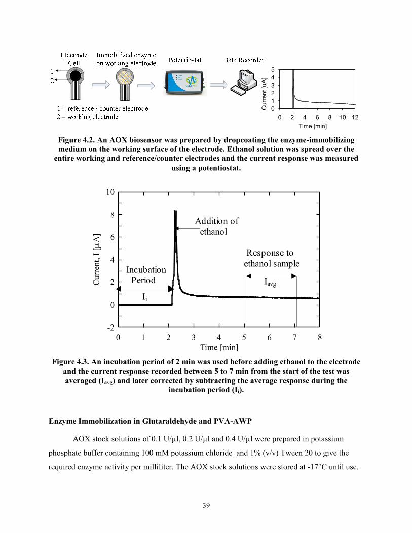
39
0
1
2
3
4
5
0 2 4 6 8 10 12
Current [uA]
Time [min]
Figure 4.2. An AOX biosensor was prepared by dropcoating the enzyme-immobilizing
medium on the working surface of the electrode. Ethanol solution was spread over the
entire working and reference/counter electrodes and the current response was measured
using a potentiostat.
Time [min]
Curr
ent,
I [µ
A]
0 1 2 3 4 5 6 7 8
-2
0
2
4
6
8
10
Incubation
Period
Ii
Response to
ethanol sample
Iavg
Addition of
ethanol
Figure 4.3. An incubation period of 2 min was used before adding ethanol to the electrode
and the current response recorded between 5 to 7 min from the start of the test was
averaged (Iavg) and later corrected by subtracting the average response during the
incubation period (Ii).
Enzyme Immobilization in Glutaraldehyde and PVA-AWP
AOX stock solutions of 0.1 U/µl, 0.2 U/µl and 0.4 U/µl were prepared in potassium
phosphate buffer containing 100 mM potassium chloride and 1% (v/v) Tween 20 to give the
required enzyme activity per milliliter. The AOX stock solutions were stored at -17°C until use.

40
Tween 20 is a surfactant that is used to lower the surface tension of a liquid, enhancing the
contact angle between liquid and the electrode surface. AOX-immobilizing medium solutions
were dropcoated over the working surface of the electrode to yield an enzyme loading of 0.5 U
(0.02 U/mm2) per electrode.
AOX-Glutaraldehyde Films
A stock solution of glutaraldehyde (1% w/v), BSA (40 mg/ml) and AOX (0.2 U/µl) was
prepared in 1:3:6 volumetric ratio and mixed using a vortex mixer. A 4 µl aliquot of the enzyme-
glutaraldehyde mixture was dropcoated on the working electrode and left to dry for 3 h at room
temperature to allow for crosslinking between the enzyme and glutaraldehyde.
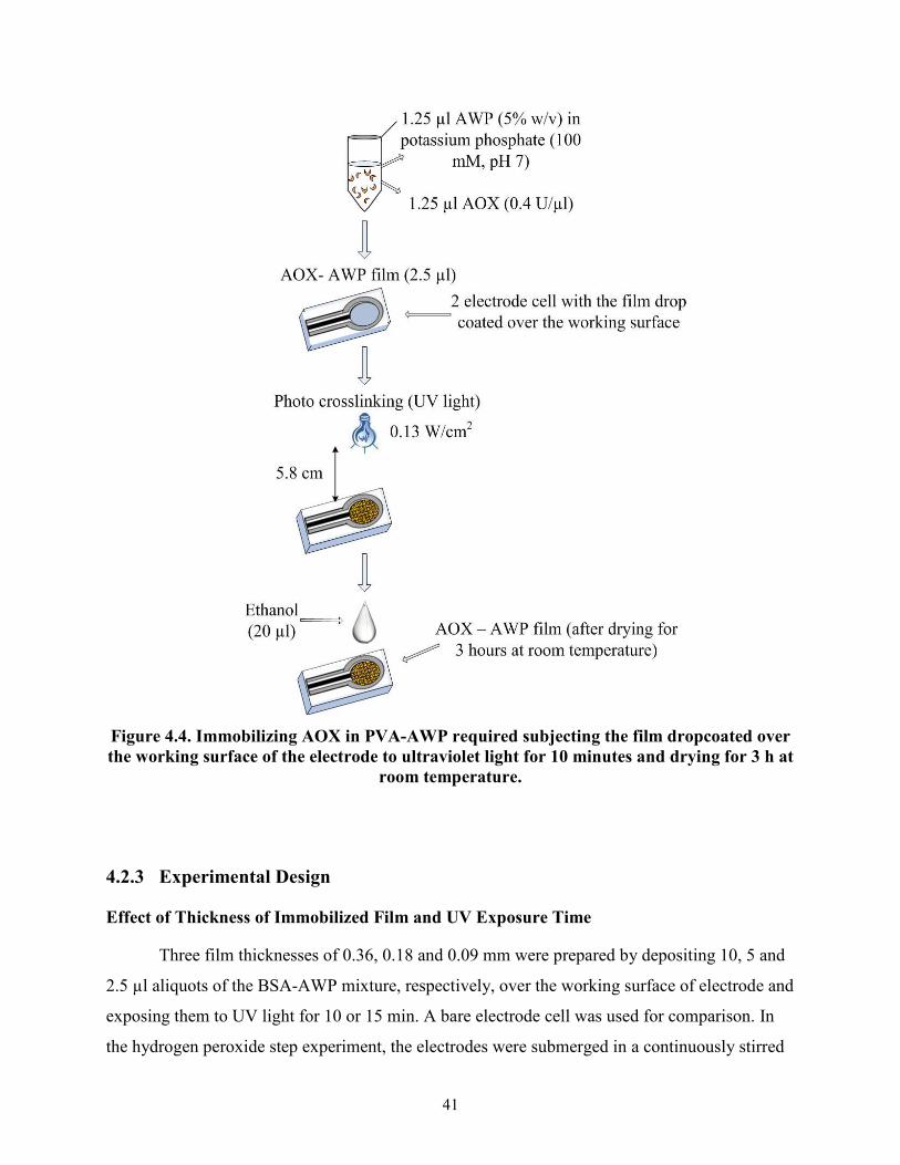
AOX-AWP Films
PVA-AWP was diluted to 5% (w/v) in potassium phosphate buffer. The PVA-AWP
solution was kept warm at 40°C to reduce its viscosity. PVA-AWP and AOX (0.4 U/µl) were
mixed in 1:1 volumetric ratio. An aliquot of the AOX-AWP mixture on the electrode was
exposed to ultraviolet light (UVG-11, UVP, Upland, CA, 4 W and 7.1 x 4.5 cm2) to promote
polymerization of the PVA-AWP medium and entrapment of AOX (Figure 4.4). The films were
left to dry for 3 h at room temperature before testing.
To determine the volume of PVA-AWP to be used as immobilization medium above and
UV exposure time, hydrogen peroxide (H2O2) step experiment was conducted. Three volumetric
aliquots of PVA-AWP (5% w/v) and BSA (40 mg/ml) in 1:1 volumetric ratio were immobilized
on the electrode using the procedure described for immobilizing AOX in PVA-AWP. BSA was
used as a noncatalytic protein in place of AOX. The films were left to dry for 3 h at room
temperature before testing.
$on-immobilized AOX Films
Non-immobilized AOX films were prepared by dropcoating 5 µl of AOX (0.1 U/µl) on
the working surface to yield an enzyme loading of 0.5 U per electrode. The films were left to dry
for 3 h at room temperature before testing.
41
Figure 4.4. Immobilizing AOX in PVA-AWP required subjecting the film dropcoated over
the working surface of the electrode to ultraviolet light for 10 minutes and drying for 3 h at
room temperature.
4.2.3 Experimental Design
Effect of Thickness of Immobilized Film and UV Exposure Time
Three film thicknesses of 0.36, 0.18 and 0.09 mm were prepared by depositing 10, 5 and
2.5 µl aliquots of the BSA-AWP mixture, respectively, over the working surface of electrode and
exposing them to UV light for 10 or 15 min. A bare electrode cell was used for comparison. In
the hydrogen peroxide step experiment, the electrodes were submerged in a continuously stirred

42
10 ml potassium phosphate buffer solution. A 10 µl aliquot of hydrogen peroxide (0.3% w/w)
was added to the solution at regular intervals of 3 min and the corresponding change in current
response was recorded (Figure 4.5). The average current response (IC) after each hydrogen
peroxide addition was calculated and plotted against hydrogen peroxide concentration. The slope
of the curve is the sensitivity of the film (Figure 4.6). The test was replicated three times.
Regression analysis (R, Version 2.9.0, St. Louis, MO) was used to compare the sensitivities.
Statistical significance was tested at 5% (p < 0.05).
Time [min]
Curr
ent,
I [µ
A]
0 5 10 15 20 25 300
5
10
15
Io
Iavg
Io = average I for 2 min for buffer
Iavg = average I for 3 min
Ic = Iavg - Io
Figure 4.5. A BSA-AWP film coated electrode was expected to respond linearly to the
addition of 10 µl aliquots of hydrogen peroxide (0.3 % w/w) at 3 min intervals. An initial
incubation period of 2 min was allowed before the addition of hydrogen peroxide.
Effect of Immobilization Medium on Biosensor Sensitivity
An AOX-based biosensor was first exposed to ambient air for a period of 2 min prior to
the addition of 20 µl ethanol sample (0.0001 - 0.01% v/v). Amperometric measurements were
recorded for an additional 6 min. The current responses between 5 and 7 min were averaged (as
shown in Figure 4.3 in biosensor preparation) and corrected by subtracting the average current
response for deionized water, which was used as a control (Figure 4.6). All tests were replicated
three times. Regression analysis (R, Version 2.9.0, St. Louis, MO) was used to compare the

43
linearity of the current responses to various ethanol concentrations for non-immobilized AOX,
AOX-glutaraldehyde, and AOX-AWP films. The slope of the regression curve is the sensitivity
of the AOX-based biosensor. Statistical significance was tested at 5% (p < 0.05).
Ethanol Concentration [% v/v]
Curr
ent,
IC [µ
A]
0.000 0.002 0.004 0.006 0.008 0.010 0.012
0.0
0.5
1.0
1.5
2.0
IC = Iavg - Ii - Io
Io = control
Figure 4.6. The corrected current response of biosensors with immobilized AOX was
expected to respond linearly with increasing levels of ethanol in the sample.
The response times were calculated for each type of film (Figure 4.7). The peak of the
current response curve, i.e., deposition of ethanol sample, coincided with the time at which the
first derivative was zero or changed from a positive value to a negative value. The current
response curve stabilizes once the ethanol substrate has diffused through the AOX layer (non-
immobilized or immobilized) and coincided when the first approximation curve was zero. The
time elapsed between the peak of the current response curve and the current stabilization point
was defined as the response time, tr (Figure 4.8).
The effect of immobilizing medium on the diffusion rate was interpreted from the
response times. Student’s t-test (R, Version 2.9.0, St. Louis, MO) was used to determine the
difference between the response times for the three types of enzyme films. The level selected to
show statistical significance was 5% (p < 0.05). Pearson correlation test (R, Version 2.9.0, St.
Louis, MO) was used to compare the effect of ethanol concentration on the response time for the
three types of enzyme films.

44
Time [min]
Curr
ent,
I [µ
A]
1 2 3 4 5-1
0
1
2
3
4
5
PVA-AWP
Glutaraldehyde
Non-immobilized
Figure 4.7. The response time, tr, is the time it takes for the current response to stabilize
after the addition of ethanol.
Time [min]
Curr
ent,
I [µ
A]
Fir
st d
eriv
ativ
e ap
pro
xim
atio
n, I'
2.0 2.1 2.2 2.3 2.4 2.5-10 -12
0 -6
10 0
20 6
30 12
40 18
50 24
Response time
(tr)
I
I'
Figure 4.8. The response time, tr, is the time between two points where the first derivative
approximation of the curve is zero.
Stability of Immobilized AOX-AWP Films
The stability of AOX-AWP films was measured by comparing the sensitivity of AOX-
based biosensors after a 24 h storage period at different temperatures. AOX-AWP coated
electrodes were prepared as usual and packed in heat sealed aluminum packs prior to storage at

45
three different temperatures, -17°C, 3°C and room temperature, which was approximately 23°C.
After 24 h, they were tested for current responses using 20 µl of ethanol solution of 0.005% (v/v)
concentration.
4.3 Results and Discussion
4.3.1 Effect of UV Exposure Time and Film Thickness of PVA-AWP on Biosensor
Sensitivity
As the volume of AOX-AWP mixtures dropcoated on the electrode increased, the
thickness of the enzyme film also increased and the sensitivity decreased (Table 4.1). Increased
film thickness was negatively correlated with biosensor sensitivity (Figure 4.9). Regression
analyses determined that sensitivity was different (p < 0.05) for UV 10 and 15 min exposure
times and film thicknesses 2.5 and 5 µl. Biosensors with 2.5 µl volume films showed sensitivities
that were higher (p < 0.05) than those with 5 and 10 µl films. Based on these results, AOX-AWP
film of 0.09 mm thickness (2.5 µl volume) and UV exposure time of 10 min was chosen in
subsequent experiments.
Table 4.1. Sensitivity and coefficient of variation for UV 10 and 15 min exposed BSA-AWP
films.
Volume of
AOX-AWP film
(µl)
Estimated
film
thickness
(mm)
UV 10 min UV 15 min
Average
Sensitivitya [A/M]
Coefficient
of variation
[R2]
Average
Sensitivityb
[A/M]
Coefficient
of variation
[R2]
0.0 0.00 0.0158a 0.99 0.0158A 0.99
2.5 0.09 0.0111b 0.99 0.0106B 0.99
5.0 0.18 0.0092c 0.99 0.0074C 0.96
10.0 0.36 0.0068d 0.99 0.0070d 0.99
a Mean sensitivity values followed by the same letter in a column (abcd, ABCD) or row (abcd,
ABCD) were not different (p < 0.05)

46
H2O2 Concentration [µM]
Cu
rren
t, I
C [µ
A]
0 200 400 600 800 10000
5
10
15
20
No film
H2O2 Concentration [µM]
Cu
rren
t, I
C [µ
A]
0 200 400 600 800 10000
5
10
15
20
2.5 µl
UV 10
H2O2 Concentration [µM]
0 200 400 600 800 10000
5
10
15
20
2.5 µl
UV 15
H2O2 Concentration [µM]
Cu
rren
t, I
C [µ
A]
0 200 400 600 800 10000
5
10
15
20
5 µl
UV 10
H2O2 Concentration [µM]
0 200 400 600 800 10000
5
10
15
20
5 µl
UV 15
Figure 4.9. Corrected average current responses from three replications were plotted
against hydrogen concentration in the buffer solution for no film, 2.5, 5 and 10 µl films
exposed to UV light for 10 and 15 min. As the film thickness increased, the sensitivity
decreased.

47
H2O2 Concentration [µM]
Cu
rren
t, I
C [µ
A]
0 200 400 600 800 10000
5
10
15
20
10 µl
UV 10
H2O2 Concentration [µM]
0 200 400 600 800 10000
5
10
15
20
10 µl
UV 15
Figure 4.9. (cont.)
4.3.2 Effect of Immobilization Medium on Biosensor Sensitivity
Current responses for ethanol solutions (0.001-0.01% v/v) catalyzed in three enzyme
films -- non-immobilized AOX, AOX immobilized in glutaraldehyde, and AOX immobilized in
PVA-AWP films were corrected and the average current responses were plotted against ethanol
concentration for each type of film (Figure 4.10). Regression analyses determined that the
current responses increased linearly with increasing levels of ethanol (Table 4.2) and the linear
response was higher for immobilized AOX films than non-immobilized AOX film. Biosensors
with AOX immobilized in glutaraldehyde had higher sensitivity (p < 0.05) followed by non-
immobilized AOX and AOX immobilized in PVA-AWP. Though the sensitivity in AOX-
glutaraldehyde film was highest, the variability was also highest. The lower limit of ethanol
detection using AOX immobilized in PVA-AWP was 171.3 µM; AOX in glutaraldehyde films
was 17.13 µM; and non-immobilized AOX was between 485-625 µM.

48
Table 4.2. Sensitivity, coefficient of variation and lower limit of detection for each type of
corrected AOX film.
Type of film Sensitivity
a
[A/M]
Coefficient of Variation
[R2]
Limit of
detection
[µM]
Non-immobilized AOX 113.62a 0.84 428-625
AOX-glutaraldehyde 154.49b 0.99 17.13
AOX-AWP 77.88c 0.98 171.3
a Mean sensitivity values followed by the same letter in a column (abcd) were not different (p <
0.05)

49
Curr
ent,
IC [µ
A]
0.0
0.5
1.0
1.5
2.0
Non-immobilized AOX
Curr
ent,
IC [µ
A]
0.0
0.5
1.0
1.5
2.0
AOX-glutaraldehyde
Ethanol Concentration [% v/v]
Curr
ent,
IC [µ
A]
0.000 0.002 0.004 0.006 0.008 0.010 0.012
0.0
0.5
1.0
1.5
2.0
AOX-AWP
Figure 4.10. For non-immobilized and immobilized AOX, the current responses increased
linearly with increasing levels of ethanol. Although sensitivity was highest with AOX-
glutaraldehyde films, the variability in the response was also greatest. Error bars are ±1
standard error.

50
The diffusion rate of the substrate into the three different enzyme films was assessed by
comparing the response times in the films for different ethanol concentrations (Table 4.3). The t-
test showed that all the response times were different (p < 0.05). The response times for non-
immobilized AOX were lower (p < 0.05) than those for immobilized AOX films. This is due to
the thickness of the immobilized AOX films. The response times for AOX-glutaraldehyde films
were lower than AOX-AWP films for most of the ethanol concentrations. The viscosity of 10%
(w/v) AWP is 2825 cP/ 25°C (Ito et al., 2005). The AOX-AWP films were based on 5% (w/v)
PVA-AWP solutions which, even after a 1:1 dilution with aqueous AOX solution, were more
viscous than 1% (w/v) glutaraldehyde in water. The viscous nature of PVA-AWP resulted in
denser enzyme films than glutaraldehyde films, and the density and thickness of the films slowed
down the diffusion of substrate to the electrodes.
Table 4.3. Average response times from three replications for the three enzyme films.
Ethanol
Concentration
[%v/v]
Average Response Time [min]
on-immobilized
AOX
AOX-glutaraldehyde AOX-AWP
0.0001 0.26 0.47 0.59
0.0005 0.24 0.57 0.62
0.0010 0.42 0.56 0.60
0.0025 0.13 0.43 0.70
0.0040 0.16 0.44 0.38
0.0050 0.29 0.39 0.43
0.0075 0.31 0.43 0.42
0.0100 0.29 0.44 0.46
Response times for AOX-glutaraldehyde and AOX-AWP films had a moderately
decreasing linear correlation relationship with increasing ethanol concentrations (Figure 4.11,
Table 4.4) but the response time for non-immobilized AOX film was not affected by ethanol
concentration. As the concentration of ethanol increased, the number of ethanol molecules

51
available for reaction increased and the time taken for them to reach the sensor surface which
had the immobilized enzyme decreased.
Table 4.4. Pearson correlation coefficients for the three enzyme films.
Type of film Correlation Coefficient
Non-immobilized AOX 0.07
AOX-glutaraldehyde -0.62
AOX-AWP -0.67
Ethanol Concentration [% v/v]
Res
ponse
Tim
e [m
in]
0.000 0.002 0.004 0.006 0.008 0.010 0.0120.0
0.1
0.2
0.3
0.4
0.5
0.6
0.7
0.8
0.9AOX-AWP
AOX-glutaraldehyde
Non-immobilized AOX
Figure 4.11. The average response times decreased moderately with increasing levels of
ethanol for AOX-AWP and AOX-glutaraldehyde films while there was no specific trend for
non-immobilized AOX film.
4.3.3 Stability of Immobilized AOX-AWP Films
Temperature and time had an effect on the current response given by immobilized AOX-
AWP films. Ethanol concentration of 0.005% (v/v) was used for comparison. Current response

52
decreased by 7%, 36% and 40% for films stored at -17°C, 3°C and 23°C, respectively (Figure
4.12, Table 4.5).
Table 4.5. Decrease in current response was calculated for AOX immobilized in PVA-AWP
films stored for 24 hours at -17°C, 3°C and 23°C.
Method Storage Temperature
[°C]
Current
[µA]
Decrease in current response
[%]
1 No Storage 0.82 -
2 -17 0.76 6.99
3 4 0.53 35.72
4 23 0.49 40.29
Curr
ent,
I [µ
A]
0 1 2 3 4 5
0.0
0.2
0.4
0.6
0.8
1.0
No storage -17°C 3°C 23°C
Figure 4.12. Average current responses from three replications are reported for stability of
AOX immobilized in PVA-AWP films. Films which were not stored were compared with
films stored at three different temperatures for 24 h and tested using ethanol solution of
0.005% (v/v) concentration. Error bars are ±1 standard error.

53
4.4 Conclusions
The current response decreased (p < 0.05) with increasing thickness of PVA-AWP films
on the electrode and UV exposure 10, 15 min. The current response of the AOX-based biosensor
varied with type of immobilization medium of the enzyme. Immobilized AOX films gave higher
current responses compared to non-immobilized AOX film for lower concentrations of ethanol
solution. The linear responses of sensors with immobilized AOX were higher than non-
immobilized AOX sensors. Biosensors with AOX immobilized in glutaraldehyde had higher
sensitivity (p < 0.05) followed by non-immobilized AOX and AOX immobilized in PVA-AWP.
The lower limit of ethanol detection using AOX immobilized in PVA-AWP was 171.3 µM;
AOX in glutaraldehyde films was 17.13 µM; and non-immobilized AOX was between 485-625
µM. The rate of diffusion of ethanol into the films was lower for immobilized AOX films
compared to non-immobilized AOX film. The rate of diffusion for immobilized AOX films had
a moderately decreasing linear relationship with increasing concentrations of ethanol. Current
response decreased by 40%, 36% and 7% when AOX immobilized in PVA-AWP films were
stored for 24 h at 23°C, 3°C and -17°C, respectively.

54
CHAPTER 5
COCLUSIOS AD FUTURE WORK
Immobilization in PVA-AWP does not change the conformation of HRP and it has higher
mechanical stability as compared to glutaraldehyde. Thickness of PVA-AWP film layer and UV
exposure time negatively affected the sensitivity of detection. Sensors with immobilized alcohol
oxidase had more linearity than those with non-immobilized AOX. The rate of diffusion of
ethanol into the immobilized enzyme films decreased with increase in thickness of film on the
electrode and had a moderately decreasing linear relationship with increase in concentration of
ethanol. Current response decreased when AOX immobilized in PVA-AWP films were stored at
24 h at different temperatures.
From these results, the effects of different parameters on the activity of HRP and
sensitivity of AOX immobilized in PVA-AWP based biosensor can be further characterized:
1. The effect of enzyme to polymer ratio on the activity of HRP immobilized in PVA-AWP
can be characterized to determine increased entrapment of enzyme.
2. A wider range of film thickness and UV exposure times could be used to determine the
effect on the AOX based biosensor immobilized in PVA-AWP.
3. The effect of pH, temperature and storage conditions in buffer on the sensitivity of the
AOX-AWP biosensor can be characterized.
4. The effect of lower UV exposure times on the sensitivity of the biosensor can be
analyzed.

55
REFERECES
Alcock, S.J., and A.P.F. Turner. 1994. Continuous analyte monitoring to aid clinical practice.
IEEE Engineering in Medicine and Biology 13: 319-325.
Amine, A., H. Mohammadi, I. Bourais, and G. Palleschi. 2006. Enzyme inhibition-based
biosensors for food safety and environmental monitoring. Biosensors and Bioelectronics 21:
1405-1423.
Azavedo A.M, D. Miguel, F. Prazeres, M. Joaquim, S. Cabral, and L.P. Fonseca. 2005. Ethanol
biosensors based on alcohol oxidase. Biosensors and Bioelectronics 21: 235-247.
Bano, B., and M. Saleemuddin. 1980. Studies on chemically aggregated trypsin using
glutaraldehyde. Indian Journal of Biochemistry and Biophysics 17: 12-17.
Bayley, H., and J. R. Knowles. 1977. Photoaffinity labeling. In Methods in Enzymology, 69-114.
New York, N.Y.: Academic Press.
Bickerstaff, G.F. 1984. Application of immobilized enzymes to fundamental studies on enzyme
structure and function. In Topics in Enzyme and Fermentation Biotechnology, 162-201.
Chichester, U.K.: Ellis Horwood.
Bickerstaff, G.F. 1995. Impact of genetic technology on enzyme technology. The Genetic
Engineer and Biotechnologist 15: 13-30.
Bickerstaff, G.F. 1997. Immobilization of enzymes and cells. Methods in Biotechnology, vol. 1.
Totowa, New Jersey: Human Press Inc.
Bommarius, A.S., M. Schwarm, and K. Drauz. 1998. Biocatalysis to amino acid based chiral
pharmaceuticals - examples and perspectives. Journal of Molecular Catalysis B: Enzymatic
5: 1-11.
Bos, E.S., A.A. van der Doelen, N. van Rooy, and A.H.W.M. Schuurs. 1981. 3,3′,5,5′-
Tetramethylbenzidine as an Ames test-negative chromogen for horseradish peroxidase in
enzyme immunoassay. Journal of Immunoassay 2: 187-204.
Boyd, S., and H. Yamazaki. 1994. Use of polyvinyl-alcohol as a stabilizer of peroxidase-
antibody conjugate for enzyme immunoassay. Biotechnology Techniques 8: 123-128.
Brodelius, P. 1985. Immobilized plant cells. In Enzyme and Immobilized Cells in Biotechnology,
109-148. London, U.K.: Benjamin Cummings.
Broun, G.B. 1976. Chemically aggregated enzymes. In Methods in Enzymology, 263-280, vol.
XLIV. New York, N.Y.: Academic Press.

56
Bucke, C. 1983. Immobilized cells. Philosophical Transactions of the Royal Society of London
series B-Biological 300: 369-389.
Cabral, J.M.S., and J.F. Kennedy. 1991. Covalent and coordination immobilization of protein. In
Protein Immobilization, 73-138. New York, N.Y.: Marcel Dekker.
Campas, M., D. Szydlowska, M. Trojanowicz, and J. Marty. 2005. Towards the protein
phosphatase-based biosensor for microcystin detection. Biosensors and Bioelectronics 20:
1520-1530.
Campbell, D.H., F.L. Luescher, and L.S. Lerman. 1951. Immologic adsorbents. I. Isolation of
antibody by means of a cellulose-protein antigen. Proceedings of the $ational Academy of
Sciences USA 37: 575-578.
Chang, K., C. Chang, S. Chou, H. Han, and C. Chen. 2007. Characterization of a planar L-
glutamate amperometric biosensor immobilized with a photo-crosslinkable polymer
membrane. Sensors and Actuators B: Chemical 122: 195-203.
Chang, T.M.S. 1977. Medical Application of Immobilized Enzymes and Proteins. Vols I and II.
New York, N.Y.: Plenum Press.
Cheetham, P.S.J. 1998. What makes a good biocatalyst? Journal of Biotechnology 66: 3-10.
Childs, R. E., and W.G. Bardsley. 1975. The steady-state kinetics of peroxidase with 2,2'-azino-
di-(3-ethyl-benzthiazoline-6-sulphonic acid) as chromogen. Biochemical Journal 145: 93-
103.
Chui, W.K., and L.S. Wan. 1997. Prolonged retention of cross-linked trypsin in calcium alginate
microspheres. Journal of Microencapsulation 14: 51-61.
Clark, D.S. 1994. Can immobilization be exploited to modify enzyme activity? Trends in
Biotechnology 12: 439-443.
Clark, L.C. Jr., and C. Lyons. 1962. Electrode systems for continuous monitoring in
cardiovascular surgery. Annals of the $ew York Academy of Sciences 102: 29-45.
Cowell, D. C., A.A. Dowman, J.D. Newman, R. Pirzad, and C. Rantle. 1992. The polycrystalline
fluoride ion-selective electrode and horseradish peroxidase--an alternative electrochemical
biosensor. Biosensors and Bioelectronics 7: 27-38.
Cristallini, C.L.L., M.G. Cascone, G. Polacco, D. Lupinacci, and N. Barbani. 1997. Enzyme-
based bioartificial polymeric materials: the α- amylase–poly(vinyl alcohol) system. Polymer
International 44: 5010–516.
de Alteriis E., P. Parascandola, S. Salvadore, and V. Sardi. 1985. Enzyme immobilization within
insolubilized gelatin. Journal of Chemical Technology and Biotechnology 35 B: 60-64.

57
Dennison, M.J., and A.P.F. Turner. 1995. Biosensors for environmental monitoring.
Biotechnology Advances 139: 1-12.
Dordick, J. S. 1992. Enzymatic and chemoenzymatic approaches to polymer synthesis. Trends in
Biotechnology 10:287-293.
Dunford, H.B. 1991. Horseradish peroxidase: Structure and kinetic properties. In Peroxidases in
Chemistry and Biology, 1-24. Boca Raton, FL.: CRC Press.
Dunlap, B.R. 1974. Immobilized Chemicals and Affinity Chromatography. New York, N.Y.:
Plenum Press.
Eaton, W.A., L.K. Hanson, P.J. Stephens, J.C. Sutherland, and J.B.R. Dunn. 1978. Optical
spectra of oxy- and deoxyhemoglobin. Journal of the American Chemical Society 100: 4991-
5003.
Eaton, W.A., and J. Hofrichter. 1981. Polarized absorption and linear dichroism spectroscopy of
hemoglobin. Methods in Enzymology 76: 175-261.
Eggins, B.R. 2002. Chemical Sensors and Biosensors. England: John Wiley & Sons.
Fink, G., H. Fasold, W. Rommel, and R. Brimacombe. 1980. Reagents suitable for the
crosslinking of nucleic-acids to proteins. Analytical Biochemistry 108: 394-401.
George, P., and G.I.H. Hanania. 1953. A spectrophotometric study of ionizations in
methaemoglobin. Biochemical Journal 55: 236-243.
Gibson, T.D. 1999. Biosensors: the stabilité problem. Analusis 27: 630-638.
Gibson, T.D., and J.R. Woodward. 1992. Protein Stabilization in Biosensor Systems. Washington
D.C.: ACS Books.
Goldstein, L. 1976. Kinetic behavior of immobilized enzyme systems. In Methods in
Enzymology, 397-443. New York, N.Y.: Academic.
Goodno, C.C., H.E. Swaisgood, and G.L. Catignani. 1981. A fluorimetric assay for available
lysine in proteins. Analytical Biochemistry 115: 203-211.
Groboillot, A., D.K. Boadi, D. Poncelot, and R.J. Neufeld. 1994. Immobilization of cells for
application in the food industry. Critical Reviews in Biotechnology 14: 75-107.
Gupta, M.N. 1993. Crosslinking techniques: application to enzyme and protein stabilization and
bioconjugate preparation. In Biocatalyst Design for Stability and Specificity, 307-324.
Washington, D.C.: American Chemical Society.
Gurban, A. M., T. Nogue, C. Bala, and L. Rotariu. 2008. Improvement of NADH detection using
prussian blue modified screen-printed electrodes and different strategies of immobilization.
Sensors and Actuators B: Chemical 128: 536-544.

58
Herskovits, T.T., and H. Jaillet. 1969. Structural stability and solvent denaturation of myoglobin.
Science 163: 282-285.
Huang, X.L., G.L. Catignani, and H.D. Swaisgood. 1995. Immobilization of biotinylated
transglutaminase by bioselective adsorption to immobilized avidin and characterization of the
immobilized activity. Journal of Agricultural and. Food Chemistry 43: 895-901.
Hyon, S.H., W.I. Cha, Y. Ikada, M. Kita, Y. Ogura, and Y. Honda. 1994. Polyvinyl (alcohol)
hydrogels as soft contact-lens material. Journal of Biomaterials Science, Polymer Edition 5:
397-406.
Ichimura K., and S. Watanabe. 1980. Immobilization of enzymes with use of photosensitive
polymers having the stilbazolium group. Journal of Polymer Science, Part A, Polymer
Chemistry 18: 891-902.
Iguchi, S., H. Kudo, T. Saito, M. Ogawa, H. Saito, K. Otsuka, A. Funakubo, and K.
Mitsubayashi. 2007. A flexible and wearable biosensor for tear glucose measurement.
Biomedical Microdevices 9: 603-609.
Ishizuka, N., Y. Hashimoto, Y. Matsuo, and K. Ijiro. 2006. Highly expansive DNA hydrogel
films prepared with photocrosslinkable polyvinyl (alcohol). Colloids and Surfaces A:
Physicochemical and Engineering Aspects 284: 440-443.
Ito, Y., M. Nogawa, M. Takeda, and T. Shibuya. 2005. Photo-reactive polyvinyl (alcohol) for
photo-immobilized microarray. Biomaterials 26: 211-216.
Janolino, V.G., J. Fontecha, and H.E. Swaisgood. 1996. A spectrophotometric assay for biotin-
binding sties of immobilized avidin. Applied Biochemistry and Biotechnology 56: 1-7.
Janolino, V.G., and H.E. Swaisgood. 1982. Analysis and optimization of methods using water-
soluble carbodiimide for immobilization of biochemicals to porous glass. Biotechnology and
Bioengineering 24: 1069-1080.
Janssen, M.H.A., L.M. van Langen, S.R.M. Pereira, F. van Rantwijk, and R.A. Sheldon. 2002.
Evaluation of the performance of immobilized penicillin G acylase using active-site titration.
Biotechnology and Bioengineering 78: 425–432.
Jeanty, G., and J. Marty. 1998. Detection of paraoxon by continuous flow system based on
enzyme sensor. Biosensors and Bioelectronics 13: 213-218.
Kamiya, N., M. Inoue, M. Goto, N. Nakamura, and Y. Naruta. 2000. Catalytic and structural
properties of surfactant – Horseradish peroxidase complex in organic media. Biotechnology
Progress 16: 52-58.
Kandimalla, V.B., V.S. Tripathi, and H. Ju. 2006. Immobilization of biomolecules in sol–gels:
biological and analytical applications. Critical Reviews in Analytical Chemistry 36: 73-106.

59
Kazandjian, R. Z., J.S. Dordick, and A.M. Klibanov. 1986. Enzymatic analyses in organic
solvents. Biotechnology and Bioengineering 28: 417-421.
Kierstan, M.P.J., and M.P. Coughlan. 1991. Immobilization of proteins by noncovalent
procedures: principles and applications. In Protein Immobilization, 13-71. New York, N.Y.:
Marcel Dekker.
Kircka, L.J., and G.H.G. Thorpe. 1986. Immobilized enzymes in analysis. Trends in
Biotechnology 4: 253–258.
Klein, M.D., and R. Langer. 1986. Immobilized enzymes in clinical medicines: an emerging
approaches to new drug therapies. Trends in Biotechnology 4: 179–186.
Kress-Rogers, E. 1996. Handbook of Biosensors and Electronic $oses: Medicine, Food and the
Environment. Boca Raton, FL: CRC Press.
Kudo, H., T. Goto, T. Saito, H. Saito, K. Otsuka, and K. Mitsubayashi. 2008. Biochemical
sniffer with choline oxidase for measurement of choline vapour. Mikrochimica Acta 160:
421-426.
Lee, J.M., and J. Woodward. 1983. Properties and applications of immobilized beta-D-
glucosidase coentrapped with zymomonas-mobilis in calcium alginate. Biotechnology and
Bioengineering 25: 2441-2451.
Li, J., N. Wang, and X. Wu. 1998. Polyvinyl (alcohol) nanoparticles prepared by freezing-
thawing process for protein/peptide drug delivery. Journal of Controlled Release 56: 117-
126.
Liese, A., and M.V. Filho. 1999. Production of fine chemicals using biocatalysis. Current
Opinions in Biotechnology 10: 595–603.
Liu, H., and N. Hu. 2003. Heme protein-gluten films: voltammetric studies and their
electrocatalytic properties. Analytica Chimica Acta 481: 91-99.
Makinen, M.W., and A.K. Churg. 1983. Structural and analytical aspects of the electronic
spectra of heme proteins. In Iron Porphyrins, 141-235, Vol I0. Reading, MA.: Addison
Wesley.
Martinek, K., and V.V. Mozhaev. 1985. Immobilization of enzymes: an approach to fundamental
studies in biochemistry. Advances in Enzymology and Related Areas of Molecular Biology
57: 179-249.
Marty, J., N. Mionetto, and R. Rouillon. 1992. Entrapped enzymes in photocrosslinkable gel for
enzyme electrodes. Analytical Letters 25: 1389-1398.
Massafera, M., and S. de Torresi. 2009. Urea amperometric biosensors based on a
multifunctional bipolymeric layer: Comparing enzyme immobilization methods. Sensors and
Actuators B: Chemical 137: 476-482.

60
Messing, R. A. 1976. Adsorption and inorganic bridge formations. In Methods in Enzymology,
vol. XLIV , pp. 148-169. New York, N.Y.: Academic Press.
Mitsubayashi, K., H. Matsunaga, G. Nishio, S. Toda, and Y. Nakanishi. 2005. Bioelectronic
sniffers for ethanol and acetaldehyde in breath air after drinking. Biosensors and
Bioelectronics 20: 1573-1579.
Neves-Petersen, M.T., S. Klitgaard, A.S.L. Carvalho, S.B. Petersen, M.R.A. de Barros, and E.P.
e Melo. 2007. Photophysics and photochemistry of horseradish peroxidase A2 upon
ultraviolet illumination. Biophysical Journal 92: 2016-2027.
Nilsson, K. 1987. Methods for immobilizing animal cells. Trends in Biotechnology 5: 73-78.
Novick, S.J., and Rozzell, J.D. 2005. Immobilization of enzymes by covalent attachment. In
Microbial Enzymes and Biotransformations, 247-271. Totowa, N.J.: Humana Press Inc.
O’Driscoll, K.F. 1976. Techniques of enzyme entrapment in gels. In Methods in Enzymology,
169-183, vol.XLIV. New York, N.Y.: Academic Press.
Ohlson, S., P.O. Larsson, and K. Mosbach. 1979. Steroid transformation by living cells
immobilized in calcium alginate. European Journal of Applied Microbiology 7: 103-110.
Okuda, K., I. Urabe, Y. Yamada, and H. Okada. 1991. Reaction of glutaraldehyde with amino
and thiol compounds. Journal of Fermentation and Bioengineering 71: 100-105.
Oliver, J. M., R.D. Berlin, and B.H. Davis. 1984. Use of horseradish peroxidase and fluorescent
dextrans to study fluid pinocytosis in leukocytes. Methods of Enzymology 108: 336-347.
Pappa, H.S., and A.E.G. Cass. 1993. A step towards understanding the folding mechanism of
horseradish peroxidase tryptophan fluorescence and circular dichroism equilibrium studies.
European Journal of Biochemistry 212: 227–235.
Patel, N.G., S. Meier, K. Cammann, and G.C. Chemnitius. 2001. Screen-printed biosensors using
different alcohol oxidases. Sensors and Actuators B: Chemical 75: 101-110.
Porath, J., and R. Axén. 1976. Immobilization of enzymes to agar, agarose and sephadex
supports. In Methods in Enzymology, 19-45, vol. XLIV. New York, N.Y.: Academic Press.
Renz, M., and C. Kurz. 1984. A colorimetric method for DNA hybridization. $ucleic Acids
Research 12: 3435-3444.
Rudge, J., and G.F. Bickerstaff. 1984. Thermal stability properties of immobilized creatine
kinase. Biochemical Society Transactions 12: 311-312.
Ryan, O., M.R. Smyth, and C. O. Fagain. 1994. Thermostabilized chemical derivatives of
horseradish peroxidase. Enzyme and Microbial Technology 16: 501-505.

61
Sanchez, D., P.T. Blanco, J.M. Fernandez-Alvarez, M.R. Smyth, and R. O'Kennedy. 1990.
Flow-injection analysis of hydrogen peroxide using a horseradish peroxidase-modified
electrode detection system. Electroanalysis 2: 303-308.
Schulze, B., and M.G. Wubbolts. 1999. Biocatalysis for industrial production of fine chemicals.
Current Opinions in Biotechnology 10: 609-615.
Sheldon, R.A. 2007. Cross-linked enzyme aggregates (CLEA’s): stable and recyclable
biocatalysts. Biochemical Society Transactions 35: 1538-1587.
Silman, I.H., and E. Katchalski. 1966. Water insoluble derivatives of enzymes, antigens and
antibodies. Annual Review of Biochemistry 35: 873-908.
Srere, P.A., and K. Uyeda. 1976. Functional groups on enzymes suitable for binding to matrices.
In Methods in Enzymology, 11-19, vol. XLIV. New York, N.Y.: Academic Press.
Stammen, J.A., S. Williams, D.N. Ku, and R.E. Guldberg. 2001. Mechanical properties of a
novel PVA hydrogel in shear and unconfined compression. Biomaterials 22: 799-806.
Staros, J.V. 1980. Aryl azide photolabels in biochemistry. Trends in Biochemical Sciences 5:
320-322.
Stetter, H. 1951. Enzymatische Analyse. Weinheim, Bergstr.: Verlag Chemie.
Straathof, A.J.J., S. Panke, and A. Schmid A. 2002. The production of fine chemicals by
biotransformations. Current Opinions in Biotechnology 13: 548-556.
Takahashi, K., H. Nishimura, T. Yoshimoto, Y. Saito, and Y. Inada. 1984. A chemical
modification to make horseradish peroxidase soluble and active in benzene. Biochemical and
Biophysical Research Communications 121: 261-265.
Taylor, R.F. 1985. A comparison of various commercially available liquid chromatographic
supports for immobilization of enzymes and immunoglobulins. Analytical Chimica Acta 172:
241-248.
Thorpe, G.H.G., L.J. Kricka, E. Gillespie, S. Moseley, R. Ames, N. Baggett, and T.P.
Whitehead. 1985. Enhancement of the horseradish peroxidase-catalyzed chemiluminescent
oxidation of cyclic diacyl hydrazides by 6-hydroxybenzothiazoles. Analytical Biochemistry
145: 96-100.
Tijssen, P. 1985. Practice and theory of enzyme immunoassays. In Laboratory Techniques in
Biochemistry and Molecular Biology, Vol. 15. Amsterdam, The Netherlands: Elsevier.
Tischer, W., and V. Kasche. 1999. Immobilized enzymes: crystals or carriers? Trends in
Biotechnology 17: 326-335.
Toher, J., A.M. Kelly, and G.F. Bickerstaff. 1990. Stability properties of two supports for
immobilization of enzymes. Biochemical Society Transactions 18: 313-314.

62
Trevan, M.D., and S. Grover. 1979. Relationship of the enzymatic activity of polyacrylamide-
gel-entrapped trypsin to pH. Biochemical Society Transactions 7: 28-30.
Turner, M.B., S.K. Spear, J.D. Holbrey, and R.D. Rogers. 2004. Production of bioactive
cellulose films reconstituted from ionic liquids. Biomacromolecules 5: 1379-1394.
Uno, T., Y. Nishimura, and M. Tsuboi. 1984. Time-resolved resonance raman-study of alkaline
isomerization of ferricytochrome-c. Biochemistry 23: 6802-6808.
Vaija J, Y.Y. Linko, and P. Linko. 1982. Citric-acid production with alginate bead entrapped
Aspergillus-niger ATCC 9142. Applied Biochemistry and Biotechnololgy 7: 51-54.
Vandamme, E.J. 1983. Peptide antibiotic production through immobilized biocatalyst
technology. Enzyme Microbial Technology 5: 403-416.
Van Roon, J., R. Beeftink, K. Schroen, and H. Tramper. 2002. Assessment of intraparticle
biocatalytic distribution as a tool in rational formulation. Current Opinions in Biotechnology
13: 398-405.
Veenhuis, M., A. Douma, W. Harder, and M. Osumi. 1983. Degradation and turnover of
peroxisomes in the yeast Hansenula polymorpha induced by selective inactivation of
peroxisomal enzymes. Archives of Microbiology 134: 193-203.
Veitch, N.C. 2004. Horseradish peroxidase: a modern view of a classic enzyme. Phytochemistry
65: 249-259.
Veitch, N.C., and R.J. Williams. 1990. Two-dimensional 1H-NMR studies of horseradish
peroxidase C and its interaction with indole-3-propionic acid. European Journal of
Biochemistry 189: 351-362.
Velasco-Garcia, M. N., and T. Mottram. 2003. Biosensor technology addressing agricultural
problems. Biosystems Engineering 84: 1-12.
Vojinovic, V., F.M.F. Esteves, J.M.S. Cabral, and L.P. Fonseca. 2006. Bienzymatic analytical
microreactors for glucose, lactate, ethanol, galactose and l-amino acid monitoring in cell
culture media. Analytica Chimica Acta 565: 240-249.
Vonck J., and E.F. van Bruggen. 1990. Electron microscopy and image analysis of two-
dimensional crystals and single molecules of alcohol oxidase from Hansenula polymorpha.
Biochimica et Biophysica Acta 1038: 74-79.
Walsh, M.K., and H.E. Swaisgood. 1994. Characterization of a chemically conjugated β-
galactosidase bioreactor. Journal of Food Biochemistry 17: 283-292.
Wan, K., J. Chovelon, N. Jaffrezic-Renault, and A. Soldatkin. 1999. Sensitive detection of
pesticide using ENFET with enzymes immobilized by crosslinking and entrapment method.
Sensors and Actuators B: Chemical 58: 399-408.

63
Wang, S., T. Chen, Z. Zhang, and D. Pang. 2007. Activity and stability of horseradish peroxidase
in hydrophilic room temperature ionic liquid and its application in non-aqueous biosensing.
Electrochemistry Communications 9: 1337-1342.
Watanabe, S., Y. Shimizu, T. Teramatsu, T. Murachi, and T. Hino. 1988. Application of
immobilized enzymes for biomaterials used in surgery. Methods in Enzymology 137: 545-
551.
Welinder, K.G. 1979. Amino acid sequence studies of horseradish peroxidase .4. Amino and
carboxyl termini, cyanogen bromide and tryptic fragments, the complete sequence, and some
structural characteristics of horseradish peroxidase-c. European Journal of Biochemistry 96:
483-502.
White, S.F., and A.P.F. Turner. 1997. Encyclopedia of Bioprocess Technology: Fermentation,
Biocatalysis and Bioseparation. New York, N.Y.: Wiley.
Whitehead, T.P., G.H.G. Thorpe, T.J.N. Carter, C. Groucutt, and L.J. Kricka. 1983. Enhanced
luminescence procedure for sensitive determination of peroxidase-labelled conjugates in
immunoassay. $ature 305: 158-159.
Woodward, J. 1985. Immobilized enzymes: adsorption and covalent coupling. In Immobilized
Cells and Enzymes: A Practical Approach, 3-17. Oxford, England: IRL.
Wring, S.A., and J.P. Hart. 1992. Chemically modified, screen-printed carbon electrodes. Analyst
117: 1281-1286.
Xu, J.J., G. Wang, Q. Zhang, X.H. Xia, and H.Y. Chen. 2005. Third generation horseradish
peroxidase biosensor based on self-assembling carbon nanotubes to gold electrode surface.
Chinese Chemical Letters 16: 523-526.
Yildiz, H., and L. Toppare. 2006. Biosensing approach for alcohol determination using
immobilized alcohol oxidase. Biosensors and Bioelectronics 21: 2306-2310.
Zhou, H., Y. Xu, and H.M. Zhou. 2002. Activity and conformational changes of horseradish
peroxidase in trifluoroethanol. Biochemistry and Cell Biology – Biochimie et Biologie
Cellulaire 80: 205-213.
Zaborsky, O.R. 1973. Immobilized Enzymes. Cleveland, OH.: CRC Press.

64
APPEDIX A
A.1. Soret Band
Soret bands were observed in HRP-agarose and HRP-AWP (3.8 and 5% w/v) films.
BSA-agarose and BSA-AWP (3.8 and 5% w/v) films were used as control. Absorbance values of
the films were corrected (Absorbancecorrected) by subtracting the absorbance values of control
(Figures A.1, A.2, and A.3).
Abso
rban
ce [
AU
]
300 340 380 420 460 500
0.0
0.2
0.4
0.6
0.8
1.0
HRP-agarose
UV 0A
bso
rban
ce [
AU
]
300 340 380 420 460 500
0.0
0.2
0.4
0.6
0.8
1.0
HRP-agarose
UV 5
Wavelength [nm]
Abso
rban
ce
[AU
]
300 340 380 420 460 500
0.0
0.2
0.4
0.6
0.8
1.0
HRP-agarose
UV 10
Wavelength [nm]
Abso
rban
ce
[AU
]
300 340 380 420 460 500
0.0
0.2
0.4
0.6
0.8
1.0
HRP-agarose
UV 15
Figure A.1. Three replications of uncorrected Soret absorption band are reported for HRP-
agarose films exposed to UV light for 0, 5, 10 and 15 min.

65
Abso
rban
ce [
AU
]
300 340 380 420 460 500
0.0
0.5
1.0
1.5
2.0
2.5
3.0
HRP-AWP (3.8% w/v)
UV 5
A
bso
rban
ce [
AU
]
300 340 380 420 460 500
0.0
0.5
1.0
1.5
2.0
2.5
3.0HRP-AWP (3.8% w/v)
UV 10
Wavelength [nm]
Abso
rban
ce [
AU
]
300 340 380 420 460 500
0.0
0.5
1.0
1.5
2.0
2.5
3.0
HRP-AWP (3.8% W/V)
UV 15
Figure A.2. Three replications of uncorrected Soret absorption band are reported for HRP-
AWP (3.8% w/v) films exposed to UV light for 5, 10 and 15 min.

66
Abso
rban
ce [
AU
]
300 340 380 420 460 500
0.0
0.5
1.0
1.5
2.0
2.5
3.0
HRP-AWP (5% w/v)
UV 5
A
bso
rban
ce [
AU
]
300 340 380 420 460 500
0.0
0.5
1.0
1.5
2.0
2.5
3.0
HRP-AWP (5% w/v)
UV 10
Wavelength [nm]
Abso
rban
ce [
AU
]
300 340 380 420 460 500
0.0
0.5
1.0
1.5
2.0
2.5
3.0
HRP-AWP (5% w/v)
UV 15
Figure A.3. Three replications of uncorrected Soret absorption band are reported for HRP-
AWP (5% w/v) films exposed to UV light for 5, 10 and 15 min.
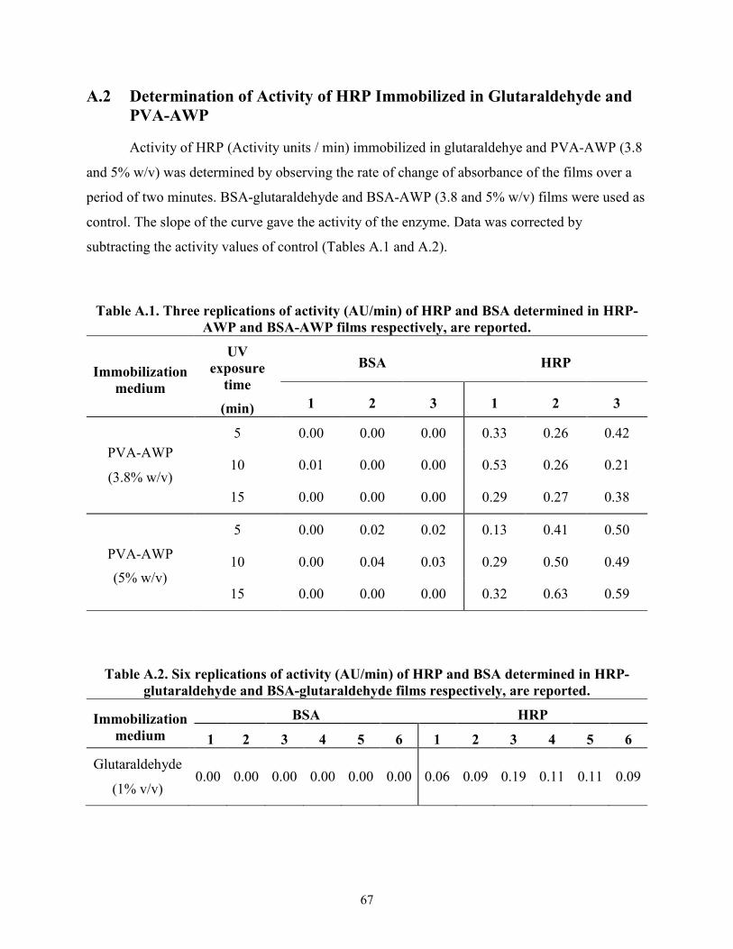
67
A.2 Determination of Activity of HRP Immobilized in Glutaraldehyde and
PVA-AWP
Activity of HRP (Activity units / min) immobilized in glutaraldehye and PVA-AWP (3.8
and 5% w/v) was determined by observing the rate of change of absorbance of the films over a
period of two minutes. BSA-glutaraldehyde and BSA-AWP (3.8 and 5% w/v) films were used as
control. The slope of the curve gave the activity of the enzyme. Data was corrected by
subtracting the activity values of control (Tables A.1 and A.2).
Table A.1. Three replications of activity (AU/min) of HRP and BSA determined in HRP-
AWP and BSA-AWP films respectively, are reported.
Immobilization
medium
UV
exposure
time
(min)
BSA HRP
1 2 3 1 2 3
PVA-AWP
(3.8% w/v)
5 0.00 0.00 0.00 0.33 0.26 0.42
10 0.01 0.00 0.00 0.53 0.26 0.21
15 0.00 0.00 0.00 0.29 0.27 0.38
PVA-AWP
(5% w/v)
5 0.00 0.02 0.02 0.13 0.41 0.50
10 0.00 0.04 0.03 0.29 0.50 0.49
15 0.00 0.00 0.00 0.32 0.63 0.59
Table A.2. Six replications of activity (AU/min) of HRP and BSA determined in HRP-
glutaraldehyde and BSA-glutaraldehyde films respectively, are reported.
Immobilization
medium
BSA HRP
1 2 3 4 5 6 1 2 3 4 5 6
Glutaraldehyde
(1% v/v) 0.00 0.00 0.00 0.00 0.00 0.00 0.06 0.09 0.19 0.11 0.11 0.09

68
A.3 Determination of activity of HRP Leached from HRP-Glutaraldehyde
and HRP-AWP films
HRP-glutaraldehyde and HRP-AWP (3.8 and 5% w/v) films were washed with buffer
and the wash was observed for enzyme activity. Activity of HRP (Activity units / min) leached
from HRP immobilized in glutaraldehye and PVA-AWP (3.8 and 5% w/v) films was determined
by observing the rate of change of absorbance of the films over a period of two minutes in the
wash. The slope of the curve gave the activity of the enzyme (Tables A.3 and A.4).
Table A.3. Three replications of activity of leached HRP (AU/min) from HRP-AWP films
are reported.
Immobilization
medium
UV
exposure
time
(min)
Concentration
(% w/v)
3.8 5
1 2 3 1 2 3
PVA-AWP
5 0.46 0.46 0.46 0.44 0.61 0.57
10 0.51 0.01 0.22 0.50 0.57 0.19
15 0.55 0.62 0.18 0.39 0.36 0.40
Table A.4. Four replications of activity of leached HRP (AU/min) from HRP-
glutaraldehyde films are reported.
Immobilization
medium 1 2 3 4
Glutaraldehyde
(1% v/v) 0.42 0.96 0.75 0.69

69
APPEDIX B
B.1 Effect of Film Thickness and UV Exposure on Sensitivity
The hydrogen peroxide step experiment was performed thrice on each BSA-AWP film of
volumes 2.5, 5.0 and 10.0 µl for UV exposure times 10 and 15 min (Figures B.1 and B.2). A bare
electrode was treated as control.
Curr
ent,
I [µ
A]
0
5
10
15
20
Control
Curr
ent,
I [µ
A]
0
5
10
15
20
2.5 µl
H2O2 Concentration [µM]
Cu
rrent,
I [µ
A]
0 200 400 600 800 1000
0
5
10
15
205.0 µl
H2O2 Concentration [µM]
Cu
rrent,
I [µ
A]
0 200 400 600 800 1000
0
5
10
15
2010.0 µl
Figure B.1. Uncorrected current responses from three replications for varying hydrogen
peroxide concentrations in the buffer solution for UV 10 min exposure are reported.

70
Curr
ent,
IC
[µ
A]
0
5
10
15
20
Control
Curr
ent,
IC [µ
A]
0
5
10
15
20
2.5 µl
H2O2 Concentration [µM]
Curr
ent,
IC [µ
A]
0 200 400 600 800 1000
0
5
10
15
205.0 µl
H2O2 Concentration [µM]
Curr
ent,
IC [µ
A]
0 200 400 600 800 1000
0
5
10
15
2010.0 µl
Figure B.2. Corrected current responses from three replications for varying hydrogen
peroxide concentrations in the buffer solution for UV 10 min exposure are reported.

71
Curr
ent,
I [µ
A]
0
5
10
15
20
Control
Curr
ent,
I [µ
A]
0
5
10
15
20
2.5 µl
H2O2 Concentration [µM]
Cu
rrent,
I [µ
A]
0 200 400 600 800 1000
0
5
10
15
205.0 µl
H2O2 Concentration [µM]
Cu
rrent,
I [µ
A]
0 200 400 600 800 1000
0
5
10
15
2010.0 µl
Figure B.3. Uncorrected current responses from three replications for varying hydrogen
peroxide concentrations in the buffer solution for UV 15 min exposure are reported.
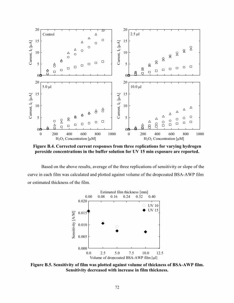
72
Curr
ent,
IC
[µ
A]
0
5
10
15
20
Control
Curr
ent,
IC
[µ
A]
0
5
10
15
20
2.5 µl
H2O2 Concentration [µM]
Curr
ent,
IC [µ
A]
0 200 400 600 800 1000
0
5
10
15
205.0 µl
H2O2 Concentration [µM]
Curr
ent,
IC [µ
A]
0 200 400 600 800 1000
0
5
10
15
2010.0 µl
Figure B.4. Corrected current responses from three replications for varying hydrogen
peroxide concentrations in the buffer solution for UV 15 min exposure are reported.
Based on the above results, average of the three replications of sensitivity or slope of the
curve in each film was calculated and plotted against volume of the dropcoated BSA-AWP film
or estimated thickness of the film.
Volume of dropcoated BSA-AWP film [µl]
Estimated film thickness [mm]
Sen
siti
vit
y [
A/M
]
0.0 2.5 5.0 7.5 10.0 12.5
0.00 0.08 0.16 0.24 0.32 0.40
0.000
0.005
0.010
0.015
0.020
UV 10
UV 15
Figure B.5. Sensitivity of film was plotted against volume of thickness of BSA-AWP film.
Sensitivity decreased with increase in film thickness.

73
B.2 Effect of Immobilization Medium on Sensitivity of Biosensor
Average current response was calculated after the addition of 20 µl of ethanol in non-
immobilized AOX, AOX-glutaraldehyde and AOX-AWP films. Three replications were
performed on each film (Figure B.6).
Curr
ent,
(I a
vg -
Ii )
[µA
]
0.000 0.002 0.004 0.006 0.008 0.010 0.012
0.0
0.5
1.0
1.5
2.0
Non-immobilized AOX
Curr
ent,
(I a
vg -
Ii )
[µA
]
0.000 0.002 0.004 0.006 0.008 0.010 0.012
0.0
0.5
1.0
1.5
2.0
AOX-glutaraldehyde
Ethanol Concentration [% v/v]
Curr
ent,
(I a
vg -
Ii)
[µ
A]
0.000 0.002 0.004 0.006 0.008 0.010 0.012
0.0
0.5
1.0
1.5
2.0
AOX-AWP
Figure B.6. Three replications of uncorrected average current responses in AOX films for
varying ethanol concentrations are reported.

74
Regression analysis was performed on above data after correcting, by subtracting the
control, for each replication in the three films. The slope of the curves was the sensitivity of the
AOX biosensor (Figure B.7).
Cu
rren
t, I
C[µ
A]
0.000 0.002 0.004 0.006 0.008 0.010 0.012
0.0
0.5
1.0
1.5
2.0
Non-immobilized AOX
Cu
rren
t, I
C[µ
A]
0.000 0.002 0.004 0.006 0.008 0.010 0.012
0.0
0.5
1.0
1.5
2.0
AOX-glutaraldehyde
Ethanol Concentration [% v/v]
Curr
ent,
IC[µ
A]
0.000 0.002 0.004 0.006 0.008 0.010 0.012
0.0
0.5
1.0
1.5
2.0
AOX-AWP
Figure B.7. Linearity of current responses with change in ethanol concentration was more
for immobilized films. Though sensitivity of AOX-glutaraldehyde films were higher,
variability was also higher.

75
B.3 Response Times
Response times were calculated in non-immobilized AOX, AOX-glutaraldehyde and
AOX-AWP films. Three replications were performed on each film and the response times
calculated in each replication are reported (Table B.1).
Table B.1. Three replications of response times measured in each type of AOX film for
different ethanol concentrations are reported.
Ethanol
Concentration
[% v/v]
Response Time [min]
on-immobilized
AOX AOX-Glutaraldehyde AOX-AWP
1 2 3 1 2 3 1 2 3
0.0001 0.13 0.45 0.20 0.48 0.53 0.41 0.45 0.70 0.62
0.0005 0.20 0.21 0.31 0.50 0.49 0.72 0.54 0.76 0.56
0.0010 0.32 0.61 0.33 0.41 0.47 0.80 0.52 0.66 0.63
0.0025 0.16 0.13 0.10 0.51 0.41 0.36 0.39 0.77 0.94
0.0040 0.24 0.12 0.13 0.45 0.42 0.47 0.36 0.46 0.32
0.0050 0.25 0.48 0.15 0.46 0.30 0.42 0.34 0.35 0.61
0.0075 0.11 0.13 0.69 0.46 0.41 0.41 0.30 0.56 0.52
0.0100 0.11 0.62 0.13 0.46 0.45 0.40 0.43 0.45 0.49